Garrett R.H., Grisham C.M. - Biochemistry (1999)(2nd ed.)(en)
.pdf
20.13 ● The TCA Cycle Provides Intermediates for Biosynthetic Pathways |
661 |
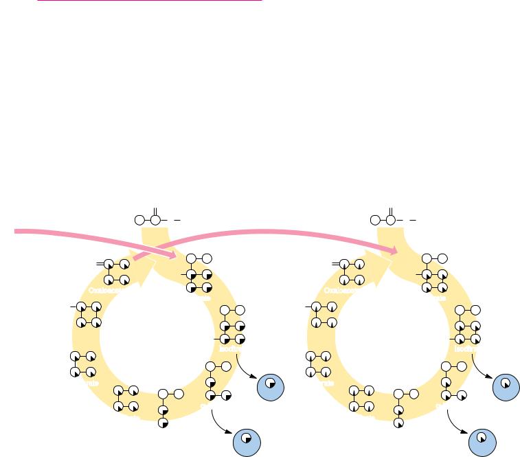
survives two full turns, then undergoes a 50% loss through each succeeding turn of the cycle.
It is worth noting that the carbon–carbon bond cleaved in the TCA pathway entered as an acetate unit in the previous turn of the cycle. Thus, the oxidative decarboxylations that cleave this bond are just a cleverly disguised acetate COC cleavage and oxidation.
20.13 ● The TCA Cycle Provides Intermediates
for Biosynthetic Pathways
Until now we have viewed the TCA cycle as a catabolic process because it oxidizes acetate units to CO2 and converts the liberated energy to ATP and reduced coenzymes. The TCA cycle is, after all, the end point for breakdown of food materials, at least in terms of carbon turnover. However, as shown in Figure 20.22, four-, five-, and six-carbon species produced in the TCA cycle also fuel a variety of biosynthetic processes. -Ketoglutarate, succinyl-CoA, fumarate, and oxaloacetate are all precursors of important cellular species. (In order to par-
FIGURE 20.21 ● |
The fate of the carbon atoms of acetate in successive TCA cycles. |
|
|
|
|
||||
(a) The carbonyl carbon of acetyl-CoA is fully retained through one turn of the cycle but |
|
|
|
|
|||||
is lost completely in a second turn of the cycle. (b) The methyl carbon of a labeled acetyl- |
|
|
|
|
|||||
CoA survives two full turns of the cycle but becomes equally distributed among the four |
|
|
|
|
|||||
carbons of oxaloacetate by the end of the second turn. In each subsequent turn of the |
|
|
|
|
|||||
cycle, one-half of this carbon (the original labeled methyl group) is lost. |
|
|
|
|
|
||||
|
O |
|
|
|
|
|
O |
|
|
|
S CoA |
|
|
|
|
|
S CoA |
|
|
O |
|
|
|
|
O |
|
|
|
|
|
HO |
|
|
|
|
|
HO |
|
|
Oxaloacetate |
|
|
|
Oxaloacetate |
|
|
|
||
|
Citrate |
|
|
|
|
Citrate |
|
||
HO |
|
|
|
|
HO |
|
|
|
|
Malate |
3rd turn |
HO |
|
|
Malate |
|
4th turn |
HO |
|
|
|
|
|
|
|
|
|
||
|
|
Isocitrate |
|
|
|
|
|
Isocitrate |
|
Fumarate |
|
|
|
1 |
Fumarate |
|
|
|
1 |
|
|
|
/4 |
|
|
|
/8 |
||
|
|
|
(CO2) |
|
|
|
|
(CO2) |
|
|
|
α -Ketoglutarate |
|
|
|
|
|
α -Ketoglutarate |
|
|
Succinate |
|
1 |
/2 Total |
Succinate |
|
|
|
|
|
|
|
|
|
|
|
|
||
|
Succinyl–CoA |
|
methyl C |
|
Succinyl–CoA |
|
|
||
|
1 |
label |
|
|
1 |
|
|||
|
|
/4 |
|
|
|
|
|
/8 |
|
|
|
(CO2) |
|
|
|
|
|
(CO2) |
|
|
|
|
|
|
|
|
|
1 |
Total |
|
|
|
|
|
|
|
|
/4 |
|
methyl C label


20.15 ● Regulation of the TCA Cycle |
665 |
|
2–O |
PO |
|
GDP |
GTP |
O |
|||||
|
3 |
C COO– |
|
|
|
|
|||||
CO2 + |
|
|
|
|
|
C |
|
COO– |
|||
|
|
|
|
|
|
||||||
|
|
|
|
|
|
|
|||||
|
|
|
|
|
|
|
|
|
|
|
|
|
|
CH2 |
|
|
|
|
|||||
|
|
|
|
|
|
H2C |
|
COO– |
|
||
|
|
|
|
|
|
|
|
||||
|
|
|
|
|
|
|
|
||||
|
|
PEP |
|
|
|
|
Oxaloacetate |
||||
FIGURE 20.25 ● The phosphoenolpyruvate carboxykinase reaction.
PEP carboxylase occurs in yeast, bacteria, and higher plants, but not in animals. The enzyme is specifically inhibited by aspartate, which is produced by transamination of oxaloacetate. Thus, organisms utilizing this enzyme control aspartate production by regulation of PEP carboxylase. Malic enzyme is found in the cytosol or mitochondria of many animal and plant cells and is an NADPHdependent enzyme.
It is worth noting that the reaction catalyzed by PEP carboxykinase (Figure 20.25) could also function as an anaplerotic reaction, were it not for the particular properties of the enzyme. CO2 binds weakly to PEP carboxykinase, whereas oxaloacetate binds very tightly (K D 2 10 6 M), and, as a result, the enzyme favors formation of PEP from oxaloacetate.
The catabolism of amino acids provides pyruvate, acetyl-CoA, oxaloacetate, fumarate, -ketoglutarate, and succinate, all of which may be oxidized by the TCA cycle. In this way, proteins may serve as excellent sources of nutrient energy, as seen in Chapter 26.
20.15 ● Regulation of the TCA Cycle
Situated as it is between glycolysis and the electron transport chain, the TCA cycle must be carefully controlled by the cell. If the cycle were permitted to run unchecked, large amounts of metabolic energy could be wasted in overproduction of reduced coenzymes and ATP; conversely, if it ran too slowly, ATP would not be produced rapidly enough to satisfy the needs of the cell. Also, as just seen, the TCA cycle is an important source of precursors for biosynthetic processes and must be able to provide them as needed.
What are the sites of regulation in the TCA cycle? Based upon our experience with glycolysis (Figure 19.31), we might anticipate that some of the reactions of the TCA cycle would operate near equilibrium under cellular conditions (with G 0), whereas others—the sites of regulation—would be characterized by large, negative G values. Estimates for the values of G in mitochondria, based on mitochondrial concentrations of metabolites, are summarized in Table 20.1. Three reactions of the cycle—citrate synthase, isocitrate dehydrogenase, and -ketoglutarate dehydrogenase—operate with large, negative G values under mitochondrial conditions and are thus the primary sites of regulation in the cycle.
The regulatory actions that control the TCA cycle are shown in Figure 20.26. As one might expect, the principal regulatory “signals” are the concentrations of acetyl-CoA, ATP, NAD , and NADH, with additional effects provided by several other metabolites. The main sites of regulation are pyruvate dehydrogenase, citrate synthase, isocitrate dehydrogenase, and -ketoglutarate dehydrogenase. All of these enzymes are inhibited by NADH, so that when the cell has produced all the NADH that can conveniently be turned into ATP, the cycle shuts down. For similar reasons, ATP is an inhibitor of pyruvate dehydrogenase and isocitrate dehydrogenase. The TCA cycle is turned on, however, when either the ADP/ATP or NAD /NADH ratio is high, an indication that the cell has run low on ATP or NADH. Regulation of the TCA cycle by NADH,

20.15 ● Regulation of the TCA Cycle |
667 |
NAD , ATP, and ADP thus reflects the energy status of the cell. On the other hand, succinyl-CoA is an intracycle regulator, inhibiting citrate synthase and-ketoglutarate dehydrogenase. Acetyl-CoA acts as a signal to the TCA cycle that glycolysis or fatty acid breakdown is producing two-carbon units. AcetylCoA activates pyruvate carboxylase, the anaplerotic reaction that provides oxaloacetate, the acceptor for increased flux of acetyl-CoA into the TCA cycle.
Regulation of Pyruvate Dehydrogenase
As we shall see in Chapter 23, most organisms can synthesize sugars such as glucose from pyruvate. However, animals cannot synthesize glucose from acetylCoA. For this reason, the pyruvate dehydrogenase complex, which converts pyruvate to acetyl-CoA, plays a pivotal role in metabolism. Conversion to acetylCoA commits nutrient carbon atoms either to oxidation in the TCA cycle or to fatty acid synthesis (see Chapter 25). Because this choice is so crucial to the organism, pyruvate dehydrogenase is a carefully regulated enzyme. It is subject to product inhibition and is further regulated by nucleotides. Finally, activity of pyruvate dehydrogenase is regulated by phosphorylation and dephosphorylation of the enzyme complex itself.
High levels of either product, acetyl-CoA or NADH, allosterically inhibit the pyruvate dehydrogenase complex. Acetyl-CoA specifically blocks dihydrolipoyl transacetylase, and NADH acts on dihydrolipoyl dehydrogenase. The mammalian pyruvate dehydrogenase is also regulated by covalent modifications. As shown in Figure 20.27, a Mg2 -dependent pyruvate dehydrogenase kinase is associated with the enzyme in mammals. This kinase is allosterically activated by NADH and acetyl-CoA, and when levels of these metabolites rise in the mitochondrion, they stimulate phosphorylation of a serine residue on the pyruvate dehydrogenase subunit, blocking the first step of the pyruvate dehydrogenase reaction, the decarboxylation of pyruvate. Inhibition of the dehydrogenase in this manner eventually lowers the levels of NADH and acetylCoA in the matrix of the mitochondrion. Reactivation of the enzyme is carried out by pyruvate dehydrogenase phosphatase, a Ca2 -activated enzyme that binds to the dehydrogenase complex and hydrolyzes the phosphoserine moiety on the dehydrogenase subunit. At low ratios of NADH to NAD and low acetyl-CoA levels, the phosphatase maintains the dehydrogenase in an activated state, but a high level of acetyl-CoA or NADH once again activates the kinase and leads to the inhibition of the dehydrogenase. Insulin and Ca2 ions activate dephosphorylation, and pyruvate inhibits the phosphorylation reaction.
Pyruvate dehydrogenase is also sensitive to the energy status of the cell. AMP activates pyruvate dehydrogenase, whereas GTP inhibits it. High levels of AMP are a sign that the cell may become energy-poor. Activation of pyruvate dehydrogenase under such conditions commits pyruvate to energy production.
Regulation of Isocitrate Dehydrogenase
The mechanism of regulation of isocitrate dehydrogenase is in some respects the reverse of pyruvate dehydrogenase. The mammalian isocitrate dehydrogenase is subject only to allosteric activation by ADP and NAD and to inhibition by ATP and NADH. Thus, high NAD /NADH and ADP/ATP ratios stimulate isocitrate dehydrogenase and TCA cycle activity. The Escherichia coli enzyme, on the other hand, is regulated by covalent modification. Serine residues on each subunit of the dimeric enzyme are phosphorylated by a protein kinase, causing inhibition of the isocitrate dehydrogenase activity. Activity is restored by the action of a specific phosphatase. When TCA cycle and glycolytic intermediates—such as isocitrate, 3-phosphoglycerate, pyruvate, PEP, and oxaloacetate—are high, the kinase is inhibited, the phosphatase is acti-


670 Chapter 20 ● The Tricarboxylic Acid Cycle
CoA to form L-malate. The net effect is to conserve carbon units, using two acetyl-CoA molecules per cycle to generate oxaloacetate. Some of this is converted to PEP and then to glucose by pathways discussed in Chapter 23.
The Glyoxylate Cycle Operates in Specialized Organelles
The enzymes of the glyoxylate cycle in plants are contained in glyoxysomes, organelles devoted to this cycle. Yeast and algae carry out the glyoxylate cycle in the cytoplasm. The enzymes common to both the TCA and glyoxylate pathways exist as isozymes, with spatially and functionally distinct enzymes operating independently in the two cycles.
Isocitrate Lyase Short-Circuits the TCA Cycle by Producing Glyoxylate and Succinate
The isocitrate lyase reaction (Figure 20.29) produces succinate, a four-carbon product of the cycle, as well as glyoxylate, which can then combine with a second molecule of acetyl-CoA. Isocitrate lyase catalyzes an aldol cleavage and is similar to the reaction mediated by aldolase in glycolysis. The malate synthase reaction (Figure 20.30), a Claisen condensation of acetyl-CoA with the aldehyde of glyoxylate to yield malate, is quite similar to the citrate synthase reaction. Compared with the TCA cycle, the glyoxylate cycle (a) contains only five steps (as opposed to eight), (b) lacks the CO2-liberating reactions, (c) consumes two molecules of acetyl-CoA per cycle, and (d) produces four-carbon units (oxaloacetate) as opposed to one-carbon units.
The Glyoxylate Cycle Helps Plants Grow in the Dark
The existence of the glyoxylate cycle explains how certain seeds grow underground (or in the dark), where photosynthesis is impossible. Many seeds (peanuts, soybeans, and castor beans, for example) are rich in lipids; and, as we see in Chapter 24, most organisms degrade the fatty acids of lipids to acetylCoA. Glyoxysomes form in seeds as germination begins, and the glyoxylate cycle uses the acetyl-CoA produced in fatty acid oxidation to provide large amounts of oxaloacetate and other intermediates for carbohydrate synthesis. Once the growing plant begins photosynthesis and can fix CO2 to produce carbohydrates (see Chapter 22), the glyoxysomes disappear.
Glyoxysomes Must Borrow Three Reactions from Mitochondria
Glyoxysomes do not contain all the enzymes needed to run the glyoxylate cycle; succinate dehydrogenase, fumarase, and malate dehydrogenase are absent. Consequently, glyoxysomes must cooperate with mitochondria to run their cycle (Figure 20.31). Succinate travels from the glyoxysomes to the mitochondria, where it is converted to oxaloacetate. Transamination to aspartate follows
|
H2C |
|
COO– |
|
|
|
|
|
|
|
|
|
|
|
|
|
|
|
|
|
||||
|
|
|
|
|
|
|
|
|
|
|
|
|
|
|
|
|
|
|
||||||
H |
|
C |
|
|
COO– |
|
|
|
|
|
|
|
|
|
|
|
|
|
|
|
|
|
||
|
|
|
+H B |
|
|
|
HC |
|
|
COO– |
+ |
H2C |
|
COO– |
B |
|||||||||
|
|
|
|
|
|
|
COO– |
|
|
|
|
|
|
|||||||||||
|
|
|
|
|
|
|
|
|
|
|
|
|
|
|
|
|
|
E |
||||||
H |
|
C |
|
|
|
|
|
|
|
|
|
|
|
|
|
|
|
|
COO– |
|||||
|
|
|
|
|
|
|
|
|
|
|
|
|
|
|
O |
|
H2C |
|
+ |
|||||
|
|
|
|
|
|
|
|
|
|
|
|
|
|
|
|
|
||||||||
|
|
|
|
|
|
|
|
|
|
|
|
|
|
|
|
|
|
|
|
|
|
|
|
H B |
|
|
O |
|
|
H B |
|
E |
|
|
|
Glyoxylate |
|
Succinate |
|
||||||||||
|
|
|
|
|
|
|
|
|
|
|
|
|
|
|
|
|
|
|
|
|
|
|
|
|
2R, 3S-Isocitrate
FIGURE 20.29 ● The isocitrate lyase reaction.

 Aspartate

 Glutamate
  Proline
 NADH
NADH
 ADP
ADP