

Formation of Memory
The nucleus of the mammillary body receives many inputs from the major memoryprocessing structures of the cerebrum, the hippocampal formation and therefore may relate to memory formation

Epithalamus
Epithalamus
The epithalamus is the posterior portion of the diencephalon
It forms the roof of the third ventricle

The Epithalamus
The epithalmus consists of one tiny group of nuclei and a small, unpaired knob called the pineal body
This gland, which derives from ependymal glial cells, is a hormone secreting organ

Epithalamus
The pineal gland extends from the posterior border of the epithalamus
The pineal gland secretes the hormone melatonin which signals the sleep wake cycle
Pinal
Body
The Epithalamus
Choroid
Plexus
A cerebrospinal fluidforming structure called a choroid plexus is also part of the epithalamus

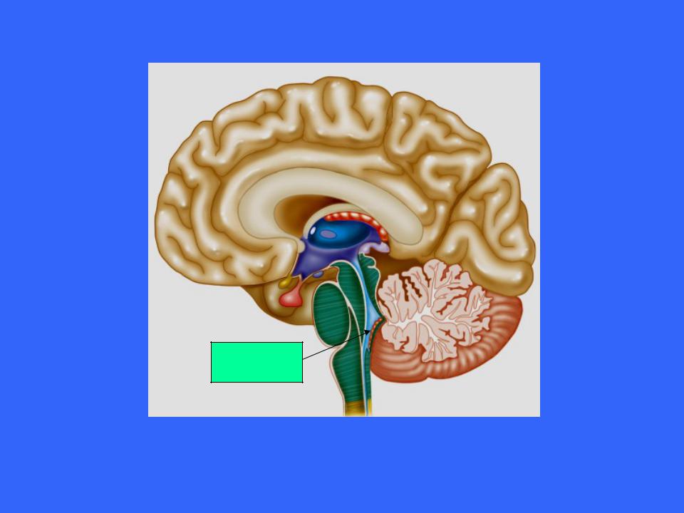
The Brain Stem
The third of the four major parts of the brain is the brain stem
From superior to inferior, the brain stem is divided into;
–Midbrain
–Pons
–Medulla oblongata
Pons
Medulla
oblongata
Midbrain

The Brain Stem
Each region is roughly an inch long
Together than constitute 2.5% of total brain mass
The brain stem has several functions
–It produce the rigidly programmed, automatic behaviors necessary for our survival
–Acts as a passageway for all the fiber tracts running between the cerebrum and spinal cord
–It is heavily involved with the innervation of the face and head as 10 of the 12 cranial nerve attach to it

The Brain Stem
The brain stem has the same structural plan as the spinal cord, with outer white matter surrounding an inner region of gray matter
However, there are also nuclei of gray matter located within the white matter

The Midbrain
The midbrain is located between the diencephalon superiorly and the pons inferiorly

The Midbrain
Its central cavity is the cerebral aqueduct, which divides it into a tectum (dorsal surface) and paired cerebral peduncles
From an anterior view the cerebral peduncles appear as columns that hold up the cerebrum