- •1. Вступ
- •2. Звуки і букви
- •3. Алфавіт
- •4. Транскрипція
- •5. Класифікація звуків
- •6. Інтонація
- •7. Наголос
- •8. Типи складів
- •9. Характеристика звуків
- •Голосні звуки
- •Дифтонги
- •10. Читання буквосполучень
- •11. "Німі" літери
- •12. Органи мовлення
- •Заняття 1 (lesson one)
- •III. Speaking: About Myself
- •Personal Information Sheet
- •About My Family and Myself
- •IV. Grammar Exercises
- •1. Наголошений склад:
- •2. Ненаголошений склад: а [з] - aside [a'said], data ['deita] ai, ay [ei] - play, rain, nail ei, ey [ei] - vein, fey еа [I:] - peace, seat, tea name, pain, may
- •II. Читання приголосних звуків
- •Exercise 13. Прочитайте cj
- •III. Speaking: Elements of Conversation
- •Exercise 2. Вивчіть наступні вирази
- •IV. Grammar Exercises
- •V. Independent Work: Introducing People
- •Introducing People
- •/ Study at the Medical College
- •III. Grammar Exercises
- •IV. Independent Work: My Medical College
- •[Зо] [d]
- •II. Speaking: My Future Profession
- •III. Grammar Exercises
- •IV. Independent Work: Where Do Nurses Work?
- •Where Do Nurses Work?
- •1. Відкритий склад
- •II. Speaking: Nurse's Working Day
- •In a Hospital
- •III. Grammar Exercises
- •IV. Independent Work: Florence Nightingale
- •Igh - [аі]
- •II. Speaking: English for Modern Medical Specialists
- •/ Study English
- •IV. Independent Work: English as Means of International Communication
- •English as Means of International Communication
- •I. Speaking
- •We Study Anatomy
- •In class:
- •III. Independent Work: From the History of Anatomy
- •Some Notes from the History of Anatomy and Physiology
- •I. Speaking: Skeleton
- •Skeleton
- •III. Independent Work: Indoor Activities or Home Interests
- •I. Speaking: Internal Organs
- •Internal Organs
- •II. Independent Work: Body Systems
- •Body Systems
- •I. Speaking: Heart
- •II. Grammar Exercises
- •It is early. - Рано. It is late. - Пізно. It is time. - Пора. It is high time. - Давно пора.
- •It's morning. - Ранок. It's evening. - Вечір. It's night. - Ніч. It's afternoon. - Полудень.
- •I. Speaking: How to Take a Pulse
- •Heart (Part 2)
- •How the Second Hand in Your Watch Was Invented
- •II. Grammar Exercises
- •III. Independent Work: My Heart Will Go on
- •My Heart Will Go on
- •I. Speaking: Blood
- •Heart and Blood
- •II. Grammar Exercises
- •III. Independent Work: Functions of Blood
- •Functions of Blood
- •I. Speaking: Blood Pressure
- •Blood Pressure
- •II. Grammar Exercises
- •III. Independent Work: Instant Blood Test
- •Instant Blood Test
- •I. Speaking: Healthy Way of Life
- •Exercise 6. Match the words/word combinations.
- •Eternal Youth Laws
- •II. Grammar Exercises
- •III. Independent Work: Are You Going to Live to 100?
- •I. Speaking: Vitamins
- •Vitamins
- •II. Grammar Exercises
- •III. Independent Work: What You Should Know about Vitamins
- •Vitamins: Their Uses
- •Vitamins: Six Cooking Tips
- •I. Control Test Variant 1
- •Variant 2
- •II. Grammar
- •Exercise 5. Translate the sentences into Ukrainian. Mind the meaning of one.
- •I. Speaking: First Medical Aid (Part I)
- •First Aid
- •II. Grammar Exercises
- •Ill. Independent Work: The Strange Doctor
- •The Strange Doctor
- •I. Speaking: First Medical Aid (Part II)
- •Text 1 Bruises
- •Text 2 Burns
- •Text 4 Spinal Injuries
- •Text 5 Unconsciousness
- •Text б Cuts, Bleeding
- •II. Independent Work: Future Tense
- •Lessons nineteen-twenty
- •І. Texts for Home Reading Text a Early Folk Medicine
- •Text в Higher Education in Ukraine
- •Text c English Universities and Colleges
- •I. Speaking: Diseases. Symptoms
- •When We Have Pain
- •II. Grammar Exercises
- •III. Independent Work: From the History of Medicine
- •I. Speaking: Things for Nursing
- •II. Grammar Exercises
- •In question - який вивчається; який обговорюється; який викликає сумнів; спірний under study - який вивчається under discussion - який обговорюється
- •III. Independent Work: Medicine
- •Medicine
- •I. Speaking: When I'm III
- •The Doctor
- •II. Grammar Exercises
- •III. Independent Work: Health
- •I. Speaking: a Visit to a Doctor
- •At the Doctor's
- •II. Grammar Exercises
- •Hi. Independent Work: Cold War
- •Cold War
- •I. Speaking: Illness
- •II. Grammar Exercises
- •III. Independent Work: Why Are British Sailors Called "Limeys"?
- •Why Are British Sailors Called "Limeys"?
- •6. What are some foods that you should eat so that you get enough vitamin c?
- •I. Speaking: Infectious Diseases
- •Infectious Diseases
- •II. Grammar Exercises
- •III. Independent Work: Bacteria
- •Bacteria
- •I. Speaking: Children Diseases
- •II. Grammar Exercises
- •Rickets
- •I. Speaking: Surgical Department
- •II. Grammar Exercises
- •III. Independent Work: Superlatives
- •Superlatives
- •I. Speaking: Operating Nurse
- •Operation
- •II. Grammar Exercises
- •III. Independent Work: Surgery
- •Surgery
- •Introduction
- •I. Speaking: Work of a Laboratory Assistant
- •Work of a Laboratory Assistant
- •Pain in the Leg
- •Your Arm Is out of Joint
- •Leg Fracture
- •A Dog Bit Me
- •II. Grammar Exercises
- •I. Speaking: Teeth
- •II. Grammar Exercises
- •III. Independent Work: What Happens to a Hamburger
- •What Happens to a Hamburger
- •I. Speaking: At the Dentist's. Dental Instruments
- •II. You are examining your patient's oral cavity. Ask the patient to perform thefollowing.
- •Dental Calculus
- •II. Grammar Exercises
- •III. Independent Work: What Happens to a Hamburger What Happens to a Hamburger
- •I. Speaking: Medicines and Their Forms
- •At the Chemist's
- •II. Grammar Exercises
- •III. Independent Work: d.I. Mendeleev
- •Outstanding Russian Chemist d.I. Mendeleev
- •I. Speaking: Chemical Elements. Properties of Substances
- •II. Grammar Exercises
- •III. Independent Work: Chemical Elements of Living Matter
- •Chemical Elements of Living Matter
- •I. Speaking: At the Chemist's
- •At the Chemist's
- •At the Chemist's
- •II. Grammar Exercises
- •III. Independent Work: Aspirin
- •Ordinary Aspirin Is Truly a Wonder Drug
- •I. Speaking: Obstetrics and Gynecology
- •II. Grammar Exercises
- •III. Independent Work: Childbirth
- •I. Speaking
- •Computers in Our Life
- •Interpreter: ...
- •Interpreter: ....
- •Interpreter: ....
- •II. Grammar Exercises
- •I. Control Test
- •Medicinal Plants
- •X All child's diseases are caused by
- •Independent Reading
- •/. Health Service in Ukraine
- •2. Doctors and Patients
- •3. Medicine and Health Care
- •Physicians
- •4. Computers Concern You
- •5. Hobbies and Leisure Time Occupations
- •Outdoor Activities or Activities outside the Home
- •6. Are You Left-Handed?
- •7. Vegetarians
- •I. Speaking: Human Body
- •II. Grammar Exercises
- •I. Speaking: Chemistry, Matter and Life
- •Chemistry, Matter, and Life
- •II. Grammar Exercises
- •I. Speaking: Cells and Their Functions
- •Cells and Their Functions
- •II. Grammar Exercises
- •I. Speaking: Tissues, Glands and Membranes
- •Voluntary ['vubntsri] довільний
- •Visceral ['visarsl] що стосується нутрощів
- •II. Independent Work: Diseases
- •Study of Diseases
- •I. Speaking: Skeleton
- •Exercise 1. Read and learn the following words, remember their Latin or Greek equivalents.
- •II. Grammar Exercises
- •IhaL cresl
- •I want I'd like He agreed The wounded asked She likes You can Exercise 2. Make up sentences, translate them. Name the forms of the infinitive.
- •111. Independent Work: Spinal Curves
- •Spinal Curves
- •I. Speaking: Bones
- •II. Grammar Exercises
- •III. Independent Work: Traumatological Case Report
- •Traumatological Case Report
- •I. Speaking: Joints
- •II. Grammar Exercises
- •III. Independent Work: Anti-Inflammatory Drugs
- •Anti-Inflammatory Drugs
- •Exercise 2. Make up a dialog: you are a traumatologist and you receive a patient with severe pain in his knee.
- •I. Speaking: Musculoskeletal System
- •Musculoskeletal System
- •II. Grammar Exercises
- •III. Independent Work: Muscle Tissues
- •I. Speaking: Skin (Integumentary System)
- •Is eczema a contagious disease?
- •9) What is urticaria?
- •II. Grammar Exercises
- •It is not difficult for the doctor to treat the disease.
- •10) What clotting disorders do you know?
- •II. Grammar Exercises
- •1.1 Can't hear a word, though he seems to be speaking. 2.1 am happy not to have failed you.
- •It is not so difficult for him to diagnose this disease.
- •III. Independent Work: Blood Tests
- •7. Bone marrow biopsy gives valuable information to diagnose bone marrow disorders,cukemia and anemia.
- •1. The most numerous cells of blood are ...
- •Interspace [,ints'speis] проміжок; інтервал
- •In the left chamber the atrium and ventricle are separated by the mitral valve. In the rig: chamber the atrium and ventricle are separated by the tricuspid valve.
- •1. Endocarditis [,endsuka:'daitis] means "inflammation of the lining of the heart cavities"but it most commonly refers to valvular disease.
- •3. Pericarditis [,peri:ka:'daitis] refers to disease of the serous membrane on the heartsurface, as well as that lining the pericardial sac.
- •II. Grammar Exercises
- •III. Independent Work: Heart Sounds
- •Visceral ['visaral] що стосується нутрощів
- •Vary ['veari] мінятись; змінюватись
- •III. Independent Work: Cardiovascular Diseases
- •1) To be of great importance; at present; scientists consider; cardiovascular diseases;
- •In the treatment of some cardiovascular diseases; to have been achieved; definite success.
- •I. Speaking: Lymphatic System and Lymphoid Tissue
- •Inguinal [irjgwm(a)l] пахвинний
- •Vessels in grey area drain into right lymphatic duel
- •I saw them walking along the street.
- •I saw Sydorenko running along the avenue.
- •III. Independent Work: Disorders of Lymphatic System and Lymphoid Tissue
- •In inflammation of the lymphatic vessels called lymphangitis, red streaks can be seen extending along an extremity. Septicemia or blood poisoning may occur because of streptococci.
- •1. Вона бачила через вікно, що йшов сильний дощ. 2. Він хотів, щоб пацієнт одужав швидше. 3. Ти вже маєш переписаний текст? 4. Медсестра вже зробила перев'язку.
- •Irritants can be bacteria, friction, chemicals, X-rays, fire, cuts or blows, etc.
- •Immunity is the power of an individual to resist or overcome the effects of a particular disease or other harmful agents.
- •I. Speaking: Respiratory System
- •1. Pulmonary ventilation is normally accomplished by inspiration and expiration.
- •2. The diffusion of gases includes the passage of oxygen from air sacs into the blood andof carbon dioxide out of the blood.
- •3. The transport of oxygen and carbon dioxide by the circulating blood.
- •II. Grammar Exercises
- •III. Independent Work: Lungs
- •In infants the lungs are of a pale rosy color, but later they become darker.
- •1. Make your analyses of blood and urine. 2. Take an electrocardiogram. 3. Your lungs should be X-rayed. 4. Go to your doctor and check your bp. 5. You need treatment. 6. You will be treated.
- •I. Speaking: Respiratory System Disorders
- •I am going to examine you.
- •1. This science studies the structure and shapeof the body and organs. Take part in respiration. A. Kidneys b. Bladders c. Lungs d. Limbs person? e. Muscles a- Ears
- •11. With the help of what do we auscultate a
- •III. Independent Work: Virus
- •I. SPeaking: Digestive System
- •Inner, serous, salivary, hard, exact (точний), vital, face, connective, pale, length, palate, coat, capacity, gland, layer.
- •Vermiform appendix
- •II. Grammar Exercises
- •3. That portion of the alimentary tract which forms the large intenstine consists of thececum, colon and rectum.
- •4. The valve that divides the atrium and the ventricle of the right chamber is called thetricuspid valve.
- •1. The left atrium and ventricle connected by the mitral valve form the left chamber of theheart.
- •In the pulmonological department there are patients with lung diseases. They suffer from pneumonia, bronchitis, asthma, etc. They complain of their bad cough, high temperature, headache.
- •I feel faint. - Мені погано.
- •Infection with the bacteria called Helicobacter pylori.
- •Home Treatment
- •1. What function does the process of digestion 3. Where does food get in at first?fulfill? a. The large intestine
- •1. A) The discussion of the report lasted two hours.
- •I. Speaking: Nervous System (Part I)
- •(Part I)
- •II. Grammar Exercises
- •It's no use crying over spilt milk.
- •I was afraid of being here in such hour.
- •10. Sterilizing the instruments, preparing the patient for the operation took me about an hour.
- •4.1 Was told that he was operated on without having been anesthetized.
- •5. After having gathered a complete clinical history, he began to examine the patient.
- •III. Independent Work: Role of Nervous System
- •It should be remembered that unlike animals, man can himself considerably change his external environment.
- •I. Pavlov demonstrated that man's so-called psychic activity is based on physiological processes operating in the cerebral cortex.
- •I. Speaking: Nervous System (Part II)
- •The functions of the autonomic nervous system.
- •31 Pairs of spinal nerves, each pair is numbered according to the level of the spinal cord from which it arises. Each spinal nerve has small posterior divisions and rather large anterior divisions.
- •II. Grammar Exercises
- •1. There is no hope of our seeing him soon.
- •1) Dura mater is the upper layer, the outmost of the three membranes, which surrounds the spinal cord and is the toughest and most fibrous substance.
- •1. Special sensory impulses, such as those for smell, taste, vision, and hearing.
- •2. General sensory impulses, such as those for pain, touch, temperature, deep musclesense, pressure, and vibration.
- •3. Somatic motor impulses, resulting in voluntary control of skeletal muscles.
- •4. Visceral motor impulses producing involuntary control of glands and muscles of theheart and smooth muscles.
- •II. Grammar Exercises
- •1 The first faculty to go and the last to appear - здатність, що першою зникає і останньою з'являється
- •1. What is cts? 2. What does the method of mri mean? 3. What is eeg? 4. What can we study with the help of the electroencephalograph?
- •Tumors of the brain are growths of brain tissues and meninges of abnormal character.
- •III. Independent Work: Pain
- •1. How can pain be relieved? 2. Can pain be localized? 3. Where are receptors for pain distributed? 4. What are nociceptors? 5. Is pain the most primitive sensation?
- •I. Speaking: Sensory System
- •Vision from receptors in the eye.
- •1. What system is responsible for all activitiesof organs and systems of organs in the humanbody?
- •10. He dared test this device without permission.11.1 cannot understand why you should do it.
- •III. Independent Work: Drugs
- •1 Awareness [s'wesnss] свідомість, усвідомлення
- •I. Speaking: Endocrine System
- •Islets of Langcrhans (in pancreas)
- •What are the parts of the endocrine system?
- •1. What substances are produced by endocrineglands?
- •1. Some glands practically limit their activity to this.
- •2. The activity of a gland is normally accompanied by a great dilation of its bloodvessels.
- •I. Speaking: Urinary System
- •10. What disorders of the urinary system do you know?
- •II. Grammar Exercises
- •Vulva is the external genitalia, it includes the vaginal lips, clitoris and vaginal and urethral orifices. The space between the vaginal orifice and the anus is called perineum.
- •Is the placenta also an organ?
- •I. Speaking: Duties of a Midwife
- •In what ways does Nicky prepare women for birth in her weekly antenatal classes?
- •II. Independent Work: Risk of Having Children in Later Life
- •Symptoms
- •In the second trimester, the uterus expands, abdominal enlargement becomes more apparent, and the woman can feel the fetus move. Many women feel their best during this trimester.
- •Immunization against chickenpox
- •Immunization against hepatitis в if you are at increased risk of getting this infection;
- •Illegal drugs;
- •1) Fertility; 2) in vitro; 3) menopause; 4) to fail; 5) to insert; 6) to increase.
- •II. Independent Work: Miscarriage
- •I. Speaking: Precautions
- •It is estimated - приблизно підраховано inherited predisposition - спадкова схильність contemplate - обдумувати; мати намір to be vulnerable - бути вразливим ingestion - ковтання, поглинання
- •I. Speaking: Fetus
- •Vary - різнитися, мінятися
- •II. Independent Work: Premature Delivery
- •I. Speaking: Labor
- •3. And sleepiness are common.
- •II. Independent Work: who. Pandemics and Tamif lu
- •Improving sanitation and water supply,
- •Deaths per day. We have lost many nurses and doctors. Special trains carry away the dead. For several days there were no coffins and the bodies piled up."
- •Viruses reproduce outside your body.
- •I Speaking
- •I. Speaking: Anesthesia
- •1. How do anesthetic drugs work?
- •2. Why do you think patients were held or strapped down before anesthetic drugs wereavailable?
- •3.What difference did anesthetic drugs make to the work of surgeons?
- •2. Read and translate the text.
- •II. Grammar Exercises
- •III. Independent Work: Major Medical Specialty Fields
- •I. Speaking: English Prescription
- •1. Name and address of the physician and his telephone number. The title m.D. (MedicalDoctor) should follow the physician's name to indicate that he or she is a physician.
- •2. Usually at the top - patient's name, address, age and the date.
- •3. Symbol Rx. This is about the same as "Dear Sir" on top of a letter. It comes from Latinand means a command for a patient "you take".
- •4. The body of the prescription contains the name of the drag and the dose.
- •5Is copy right - на неї (назву) розповсюджується авторське право 6to capitalize - писати з великої літери 7it goes without saying - нічого й говорити 8dose specification - інструкція з дозування
- •230 Broad Street
- •1. Dioscorides, a Greek physician of the second century who accompanied the Romanarmies
- •In his estate he cultivated his orchard and worked there being in old age.
- •13 Sliding palpation - ковзна пальпація
- •14 To win (won) recognition - здобути визнання
- •1 Disaster [di'za:sts] катастрофа, лихо
- •1 Sensor ['sensa] an instrument which reacts to certain physical conditions or impressions such as beat, and which is used to provide information
- •2 Set up [set'Apj here, make the necessary preparations for something to be used in a certain way
- •3 Quality control ['kwolitikan'traul] activity of checking that products are all of a satisfactory standard
- •4 Batch [bsetf] group of things or people
- •§ 1. Спонукальні речення
- •§ 2. Відмінювання іменників і займенників
- •§ 3. Множина іменників (вимова)
- •§ 4. Артикль
- •§ 5. Наказові речення (інтонація)
- •§ 6. Рід іменників
- •§ 7. Дієслова to be, to have у часах групи Indefinite
- •§ 8. Множина іменників (способи утворення, правопис)
- •§ 9. Вказівні займенники this - цей, ця, це these - ці that - той, та, те those - ті such - такий the same - той самий
- •§ 10. Загальне питання та його інтонація
- •§ 11. Особливості однини та множини іменників
- •§ 12. Множина іменників латинського і грецького походження
- •§ 13. Альтернативні питання та їх інтонація
- •§ 14. Особові займенники
- •§15. Прийменники місця і напрямку
- •§ 16. Спеціальні питання
- •§ 17. Питальні слова
- •§ 18. Присвійні займенники
- •§ 19. Прийменниковий додаток
- •§ 20. Родовий відмінок іменника
- •§ 21. Слова, що замінюють артиклі
- •§ 22. Дієслово у Present Simple (Теперішній неозначений час)
- •§ 23. Порядок слів у стверджувальному розповідному реченні
- •§ 24. Слова-замінники іменника:
- •§ 25. Третя особа однини дієслова у Present Indefinite
- •§ 26. Прислівники неозначеного часу
- •§ 27. Спеціальні питання до підмета та його означення
- •§ 28. Ланцюг іменників (правило ряду)
- •§ 29. Звороти there is/are
- •§ 31. Парні сполучники
- •§ 32. Об'єктний відмінок особових займенників
- •§ 33. Основні форми дієслова
- •§ 34. Вживання займенника it
- •§ 35. Вживання слів much - many, little - few
- •§ 36. Питальна і заперечна форма дієслова у Present Simple
- •§ 37. Схеми загального і спеціального питань
- •§ 38. Присвійний відмінок іменників
- •§ 39. Числівники
- •§ 49. Неозначені займенники some, any
- •2. Порядкові числівники.
- •3. Дроби,
- •§ 40. Конверсія
- •§ 41. Місце прямого і непрямого додатків у реченні
- •§ 42. Дієслово у Past Indefinite (Минулий неозначений час)
- •§ 44. Дієслово to be у Past Indefinite Теперішній час Минулий
- •§ 45. Дати, місяці, дні тижня
- •§ 46. Participle II (Past Participle) (Дієприкметник минулого часу)
- •§ 47. Дієслово to have і зворот to have got
- •§ 48. Вираження відмінків у реченні
- •§ 49. Неозначені займенники some, any
- •§ 50. Зворотні займенники
- •§ 51. Модальне дієслово сап і вирази, що його замінюють
- •§ 52. Способи перекладу слова as
- •§ 53. Пасивний стан дієслова
- •§ 54. The Present Simple and Present Continuous Tenses (Теперішній неозначений та теперішній тривалий часи)
- •§ 55. Способи перекладу слова one
- •§ 57. Дієслово у Past Simple, Past Continuous (Минулий неозначений та тривалий часи)
- •§ 58. Infinitive (Інфінітив)
- •§ 59. Прийменники і сполучники часу
- •§ 60. Зворот to be going to
- •§ 61. Дієслово у Future Simple, Future Continuous (Майбутній неозначений та тривалий часи)
- •§ 62. Питально-заперечні речення
- •§ 63. Дієслово у Present Perfect (Теперішній завершений час)
- •§ 64. Вживання Present Perfect
- •§ 65. Складнопідрядні означальні речення
- •3. Схема безсполучникового підрядного означального речення:
- •I had rung him up before I went home. (я подзвонив йому перед тим, як пішов додому.)
- •§ 69. Підсилювальна конструкція it's... That (who)
- •§ 70. Узгодження часів (Sequence of Tenses)
- •§ 71. Розділові запитання (Disjunctive Questions)
- •§ 72. Висловлення прохання чи наказу першій чи третій особі
- •§ 73. Дієслово у Future Continuous (Майбутній тривалий час)
- •I shall be doing exercises tomorrow morning. (я робитиму вправи завтра вранці.)
- •I'll be speaking to you again at 5 tomorrow. (я переговорю з тобою знову о 5й завтра.) I shall be working when my sister visits me. (Коли моя сестра прийде до мене, я буду працювати.)
- •§ 74. Передача наказу чи прохання у непрямій мові
- •§ 75. Підрядні речення наслідку
- •§ 76. Непряма мова (Indirect Speech)
- •§ 77. Непрямі питання (indirect Questions) Загальне питання:
- •§ 79. Ступені порівняння прикметників (Degrees of Comparison)
- •§ 80. Безособові речення (Impersonal Sentences)
- •§ 81. Дієслово у Future-in-the-Past Indefinite (Майбутній час з точки зору минулого)
- •4. Пасивний стан:
- •§ 84. Ступені порівняння прислівників (Degrees of Comparison of Adverbs)
- •§ 93. Скорочені стверджувальні і заперечні речення типу
- •§ 94. Місце прислівників у реченні
- •§ 95. Infinitive (Інфінітив, неозначена форма дієслова)
- •§ 96. Функції інфінітива в реченні
- •§ 97. Objective Infinitive Complex (Об'єктний інфінітивний комплекс)
- •§ 98. Subjective Infinitive Complex (Суб'єктний інфінітивний комплекс)
- •§ 99. Prepositional Infinitive Complex (Інфінітивний прийменниковий комплекс)
- •§ 100. Особливості перекладу інфінітивних комплексів
- •§ 101. Participle (I) (Present), Participle II (Past), Perfect Participle (Дієприкметники)
- •Що формує, формувальний
- •§ 102. Значення й вживання дієприкметників
- •§ 103. Objective Participial Construction (Об'єктний дієприкметниковий комплекс
- •§ 104. Subjective Participial Construction (Суб'єктний дієприкметниковий комплекс)
- •§ 105. Absolute Participle Complex (Незалежний дієприкметниковий комплекс)
- •§ 106, Gerund (Герундій)
- •§ 107. Вживання герундія
- •§ 108. Gerund Constructions (Комплекси з герундієм)
- •§ 109. Переклад комплексів із герундієм
- •§ 110. Gerund and Verbal Noun (Герундій та віддієслівний іменник)
- •§ 111. Conditional Clauses (Підрядні умовні речення)
- •§ 112. Subjunctive Mood (Умовний спосіб)
- •§ 113. Conjunction (Сполучник)
- •§ 114. Складені прийменники та сполучники
- •Додаток 2 modal verbs (модальні дієслова)
the branches of the spinal nerves;
the disorders of the spinal nerves;
the parts of the autonomic nervous system;
The functions of the autonomic nervous system.
Text 1. Spinal Cord
The spinal cord occupies the spinal canal, but not all of it in the lower part. In adult the cord ends in the region just below the area to which the last rib attaches (between the first and the second lumbar vertebrae).
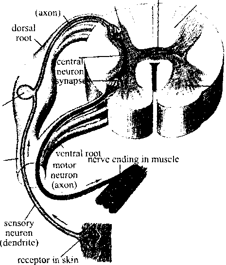
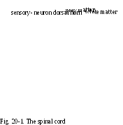
The spinal cord consists of gray matter (nerve cell bodies) and a larger area surrounding this gray part that consists of white matter (nerve fibers). The structure of the spinal cord is shown in Fig. 20-1.
You can see that the gray matter has the form of the letter H or a butterfly (метелика). The functions of the spinal cord may be divided into three categories:
reflex activities;
conduction of sensory impulses (to the brain);
conduction of motor impulses (from the brain).
All the nervous activities are taking place through the reflex arcs. The reflex arc consists of five components: the receptor, the afferent pathway, the nervous center, the efferent pathway and the effector.
The diseases of the spinal cord are: poliomyelitis (an infectious disease, which commonly occurs in children; polio virus enters the body through the nose and throat; the virus destroys
dorsal root /. gangliorTy^
ventral - horn
cell-body

the motor nerve cells in the spinal cord; the preventive medicine is the oral Sabin vaccine), paraplegia (the loss of sensation and motion in the lower part of the body), tumors that grow from within the cord or that compress the cord from outside.
spinal nerve

Text 2. Spinal Nerves
Look at Fig. 19-1. There are
31 Pairs of spinal nerves, each pair is numbered according to the level of the spinal cord from which it arises. Each spinal nerve has small posterior divisions and rather large anterior divisions.
The larger anterior branches
form networks called plexuses, which innervate (іннервують) different parts of the body.
The three main plexuses are described as follows:
The cervical plexus supplies motor impulses to the muscles of the neck and receives sensory impulses from the neck and the back of the head. The phrenic nerve, which activates the diaphragm, arises from this plexus.
The brachial ['braskial] plexus sends numerous branches to the shoulder, arm, forearm, wrist and hand. The radial nerve emerges from the brachial plexus.
The lumbosacral [^Amba'seikral] plexus supplies nerves to the lower extremities. The largest of these branches is the sciatic [sai'aetik] nerve, which leaves the dorsal part of the pelvis, passes the glutaeus maximus muscle and then to the thigh, and lower leg, and foot.
The disorders of the spinal nerves are:
- neuritis (inflammation of a nerve);
sciatica [sai'astkka] (kind of neuritis characterized by severe pain along the sciatic nerve);
herpes zoster (known as shingles) is characterized by numerous blisters along the course of certain nerves (the cause is the chickenpox virus).
Text 3. Autonomic Nervous System
Look at Fig. 20-2. The autonomic nervous system is the motor (efferent) division of the visceral (involuntary) nervous system. It has many ganglia that serve as relay stations. In these ganglia each message is transferred at a synapse from the first neuron to the second one from there to the muscle or gland cell. This differs from the voluntary (somatic) nervous system, in which each motor nerve fiber extends all the way from the spinal cord to the skeletal muscle with no intervening synapse.
The autonomic nervous system has sympathetic pathways and parasympathetic pathways.
The sympathetic pathways begin in the spinal cord with cell bodies in the thoracolumbar area and then their nerve fibers extend to ganglia where they form a synapse with a second set of neurons, whose fibers extend to the glands and involuntary muscle tissues. These second neurons act on the effectors by releasing the neurotransmitter epinephrine (adrenaline), so the sympathetic system can also be called adrenergic [aedri пз:дзік].
The parasympathetic pathways begin in the craniosacral areas, with fibers arising from cell bodies of midbrain, medulla, and sacral part of the spinal cord, and stimulate the visceral tissues. The neurons of this part of the nervous system release the neurotransmitter acetylcholine, so it is called cholinergic [кт)1і'пз:дзік].
The autonomic nervous system regulates the action of the glands, the smooth muscles of hollow organs, and the heart. The sympathetic part tends to act as an accelerator for the organs needed to meet a stressful situation.
The parasympathetic part normally acts as a balance for the sympathetic system once a crisis has passed.
The autonomic nervous system, together with the endocrine system, regulates our responses to stress.
Exercise 3. Write out all terms of Greek origin form Texts 1, 2, 3.
Exercise 4. Define the following terms.
Plexus, ganglia, autonomic nervous system, somatic nervous system, neuritis, neuron, herpes zoster, synapse, nervous system.
Exercise 5. Differentiate between the sympathetic and parasympathetic nervous systems.
