

3 курс / Фармакология / Essential_Psychopharmacology_2nd_edition
.pdfFIGURE 10—12. The nigrostriatal dopamine pathway is part of the extrapyramidal nervous system and plays a key role in regulating movements. When dopamine is deficient, it can cause parkinsonism with tremor, rigidity, and akinesia/bradykinesia. When DA is in excess, it can cause hyperkinetic movements such as tics and dyskinesias.
FIGURE 10 — 13. The tuberoinfundibular dopamine pathway from hypothalamus to anterior pituitary regulates prolactin secretion into the circulation. Dopamine inhibits prolactin secretion.
379
380Essential Psychopharmacology
and possibly other problems, such as sexual dysfunction. Such problems can occur after treatment with many antipsychotic drugs that block dopamine 2 receptors, as will be discussed further in Chapter 11.
Neurodevelopmental Hypotheses of Schizophrenia
One leading hypothesis for the etiology of schizophrenia is that this illness originates from abnormalities in fetal brain development during the early stages of neuronal selection (Fig. 4—6) and migration (Fig. 4—7). Although the symptoms of schizophrenia are usually not evident until the late teens to the twenties it may be that "the die is cast" much earlier. That is, an abnormal degenerative process may be "turned on" genetically very early in fetal brain development. However, symptoms do not occur until the brain extensively revises its synapses in adolescence, and it is hypothetically this normal restructuring process that unmasks the problems of neuronal selection and migration that were previously hidden. Although one idea is that the degenerative process may only do this type of fetal "hit and run" damage, it is also possible that the degenerative process continues during the symptomatic phase of schizophrenia, as discussed below in relation to the neurodegenerative hypothesis and combined neurodevelopmental/neurodegenerative hypothesis of schizophrenia.
Other support for the possibility that schizophrenia could have a neurodevelopmental basis includes observations that schizophrenia is increased in those with a fetal history of obstetric complications ranging from viral infections to starvation to autoimmune processes and other such problems in the pregnant mother. These observations suggest that an insult to the brain early in fetal development could contribute to the cause of schizophrenia. These risk factors may all have the final common pathway of reducing nerve growth factors (see Figs. 1—22 and 5 — 64), and also stimulating certain noxious processes that kill off critical neurons, such as cytokines, viral infection, hypoxia, trauma, starvation, or stress. This may be mediated either by apoptosis or by necrosis (Fig. 1 — 18). The result (reviewed in Fig. 10—14) could be either overt structural abnormalities or more subtle problems, including selection of the wrong neurons to survive in the fetal brain (Fig. 4—6), neuron migration to the wrong places (Fig. 4—7), neuron innervation of the wrong targets (Figs. 4—8 and 4—9), and mixup of the nurturing signals so that what innervates these neurons is also mixed up (Figs. 1 — 19, 1—20, and 1—21). Problems with proteins involved in the structural matrix of synapses (such as synapsins) may occur in schizophrenia, leading to reduced numbers of synaptic vesicles, aberrant synapse formation, and delays or reduction in synapse formation.
If schizophrenia is caused by abnormal early brain development (cf. Figs. 10—15 and 10—16), it may be virtually impossible to reverse such abnormalities in adulthood. On the other hand, some day it may be possible to compensate for such postulated neurodevelopmental difficulties by other mechanisms or to interrupt an ongoing mechanism still present in the symptomatic patient. Therefore, it will be critical to learn what neurodevelopmental abnormalities may exist in schizophrenia in order to devise strategies for reducing their potential impact. It may even be possible to identify such abnormalities in presymptomatic individuals or to exploit the plasticity of adult neurons to compensate for neurodevelopmentally endowed dysfunction. These are bold and unsubstantiated theoretical extrapolations based on

Psychosis and Schizophrenia |
381 |
FIGURE 10 — 14. Neurodevelopmental abnormalities in schizophrenia may include toxic or genetic insults to neurons, either killing them or rendering their functioning inadequate; poor neuronal migration during fetal brain development; inadequate and improper selection of synaptic targets during synaptogenesis, especially before the age of 6; and/or inadequate innervation received from inputs of other neurons.
the most optimistic therapeutic visions; current molecular and neurodevelopmental approaches have not yet evolved into successful therapeutic strategies.
Strong evidence for a genetic basis of schizophrenia comes from twin studies, as already discussed in Chapter 4. Scientists have been trying for a long time to identify abnormal genes in schizophrenia (Fig. 10—17) and the consequences that such abnormal genes could have on molecular regulation of neuronal functioning in schizophrenic patients (Fig. 10 — 18). It is already clear that the causes of psychotic illnesses
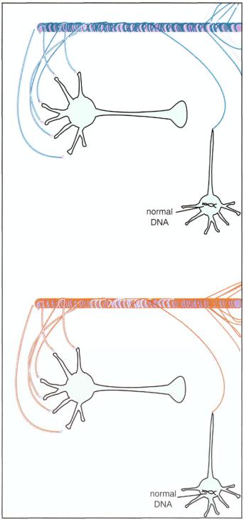
FIGURE 10—15. Neurodevelopmental theories of schizophrenia suggest that something goes wrong with the genetic program for the normal formation of synapses and migration of neurons in the brain during the prenatal and early childhood formation of the brain and its connections. Depicted here is a concept of how a neuron with normal genetic programming would develop and form synaptic connections.
382

FIGURE 10 — 16. According to neurodevelopmental theories of schizophrenia, an abnormality in the DNA of a schizophrenic patient may cause the wrong synaptic connections to be made during the prenatal and early childhood formation of the brain and its connections. Schizophrenia may be the result of abnormal development of the brain from the beginning of life either because the wrong neurons are selected to survive into adulthood or because those neurons that do survive fail to migrate to the correct parts of the brain, fail to form appropriate connections, and then are subject to breakdown when put to use by the individual in late adolescence and adulthood.

384 Essential Psychopharmacology
FIGURE 10—17. This figure shows one of the several postulated abnormal genes in schizophrenia that may contribute to the risk of this illness. Here it is lying dormant in the cell. In this case, it does not produce abnormal gene products or cause schizophrenia. Thus, it is not contributing to the risk of illness.
FIGURE 10-18. Here, the postulated abnormal gene for schizophrenia is being expressed, leading to an abnormal gene product that contributes to the risk of schizophrenia because it causes disruption in the functioning of the neuron. The manner of the disruption is additive with other risks from other genes and other environmental factors, with just the right timing and in just the right sequence; this, in turn, leads to psychosis and the other symptoms of schizophrenia.
such as schizophrenia and bipolar disorder are not going to be single abnormalities in a major genetic locus of DNA, like those already proven for diseases such as Huntington's disease. Rather, multiple genetic abnormalities are likely to each contribute in complex ways to a vulnerability to schizophrenia and other psychotic illnesses, perhaps only when other critical environmental inputs are also present. Thus, the genetic basis of schizophrenia is not likely to be as simple as depicted in Figures 10—17 and 10—18; rather, a whole list of abnormally acting genes and their corresponding gene products, triggered from both inherited and acquired risk factors, are hypothesized to act together or in just the right sequence to cause the evolution of the symptom clusters known as schizophrenia. It will be important to

Psychosis and Schizophrenia |
385 |
FIGURE 10—19. A highly theoretical direct genetic approach to therapeutics in schizophrenia is based on the notion that if dormant risk factors could be identified in the genome, perhaps drugs could prevent the expression of such genes and thus prevent the triggering of the disease process leading to schizophrenia.
determine just how these gene products participate in mediating the symptoms of schizophrenia, because only then could a logical biochemical rationale be found for preventing or interrupting these abnormalities by interfering with gene transcription, for example (Fig. 10—19), by blocking the action of unwanted gene products, or by substituting for the action of missing gene products. This is not likely to be simple, as multiple simultaneous drugs acting to compensate for each genetic abnormality might prove to be necessary, and treatments based on this approach do not appear to be imminent.
Neurodegenerative Hypotheses of Schizophrenia
The presence of both functional and structural abnormalities demonstrated in neuroimaging studies of the brain of schizophrenics suggests that a neurodegenerative process with progressive loss of neuronal function may be ongoing during the course of the disease. A neurodegenerative condition is also suggested by the progressive nature of the course of illness in schizophrenia (Fig. 10—20). Such a course of illness is not consistent with simply being the result of a static and previously completed pathological process.
Schizophrenia progresses from a largely asymptomatic stage prior to the teen years (phase I in Fig. 10—20), to a prodromal stage of "oddness" and the onset of subtle negative symptoms in the late teens to early twenties (phase II in Fig. 10—20). The active phase of the illness begins and continues throughout the twenties and thirties with destructive positive symptoms, characterized by an up-and-down course with treatment and relapse, with the patient never quite returning to the same level of functioning following acute relapses or exacerbations (phase III in Fig. 10—20). Finally, the disease can reach a largely stable level of poor social functioning and prominent negative and cognitive symptoms, with some ups and downs but at a considerable step-off from baseline functioning, suggesting a more static phase of illness sometimes called "burnout" in the forties or later in life (phase IV in Fig. 1020).

386
Essential Psychopharmacology
FIGURE 10—20. The stages of schizophrenia are shown here over a lifetime. The patient has full functioning (100%) early in life and is virtually asymptomatic (stage I). However, during a prodromal phase (stage II) starting in the teens, there may be odd behaviors and subtle negative symptoms. The acute phase of the illness usually announces itself fairly dramatically in the twenties (stage III), with positive symptoms, remissions, and relapses but never a complete return to previous levels of functioning. This is often a chaotic stage of the illness, with a progressive downhill course. The final phase of the illness (stage IV) may begin in the forties or later, with prominent negative and cognitive symptoms and some waxing and waning during its course, but often more of a burnout stage of continuing disability. There may not necessarily be a continuing and relentless downhill course, but the patient may become progressively resistant to treatment with antipsychotic medications during this stage.
The fact that a schizophrenic patient's responsiveness to antipsychotic treatment can change (and lessen) over the course of illness also suggests an ongoing neurodegenerative process of some kind. For example, the time it takes for a schizophrenic patient to go into remission increases in each successive psychotic relapse. A patient may be less responsive to antipsychotic treatment during successive episodes or exacerbations, so that residual symptoms remain as well as decrements in the patient's functional capacities. This development of treatment resistance during successive episodes of the illness suggests that "psychosis is hazardous to the brain." It thus seems possible that patients who receive early and effective continuous treatment may prevent disease progression or at least the development of treatment resistance.
Excitotoxicity
One major idea proposed to explain the downhill course of schizophrenia and the development of treatment resistance is that neurodegenerative events in schizophrenia may be mediated by a type of excessive action of the neurotransmitter glutamate that has come to be known as excitotoxicity. The excitotoxic hypothesis of schizophrenia proposes that neurons degenerate because of excessive excitatory neurotrans-mission at glutamate neurons. This process of excitotoxicity, already discussed in Chapter 4, not only is a hypothesis to explain neurodegeneration in schizophrenia but also has been invoked as an explanation for neurodegeneration in any number of neurological and psychiatric conditions, including Alzheimer's disease and other
Psychosis and Schizophrenia |
387 |
degenerative dementias, Parkinson's disease, amytrophic lateral sclerosis (Lou Gehrig's disease), and even stroke.
In order to understand the hypothesis of excessive excitation of neurons by glutamate, it is necessary to understand glutamatergic neurotransmission.
Glutamatergic Neurotransmission
Glutamate synthesis. The amino acid glutamate or glutamic acid is a neurotransmitter, but its predominant use is as an amino acid building block for protein biosynthesis. When used as a neurotransmitter, it is synthesized from glutamine (Fig. 10—21), which is converted to glutamate by an enzyme in mitochondria called glutaminase. It is then stored in synaptic vesicles for subsequent release during neurotransmission. Glutamine itself can be obtained from glial cells adjacent to neurons. The glial cells help to support neurons both structurally and metabolically. In the case of glutamate neurons, nearby glia can provide glutamine for neurotransmitter glutamate synthesis. In this case, glutamate from metabolic pools in the glia is converted into glutamate for use as a neurotransmitter. This is accomplished by first converting glutamate into glutamine in the glial cell via the enzyme glutamine synthetase. Glutamine is then transported into the neuron for conversion into glutamate for use as a neurotransmitter (Fig. 10—21).
Glutamate removal. Glutamate's actions are stopped not by enzymatic breakdown, as in other neurotransmitter systems, but by removal by two transport pumps. The first of these pumps is a presynaptic glutamate transporter, which works as do all the other neurotransmitter transporters already discussed for monoamine neurotransmitter systems such as dopamine, norepinephrine, and serotonin. The second transport pump, located on nearby glia, removes glutamate from the synapse and terminates its actions there. Glutamate removal is summarized in Figure 10—22.
Glutamate receptors. There are several types of glutamate receptors (Fig. 10—23), including N-methyl-d-asparate (NMDA), alpha-amino-3-hydroxy-5-methyl-4-isoxa- zole-propionic acid (AMPA), and kainate, all named after the agonists that selectively bind to them. Another type of glutamate receptor is the metabotropic glutamate receptor, which may mediate long-lasting electrical signals in the brain by a process called long-term potentiation which appears to have a key role in memory functions.
The NMDA, AMPA, and kainate subtypes of glutamate receptors are probably all linked to an ion channel. The metabotropic glutamate receptor subtype, however, belongs to the G protein —linked superfamily of receptors. The specific functioning of the various subtypes of glutamate receptors is the focus of intense debate. The actions at NMDA receptors will be emphasized here in our discussions on excitotoxicity.
Just as does the GABA-benzodiazepine receptor complex discussed in Chapter 8 (see Figs. 8 — 18 to 8-20), the NMDA glutamate—calcium channel complex also has multiple receptors surrounding the ion channel, which act in concert as allosteric modulators (Fig. 10—24). One modulatory site is for the neurotransmitter glycine; another is for polyamines, and yet another is for zinc (Fig. 10 — 24). The magnesium ion can block the calcium channel at yet another modulatory site, which is presumably inside the ion channel or closely related to it. Another inhibitory modulatory site,

388 Essential Psychopharmacology
FIGURE 10—21. Glutamate is produced (synthesized). Glutamate or glutamic acid (glu) is a neurotransmitter that is an amino acid. Its predominant use is not as a neurotransmitter but as an amino acid building block of protein synthesis. When used as a neurotransmitter, it is synthesized from glutamine. Glutamine is turned into glutamate by an enzyme present in mitochondria called glutaminase. It is then stored in synaptic vesicles for subsequent release during neurotransmission. Glutamine itself can be obtained from glial cells adjacent to neurons. Glial cells have a supportive role to neurons, helping to support them both structurally and metabolically. In the case of glutamate neurons, nearby glia can provide glutamine for neurotransmitter glutamate synthesis. In this case, glutamate from metabolic pools in the glia is converted into glutamate for use as a neurotransmitter. This is accomplished by first converting glutamate into glutamine in the glial cell via the enzyme glutamine synthetase. Glutamine is then transported into the neuron for conversion into glutamate for use as a neurotransmitter.
located inside the ion channel, is sometimes called the PCP site since the psychotomimic agent phencylclidine (PCP) binds to this site (Fig. 10 — 24). Since PCP induces a psychotic state with some similarities to schizophrenia (see Chapter 13 on drug abuse), it is possible that such psychotic symptoms in schizophrenia may be modulated by dysfunction in the NMDA subtype of glutamate receptor.
Antagonists for any of the various modulatory sites around the NMDA—calcium channel complex would possibly restrict the flow of calcium and close the channel and therefore be candidates for neuroprotective agents. Such antagonists are being