

5 курс / Пульмонология и фтизиатрия / Clinical_Tuberculosis_Friedman_Lloyd_N_,_Dedicoat
.pdf350 Tuberculosis in Childhood and Pregnancy
organisms. Lesions are often larger and more numerous in the lungs, spleen, liver, and bone marrow than other tissues.65 The distribution may correlate with both the blood supply and the number of reticuloendothelial cells and tissue phagocytes.
The onset of miliary tuberculosis can be explosive when the patient becomes gravely ill in several days.66 More often, the onset is insidious and the patient may not be able to accurately pinpoint the time of initial symptoms. Early systemic signs include malaise, anorexia, weight loss, and low-grade fever. At this time, abnormal physical signs are usually absent. Within weeks, generalized lymphadenopathy and hepatosplenomegaly develop in approximately 50% of cases. The fever can become higher and more sustained although the chest radiograph at this stage is usually normal and respiratory symptoms are few. Within several more weeks, the lungs can become filled with tubercles, accompanied by dyspnea, cough, rales, or wheezing. As the pulmonary disease progresses, an alveolar-air block syndrome can result in frank respiratory distress, hypoxia, and pneumothorax, or pneumomediastinum. Signs or symptoms of meningitis or peritonitis are found in 20%–40% of patients with advanced disease.65,66 Chronic or recurrent headache in a child with miliary tuberculosis usually indicates the presence of central nervous system (CNS) involvement, whereas the onset of abdominal pain or tenderness is a sign of tuberculous peritonitis. Cutaneous lesions include papulonecrotic tuberculids, nodules, or purpura; which often occur in crops. Choroid tubercles occur in 13%–87% of patients and are highly specific for the diagnosis of miliary tuberculosis.
The early diagnosis of disseminated tuberculosis can be difficult, requiring a high index of suspicion by the clinician. Often the patient presents with fever of unknown origin. Mycobacterial blood cultures are rarely, if ever, positive. Early sputum or gastric aspirate cultures have a low sensitivity. Biopsy of the liver or bone marrow are the best methods to attempt to establish an early diagnosis.
With proper treatment, the prognosis of miliary tuberculosis in children is excellent. However, resolution of signs and symptoms may be slow with fever declining in 2–3 weeks of starting chemotherapy, and chest radiographic abnormalities persisting for several months.
Lymphatic
Tuberculosis of the superficial lymph nodes, historically referred to as scrofula, is the most common form of extrapulmonary tuberculosis in children, accounting for approximately 67% of cases.63,67 Historically, scrofula was usually caused by drinking unpasteurized cow’s milk laden with Mycobacterium bovis. However, through effective veterinary control, M. bovis has been nearly eliminated from North America. Most current cases of tuberculous lymphadenitis occur within 6–9 months of the initial infection, although some cases arise years later. The tonsillar, anterior cervical, submandibular, and supraclavicular nodes become involved secondary to extension of a primary lesion of the upper lung fields or abdomen. Infected lymph nodes in the inguinal, epitrochlear, or axillary regions result from regional adenitis associated with tuberculosis of the skin or skeletal system.
In the early stages of infection, the lymph nodes enlarge gradually. The nodes are discrete, nontender, and firm but not hard. The
nodes often feel fixed to underlying or overlying tissue. Disease is most often unilateral, but because of the crossover drainage patterns of lymphatic vessels in the chest and lower neck, can present bilaterally. As infection progresses, multiple nodes can become infected, resulting in a mass of matted nodes. Other than lowgrade fever, systemic signs and symptoms are usually absent. The TST or IGRA is often positive but the chest radiograph is normal in up to 70% of cases. The onset of illness occasionally is more acute, with rapid enlargement of lymph nodes associated with high fever, tenderness, and fluctuance. Rarely children will present with a tender fluctuant mass with overlying cellulitis or skin discoloration.
If left untreated, lymph node tuberculosis can resolve spontaneously, but more often progresses to caseation and lymph node necrosis.68 The capsule of the node breaks down, resulting in the spread of infection to adjacent nodes. The skin overlying the mass of nodes becomes thin, shiny, and erythematous. Rupture through the skin results in a draining sinus tract that may require surgical removal.
The most difficult diagnostic dilemma in the differential diagnosis of tuberculous adenitis is distinguishing this condition from lymphadenitis caused by nontuberculous mycobacteria. Both conditions can cause chronic, nontender adenopathy with overlying skin changes, and the eventual creation of tissue breakdown and sinus tracts.7 The chest radiograph is usually normal in both conditions and the TST reaction may be positive with either infection. The most important diagnostic clue for the diagnosis of tuberculous adenitis in a child is finding an adult source case in the child’s environment. Fine needle aspiration or excisional biopsy and culture of the lymph nodes are often required to definitively establish the etiology. However, the cultures can be negative in up to 50% of reported cases of both tuberculous and nontuberculous mycobacterial lymphadenitis.
Central nervous system
Meningitis
CNS tuberculosis is the most serious complication in children and is universally fatal without effective treatment. It usually arises from the formation of a metastatic caseous lesion in the cerebral cortex or meninges that develops during the lymphohematogenous dissemination of the primary infection.69 This lesion, often called a Rich focus, increases in size and discharges small numbers of tubercle bacilli into the subarachnoid space. The resulting gelatinous exudate can infiltrate the cortical or meningeal blood vessels, producing inflammation, obstruction, and subsequent infarction of the cerebral cortex. The brainstem is the area most commonly affected, accounting for the frequent involvement of cranial nerves III, VI, and VII. The exudate interferes with the normal flow of cerebrospinal fluid (CSF) in and out of the ventricular system at the level of the basilar cisterns, leading to a communicating hydrocephalus. This combination of vasculitis, infarction, cerebral edema, and hydrocephalus results in severe damage that can occur gradually or rapidly with this disease. Profound abnormalities in electrolyte metabolism, especially hyponatremia secondary to inappropriate secretion of antidiuretic hormone or salt wasting, are common and may contribute to the pathophysiology.
Книга в списке рекомендаций к покупке и прочтению сайта https://meduniver.com/

Tuberculosis in children 351
Salt wasting may make correction of the electrolyte disturbances difficult.70
Tuberculous meningitis complicates approximately 0.5% of untreated primary infections in children of all ages, but up to 10% of children less than 1 year of age. It is most common in children between 6 months and 4 years of age.71,72 It is extremely rare in infants less than 4 months of age because, in general, it takes that long for the pathologic events to take place. Since it is an early manifestation of the primary infection, the initial exposure history is negative but the adult source case can be identified soon after the diagnosis of tuberculosis meningitis in the child is suspected.73
The clinical progression of tuberculous meningitis may be rapid or gradual. Rapid progression tends to occur more often in infants and young children, who can experience symptoms for only several days before the onset of acute hydrocephalus, seizure activity, and cerebral edema.74 More often, the signs and symptoms progress slowly over weeks and are divided into three clinical stages. The first stage typically lasts 1–2 weeks and is characterized by nonspecific symptoms such as fever, headache, irritability, drowsiness, and malaise. Focal neurologic signs are absent, but infants can experience a stagnation or loss of developmental milestones. The second stage usually begins more abruptly with lethargy, nuchal rigidity, seizures, positive Kernig and Brudzinski signs, hypertonia, vomiting, cranial nerve palsies, and other focal neurologic signs. The accelerating clinical illness usually correlates with the development of hydrocephalus with subsequent increased intracranial pressure and vasculitis. Some children do not have evidence of meningeal irritation but have signs of encephalitis such as disorientation, abnormal movement, or speech impairment.75 The third stage is marked by coma, hemiplegia or paraplegia, hypertension, decerebrate posturing, deterioration of vital signs, and eventually death. The prognosis of tuberculous meningitis correlates most closely with the clinical stage of illness at the time treatment is initiated. The majority of patients in the first stage have an excellent outcome, whereas most patients in the third stage who survive have permanent disabilities which include blindness, deafness, paraplegia, diabetes insipidus, or mental retardation. It is imperative that antituberculosis treatment be considered strongly for any child with meningitis and no other established etiology who develops basilar meningitis and hydrocephalus, cranial nerve palsy, or stroke. The key to the diagnosis of tuberculous meningitis in children is often identifying the adult source case.
Confirming a diagnosis of tuberculous meningitis can be extremely difficult. The TST and IGRAs are negative in up to 50% of cases, and 20%–50% of children have a normal chest radiograph.76 The most important laboratory test for the diagnosis of tuberculous meningitis is examination and culture of the lumbar cerebrospinal fluid (CSF). The CSF leukocyte count usually ranges from 10 to 500 cells/µL. Polymorphonuclear leukocytes may be present initially and may portend a poorer prognosis, but a lymphocyte predominance is more typical. The CSF glucose is usually between 20 and 40 mg/dL, whereas the CSF protein level is elevated and may be markedly high (400–5,000 mg/dL). The success of the microscopic examination of stained CSF and mycobacterial cultures correlates with the volume of the CSF sample. When a minimum of 10 mL of lumbar CSF is available, the acid-fast stain
of the CSF sediment is positive in up to 30% of cases and the culture is positive in up to 70% of cases. Unfortunately, a volume of 1–2 mL is usually all that can be obtained from a young child. Polymerase chain reaction (PCR) testing of the CSF can improve diagnosis. Cultures of other body fluids can help confirm the diagnosis.
Computed tomography or magnetic resonance scans often help establish the diagnosis of tuberculous meningitis and can aid in evaluating the success of therapy. The classic imaging finding of tuberculous meningitis is abnormal enhancement in the posterior fossa (basal enhancement), which may involve the meninges or cisterns.77 Both computed tomography and magnetic resonance imaging are capable of demonstrating hydrocephalus, the most common complication of tuberculous meningitis. Computed tomography is capable of identifying abnormal enhancement in the basal cisterns but cannot distinguish between vessels and cistern enhancement as with magnetic resonance imaging. Contrasted magnetic resonance imaging is more sensitive for the identification of miliary foci/nodules in the leptomeninges and inflammatory pan-arteritis (and associated cerebral infarction) which are rarely identified on computed tomography imaging.78–80 Any child with a triad of imaging findings including basal enhancement, hydrocephalus, and cerebral infarction has tuberculous meningitis until proven otherwise.
Tuberculoma
Another manifestation of CNS tuberculosis is the tuberculoma which usually presents clinically as a brain tumor. Tuberculomas account for up to 30% of brain tumors in some areas of the world. They occur most often in children less than 10 years of age and most often present as a solitary lesion. While tuberculoma lesions in adults are most often supratentorial, in children, they are often infratentorial, located at the base of the brain near the cerebellum (Figure 18.4). The most common
Figure 18.4 Magnetic resonance imaging: infratentorial tuberculosis.
352 Tuberculosis in Childhood and Pregnancy
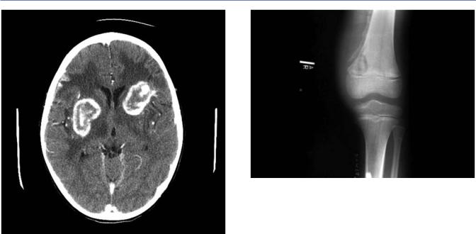
Figure 18.5 Computed tomography: tuberculoma.
symptoms in children include headache, fever, focal neurologic findings, and seizures. The TST or IGRA is usually positive, but the chest radiograph is often normal. Advanced imaging of the brain reveals tuberculomas as discrete lesions with a significant amount of surrounding edema. Contrast enhancement is often impressive and can reveal a ring-like lesion (Figure 18.5). Surgical excision of tuberculomas may be necessary to distinguish tuberculoma from other causes of brain tumor or other ring enhancing lesions. However, if the clinical suspicion for tuberculoma is high, surgical removal is not necessary as most tuberculomas resolve with medical management alone. These lesions can persist for months or years.
Since the advent of contrast-enhanced brain imaging, the paradoxical development of tuberculomas has been recognized in children with and without HIV infection who have an initial diagnosis of tuberculous meningitis while they are receiving effective chemotherapy.81,82 The cause and nature of these tuberculomas are poorly understood but they likely represent a form of immune reconstitution inflammatory syndrome (IRIS). Their development is not thought to be a failure of drug treatment and does not necessitate a change in therapeutic regimen. This phenomenon should be considered whenever a child with tuberculous meningitis deteriorates or develops focal neurologic findings while on treatment. Corticosteroids can alleviate the occasionally severe clinical signs and symptoms that occur. Thalidomide, a potent TNF-α inhibitor, is now also being used by experts in the treatment of refractory tuberculomas.83
Skeletal tuberculosis
Skeletal tuberculosis usually results from lymphohematogenous seeding of tubercle bacilli at the time of a primary infection. Bone infection may also originate by direct extension from a caseous regional lymph node or by extension from a neighboring infected tissue. The time interval between infection and disease can be as
Figure 18.6 Tuberculous lesion in metaphysis of femur.
short as 1 month in cases of tuberculous dactylitis or years for tuberculosis of the hip. Joints of weight bearing bones are most commonly affected. The infection usually begins in the metaphysis (Figure 18.6). Granulation tissue and caseation develop, which can destroy bone both by direct infection and pressure necrosis. Soft tissue abscess and extension of the infection through the epiphysis into the nearby joint often complicate the bone lesion. The infection frequently becomes clinically apparent when the joint involvement progresses.
Most cases of bone tuberculosis occur in the vertebrae causing tuberculosis of the spine, known as Pott’s disease.84 Although any vertebral body can be involved, there is a predilection for the lower thoracic and upper lumbar vertebrae. Involvement of two or more vertebrae is common; they usually are continuous but there may be skip areas between lesions. The infection is in the body of the vertebra leading to bony destruction and collapse. The usual progression of tuberculous spondylitis is from initial narrowing of one or several disc spaces to collapse and wedging of the vertebral body with subsequent angulation of the spine (gibbus) or kyphosis. The infection may extend out from the bone causing paraspinal (Pott’s), psoas, or retropharyngeal abscess. The most frequent clinical signs and symptoms of tuberculous spondylitis in children are low-grade fever, irritability and restlessness (especially at night), back pain usually without significant tenderness, and abnormal positioning and gait or refusal to walk. Rigidity of the spine may be caused by profound muscle spasm resulting from the child’s involuntary effort to immobilize the spine.
Other sites of skeletal tuberculosis, in approximate order of frequency, are the knee, elbow, and ankle.85 The degree of involvement ranges from joint effusion without bone destruction to frank destruction of bone and restriction of the joint caused by chronic fibrosis of the synovial membrane. The process usually evolves over months to years, most commonly causing mild pain, stiffness, limping, and restricted movement. The TST or IGRA is positive in 80%–90% of cases. In most cases, culture of the joint fluid or bone biopsy yields the organism. Tuberculosis should be considered in any child with risk factors and a persistent bone or joint lesion.
Книга в списке рекомендаций к покупке и прочтению сайта https://meduniver.com/

Tuberculosis in children 353
One form of bony tuberculosis peculiar to infants is tuberculous dactylitis. Affected children develop distal endarteritis followed by painless swelling and cystic bone lesions. Abscesses are rare.
Abdominal tuberculosis
Abdominal tuberculosis can result from ingestion of mycobacterium, hematogenous dissemination or lymphatic spread from a pulmonary focus. Abdominal tuberculosis is one of the most common sites for extrapulmonary infection; and it may be underdiagnosed in children. A retrospective study from a high-tuberculosis burden country found that among 47 children with culture positive pulmonary tuberculosis (20 of whom were HIV infected), 23% of children had ultrasound findings consistent with abdominal infection.86 The most common manifestation was lymphadenopathy followed by hepatosplenomegaly, hepatic and splenic lesions, and ascites. The presence of abdominal adenopathy correlated with thoracic adenopathy. Children with gastrointestinal tuberculosis can present with intestinal inflammation with or without peritonitis.87 A primary intestinal infection rarely will occur after ingestion of a bacilli laden sputum, which is more common in adolescents with cavitary tuberculosis. M. bovis also causes a primary intestinal infection following the ingestion of contaminated milk products.
The most common manifestations of abdominal tuberculosis include abdominal pain, abdominal distension, malnutrition, and prolonged fever.85–88 It is uncommon for a child to have a palpable abdominal mass. The TST or IGRA is likely to be negative if a child is severely malnourished. If present, ascitic fluid should be collected and analyzed for infection. The fluid is exudative with a high albumin level and lymphocytic predominance.89 Microbiologic studies, including acid-fast smear, culture, PCR, and drug-suscep- tibility testing (DST) should be obtained. Laparoscopic examination allows for direct visualization of the peritoneal space, which often reveals ascites, fibrous bands, mesenteric adhesions, nodules throughout the peritoneum, lymphadenopathy, and edematous loops of bowel. Tissue samples can be sent for histologic and bacteriologic examination. Colonoscopy may be helpful in the evaluation of intestinal tuberculosis, which most often reveals mucosal ulceration in the ileocecal region. These ulcers should be biopsied for histopathologic examination and culture. Intestinal tuberculosis remains on the differential diagnosis for inflammatory bowel disease in children.90
Female genitourinary
Tuberculosis of the female genital tract is uncommon in prepubescent girls. This condition usually originates from lymphohematogenous spread, although it can be caused by direct spread from the intestinal tract or bone.91 Adolescent girls can develop genital tract tuberculosis during a primary infection. The fallopian tubes are most often involved (90%–100% of cases), followed by the endometrium (50%), ovaries (25%), and cervix (5%). Tuberculous involvement of the fallopian tubes may lead to distension and obstruction; progression may lead to the development of a tubo-ovarian abscess. With uterine involvement, fluid accumulation will occur and may cause cervical obstruction. Extra-pelvic spread can lead to inflammation of the peritoneum,
omentum, mesentery, and bowel. The most common symptoms are lower abdominal pain, dysmenorrhea or amenorrhea, or infertility. Systemic manifestations are usually absent, and most patients have a normal chest radiograph. The TST or IGRA is usually positive. Imaging findings are nonspecific as the differential diagnosis includes sexually transmitted infections and malignant conditions. Clinicians should consider genital tuberculosis in asexual adolescent females at risk for tuberculosis who have a negative infectious or pathologic work-up for alternative diagnoses and have a positive TST or IGRA.
Diagnostic evaluation
Many children with tuberculosis in low-burden areas are discovered through contact tracing of adults with infectious pulmonary tuberculosis. Most of these children have tuberculosis infection or asymptomatic disease that would have progressed or escaped detection if the contact tracing had not occurred. In most hightuberculosis burden areas, children with pulmonary tuberculosis are discovered only after a symptomatic illness begins. A strong index of suspicion for tuberculosis is required to correctly identify the cause of illness in children since the signs and symptoms of most forms of tuberculosis are similar to those of other infections and conditions. The importance of the epidemiologic setting of the child in establishing the diagnosis of tuberculosis cannot be overemphasized.
Routine laboratory tests such as complete blood count and differential, C-reactive protein (CRP), erythrocyte sedimentation rate (ESR), urinalysis, and blood chemistries are usually normal in children with early manifestations of tuberculosis disease. Anemia, hypoalbuminemia, and abnormalities in liver serum enzyme tests may indicate more severe forms of or disseminated tuberculosis.
IMMUNE-BASED TESTING “TESTS OF TUBERCULOSIS INFECTION”
Tuberculin skin test
The principles for tuberculin skin testing of children are the same as those for adults.92 The tuberculin skin test (TST) measures a delayed-type hypersensitivity reaction to tuberculous antigens. T-lymphocytes sensitized by prior tuberculosis infection are recruited to the skin, where they release lymphokines that induce induration through local vasodilation, edema, fibrin deposition, and recruitment of other inflammatory cells to the area. The amount of induration in response to the test should be measured by a trained person 48–72 hours after administration. In some children the onset of induration is longer than 72 hours after placement; this is also a positive result. Immediate hypersensitivity reactions to tuberculin or other constituents of the preparation are short-lived (<24 hours) and not considered a positive result. Tuberculin sensitivity develops between 3 weeks and 3 months (most often in 4–8 weeks) after the inhalation of organisms. Approximately 10% of immunocompetent children with culture-documented tuberculosis do not react initially to tuberculin, although most become reactive after several months of therapy.93,94 The tuberculin reaction persists for many years, even after successful completion of chemotherapy.94

354 Tuberculosis in Childhood and Pregnancy
Young infants generally produce less induration and response to tuberculin than older children. Additional host-related factors, including malnutrition, immunosuppression by disease or drugs, viral infections (measles, mumps, varicella, or influenza), vaccination with live-virus vaccines, and overwhelming tuberculosis, can depress the TST reaction in an infected child. Corticosteroid therapy can decrease the reaction to the tuberculin proteins, but the effect is variable; in general, TST placement at the time of initiating corticosteroid therapy is reliable. False-positive reactions to tuberculin can be caused by cross-sensitization to antigens of nontuberculous mycobacteria. These cross-reactions are usually transient over months to years and produce <10–12 mm of induration, but larger areas of induration can occur. Previous vaccination with bacille Calmette-Guérin (BCG) also can cause a reaction to a TST, which is partly related to the strain of BCG used.95–97 This reaction is most prominent if a person has received two or more BCG vaccinations (at birth and then later in childhood). At least 50% of the infants who receive a BCG vaccine do not develop a positive TST and 80%–90% of those who have a positive reaction initially have a negative TST within 2–5 years. When BCGinduced TST reactivity is present, it usually causes <15 mm of induration, although larger reactions occur in some persons.
Routine testing of all children for tuberculosis infection in lowburden settings is no longer recommended; this practice has been replaced by targeted testing of children with specific risk tuberculosis factors.7,98 In children with no tuberculosis risk factors, the vast majority of “positive” TST results will be false-positive results. The appropriate size of induration indicating a positive TST result varies with related epidemiologic risk factors (Table 18.3).7 For children at the highest risk of progression to tuberculosis disease, TST sensitivity is most important whereas specificity is more important for children at low risk of progression.
Table 18.3 Definitions of positive TST results in infants, children, and adolescents7
Induration ≥5 mm
Children in close contact with a known or suspected source case with tuberculosis disease
Children suspected to have tuberculosis disease:
•Chest radiograph findings consistent with active tuberculosis disease
•Clinical signs or symptoms of tuberculosis disease
Children receiving immunosuppressive therapy (including high-dose corticosteroids or TNF-α antagonists), or immunosuppressive conditions, including HIV
Induration ≥10 mm
Children at increased risk of disseminated tuberculosis disease:
•Children younger than 4 years
•Children with a chronic medical condition, including lymphoma (including Hodgkin’s disease ), diabetes mellitus, chronic renal failure, or malnutrition
•Children born in high-prevalence regions of the world
•Children frequently exposed to adults who are HIV infected, homeless, users of illicit drugs, or incarcerated
Induration ≥15 mm
Children aged 4 years or older without any risk factors
Interferon-γ release assays
There are two interferon-γ release assays (IGRAs) available for commercial use in the United States: (T-SPOT.TB [Oxford Immunotec; Marlborough, MA] and QuantiFERON-TB [QFT] [QIAGEN; Germantown, MD]). They have clear advantages for the diagnosis of tuberculosis infection over the TST. Both IGRAs detect IFN-γ production by T-lymphocytes in response to specific tuberculosis antigens. The QFT test measures whole blood concentrations of IFN-γ to three specific antigens (ESAT-6, CFP-10, and TB7.7) while the T-SPOT.TB test measures the number of T-lymphocytes/ monocytes producing IFN-γ in response to two antigens (ESAT-6 and CFP-10). The test antigens are not present on M. bovis- BCG and most species of environmental mycobacteria (including Mycobacterium avium complex). This improves the IGRA specificity compared with the TST, leading to fewer false-positive results. Both IGRAs have internal positive and negative controls. Indeterminate (QuantiFERON-TB)/invalid (T-SPOT.TB) responses occur when the test sample is negative but the positive control has insufficient activity, or if the negative control has high background activity. Indeterminate/invalid results may be caused by technical factors (such as insufficient shaking of QFT tubes or delayed processing time).99–101 Most studies report indeterminate/invalid rates in children from 0%-10%, which is influenced by a child’s age and immune status. In children <2 years of age, indeterminate rates can be as high as 8.1%, compared to 2.7% in older children, although more recent studies generally report much lower rates.102–106 An indeterminate or invalid IGRA result is neither negative nor positive and should not guide treatment decisions. Neither IGRA test has proven to be superior to the other in children.
Similar to the TST, IGRAs cannot differentiate between tuberculosis infection and disease. Studies comparing IGRA and TST performance in children have shown comparable sensitivity (85% in culture-confirmed children) but superior IGRA specificity (95% vs. 49%) in BCG-immunized, low-risk children. Neither the TST nor the IGRAs perform well in infants and young children who are malnourished, severely immunocompromised, or have disseminated tuberculosis disease. Most experts use an IGRA in the evaluation of healthy young children down to 2 years of age who are at low risk of tuberculosis infection, especially in those who have received a BCG vaccine.7,107 Both TST and IGRA testing should be considered in children whose initial TST or IGRA result is negative for whom the risk of tuberculosis is high (to enhance the sensitivity of the combination of the two tests).
Technical advantages of the IGRAs over the TST include: the need for a single patient encounter (vs. 2 spaced in time with the TST) and the lack of cross-reaction with BCG vaccination and most environmental mycobacteria. IGRAs are also useful for those: who are unlikely to return for TST interpretation; whose family is reluctant to treat a child with tuberculosis infection based on a TST result alone; and, with a positive TST result in whom nontuberculous mycobacterial disease is suspected.104
MICROBIOLOGIC TESTING
Acid-fast stain, culture, and drug susceptibility testing
The isolation of M. tuberculosis from a clinical sample remains the gold standard for the diagnosis of tuberculosis disease. The
Книга в списке рекомендаций к покупке и прочтению сайта https://meduniver.com/

Tuberculosis in children 355
collection of a respiratory specimen from a child with suspected pulmonary tuberculosis provides samples for acid-fast bacilli staining, PCR, culture, and DST. Sputum specimens for culture should be collected from adolescents and older children who are able to expectorate spontaneously. Induced sputum with a jet nebulizer, inhaled saline, and chest percussion followed by nasopharyngeal suctioning are effective methods to obtain a respiratory specimen from children as young as 12 months of age.107,108 The most commonly obtained culture specimen for young children with suspected pulmonary tuberculosis is the early morning gastric aspirate obtained before the child has arisen and peristalsis has emptied the stomach of the pooled, swallowed overnight respiratory secretions.108 Unfortunately, even under optimal conditions, three consecutive induced sputum samples or gastric aspirates yield the organism in fewer than 50% of clinically diagnosed cases; negative cultures never exclude the diagnosis of tuberculosis in a child.109 The yield from culture obtained via flexible bronchoscopy is significantly less than that from properly obtained gastric aspirates.110 However, bronchoscopy can be useful to examine the anatomy of the bronchial tree when the diagnosis is uncertain.
Fortunately, there is often little need for culture confirmation for many children with suspected pulmonary tuberculosis, especially in low-burden settings. If the child has a positive TST or IGRA, clinical or radiographic findings suggestive of tuberculosis and known recent contact with an adult case with infectious tuberculosis, the child should be treated for tuberculosis disease. The drug-susceptibility test results from the source case’s isolate can be used to determine the best treatment regimen for the child. However, PCR, cultures, and DST should always be performed on specimens from the child under four conditions: (1) the source case is unknown, (2) the source case has a drug-resistant organism, (3) the child has rapidly progressive disease or severe disseminated disease, (4) the child has extrapulmonary tuberculosis (which has a broader differential diagnosis than is usual for pulmonary tuberculosis). Unfortunately, while acid-fast stains of gastric contents may have a specificity for tuberculosis greater than 90%, the sensitivity is usually less than 10%.
Molecular techniques
Molecular techniques to identify M. tuberculosis specific DNA and RNA genomic targets have clear advantages over the traditional microbiologic methods including: increased sensitivity compared to smear microscopy in respiratory specimens, decreased turnaround time compared to traditional culture, and more rapid drug-susceptibility information. These techniques are increasingly popular to confirm or support the diagnosis of tuberculosis in adults and children in both high-burden and low-burden set- tings.111–114 The application of molecular testing of clinical samples from children to confirm tuberculosis disease has the potential to improve estimates of childhood tuberculosis in high-burden settings.
The first form of nucleic amplification studied in children with tuberculosis was the traditional PCR technique, which identifies M. tuberculosis specific DNA sequences as markers for microorganisms in clinical specimens. Epidemiologic studies have
revealed that most strains of M. tuberculosis carry 10–15 randomly distributed repetitive DNA sequences of IS6110 restriction fragment length polymorphism (RFLP) along the chromosome. Most PCR techniques identify insertion element IS6110 as the DNA marker for M. tuberculosis complex organisms and have a sensitivity of more than 90% compared with sputum culture for detecting pulmonary tuberculosis in adults. Unfortunately, this test is expensive, requires sophisticated equipment and technical competence to avoid cross-contamination of specimens, and is, therefore, restricted to reference laboratories and clinical studies.
Compared with a clinical diagnosis of pulmonary tuberculosis in children, the sensitivity of DNA PCR has varied from 25% to 83%, and specificity has varied from 80% to 100%.111–114 The DNA PCR of gastric aspirates may be positive in a recently infected child even when the child is asymptomatic and the chest radiograph is normal, demonstrating the occasional difficulty in distinguishing between tuberculosis infection and disease in children. A negative PCR result never eliminates the diagnosis of tuberculosis, and a positive PCR result supports, but never confirms, the diagnosis.
GeneXpert MTB/RIF (Xpert) (Cepheid; Sunnyvale, CA) is a cartridge-based nucleic acid amplification test that simultaneously identifies M. tuberculosis DNA and rifampin resistance. GeneXpert is often used as a proxy for identifying multidrugresistant tuberculosis. This assay uses a self-contained cartridge system, which yields results from direct specimens in 2 hours and is less operator-dependent than traditional PCR detection methods. Multiple meta-analyses of studies involving children with a confirmed or suspected diagnosis of pulmonary tuberculosis have demonstrated improved sensitivity (62%–98%) and specificity (66%–98%) of Xpert on induced or expectorated sputa compared to smear microscopy for the diagnosis of pulmonary tubercu- losis.115–117 The sensitivity of Xpert is similar on acid-fast smear gastric aspirate samples from children with suspected pulmonary tuberculosis and smear positive-induced sputum samples (77% vs. 86%). However, the combination of the two methods on smear positive samples improves sensitivity (96% for smear and culture positive samples and 63% for smear negative-culture positive samples). Although cartridges for the Xpert system are expensive, it offers advantages in rapid detection of MDR tuberculosis and is especially useful in settings lacking more sophisticated laboratory infrastructure. In low-resource settings, Xpert may replace smear microscopy; however, it should never replace mycobacterial culture and drug-susceptibility testing.
An additional application of molecular testing is a genomewide analysis of RNA expression of M. tuberculosis genomic targets in host blood. This has potential to distinguish tuberculosis from other diseases in high-burden settings, which is especially promising in areas with a high prevalence of HIV infection. A multicenter study enrolled 346 children from three high-tuber- culosis burden countries to evaluate the performance of genomewide RNA expression.118 Among the children enrolled, 114 had culture-confirmed tuberculosis disease (32%). Fifty-one RNA transcripts were identified that distinguished tuberculosis from other diseases and 42 transcripts distinguished between tuberculosis disease and infection. In a validation cohort of Kenyan children with culture-confirmed disease, the transcript signature was 82.9% sensitive [CI: 69%–94%], and 84% specific [CI: 75%–93%].

356 Tuberculosis in Childhood and Pregnancy
An additional application of whole-genome sequencing is to identify molecular drug resistance gene targets and offers more timely information (days compared to weeks) on drug susceptibility compared with traditional phenotypic DST.119 This technology is especially useful in providing treatment recommendations for children who are contacts of adult source cases who have been treated previously for tuberculosis or who have failed to improve on their tuberculosis treatment regimen. The availability of this technology is currently limited to reference laboratories.
Treatment
The general principles that have governed the development of treatment regimens for adults with tuberculosis apply to children. Most childhood tuberculosis experts believe that if a regimen is efficacious for treating adults with tuberculosis, it will be effective for childhood tuberculosis. However, there are several special considerations for children with tuberculosis based on the natural history of the disease, the lower bacillary burden in young children, drug pharmacokinetics and dynamics, safety and tolerability, and the formulations of the available drugs. First, children usually develop tuberculosis disease as an immediate complication of a primary infection. Children typically have low numbers of bacteria enclosed in caseous lesions with relatively few mycobacteria (termed paucibacillary disease). The large bacterial populations found within cavities or infiltrates that are characteristic of adult pulmonary tuberculosis can be seen in adolescents, but are usually absent in young children. Since the likelihood of M. tuberculosis developing resistance to any antimycobacterial drug is proportional to the mycobacterial population size, in general, children are less likely than adults to develop secondary drug resistance while receiving therapy.120
A related problem concerns the natural history of primary tuberculosis in children. While asymptomatic infection and pulmonary disease are easily distinguishable events in adults, the range of microbiologic and host response events exists on a continuum in children. In asymptomatic children, with a positive TST or IGRA, who are identified at the time of a contact investigation with a known exposure to a symptomatic adult, it may be difficult for clinicians to distinguish between infection and disease. Furthermore, pediatric radiographs can be difficult to interpret and there are no standards for what constitutes hilar or mediastinal adenopathy in a child. In general, a clinician should consider a child to have tuberculosis disease if adenopathy is readily visible on chest radiograph, even if the child has no signs or symptoms of tuberculosis. When determining the best treatment regimen for a child (which in most cases is very well tolerated), in general, it is safer to overestimate rather than underestimate the extent of disease, particularly in a child known to be at high risk for recent acquisition of tuberculosis infection.
Third, young children have a higher propensity than adults to develop extrapulmonary forms of tuberculosis, especially disseminated tuberculosis and meningitis. It is important that antituberculosis drugs penetrate into a variety of tissues and fluids, especially across the blood brain barrier into the meninges. Isoniazid, rifampin, pyrazinamide, ethionamide, fluoroquinolones, and linezolid cross both inflamed an uninflamed
meninges adequately to kill virtually all strains of drug-suscep- tible M. tuberculosis.
Fourth, the pharmacokinetics and dynamics and safety and tolerability of antituberculosis drugs differ between children and adults.121 In general, children tolerate larger doses per kilogram of body weight and have fewer adverse events than adults.122,123 It is unclear whether the higher serum concentration of drugs achieved in children has any therapeutic advantage.124 The lower rates of toxicity in children usually correlate with fewer interruptions of treatment.
Finally, the most important difference between children and adults concerns the formulations, and, therefore, the administration of antituberculosis medications. Most available dosage formulations worldwide are in pill or tablet form, and were designed for use in adults. The administration of these preparations to children often involves crushing pills or constituting suspensions, which may lead to inadequate absorption of oral medications.125 More recent pharmacokinetic studies using increased doses of commonly used drugs have demonstrated higher blood levels of antituberculosis drugs than in previous studies without increased toxicity, which has guided new dosing recommendations by the WHO for the management of childhood tuberculosis.126–128 The “pill burden” may be difficult for young children at the start of therapy. If these problems are not anticipated or discussed with families, they may cause significant delays and interruptions of treatment. To overcome some of these barriers, fixed-dose multidrug combinations (FDCs) tablets have been developed recently which easily dissolve in water and have improved palatability. These FDCs have improved childhood tuberculosis disease treatment completion rates.125 In the United States, FDCs are not readily available for the treatment of childhood tuberculosis. The most effective intervention in the United States that has been shown to improve childhood tuberculosis treatment completion rates is the administration of treatment via directly observed therapy (DOT) with trained health-care workers who are educated and trained in techniques to assist parents in the administration of the treatment.129–131
ANTITUBERCULOSIS DRUGS FOR CHILDREN
Several antituberculosis drugs are used to achieve a relatively rapid cure and prevent the emergence of secondary drug resistance during therapy (Table 18.4).7 The choice of regimen depends on the extent of tuberculosis disease, the host, and the likelihood of drug resistance. Isoniazid is familiar to pediatricians, as it is effective and well tolerated by children. It is metabolized by acetylation in the liver, however, there is no correlation in children between the acetylation rate and either drug efficacy or adverse reactions.132 The major toxic effects of isoniazid in adults are very rare in children. Hepatotoxicity among children taking isoniazid is exceedingly rare.122,133 Only 3%–10% of children taking isoniazid experience a transient elevation in liver enzyme levels; clinically significant hepatitis occurs in far less than 1% of children.122 Adolescents are more likely than younger children to experience hepatotoxicity.134 For most children and adolescents, toxicity can be monitored using only clinical signs and symptoms of hepatitis; routine biochemical monitoring is not necessary. Pyridoxine levels are decreased in children taking isoniazid, but symptomatic
Книга в списке рекомендаций к покупке и прочтению сайта https://meduniver.com/

|
|
|
|
|
|
Tuberculosis in children 357 |
|
|
|||||
Table 18.4 First-line drugs for the treatment of childhood tuberculosis infection and disease |
|
|||||
|
|
|
|
Twice weekly |
|
|
|
|
|
Daily dosage |
dosage |
Weekly dosage |
|
Drug |
Dosage forms |
(mg/kg/day) |
(mg/kg/dose) |
(mg/kg/dose) |
Maximum dose |
|
Ethambutol |
Tablets |
20–25 |
50 |
|
2.5 g |
|
|
|
100 mg |
|
|
|
|
|
|
400 mg |
|
|
|
|
Isoniazid |
Scored tablets |
10–15 |
20–30 |
Age 2–11 years, ≥10 kg: 25 |
Daily, 300 mg |
|
|
|
100 mg |
|
|
Age ≥12 years: 15 |
Once weekly or twice |
|
|
300 mg |
|
|
|
weekly, 900 mg |
Pyrazinamide |
Scored tablets |
30–40 |
50 |
|
2 g |
|
|
|
500 mg |
|
|
|
|
Rifampin |
Capsules |
15–20 |
15–20a |
|
Daily, 600 mg |
|
|
|
150 mg |
|
|
|
Twice weekly, 600 mg |
|
|
300 mg |
|
|
|
|
Rifapentine |
Tablets |
|
|
Wt 10–14.0 kg: 300 mg/dose |
Weekly: 900 mg |
|
|
150 mg |
|
|
Wt 14.1–25.0 kg: 450 mg/dose |
|
|
|
|
|
|
|
Wt 25.1–32.0 kg: 600 mg/dose |
|
Wt 32.1–49.9 kg: 750 mg/dose ≥50.0 kg: 900 mg maximum
a For infants and toddlers, and children of any age with tuberculous meningitis, higher oral doses (20–30 mg/kg/day) of rifampin should be considered.
peripheral neuritis is exceedingly rare.133 However, certain children—especially adolescents with poor nutritional habits, children with diets low in meat and milk intake, HIV-infected children, and breastfeeding infants—should receive pyridoxine supplementation.7
Rifampin and rifapentine are rifamycins that are also well tolerated by children. Hepatotoxicity is infrequent and other adverse reactions that occur in adults, including leukopenia, thrombocytopenia and immunologically mediated flu-like syndrome, are rare. Clinicians should consider the risk of drug-drug interactions as rifamycins may influence the metabolism of various classes of medications (including antiretroviral medications, antiepileptic medications, and antihypertensive medications). In addition, the effectiveness of most oral contraceptives will be decreased, and alternative birth control methods should be used.
Pyrazinamide has been used extensively in children; hepatitis and complications of hyperuricemia are exceedingly rare events. Ethambutol is also safe in children and is widely used in the firstline regimen for the treatment of childhood tuberculosis. Optic toxicity is rare in children, and formal ophthalmologic evaluation of asymptomatic children is not necessary. Ethionamide is well tolerated in children and is considered a first-line therapy for tuberculous meningitis. Children may experience nausea and vomiting, but less frequently than adults. Baseline thyroid function testing followed by monitoring 6-month intervals is recommended for children receiving long-term ethionamide therapy.
Fluoroquinolones are the major class of medications used in children with known or suspected drug resistance.135 Tendon rupture, which has been observed in adults and rarely in adolescents, has not been observed in children. Fluoroquinolones are well tolerated by children and are safe.136–138 Hepatoxicity is exceedingly rare, including with long-term administration.135,138
SPECIFIC TREATMENT
Thoracic disease
The standard therapy of intrathoracic, drug-susceptible tuberculosis (non-cavitary pulmonary disease and/or hilar lymphadenopathy) in children, as recommended by the AAP and the CDC, is a minimum of 6 months which includes isoniazid and rifampin supplemented in the first 2 months by pyrazinamide and ethambutol. Several clinical trials have shown that this regimen yields a success rate approaching 100%, with an incidence of clinically significant adverse reactions of <2%. Nine-month regimens of only isoniazid and rifampin are also effective for drug-susceptible tuberculosis, but the necessary length of treatment, the need for good adherence by the patient, and the relative lack of protection against possible initial drug resistance have led to the favoring of treatment regimens with additional drugs for a short time period. Most experts recommend that children receive their antituberculosis drugs via DOT, when a health-care worker is present during medication administration (often by a parent or adult caregiver). When DOT is used, intermittent (twice or thrice weekly) administration of drugs after an initial period as short as 2 weeks of daily therapy has been shown to be as effective as daily therapy for the treatment of drug-susceptible tuberculosis in children.139–141 Children or adolescents with disseminated (miliary) tuberculosis or adult-type cavitary tuberculosis are treated for a minimum of 6 months from culture conversion, and the medications are given daily.140
Extrathoracic disease
In general, extrathoracic tuberculosis in children is caused by small numbers of mycobacteria. Therefore, the treatment of most manifestations of extrathoracic tuberculosis in children, including cervical lymphadenopathy, intra-abdominal tuberculosis and

358 Tuberculosis in Childhood and Pregnancy
tuberculosis of the genital tract, is the same as for pulmonary tuberculosis. Exceptions are bone and joint, disseminated, and tuberculous meningitis/tuberculomas, for which there are inadequate data to recommend a 6-month duration of therapy. These conditions are treated for a minimum of 9–12 months. Surgical debridement of bone and joint disease may be necessary in addition to medical therapy.142
Children with tuberculous meningitis are treated initially with four-drug therapy (isoniazid, rifampin, pyrazinamide, and ethionamide or an injectable aminoglycoside). In most cases, the pyrazinamide and fourth medication are discontinued after 2 months, and isoniazid and rifampin are continued for an additional 7–10 months.143,144 In adults, the use of linezolid or a fluoroquinolone has shown clinical benefit, especially when drug resistance is expected or if other medications are poorly tolerated.145,146 Corticosteroids are administered in conjunction with antituberculosis treatment for tuberculous meningitis, and reduce the associated morbidity and mortality.147 In children who have refractory inflammation, thalidomide has been used successfully, on a case-by-case basis.83,148 Neurosurgical ventriculoperitonel shunting is frequently required when significant hydrocephalus is present. When surgical shunting cannot be performed safely or urgently, acetazolamide with or without a diuretic medication has been used to decrease CSF production, as a medical adjunct to decrease intracranial pressure.
Tuberculous lymphadenitis responds well to antituberculosis chemotherapy alone.149 Surgical resection of involved lymph nodes usually is not necessary and may lead to the formation of cutaneous fistula tracts or severe scarring. If performed, excisional biopsy is preferred over incisional biopsy. However, surgical biopsy and mycobacterial culture may be necessary to distinguish tuberculous adenitis from other entities, most notably infection due to Bartonella henselae or nontuberculous mycobacteria.
Tuberculosis treatment in HIV-infected children
Treatment of tuberculosis in HIV-infected children is often empiric based on epidemiologic and radiographic information, because the radiographic appearance of other pulmonary complications of HIV in children, such as bacterial pneumonia, lymphoid interstitial pneumonitis, and nontuberculous mycobacterial infection, may be similar to that of tuberculosis. In HIV-infected children who do not respond to treatment for community-acquired pneumonia and tuberculosis cannot be excluded, antituberculosis therapy should be considered.
Children with suspected or confirmed tuberculosis and newly diagnosed or previously untreated HIV infection should be evaluated for starting early combined antiretroviral therapy (cART), preferably within 2–8 weeks of starting tuberculosis treatment, irrespective of their immune status.150,151 HIV-infected children who are severely immunocompromised (CD4+ count <50 cells/ mL), should be monitored closely for a paradoxical worsening of their tuberculosis disease, which supports a diagnosis of IRIS.149–152 IRIS occurs when the initial cART therapy causes a rapid decrease in the HIV load and an increase in CD4+ cell counts, resulting in an exaggerated inflammatory reaction to M. tuberculosis. IRIS manifests in two forms: “unmasking IRIS” occurs when a child
develops symptoms from an undiagnosed tuberculosis infection soon after cART is started and “paradoxical IRIS” occurs when a child who is receiving cART and antituberculosis therapy develops a worsening of tuberculosis symptoms despite previous clinical improvement and evidence of HIV control (improving CD4+ count and reduced HIV viral load).150 HIV-infected children who develop tuberculosis-related IRIS most commonly present with fever, cough, new skin lesions, enlarging cervical or thoracic lymph nodes, and new or enlarging brain tuberculomas, with or without associated meningitis.150–155 Treatment of tuberculosisrelated IRIS in HIV-infected children should occur in consultation with a physician who has expertise in the management of both diseases.
Following receipt of the M. bovis vaccine (BCG vaccine), HIVinfected infants are at increased risk of developing disseminated M. bovis-BCG disease. Studies have revealed that HIV-infected infants have more than a 100-fold increased risk of developing disseminated M. bovis disease following their receipt of the vaccine compared to HIV-uninfected children.156,157 This has led to the recommendation that the BCG vaccine should not be administered to infants born to women with HIV or to infants and children with a confirmed HIV diagnosis.150,158
HIV-infected children are usually started on standard antituberculosis regimens that include isoniazid, rifampin, pyrazinamide, and ethambutol. All treatment should be administered daily, not intermittently. Most experts believe that HIV-infected children with drug-susceptible tuberculosis should receive the standard four-drug regimen for the first 2 months followed by isoniazid and rifampin for a total duration of at least 9 months.147 A major consideration prior to choosing a cART regimen is that the coadministration of rifampin and some antiretroviral agents results in subtherapeutic blood levels of protease inhibitors and nonnucleoside reverse transcriptase inhibitors. Concomitant administration of these drugs should be avoided. Based on the child’s age, HIV drug resistance profile and previous cART exposure, an efavirenz-based regimen is preferred. If nevirapine is used, the dose must be adjusted for concomitant rifampin administration. If a protease inhibitor combination is used, a higher dose of ritonavir (termed “superboosting”) equal to that of the lopinavir dose, for the full duration of the rifampin treatment plus an additional 2 weeks is recommended. Pyridoxine supplementation (1–2 mg/kg/day, maximum dose 50 mg/day) should be provided to all HIV-infected children who are receiving isoniazid.7,147 HIV-infected children with extrapulmonary disease, including tuberculous meningitis, miliary/disseminated tuberculosis, or musculoskeletal disease, should be treated for a minimum of 12 months. For children with tuberculous meningitis, ethambutol is replaced with ethionamide or an injectable aminoglycoside due to their superior CNS penetration. The addition of corticosteroids is indicated for HIV-infected children with tuberculous meningitis, pleural effusion, airway compromise, or severe IRIS. HIV-infected children appear to have more frequent adverse reactions to antituberculosis drugs and must be monitored closely during therapy.159 Liver enzyme testing should be obtained after 2, 4, and 8 weeks of treatment for tuberculosis. After 2 months, routine testing can be spaced to every 2–3 months in asymptomatic children, or more frequently as clinically indicated.150
Книга в списке рекомендаций к покупке и прочтению сайта https://meduniver.com/

Tuberculosis in children 359
Drug-resistant tuberculosis
There are two major types of drug resistance: primary resistance and secondary resistance. Primary resistance occurs when a child is infected with M. tuberculosis that is already resistant to a particular drug. Secondary resistance occurs when drug-resistant organisms emerge as the dominant population during a treatment course. The major causes of secondary drug resistance are poor adherence to the medication by the patient or inadequate treatment regimens prescribed by the physician. Nonadherence to one drug is more likely to lead to secondary resistance than is failure to take all drugs. Secondary resistance is rare in young children because of the small size of the mycobacterial population. Consequently, most drug resistance in children is primary, and patterns of drug resistance among children tend to mirror those found among adults in the same population.160,161 The main predictors of drug-resistant tuberculosis among adults are history of previous antituberculosis treatment, coinfection with HIV, and exposure to another adult with infectious drug-resistant tuberculosis.
Treatment of drug-resistant tuberculosis is successful only when at least two bactericidal drugs to which the infecting strain of M. tuberculosis is susceptible are given.162,163 When a child has suspected drug-resistant tuberculosis, usually at least four or five drugs should be administered initially until the susceptibility pattern is determined and a more specific regimen
Table 18.5 Drugs used for treatment of MDR tuberculosis in children
Fluoroquinolones |
Levofloxacin |
|
Moxifloxacin |
|
Gatifloxacin |
Second-line injectable |
Amikacin |
agents |
Capreomycin |
|
Kanamycin |
|
Streptomycin |
Other core second-line |
Ethionamide/prothionamide |
agents |
Cycloserine/terizidone |
|
Linezolid |
|
Clofazimine |
Add-on agents |
Pyrazinamidea |
|
Ethambutola |
|
High-dose isoniazida |
|
Bedaquilineb |
|
Delamanidb |
|
p-aminosalicylic acidc |
|
Imipenem-cilastainc |
|
Meropenemc,d |
|
Amoxicillin-clavulanatec,d |
|
(Thioacetazone)c,e |
Source: World Health Organization: WHO treatment guidelines for drug-resistant tuberculosis, 2016 update. Geneva, Switzerland. http://apps.who.int/iris/bitstream/10665/250125/1/9789241549639eng.pdf
aConsidered first-line add-on agent.
bConsidered second-line add-on agent.
cConsidered third-line add-on agent.
dCarbapenems and amoxicillin-clavulanate are given together.
eConfirm patient is HIV negative prior to starting thioacetazone.
can be designed (Table 18.5).164–166 The specific treatment plan must be individualized for each patient according to the results of susceptibility testing on the isolates from the child or her adult source case. When isoniazid mono-resistance is present, a total treatment duration of 9 months with rifampin, pyrazinamide, and ethambutol is usually adequate. When resistance to both isoniazid and rifampin is present, the total duration of therapy is often extended to 12–24 months. Daily therapy should be administered to any child with drug-resistant tuberculosis. In 2016, the WHO endorsed a 9–12 month treatment regimen for adults and children with MDR-TB who were not previously treated with second-line drugs or who were unlikely to have a strain with resistance to second-line injectable agents or fluoroquinolones.163 This recommendation was based on the results of adult observational studies and extrapolated for use in children. Furthermore, as second-line treatment options for MDR-TB in children, there is increasing use of new antituberculosis medications (bedaquiline and delamanid) and repurposed drugs (linezolid and clofazimine).167,168 Delamanid is a nitroimidazole that has been studied for the treatment of MDR-TB in children since 2013. This includes two pharmacokinetic studies in HIVuninfected children down to age 6 years.168,169 Based on these studies, delamanid is endorsed for use in children ≥6 years and ≥20 kg in whom a four-drug regimen cannot be constructed due to drug resistance, the child experiences significant drug intolerance, or is at high risk of treatment failure. Further studies are ongoing to evaluate the safety and efficacy of delamanid in children with MDR-TB who are HIV-infected and those who are 3–5 years of age. Bedaquiline is considered acceptable for treatment of children ≥12 years of age and >33 kg with the same tuberculosis treatment indications specified for delamanid. Bedaquiline should not be given with efavirenz due to reduced bedaquiline levels with coadministration. For HIV-infected children, efavirenz should be replaced with nevirapine or raltegravir while they are receiving bedaquiline. A baseline EKG and monitoring of QTc is recommended in patients receiving bedaquiline and/ or delamanid. Children with a baseline QTc interval ≥500 ms should not be started on bedaquiline or delamanid until the QTc interval is corrected.169,170 In addition, a child’s baseline albumin should be ≥2.8 g/dL prior to starting delamanid. Trials evaluating the clinical efficacy of the use of combined treatment with bedaquiline and delamanid for the treatment of MDR-TB in children are lacking. Both linezolid and clofazimine are now included as core second-line agents in treatment regimens for children with MDR-TB. Both drugs require close monitoring for adverse effects and toxicity.167
The prognosis of single drug or MDR tuberculosis in children is usually good if the drug resistance is identified early in the treatment, appropriate drugs are administered under DOT, adverse reactions from the drugs are minor, and the child and family are in a supportive environment. The treatment of drug-resistant tuberculosis in children should always be undertaken by a clinician with specific expertise in the treatment of tuberculosis. Clinical trials to evaluate the efficacy, safety, pharmacokinetics, and pharmacodynamics of the administration of combinations and shorter regimens of new and repurposed antituberculosis medications for the treatment of childhood tuberculosis are needed.171