

5 курс / Пульмонология и фтизиатрия / Clinical_Tuberculosis_Friedman_Lloyd_N_,_Dedicoat
.pdf240 Pulmonary Tuberculosis
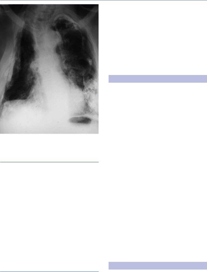
Figure 13.4 Chest x-ray of 40-year-old man showing characteristic changes of post-primary TB with soft cavitating lesions in the upper zones, particularly the left.
this will also detect those with sputum smear-negative cul- ture-positive disease. A full blood count is usually helpful by showing a normal neutrophil count and thereby excluding other bacterial infections as the cause of the chest x-ray appearance. Raised inflammatory markers (C-reactive protein, erythrocyte sedimentation rate) and a high globulin with a low albumin can often alert the attending physician to the possibility of pulmonary TB.
Fibrosis is a late feature of pulmonary TB, although the prevalence is probably overestimated due to selective reporting.49,50 Untreated, smear-positive pulmonary TB has a case fatality rate of 0.34, with an average duration of 3 years and a 10-year mortality of 70% (range 58%–80%), as has been estimated in a review of TB epidemiology in the pre-antibiotic era.51 Considerable individual variation existed and persistently sputum smear-positive individuals constitute a sizeable proportion (13%) even in the antibiotic era.52
COMPLICATIONS
Massive hemoptysis
Massive hemoptysis is defined as expectoration of approximately 150 mL of blood or more. The choice of this relatively small amount, compared to blood loss from the gastro-intestinal tract, is that it is sufficient to cause clotting of the central airways and death by asphyxiation rather than exsanguination. Before antibiotic treatment, it was the commonest cause of death from TB.
The tissue destruction associated with cavitating post-primary TB, e.g., bronchiolar erosion with necrosis of adjacent blood
vessels,53 Rasmussen’s aneurysm54,55 and post-tuberculous bronchiectasis56 may cause massive hemoptysis, as can an aspergilloma (see later). The vascular supply of the lung consists of the low-pres- sure pulmonary circulation and the high-pressure bronchial circulation, from which most significant hemoptyses arise.57
An urgent chest x-ray may localize the lung from which bleeding is occurring. CT scans replace an urgent chest x-ray where possible.58 The patient can then be placed with the affected lung downwards, whilst localizing the source of bleeding more accurately. Intubation may be required in order to remove blood from the main airways and can be used to block off the lung from which bleeding arises by placing the endotracheal tube beyond the carina. Bronchoscopy will define the site of bleeding and a rigid bronchoscope permits more effective suction to remove blood and clot if there is danger of asphyxia; otherwise the blood should be allowed to clot in order to arrest further bleeding. Irrigation with cold saline can reduce the bleeding. If bronchoscopy is unsuccessful angiography can localize the source of bleeding.
Bronchial artery embolization is the first mode of treatment where conservative measures fail to halt the hemoptysis.59 Cannulation of bronchial arteries is technically difficult and requires an experienced interventional radiologist.
If the bleeding fails to stop, surgery is required (see Chapter 17). Lobectomy is to be preferred to pneumonectomy to reduce postsurgical morbidity. As noted later post-tuberculous bronchiectasis is most commonly confined to the right middle lobe, and removal of this lobe has the least mortality and post-operative morbidity.
Aspergilloma
Aspergillus fumigatus colonizes lung cavities and this is especially the case in large cavities related to previous pulmonary TB.60 Fungal balls (aspergillomas) can be seen on chest radiographs within a cavity (Figure 13.5). CT appearances are characterized by a dependent mycetoma with surrounding air in the form of a crescent. Most are coincidental findings and cause no problems. Treatment with anti-fungal agents, such as itraconazole, has been suggested,61 but no randomized-controlled trial comparing such treatment with observation has been conducted. Some aspergillomas cause erosion of the cavity wall and significant hemoptysis.62
As with massive hemoptysis (see earlier), the treatment of choice is bronchial artery embolization.5,63 Some have used radiotherapy to induce hemostasis.64 Surgical series show significant post-sur- gical morbidity (16%–30%) and mortality (1.2%–3.3%).57,65–67
Bronchiectasis
Bronchiectasis was originally defined as the production of more than a cupful of purulent sputum daily, usually with finger clubbing to indicate chronic sepsis. A radiological definition correlating with the pathological finding of dilated bronchial tubes was achieved by lipoidal bronchography.68 As high-resolution CT scanning with 1–3 mm sections became more widespread, a radiological definition of an airway greater than the accompanying pulmonary artery became the standard,69 usually with bronchial wall thickening, air-fluid levels or mucus impaction,
Книга в списке рекомендаций к покупке и прочтению сайта https://meduniver.com/

Complications 241
Figure 13.5 Posteranterior chest x-ray showing an aspergilloma in the left apex.
loss of definition of blood vessels due to peri-bronchial fibrosis and evidence of infection.70 Most CT scans were performed for minor hemoptysis or a persistent cough. Transitory bronchiectasis in pneumonia and atelectasis occurs, so that a single CT scan may over-estimate the presence of bronchiectasis. The cause of bronchiectasis appears to be obstruction of the airway such that infected mucus cannot be expectorated, leading to a cycle of chronic inflammation. The relative contribution of TB to bronchiectasis varies according to its local incidence.71
Tuberculous bronchiectasis, thus, has several forms. In focal TB, the areas of pneumonic consolidation may be accompanied by local bronchiectasis; this is considered temporary, but long-term bronchiectasis may occur if bronchial obstruction by caseous material is prolonged.72 Fibrosis in self-healed TB may cause traction bronchiectasis. Obstruction of airways by enlarged mediastinal lymph nodes may prevent removal of infected mucus. As the entrance to the right middle lobe is the smallest in size of all the lobes and there is little collateral aeration, the “middle lobe syndrome” (Brock’s syndrome) with lobar bronchiectasis is a common cause of post-tuberculous bronchiectasis.73 Prevention can be achieved by early diagnosis and treatment of TB.
Pneumothorax
Pneumothorax may be due to either active or healed TB, with rupture of a cavity into the pleural space. If there is sufficient caseous material, a tuberculous empyema and a bronchopleural fistula can result (see later). Self-healed TB, before the advent of specific treatment, was considered to be one of the commonest causes
of pneumothorax. TB remains a significant cause of spontaneous pneumothorax in children74 and adults75 in high incidence areas. The time required to achieve resolution of a pneumothorax appeared higher in those with TB than in others, but this could be an age-related phenomenon.76 If the lung fails to inflate, a broncho-pleural fistula is suspected (see next).
Artificial pneumothoraces were prescribed as treatment for TB before the antibiotic era, in an attempt to “rest” the lung and were probably effective by reducing oxygen levels close to dividing tubercle bacilli.77
Bronchopleural fistula
Most bronchopleural fistulae occur after surgery (see Chapter 17). Anatomical delineation by CT scanning is essential before making the diagnosis of a spontaneous bronchopleural fistula as large cavities with fluid levels can give the same radiographic appearance, but attempts to drain such a cavity will cause an iatrogenic bronchopleural fistula.78 Surgical management (Chapter 17) of persistent bronchopleural fistulae, despite intercostal drainage and antibiotic treatment, has improved greatly since the times of thoracoplasty, omental flaps and muscle flaps. Glues, coils and sealants via endoscopic procedures are effective and have much less post-operative morbidity.79
Tuberculous empyema
The connection of a tuberculous cavity in the lung parenchyma with the pleural space can lead to empyema and a persisting connection between the airways and the pleural space. Empyema is diagnosed by aspiration of pleural fluid showing a significant cellular content. Bacterial causes are common and placement of an intercostal drain is required in order to remove the pus and prevent the need for later surgical decortication. A diagnosis of tuberculous empyema usually occurs after placement of the drain. There are therefore no studies to indicate whether, unlike other bacterial infections, antibiotic treatment for TB might be sufficient of itself, as it is in those with a primary tuberculous pleural effusion. The lack of an inflammatory response and the slower response to treatment means that an intercostal drain is required for longer in those with a tuberculous etiology.80,81 Even after drainage, pleural fibrosis, and later calcification, can be significant (Figure 13.6).
Endobronchial TB and bronchial stenosis
Rarely, TB may present at bronchoscopy as a nodule or caseating lesion in the bronchial airways, due to tissue necrosis either from a mediastinal lymph node, adjacent lung parenchyma or in relation to hematogenous spread. The diagnosis is made by biopsy and mycobacterial culture. Bronchial stenosis used to be a common outcome, and corticosteroids with standard TB treatment are often recommended to prevent this (see Treatment section).82 Stenting or sleeve resection (see Chapter 17) may be required.
242 Pulmonary Tuberculosis
Figure 13.6 Healed (old) TB showing extensive pleural and parenchymal calcification.
Chronic lung function impairment
Smoking is the commonest cause of chronic obstructive pulmonary disease (COPD), but an independent association with previous TB has been observed.83 In the absence of pneumoconiosis (see later), TB in South African miners reduced forced expiratory volume (FEV1) and forced vital capacity (FVC) with a dose effect for the number of episodes; both restrictive and obstructive defects were observed.84 Case-control studies, especially in subSaharan Africa, need to exclude wood burning stoves as a feature of poorer communities with a higher incidence of TB. In Texas, patients seen 6 months after completing TB treatment were more likely to have lung impairment than those with LTBI, with a lesser effect of smoking and younger age.85 In a cohort of patients with COPD, those with previous TB were more likely to have bronchiectasis and more severe emphysema.86 The most frequently missed variable was the time to diagnosis of TB—more lung destruction would be expected the longer the period of time before adequate treatment. Unilateral tuberculous lung destruction is a known feature of chronic TB with significantly delayed treatment.87
Non-tuberculous mycobacterial disease
Non-tuberculous mycobacterial diseases (NTMDs) can affect patients who have had TB. The diagnosis of NTMD requires
symptoms, the isolation of the same species of mycobacteria on two or more occasions (or one from a protected bronchoalveolar lavage) with radiological evidence of disease, other diseases being excluded.88 The difficulty in making the diagnosis is that a substantial minority of patients with TB will have radiological abnormalities and that atypical mycobacteria are frequently cultured in the absence of disease.89 Equally, NTMD is often found where the anatomy of the lung has been distorted, e.g. by bronchiectasis or fibrosis. The development of 16S rRNA species identification without the need for culture has permitted identification of the “lung microbiome.”90 Such techniques will likely play an important role in defining the role of non-tuberculous mycobacteria after successful treatment of TB.
GOALS OF TREATMENT
Providers and programs which treat persons with TB are responsible for both the care of the patient and public health outcomes. They must aim to (1) cure the individual while providing patientcentered therapy and (2) prevent transmission in the community by rapidly rendering them non-infectious. These are the dual charges for all TB programs and providers who accept the responsibility for treatment of TB.
Treatment regimens are designed to provide a rapid clinical and bacteriological response which limits lung damage, prevents death, and ensures a lasting cure. Multi-drug regimens are needed to prevent acquisition of drug resistance, treatment failure, and relapse.91 The globally recommended initial treatment regimen for all forms of drug-susceptible TB includes four drugs: isoniazid (INH), rifampin (RMP), ethambutol (EMB), and pyrazinamide (PZA). Each has a specific indication. INH has excellent early bactericidal activity, it is primarily active against rapidly dividing bacteria such as those in the walls of cavitary lesions. Rapidly decreasing the size of the bacterial population limits infectiousness and the risk of acquired resistance. Persons with isolates that are susceptible to INH are reported to have a >90% decrease in the concentration of viable bacilli in the sputum after 2 days of treatment.92 Further reports suggest that after the first 2–3 weeks of treatment that the degree of infectiousness is <1% of the original level. This is also attributed to the effects of rifampin.93 Rifampin is both a strong bactericidal drug and the key sterilizing drug in the standard regimen through its activity against both rapidly dividing mycobacteria and persisters.94 The action of anti-TB drugs in short-course chemotherapy.94 Ethambutol is added to the regimen pending drug susceptibility tests to prevent the development of resistance should the isolate be resistant to either INH or RMP. Ethambutol has little bactericidal activity but can protect against acquired resistance during the early weeks of treatment.93 PZA targets slowly growing or semi-dormant bacilli especially in acidic environments and is
responsible for decreasing the risk of relapse.95
TREATMENT REGIMENS
Treatment consists of two phases: the initial (intensive) phase and the continuation phase. The initial phase includes INH, RIF, EMB
Книга в списке рекомендаций к покупке и прочтению сайта https://meduniver.com/

Treatment regimens 243
and PZA. If the tuberculous isolate is already known to be susceptible to both INH and rifampin then ethambutol does not need to be included. The initial phase is given for the first 2 months. The continuation phase includes INH and RMP and is usually given for another 4 months; a total of 6 months of treatment (see also Chapter 21).96,97 Treatment should be supplemented with pyridoxine at 25–50 mg daily for all patients with a risk of neuropathy due to dietary deficiency (diabetics, pregnant women, breastfeeding infants, and individuals with HIV, chronic renal disease, alcoholism, and malnutrition). Pyridoxine may be increased to 100 mg daily in people with the previously listed medical conditions.98,99
Following the initial phase, PZA is stopped and if drug susceptibility is known then ethambutol can also be stopped.96 In the United States, if drug susceptibility test results are delayed, ethambutol should be continued into the second phase and many experts would also continue the PZA pending receipt of susceptibility reports. This is done to limit the possibility of rifampin monotherapy in persons with unknown INH resistance which could lead to acquisition of rifampin resistance, hence acquired multiple drug-resistant TB (MDR-TB). WHO guidelines allow for the continued use of ethambutol in the continuation phase of treatment when there is a suspicion of INH resistance or in populations with known or suspected elevated levels of INH resistance in new patients. In patients with prior treatment, susceptibility testing should be done to guide treatment.97
Treatment completion is determined by the number of directly observed therapy (DOT) doses taken by the patient within a given time. In general, to complete the 6-month standard regimen (often given 5 days per week, Monday through Friday) requires a minimum of 40 doses of daily PZA and 130 doses of medication in total. The 6-month regimen should be completed in 9 months. Poor adherence is associated with a significantly increased likelihood of relapse.100
When PZA is not tolerated, patients whose isolates are susceptible to both INH and RMP can be successfully treated with these two drugs but treatment must be continued for 9 months. Ethambutol is added until drug susceptibility is confirmed. This alternate regimen is recommended by the ATS/CDC/IDSA for most persons 70 and over to avoid the toxicity from PZA.
When RMP cannot be tolerated or cannot be used due to drug interactions, treatment must include additional drugs and the duration extended to 12–18 months or longer (see Chapter 16).
Treatment is usually completed after a total of 6 months but in the event of cavitary disease and/or a delayed response (described later), treatment should be extended for an additional 3 months or a total duration of 9 months.96 Treatment can be completed in 6 months when INH is not included due to either resistance or intolerance if RMP, ETH and PZA are given daily for 6 months or if a fluoroquinolone is added to the regimen and PZA is given daily for at least 3–4 months.101
Delivery of treatment
Treatment often requires cooperation between the provider and a public health agency. Responsibility for coordination between these sectors and the patient is needed, and is often given to the
patient’s case manager, who seeks to ensure that treatment is individualized and patient centered. The least restrictive public health interventions needed to assure adherence are preferred.102 DOT, the practice of observing the patient swallow anti-TB medication, is the standard of care in the United States,96,103,104 Europe,105 and many other countries. DOT is endorsed by the World Health Organization (WHO).97 DOT rather than self-administered therapy (SAT) is suggested for the routine treatment of patients with all forms of TB along with case management interventions during treatment by the American Thoracic Society (ATS), Centers for Disease Control and Prevention (CDC) and Infectious Disease Society of American’s (IDSA) guidelines for drug-susceptible pulmonary TB.96 Although a systematic review did not identify a significant difference between SAT and DOT for mortality, treatment completion or relapse, DOT was significantly associated with improved treatment success and with increased sputum smear conversion during treatment, when compared to SAT.96 Resource limits may dictate patient populations prioritized for DOT such as those with drug resistance, extensive disease, at risk for toxicity, poor adherence or poor outcomes and children. Although no controlled studies exist, extensive programmatic experience in the United States supports the use of 5 times per week daily DOT.
Treatment should be given on an empty stomach. If the patient has difficulty taking medications when fasting, a small snack can be given such as toast, a piece of fruit, tea, crackers or a similar food which is culturally appropriate. The entire dose of the medications should be given at once. Doses of individual first-line medications used in treatment of drug-susceptible TB cannot not be split. If the patient is not able to tolerate all medications taken at once, individual medications can be separated, with the entire dose of each medication taken at one time, if DOT can be arranged twice daily.
Intensity of dosing
Both the WHO and ATS/CDC/IDSA guidelines recommend daily dosing throughout treatment as the preferred approach.96,97 Daily dosing during the intensive period improves all treatment outcomes. These recommendations are based on a systematic review commissioned for the 2010 WHO guidelines and updated for the 2017 document.97,106 The recommendation from WHO is conditional but leaves little room for alternate approaches. ATS/CDC/ IDSA’s recommendation is categorized as “strong with moderate certainty in the evidence” but notes subgroups who may be considered for the three times per week treatment, even in the initiation phase, after several weeks of daily treatment and during the continuation phase for those with good prognostic indicators when programmatic or patient factors do not allow daily dosing.
Patients who may be considered for three times per week dosing should have excellent adherence, be HIV-uninfected, noncavitary and/or be smear negative, and have drug-susceptible TB.96 The WHO notes that with three times per week intermittent dosing during the continuation phase “no doses must be missed because the rates of unfavorable outcomes then rise.”97
Three times per week dosing throughout treatment was associated with a higher rate of treatment failure, relapse and acquired drug resistance when compared to daily treatment in patients both with known drug susceptibility and those with unknown

244 Pulmonary Tuberculosis
strain susceptibility.107,108 Poor outcomes were higher in HIVinfected persons, especially those not on highly active antiretroviral therapy (ART), those with cavitary disease or baseline drug resistance.109–112
Twice weekly dosing is generally not recommended in either the initial or continuation phase. When compared to three times per week dosing it is associated with higher rates of treatment failure, relapse, and acquired drug resistance. The WHO notes that “twice-weekly dosing should never be used during any part of TB therapy.”106
Length of treatment
Six months remain the minimum duration of treatment for TB disease. The success of the 6-month regimen depends on the presence of a rifamycin throughout and PZA for the initial 2 months of treatment. Treatment can be completed in 6 months when the patient cannot tolerate INH, or is resistant, if a rifamycin, PZA and ethambutol are given daily for 6 months. New guidelines from WHO recommend using 6 months of either levofloxacin or moxifloxacin along with RMP, PZA and ethambutol.101 Fourmonth treatment regimens employing a second-generation fluoroquinolone, either levofloxacin or moxifloxacin, substituted for either ethambutol or INH, with or without rifapentine substituted for RMP have had unacceptable relapse rates.112–116 With a fluoroquinolone in the regimen culture conversion occurs earlier but this has not translated to the long-term good outcomes seen in the 6-month standard regimen described previously.
Reanalysis of studies of 4-month regimens shows that some groups of low-risk individuals can be identified that have a good response. These include those who have non-cavitary disease, are smear negative and, are HIV negative.100
Treatment extension is recommended when those with cavitary disease have positive sputum cultures after completing 2 months of the initiation phase of treatment. These individuals with both cavitary disease and a culture that remains positive at 2 months of treatment have rates of relapse of approximately 20% compared with 2% among persons with neither factor.109–117 Treatment should be extended in these persons. After the initial phase of daily INH, RMP, PZA and ETH, INH and RMP should continue daily for an additional 7 months. The total treatment duration will be 9 months.105,106 Cavitation on the initial chest radiograph (CXR) is itself a risk factor for relapse even with culture conversion by the end of the second month of treatment.118 A meta-analysis to assess the sensitivity of sputum smear and culture alone at 2 months had a relatively low sensitivity to predict treatment failure or relapse.119
There are limited data to identify the duration of treatment in HIV-infected individuals who are receiving ART but the ATS/ CDC/ISDA guidelines note “it is widely believed that the standard 6-month regimen is effective and achieves TB cure rates comparable to those reported for HIV-uninfected patients.”96 They further recommend that in the rare situation where the individual is not receiving ART, the continuation phase of treatment should be extended by 3 months for a total of 9 months. The WHO also recommends a standard duration of treatment for those with TB and HIV.97
Treatment extension is not routinely recommended for adults with non-CNS extra-pulmonary or disseminated disease. Many experts extend treatment where the extra-pulmonary site includes the central nervous system. Clinical assessment of a slow or delayed response, especially in persons with extensive disease and other risk factors associated with a poor outcome (smokers, diabetes, chronic kidney disease, and other immune suppression) should prompt consideration of treatment extension. Other factors associated with a delayed response should also be investigated including an assessment of adherence, absorption, and drug susceptibility. If poor absorption is identified, treatment may be extended once the situation is resolved.
Culture-negative TB
Patients who present with classical symptoms of TB, who have an abnormal CXR consistent with TB disease and have a positive interferon gamma release assay (IGRA) but have negative acidfast bacilli (AFB) smears and a negative nucleic amplification test (NAAT) should be evaluated for clinical TB. If there is not another diagnosis, the standard treatment for TB should be started. After 2 months of treatment, they should be monitored for evidence of clinical improvement (decreased cough, increased energy, increased appetite, and weight) and radiographic improvement. A repeat CXR done at 2 months and compared to baseline film is key to the diagnosis of culture negative TB. If either clinical or radiographic improvement is noted, the diagnosis should be confirmed. Treatment for a total of 4 months is adequate. The full four drug regimen may be continued for 4 months if there is a possibility of undetected INH resistance.96
Adjunctive corticosteroids
Adjunct corticosteroid use has not been shown to reduce all-cause mortality or result in higher sputum conversion in patients with pulmonary TB.120 However, the addition of corticosteroid therapy to anti-mycobacterial treatment has been considered for some HIV-infected patients who develop clinical manifestations suggestive of an immune reconstitution inflammatory syndrome (IRIS) after treatment with anti-TB and antiretroviral medications. Signs and symptoms include high fevers, new or worsening pulmonary and pleural disease, lymphadenopathy and/or CNS manifestations, These reactions are felt to develop due to the reconstitution of the immune response from the antiretroviral treatment.121 For patients with TB, IRIS is more commonly seen in patients with a CD4 cell count of less than 50 cell/mm3 early on during their antiretroviral treatment.
Treatment with adjunctive corticosteroids for IRIS-related TB manifestations is considered only after other possible causes such as treatment failure from drug-resistant TB or other opportunistic infections or non-infectious disease such as lymphoma. For most patients with mild IRIS, treatment for both TB and HIV can be continued with the addition of non-steroidal anti-inflammatory drugs.
For more severe cases of IRIS, it has been suggested that prednisone at the dose of 1.25 mg/kg/day or 50–80 mg/day can be used for patients who develop IRIS. Typically, prednisone is used for 2–4
Книга в списке рекомендаций к покупке и прочтению сайта https://meduniver.com/

Treatment regimens 245
weeks with the goal for tapering of dose over 6–12 weeks or longer.96 In the absence of HIV co-infection, corticosteroids are used if there is lobar collapse due to paradoxical enlargement of mediastinal lymph nodes to prevent loss of function or bronchiectasis, as well as in some forms of extrapulmonary TB (see Chapter 14).120,122
Baseline assessment and monitoring during treatment
Initial assessment of the newly diagnosed patient with TB should include a medical assessment which identifies risk factors for a poor or delayed outcome, weight should be recorded at baseline and repeated at least monthly. If significant changes occur doses may need to be changed. Visual acuity testing and assessment of Ishihara Plates is done prior to initiation of treatment and repeated monthly while the patient is on ethambutol. Baseline assessment of neuropathy should be documented and repeated monthly while the patient is on INH.
All patients, even those thought to have only extrapulmonary disease, should have an initial CXR. The radiograph should be repeated at 2 months, especially in those with a negative culture. Many experts obtain a CXR at the end of treatment. Prior to starting treatment three sputum specimens should be collected at least 8 hours apart, one of which should be a first morning and preferably one induced and/or observed.123 A NAAT should be done on one of the specimens, AFB smears and culture should be done on all three. Molecular testing for rifampin resistance should be done on the initial specimen if the NAAT is positive and is a routine part of some NAAT tests. Sputum should be collected monthly during treatment for AFB smear and culture until two consecutive cultures are negative. If a culture is positive after completing 3 months of treatment, culture with susceptibility testing should be repeated; earlier if there is concern regarding possible treatment failure.
Initial laboratory testing should include an HIV test, complete blood count (CBC), liver enzymes, total bilirubin, glucose, and serum creatinine. If the patient is at risk of hepatitis, a baseline hepatitis panel should be included. If the initial glucose is high, a baseline hemoglobin A1c is helpful. Repeat liver enzymes should be done at least monthly in persons with abnormal enzymes at baseline and those who are pregnant, HIV infected, are heavy users of alcohol, have underlying liver disease, other chronic medical problems or taking medication with potential for druginduced liver toxicity.
Some experts recommend doing therapeutic serum drug level testing for persons at risk of poor absorption, especially those with HIV infection, poorly controlled diabetes, other gastrointestinal diagnosis and persons with delayed response to treatment.
Drug interactions with the patients’ other medications should be assessed and, if present, discussed with the primary care provider. Most interactions are related to RMP’s effect on the cytochrome P450 metabolic pathway in the liver. A list of common drug interactions can be found in the ATS/CDC/IDSA guidelines96 and a variety of drug interaction checkers are available on various websites. In many situations where RMP causes significant enhanced metabolism of a needed medication, it can be replaced in the standard regimen with rifabutin 300 mg daily.
Adverse events
Patients should be encouraged to identify difficulties with the medical regimen and discuss them with their case manager or provider. Rashes are the most common adverse effect. A flush after the first dose of PZA is frequent, but not repeated. Antihistamines are usually effective in controlling rashes, other than those associated with Stevens-Johnson syndrome, most commonly due to RMP.
Gastrointestinal upset is the next most common problem. The patient should be educated that this is quite common in the initial weeks but usually improves. The most important thing is to exclude drug-induced liver toxicity. Whenever this is a consideration, medication should be withheld until liver enzymes are documented to be less than twice the upper limit of normal.
If elevated liver enzymes ≥3 times the upper limit of normal in the presence of symptoms or ≥5 times the upper limit of normal in the absence of symptoms and/or a total bilirubin >2 are found, anti-TB medications should be stopped. Other causes of liver disease should be excluded (biliary disease, pancreatitis, gastritis, infectious hepatitis), but often drug-induced hepatitis from one of the anti-TB medications is likely. Treatment with drugs which are hepatotoxic should be held until liver enzymes return to less than twice the upper limit of normal at which time they can be re-introduced simultaneously with careful follow up of liver enzymes for 5–7 days.124 In 90%, there is no repeated rise in liver enzymes. For the 10% who have repeated hepatotoxicity, a predominant hepatitis picture is associated with INH and a predominant obstructive picture, especially with a raised bilirubin, is associated with RMP. Patients may prefer the shortest course possible and therefore wish for a re-challenge with PZA to limit treatment to 6 months, rather than take a 9-month course without PZA. Using an injectable drug and ethambutol can protect against drug resistance when re-introducing a single drug. The order of introduction of an individual drug varies widely among different guidelines and none has an evidence base.
Once liver toxicity is excluded as the cause of gastrointestinal upset, a variety of approaches can be used. Patients can be encouraged to take a short nap after the medication as most often they are still on home isolation when initially starting medication. Changing the time of the dose may be helpful for some patients. An antacid may be helpful for some patients, especially those with reflux (antacids cannot be given within two hours of the fluoroquinolone dose; aluminum hydroxide reduces the effective RMP dose; proton pump inhibitors may reduce the conversion of PZA to its effective metabolite pyrazinoic acid). Alternatively, medications can be given with a small snack such as toast without butter or margarine, crackers, or a small piece of fruit. When these simple measures fail, an antiemetic can be given ½ hour prior to the medication dose. Usually patients become more tolerant of medication after several weeks. PZA often is the drug most likely to be responsible for gastrointestinal complaints and the patient may improve after the initiation phase of treatment with PZA is completed.125
Patients on EMB must have baseline vision assessments including visual acuity and testing for red-green color blindness for

246 Pulmonary Tuberculosis
each eye. This testing should be repeated monthly while they are receiving EMB. If changes in vision occur, EMB should be stopped and the patient should be referred to an ophthalmologist. Visual changes are reversible when recognized early and the offending drug is discontinued.
Other common adverse effects with the first-line medications include arthralgia with PZA. Hematologic abnormalities and neuropathy are important to recognize and address. An extensive approach to the evaluation and management of adverse effects and drug toxicities is available from the Curry International TB Center’s “Drug-Resistant Tuberculosis: A Survival Guide for Clinicians, 3rd edition,” Chapter 8.126
REFERENCES
\1.\ Marais BJ et al. The spectrum of disease in children treated for tuberculosis in a highly endemic area. Int J Tuberc Lung Dis. 2006;10(7):732–8.
\2.\ Wallgren A. The time-table of tuberculosis. Tubercle 1948;29:245–51.
\3.\ Jones BE et al. Relationship of the manifestations of tuberculosis to CD4 cell counts in patients with human immunodeficiency virus infection. Am Rev Respir Dis. 1993;148:1292–7.
\ 4.\ Keane J et al. Tuberculosis associated with infliximab, a tumor necrosis factor α-neutralizing agent. New Engl J Med. 2001;345(15):1098–104.
\5.\ Perez-Guzman C et al. progressive age-related changes in pulmonary tuberculosis images and the effect of diabetes. Am J Respir Crit Care Med. 2000;162:1738–40.
\6.\ Boisson-Dupuis S et al. Inherited and acquired immunodeficiencies underlying tuberculosis in childhood. Immunol Rev. 2015;264(1):103–20.
\7.\ Armstrong JA, and Hart PD. Responses of cultured macrophages to Mycobacterium tuberculosis, with observations on fusion of lysosomes with phagosomes. J Exp Med. 1971;134(3 Pt 1):713–40.
\ 8.\ Cliff JM et al. The human immune response to tuberculosis and its treatment: A view from the blood. Immunol Rev. 2015;264(1):88–102.
\9.\ Malm S et al. In vivo virulence of Mycobacterium tuberculosis depends on a single homologue of the LytR-CPs-A-Psr proteins. Sci Rep. 2017;8(1):3936.
\10.\ Jacobsen M et al. Candidate biomarkers for discrimination between infection and disease caused by Mycobacterium tuberculosis. J Mol Med. 2007;85(6):613–21.
\11.\ Malhotra H et al. Mycobacterium tuberculosis glyceraldehyde- 3-phosphate dehydrogenase (GAPDH) functions as a receptor for human lactoferrin. Front Cell Infect Microbiol. 2017;7:245.
\12.\ Azad AK, Sadee W, and Schlesinger LS. Innate immune gene polymorphisms in tuberculosis. Infect Immun. 2012;80(10):3343–59.
\13.\ Bardana EJ et al. Universal occurrence of antibodies to tubercle bacilli in sera from non-tuberculous and tuberculous individuals. Clin Exp Immunol. 1973;13:65–77.
\14.\ Davis JM et al. Real-time visualization of mycobacterium macrophage interactions leading to initiation of granuloma formation in zebrafish embryos. Immunity. 2002;17(6):693–702.
\15.\ Miller FJW. The evolution of primary infection with Mycobacterium tuberculosis. In: Miller FJW (ed.). Tuberculosis in Children. Edinburgh: Churchill Livingstone, 1981, 1–17.
\16.\ Zak DE et al. A blood RNA signature for tuberculosis disease risk: A prospective cohort study. Lancet. 2016;387(10035):2312–22.
\17.\ Tomlinson G et al. Transcriptional profiling of endobronchial ultrasound-guided lymph node samples aids diagnosis of mediastinal lymphadenopathy. Chest. 2016;149(2):535–44.
\18.\ Toman K. How many bacilli are present in a sputum smear specimen found positive by smear microscopy? In: Frieden T (ed.).
Toman’s Tuberculosis Case Detection, Treatment and Monitoring: Questions and Answers. 2nd ed. Geneva: WHO, 2004, 11–3.
\19.\ Light RW. Pleural effusion. N Engl J Med. 2002;346(25):1971–7.
\20.\ Levine H et al. Diagnosis of tuberculous pleurisy by culture of pleural biopsy specimen. Arch Intern Med. 1970;126:269–71.
\21.\ Scharer L, and McClement JH. Isolation of tubercle bacilli from needle biopsy specimens of the parietal pleural. A, Rev Respir Dis. 1968;97:466–8.
\22.\ Sihoe AD, Shiraishi Y, and Yew YY. The current role of thoracic surgery in tuberculosis management. Respirology. 2009;14(7):954–68.
\23.\ Roper WH, and Waring JJ. Primary serofibrinous pleural effusion in military personnel. Am Rev Tuberc. 1955;71:616–34.
\24.\ Patiala J, and Matilla M. Effect of chemotherapy of exudative tuberculous pleurisy on the incidence of post-pleuritic tuberculosis.
Acta Tuberc Scand. 1964;44:290–6.
\25.\ McGuinness G et al. High resolution CT findings in miliary lung disease. J Coumput Assist Tomogr. 1992;16(3):384–90.
\26.\ Sharma SK et al. Computed tomography in miliary tuberculosis: Comparison with plain films, bronchoalveolar lavage, pulmonary functions and gas exchange. Australs Radiol. 1996;40(2):113–8.
\27.\ Maartens G, Willcox PA, and Benatar SR. Miliary tuberculosis: Rapid diagnosis, hematologic abnormalities, and outcome in 109 treated adults. Am J Med. 1990;89:291–6.
\28.\ Colditz GA et al. Efficacy of BCG vaccine in the prevention of tuberculosis: Meta-analysis of the published literature. JAMA. 1994;27(9):698–702.
\29.\ Mack U et al. LTBI: Latent tuberculosis infection or lasting tuberculosis immune responses? Eur Resp J. 2009;33:956–73.
\30.\ Public Health England. Tuberculosis in England: 2017. London: Public Health England, 2017, 29–32.
\31.\ Tostman A et al. Tuberculosis transmission by patients with smear-negative pulmonary tuberculosis in a large cohort in the Netherlands. Clin Infect Dis. 2008;47:1135–42.
\32.\ Horton KC et al. Sex differences in tuberculosis burden and notification in lowand middle-income countries: A systematic review and meta-analysis. PLoS Med. 2016;13(9):e1002119.
\33.\ Murray CJ et al. Global, regional and national incidence and mortality of HIV, tuberculosis, and malaria during 1990–2013: A systematic analysis for the Global Burden of Disease Study 2013. Lancet. 2014;384(9947):1005–70.
\34.\ Riley RL et al. Aerial dissemination of pulmonary tuberculosis: A two-year study of contagion in a tuberculosis ward. Am J Hyg. 1959;70:185–96. (reprinted Am J Epidemiol. 1995; 142(1):3–14).
\35.\ Houk VN et al. The epidemiology of tuberculosis in a closed environment. Arch Environ Health. 1968;16:26–35.
\36.\ Escombe AR et al. Natural ventilation for the prevention of airborne contagion. PLoS Med. 2007;4(2):e68.
\37.\ Lalor MK et al. Recent household transmission of tuberculosis in England, 2010–12: Retrospective national cohort study combining epidemiological and molecular strain typing data. BMC Med. 2017;15:105.
\38.\ Mukadi Y et al. Spectrum of immunodeficiency in HIV-1 infected patients with pulmonary tuberculosis in Zaire. Lancet. 1993;342(8864):143–6.
\39.\ Balusubramanian V, Wiegeshaus EH, Taylor BT, and Smith DW. Pathogenesis of tuberculosis: Pathway to apical localization. Tubercle Lung Dis. 1994;75:168–78.
Книга в списке рекомендаций к покупке и прочтению сайта https://meduniver.com/

References 247
40.Aktoğu S et al. Clinical spectrum of pulmonary and pleural tuberculosis: A report of 5,480 cases. Eur Respir J. 1996;9:2031–5.
41.Hunter RL. Tuberculosis, as a three-act play: A new paradigm for the pathogenesis of pulmonary tuberculosis. Tuberculosis 2016;97:8–17.
42.Dorhoi A, and Kaufmann SH. Pathology and immune reactivity: Understanding multidimensionality in pulmonary tuberculosis. Semin Immunopathol. 2016;38(2):153–66.
43.Hunter RL. Pathology of post primary tuberculosis of the lung: An illustrated critical review. Tuberculosis 2011;91(6):497–509.
44.Toman K. How does pulmonary tuberculosis develop and how can it be detected at an early stage. In: Frieden T (ed.). Toman’s Tuberculosis. 2nd ed. Geneva: WHO (WHO/HTM/TB/2004.334), 2004, 66–71.
45.Rieder H. What is the role of case detection by periodic mass radiographic examination in tuberculosis control. In: Frieden T (ed.). Toman’s Tuberculosis. 2nd ed. Geneva: WHO (WHO/HTM/ TB/2004.334), 2004, 72–8.
46.Getnet F et al. Delay in diagnosis of pulmonary tuberculosis in lowand middle-income settings: Systematic review and meta-analysis. BMC Pulm Med. 2017;17(1):202.
47.Field SK et al. Cough due to TB and other chronic infections: CHEST guideline and expert panel report. Chest. 2018;153(2):467–97.
48.World Health Organization. Automated real-time nucleic acid amplification technology for rapid and simultaneous detection of tuberculosis and rifampicin resistance: Xpert MTB/RIF assay for the diagnosis of pulmonary and extrapulmonary TB in adults and children. Policy update. World Health Organization, Geneva, 2013, and The use of Xpert MTB/RIF assay for the diagnosis of TB Meeting Report 2016.
49.Meghji J, Simpson H, Squire SB, and Mortimer K. A systematic review of the prevalence and pattern of imaging post-TB lung disease. PLOS ONE 2016;11(8):e0161176.
50.Hatipoglu ON et al. High resolution computed tomographic findings in pulmonary tuberculosis. Thorax. 1996;51:397–402.
51.Tiemersma EW et al. Natural history of tuberculosis: Duration and fatality of untreated pulmonary tuberculosis in HIV negative patients: A systematic review. PLOS ONE 2011;6(4):e17601.
52.Mlotshwa M et al. Risk factors for tuberculosis smear non-conver- sion in Eden district, Western Cape, South Africa, 2007–13: A retrospective cohort study. BMC Infect Dis. 2016;16:365.
53.Cahill BC, and Ingbar DH. Massive hemoptysis: Assessment and management. Clin Chest Med. 1994;15:147–67.
54.Rasmussen V. On hemoptysis, especially when fatal, in its anatomical and clinical aspects. Edinburgh Med J. 1868;14:385–401.
55.Bartter T, Irwin RS, and Nash G. Aneurysms of the pulmonary arteries. Chest. 1988;94:1065–75.
56.Fartoukh M et al. Early prediction of in-hospital mortality of patients with hemoptysis: An approach to defining severe hemoptysis. Respiration. 2012;83:106–14.
57.Radchenko C, Alraiyes AH, and Shojaee S. A systematic approach to the management of massive hemoptysis. J Thorac Dis. 2017;9(Suppl 10):S1069–86.
58.Revel MP et al. Can CT replace bronchoscopy in the detection of the site of bleeding in patients with large or massive hemoptysis? Am J Roentgenol. 2002;179:1217–24.
59.Panda A, Bhalla AS, and Goyal A. bronchial artery embolization in hemoptysis: A systematic review. Diagn Interv Radiol. 2017;23(4):307–17.
60.Dhooria S et al. Prevalence of Aspergillus sensitization in pulmo- nary-tuberculosis-related fibrocavitary disease. Int J Tuberc Lung Dis. 2014;18(7):850–5.
61.Campbell JH et al. Treatment of pulmonary aspergilloma with itraconazole. Thorax. 1991;46:839–41.
62.Lee JK et al. Clinical course and prognostic factors of pulmonary aspergilloma. Respirology. 2014;19(7):1066–72.
63.He G et al. Intervention treatment on massive hemoptysis of pulmonary aspergilloma. Exp Ther Med. 2017;13(5):2259–62.
64.Sapienza LG, Gomes MJ, Maliska C, and Norberg AN. Hemoptysis due to fungus ball after tuberculosis: A series of 21 cases treated with hemostatic radiotherapy. BMC Infect Dis. 2015;15:546.
65.Brik A et al. Surgical outcome of pulmonary aspergilloma. Eur J Cardiothorac Surg. 2008;34(4):882–5.
66.Chen QK, Jiang GN, and Ding JA. Surgical treatment for pulmonary aspergilloma: A 35-year experience in the Chinese population. Interact Cardiovasc Thorac Surg. 2012;15(1):77–80.
67.Muniappan A et al. Surgical therapy of pulmonary aspergillomas: A 30-year North American experience. Ann Thorac Surg. 2014;97(2):432–8.
68.Sicard JA, and Forestier J. General method of radiologic exploration with iodized oil. Bull Med Soc Hop Paris 1922;46:463–8.
69.Heard BE et al. The morphology of emphysema, chronic bronchitis, and bronchiectasis: Definition, nomenclature, and classification. J Clin Pathol. 1979;32:882–92.
70.Naidich DP et al. Computed tomography of bronchiectasis. J Comput Assist Tomogr. 1982;6:437–44.
71.Gao Y et al. Aetiology of bronchiectasis in adults: A systematic literature review. Respirology. 2016;21(8):1376–83.
72.Ko JM, Kim KJ, Park SH, and Park HJ. Bronchiectasis in active tuberculosis. Acta Radiol. 2013;54(4):412–7.
73.Graham EA, Burford TH, and Mayer JH. Middle lobe syndrome. Postgrad Med. 1948;4(1):29–34.
74.Beg MH et al. Spontaneous pneumothorax in children—A review of 95 cases. Ann Trop Paediatr. 1988;8(1):18–21.
75.Hussain SF, Aziz A, and Fatima H. Pneumothorax: A review of 146 adult cases admitted at a university teaching hospital in Pakistan. J Pak Med Assoc. 1999;49(10):243–6.
76.Lee SC, and Lee DH. Influence of old pulmonary tuberculosis on the management of secondary spontaneous pneumothorax in patients over the age of 70. J Thorac Dis. 2016;8(10):2903–10.
77.Allinson JP, Mackay AJ, and Shah PL. AJRCCM: 100-year anniversary, special historical image section: Tuberculosis then and now. Am J Respir Crit Care Med. 2017;195(9):1118–23.
78.Tam JK, and Lim KS. Massive pulmonary tuberculosis cavity misdiagnosed as pneumothorax. Respirol Case Rep. 2013;1(2):23–5.
79.Lois M, and Noppen M. Bronchopleural fistulas: An overview of the problem with special focus on endoscopic management. Chest. 2005;128(6):3955–65.
80.Kundu S, Mitra S, Mukherjee S, and Das S. Adult thoracic empyema: A comparative analysis of tuberculous and nontuberculous etiology in 75 patients. Lung India 2010;27(4):196–201.
81.Malhotra P et al. Clinical characteristics and outcomes of empyema thoracis in 117 patients: A comparative analysis of tuberculous vs. non-tuberculous aetiologies. Respir Med. 2007;101(3):423–30.
82.Slow WT, and Lee P. Endobronchial tuberculosis: A clinical review. J Thorac Dis. 2017;9(1):E71–7.
83.Allwood BW, Myer L, and Bateman ED. A systematic review of the association between pulmonary tuberculosis and the development of chronic airflow obstruction in adults. Respiration. 2013;86(1):76–85.
84.Hnizdo E, Singh T, and Churchyard G. Chronic pulmonary function impairment caused by initial and recurrent pulmonary tuberculosis following treatment. Thorax. 2000;55:32–8.
85.Pasipanodya JG et al. Pulmonary impairment after tuberculosis. Chest. 2007;131(6):1817–24.

248 Pulmonary Tuberculosis
86.Jin J et al. Emphysema and bronchiectasis in COPD patients with previous tuberculosis: Computed tomography features and clinical implications. Int J Chron Obstruct Pulmon Dis. 2018;13:375–84.
87.Varona Porreas D et al. Radiological findings of unilateral tuberculous lung destruction. Insights Imaging 2017;8(2):271–7.
88.Griffith DE et al. An official ATS/IDSA statement: Diagnosis, treatment, and prevention of nontuberculous mycobacterial diseases.
Am J Respir Crit Care Med. 2007;175:367–416.
89.Tsukamura M. Diagnosis of disease caused by Mycobacterium avium complex. Chest. 1991;99:667–9.
90.O’Dwyer D, Dickson RP, and Moore BB. The lung microbiome, immunity, and the pathogenesis of chronic lung disease. J Immunol. 2016;196(12):4839–47.
91.Fox W, Ellard GA, and Mitchison DA. Studies on the treatment of tuberculosis undertaken by the British Medical Research Council tuberculosis units, 1946–1986, with relevant subsequent publications. Int J Tuberc Lung Dis. 1999;3(10 Suppl 2):S231–79.
92.Jindani A, Aber VR, Edwards EA, and Mitchison DA. The early bactericidal activity of drugs in patients with pulmonary tuberculosis. Am Rev Respir Dis. 1980;121(6):939–49.
93.Jindani A, Dore CJ, and Mitchison DA. Bactericidal and sterilizing activities of antituberculosis drugs during the first 14 days. Am J Respir Crit Care Med. 2003;167(10):1348–54.
94.Mitchison DA. The action of antituberculosis drugs in short-course chemotherapy. Tubercle. 1985 Sep;66(3):219–25.
95.Mitchison DA, and Dickinson JM. Bactericidal mechanisms in short course chemotherapy. Proceedings of the XXIV International Tuberculosis.
96.Nahid P et al. Official American Thoracic Society/Centers for Disease Control and Prevention/Infectious Diseases Society of America Clinical Practice Guidelines: Treatment of drug-suscep- tible tuberculosis. Clin Infect Dis. 2016;63(7):e147–e95.
97.Guidelines for treatment of drug susceptible tuberculosis and patient care, 2017 update. Geneva. World Heatlh Organization, 2017. License CC B&-NC-SA 3.0 IGO: CDC, 2014.
98.Snider DE, Jr. Pyridoxine supplementation during isoniazid therapy. Tubercle. 1980;61(4):191–6.
99.Visser ME, Texeira-Swiegelaar C, and Maartens G. The short-term effects of anti-tuberculosis therapy on plasma pyridoxine levels in patients with pulmonary tuberculosis. Int J Tuberc Lung Dis. 2004;8(2):260–2.
100.Imperial MZ et al. A patient-level pooled analysis of treatmentshortening regimens for drug-susceptible pulmonary tuberculosis. Nat Med. 2019;24(11):1708–15.
101.Organization WH. Guidelines for INH resistant tuberculosis, Geneva. 2018.
102.Prevention CfDCa. Managing Tuberculosis Patients and Improving Adherence. Atlanta, GA: CDC, 2014.
103.Chaulk CP, and Kazandjian VA. Directly observed therapy for treatment completion of pulmonary tuberculosis: Consensus Statement of the Public Health Tuberculosis Guidelines Panel. JAMA. 1998;279(12):943–8.
104.Stop TB USA Tuberculosis Elimination Plan Committee A call for action on the tuberculosis elimination plan for the United States. Available at: http://wwwthoracicorg/advocacy/stop-tb/eliminate_ TB_USApdf. 2010.
105.Migliori GB et al. European Union standards for tuberculosis care. Eur Respir J. 2012;39(4):807–19.
106.Guidelines for Treatment of Tuberculosis, 4th ed. Geneva: World Health Organization, 2010. Available at: http://who.int/tb/publi cations/2010/97892415478833/en/
107.Menzies D et al. Effect of duration and intermittency of rifampin on tuberculosis treatment outcomes: A systematic review and meta-analysis. PLoS Med. 2009;6(9):e1000146.
108.Chang KC, Leung CC, Yew WW, Chan SL, and Tam CM. Dosing schedules of 6-month regimens and relapse for pulmonary tuberculosis. Am J Respir Crit Care Med. 2006;174(10):1153–8.
109.Benator D et al. Rifapentine and isoniazid once a week versus rifampicin and isoniazid twice a week for treatment of drug-sus- ceptible pulmonary tuberculosis in HIV-negative patients: A randomised clinical trial. Lancet. 2002;360(9332):528–34.
110.Vernon A, Burman W, Benator D, Khan A, and Bozeman L. Acquired rifamycin monoresistance in patients with HIV-related tuberculosis treated with once-weekly rifapentine and isoniazid. Tuberculosis Trials Consortium. Lancet. 1999;353(9167):1843–7.
111.Burman W et al. Acquired rifamycin resistance with twice-weekly treatment of HIV-related tuberculosis. Am J Respir Crit Care Med. 2006;173(3):350–6.
112.Narendran G et al. Acquired rifampicin resistance in thrice-weekly antituberculosis therapy: Impact of HIV and antiretroviral therapy. Clin Infect Dis. 2014;59(12):1798–804.
113.Gillespie SH et al. Four-month moxifloxacin-based regimens for drug-sensitive tuberculosis. N Engl J Med. 2014;371(17):1577–87.
114.Merle CS et al. A four-month gatifloxacin-containing regimen for treating tuberculosis. N Engl J Med. 2014;371(17):1588–98.
115.Jindani A et al. High-dose rifapentine with moxifloxacin for pulmonary tuberculosis. N Engl J Med. 2014;371(17):1599–608.
116.Jawahar MS et al. Randomized clinical trial of thrice-weekly 4-month moxifloxacin or gatifloxacin containing regimens in the treatment of new sputum positive pulmonary tuberculosis patients. PLOS ONE 2013;8(7):e67030.
117.Jo KW et al. Risk factors for 1-year relapse of pulmonary tuberculosis treated with a 6-month daily regimen. Respir Med. 2014;108(4):654–9.
118.Chang KC, Leung CC, Yew WW, Ho SC, and Tam CM. A nested case-control study on treatment-related risk factors for early relapse of tuberculosis. Am J Respir Crit Care Med. 2004;170(10):1124–30.
119.Horne DJ et al. Sputum monitoring during tuberculosis treatment for predicting outcome: Systematic review and meta-analysis. Lancet Infect Dis. 2010;10(6):387–94.
120.Critchley JA, Orton LC, and Pearson F. Adjunctive steroid therapy for managing pulmonary tuberculosis. Cochrane Database Syst Rev. 2014.
121.Meintjes G et al. Tuberculosis-associated immune reconstitution inflammatory syndrome: Case definitions for use in resource-lim- ited settings. Lancet Infect Dis. 2008;8(8):516–23.
122.Prasad K, and Singh MB. Corticosteroids for managing tuberculous meningitis. Cochrane Database Syst Rev. 2008;(1):CD002244.
123.Lewinsohn DM et al. Official American Thoracic Society/Infectious Diseases Society of America/Centers for Disease Control and Prevention Clinical Practice Guidelines: Diagnosis of Tuberculosis in Adults and Children. Clin Infect Dis. 2017;64(2):111–5.
124.Sharma SK, Singla R, Sarda P, Mohan A, Makharia G, Jayaswal A, Sreenivas V, and Singh S. Safety of three different re-introduction regimens compared with separate drugs for treatment of pulmonary tuberculosis: A randomised controlled trial. Clin Infect Dis. 2010 Mar 15;50(6):833–9.
125.Saukkonen JJ et al. An official ATS statement: Hepatotoxicity of antituberculosis therapy. Am J Respir Crit Care Med. 2006;174(8):935–52.
126.http://wwwcurrytbcenterucsfedu/proucts/drug-resistant-tuber culosis-survival-guide-clinicians-3rd-edition/chapter-8-moni toring-case.
Книга в списке рекомендаций к покупке и прочтению сайта https://meduniver.com/

14
Extrapulmonary Tuberculosis
CHARLES L. DALEY
Introduction
Epidemiology
Pathogenesis of extrapulmonary disease
Clinical manifestations and diagnosis: General comments Treatment: General comments
Extrapulmonary TB: Specific sites of disease (alphabetical order) References
INTRODUCTION
Mycobacterium tuberculosis can infect and cause disease at any site in the body. When tuberculosis (TB) occurs outside of the lung parenchyma, it is referred to as extrapulmonary TB, and results from the spread of tubercle bacilli throughout the body during the initial tuberculous infection. Approximately 20% of HIV-uninfected patients with TB have an extrapulmonary form of disease only, although the frequency varies between geographic areas and different populations.1,2 HIV-infected patients may be more likely to have an extrapulmonary site of disease than HIV-uninfected persons, and the risk of extrapulmonary TB increases as the CD4 lymphocyte count decreases.3,4 The two most commonly involved extrapulmonary sites are peripheral lymph nodes and the pleura, but any site or organ can be involved.5,6 Other common sites for extrapulmonary TB are those in well-vascularized areas such as the kidney, the meninges, the spine, and the growing ends of long bones.
Diagnosis of extrapulmonary TB can be challenging given the nonspecific clinical presentations, difficulty in obtaining specimens for testing, and variable sensitivity and specificity of diagnostic tests. Treatment can also be challenging but the general principles for treatment of pulmonary TB are still followed with extrapulmonary disease although treatment durations may be longer and use of corticosteroids adjunctively are recommended in some settings, such as TB involving the central nervous system (CNS). This chapter reviews the clinical presentation, diagnosis, and treatment of the more common forms of extrapulmonary TB.
EPIDEMIOLOGY
The World Health Organization reported that 14% of incident cases of TB (new and relapses) reported in 2017 were due to
249
249
251
251
252
252
260
extrapulmonary disease.2 The percentage of extrapulmonary TB cases varied significantly across regions and countries (Figure 14.1) ranging from 8% in the WHO Western Pacific Region to 24% in the Eastern Mediterranean Region. Some of the variations in prevalence are due to the large number of undiagnosed cases in many developing countries and large variation in HIV prevalence.
TB has been declining in the United States but the proportion of TB due to extrapulmonary TB has increased (Figure 14.2). Among the 253,299 reported TB cases in the United States between 1993 and 2006, extrapulmonary TB increased from 15.7% of TB cases in 1993 to 21.0% in 2006. During the study period, 73.6% had pulmonary TB and 18.7% had extrapulmonary TB: 40.4% lymphatic, 19.8% pleural, 11.3% musculoskeletal, 6.5% genitourinary, 5.4% meningeal, 4.9% peritoneal, and 11.8% unclassified.6 Extrapulmonary TB was associated with female sex, foreign birth, was almost equal for HIV infection, and was negatively associated with multidrug-resistant TB (MDR-TB), homelessness, and excess alcohol use. In 2017, the CDC (Centers for Disease Control and Prevention) reported 9105 cases of TB in the United States of whom 6271 (68.9%) had pulmonary TB, 1887 (20.7%) had extrapulmonary TB, and 933 (10.2%) had both (Table 14.1).1 The proportion of extrapulmonary cases varied significantly by state. More recently, the CDC reported a total of 1828 cases of extrapulmonary TB which represents 20.3% of all TB cases in the United States.7
Of 55,337 TB cases notified in the European Union and European Economic Area in 2017, 68.8% were diagnosed with pulmonary disease, 22.6% with extrapulmonary disease, and 7.8% with both.8 Eight countries reported over 30% of their TB cases having extrapulmonary disease and the proportion was highest in the Netherlands (41.7%), Norway (39.5%), and the United Kingdom (45.0%), and lowest in Hungary (3.9%) and Liechtenstein (0%). In a study reporting extrapulmonary disease between 2002 and 2011, the most frequent sites of extrapulmonary disease were pleural (36.7%) and peripheral lymphatic (20.1%).5 In another study including over
249