

- •Preface
- •Acknowledgments
- •Reviewers
- •Contents
- •CHAPTER OUTLINE
- •CYTOPLASM
- •Plasmalemma
- •Mitochondria
- •Ribosomes
- •Endoplasmic Reticulum
- •Golgi Apparatus, cis-Golgi Network, and the trans-Golgi Network
- •Endosomes
- •Lysosomes
- •Peroxisomes
- •Proteasomes
- •Cytoskeleton
- •Inclusions
- •NUCLEUS
- •CELL CYCLE
- •CHAPTER OUTLINE
- •EPITHELIUM
- •Epithelial Membranes
- •GLANDS
- •Chapter Summary
- •CHAPTER OUTLINE
- •EXTRACELLULAR MATRIX
- •Fibers
- •Amorphous Ground Substance
- •Extracellular Fluid
- •CELLS
- •CONNECTIVE TISSUE TYPES
- •Chapter Summary
- •CHAPTER OUTLINE
- •CARTILAGE
- •BONE
- •Cells of Bone
- •Osteogenesis
- •Bone Remodeling
- •Chapter Summary
- •CHAPTER OUTLINE
- •FORMED ELEMENTS OF BLOOD
- •Lymphocytes
- •Neutrophils
- •PLASMA
- •COAGULATION
- •HEMOPOIESIS
- •Erythrocytic Series
- •Granulocytic Series
- •Chapter Summary
- •CHAPTER OUTLINE
- •SKELETAL MUSCLE
- •Sliding Filament Model of Muscle Contraction
- •CARDIAC MUSCLE
- •SMOOTH MUSCLE
- •Chapter Summary
- •CHAPTER OUTLINE
- •BLOOD-BRAIN BARRIER
- •NEURONS
- •Membrane Resting Potential
- •Action Potential
- •Myoneural Junctions
- •Neurotransmitter Substances
- •SUPPORTING CELLS
- •PERIPHERAL NERVES
- •Chapter Summary
- •CHAPTER OUTLINE
- •BLOOD VASCULAR SYSTEM
- •HEART
- •ARTERIES
- •Capillary Permeability
- •Endothelial Cell Functions
- •VEINS
- •LYMPH VASCULAR SYSTEM
- •Chapter Summary
- •CHAPTER OUTLINE
- •CELLS OF THE IMMUNE SYSTEM
- •Antigen-Presenting Cells
- •DIFFUSE LYMPHOID TISSUE
- •LYMPH NODES
- •TONSILS
- •SPLEEN
- •THYMUS
- •Chapter Summary
- •CHAPTER OUTLINE
- •PITUITARY GLAND
- •Pars Intermedia
- •Pars Nervosa and Infundibular Stalk
- •Pars Tuberalis
- •THYROID GLAND
- •Parathyroid Glands
- •Suprarenal Glands
- •Cortex
- •Medulla
- •Pineal Body
- •Chapter Summary
- •CHAPTER OUTLINE
- •SKIN
- •Epidermis of Thick Skin
- •Dermis
- •DERIVATIVES OF SKIN
- •Chapter Summary
- •CHAPTER OUTLINE
- •CONDUCTING PORTION OF THE RESPIRATORY SYSTEM
- •Extrapulmonary Region
- •Intrapulmonary Region
- •RESPIRATORY PORTION OF THE RESPIRATORY SYSTEM
- •MECHANISM OF RESPIRATION
- •Chapter Summary
- •CHAPTER OUTLINE
- •ORAL CAVITY AND ORAL MUCOSA
- •Oral Mucosa
- •Tongue
- •Teeth
- •Odontogenesis (See Graphic 13-2)
- •Chapter Summary
- •CHAPTER OUTLINE
- •REGIONS OF THE DIGESTIVE TRACT
- •Esophagus
- •Stomach
- •Small Intestine
- •Large Intestine
- •GUT-ASSOCIATED LYMPHOID TISSUE
- •DIGESTION AND ABSORPTION
- •Carbohydrates
- •Proteins
- •Lipids
- •Water and Ions
- •Chapter Summary
- •CHAPTER OUTLINE
- •MAJOR SALIVARY GLANDS
- •PANCREAS
- •LIVER
- •Exocrine Function of the Liver
- •Endocrine and Other Functions of the Liver
- •GALLBLADDER
- •Chapter Summary
- •CHAPTER OUTLINE
- •KIDNEY
- •Uriniferous Tubule
- •Nephron
- •Collecting Tubules
- •FORMATION OF URINE FROM ULTRAFILTRATE
- •EXTRARENAL EXCRETORY PASSAGES
- •Chapter Summary
- •CHAPTER OUTLINE
- •OVARY
- •Ovarian Follicles
- •Regulation of Follicle Maturation and Ovulation
- •Corpus Luteum and Corpus Albicans
- •GENITAL DUCTS
- •Oviduct
- •Uterus
- •FERTILIZATION, IMPLANTATION, AND THE PLACENTA
- •Fertilization and Implantation
- •Placenta
- •VAGINA
- •EXTERNAL GENITALIA
- •MAMMARY GLANDS
- •Chapter Summary
- •CHAPTER OUTLINE
- •TESTES
- •Spermatogenesis
- •GENITAL DUCTS
- •ACCESSORY GLANDS
- •PENIS
- •Erection and Ejaculation
- •Chapter Summary
- •CHAPTER OUTLINE
- •SENSORY ENDINGS
- •Chapter Summary
- •Terminology of Staining
- •Common Stains Used in Histology
- •Hematoxylin and Eosin
- •Wright Stain
- •Weigert Method for Elastic Fibers and Elastic van Gieson Stain
- •Silver Stain
- •Iron Hematoxylin
- •Bielschowsky Silver Stain
- •Masson Trichrome
- •Periodic Acid-Schiff Reaction (PAS)
- •Alcian Blue
- •von Kossa Stain
- •Sudan Red
- •Mucicarmine Stain
- •Safranin-O
- •Toluidine Blue

•Where three classical lobules meet, their slender connective tissue elements merge to form portal areas that house branches of the hepatic artery, portal vein, bile duct, and lymph vessel.
•The center of each classical lobule houses a single central vein, which receives blood from the numerous hepatic sinusoids, thus forming the beginning of the blood drainage system.
•Central veins lead to sublobular veins that merge with other sublobular veins forming larger veins that eventually drain into the right and left hepatic veins that deliver their blood into the inferior vena cava.
In addition to the classical lobule, two other conceptual lobulations have been suggested for the liver, portal lobule, a triangular structure whose three apices are three neighboring central veins (see Graphic 15-2), and liver acinus (of Rappaport), a diamond-shaped structure whose long axis connects two adjacent central veins and short axis connects two portal areas (see Graphic 15-2).
•Portal lobules were suggested since in a classical lobule blood flows toward the center of the lobule and bile flows to the periphery of the lobule.Whereas in the portal lobule concept the bile flows to the center of the lobule.
•Liver acinus was suggested to describe blood flow and oxygen supply of the hepatic lobule because it reflects pathological changes in the liver during hypoxia and toxin-induced alterations.
Each acinus is subdivided into three more or less equal zones,
the zone in the vicinity of the central vein (zone 3) receives the least amount of oxygen,
the zone in the vicinity of the short axis, between the two portal areas (zone 1), receives the most oxygen, and
zone 2, the region between zones 1 and 3, receives an intermediate amount of oxygen.
Exocrine Function of the Liver
•The liver forms about 1 L of bile per day, which is its exocrine secretion.
Bile is delivered into a system of conduits: bile canaliculi, cholangioles, canals of Hering, interlobular bile ducts, and right and left hepatic ducts, which then directs the bile into the common hepatic duct and from there, via the cystic duct into the gallbladder, a storage organ associated with the liver.
The release of concentrated bile into the duodenum via the cystic and common bile ducts is regulated by hormones of the DNES cells in the alimentary tract.
D I G E S T I V E S Y S T E M I I I 359
Bile is a green, somewhat viscous fluid composed of water, ions, cholesterol, phospholipids, bilirubin glucuronide, and bile acids.
One of these components, bilirubin glucuronide, is a water-soluble conjugate of nonsoluble bilirubin, a toxic breakdown product of hemoglobin.
It is in the smooth endoplasmic reticulum (sER) of the hepatocytes that detoxification of bilirubin occurs.
Endocrine and Other Functions of the Liver
•the synthesis and release of numerous plasma proteins and components, such as fibrinogen, urea, albumin, prothrombin, and lipoproteins;
•manufacture of proteins that regulate the transfer and metabolism of iron;
•storage of glycogen and lipids for release during intervals between eating;
•synthesis of glucose;
•synthesis of the five classes of lipoproteins (see Table 15-3)
•gluconeogenesis from noncarbohydrate sources (amino acids and lipids); and
•transport of IgA into the bile and, subsequently, into the lumen of the small intestine.
•Detoxification of various drugs, toxins, metabolic byproducts, and chemicals occurs either by the microsomal mixed-function oxidase system of the sER or by peroxidases of peroxisomes.
GALLBLADDER
The gallbladder is a small, pear-shaped organ that receives bile from the liver.
•It not only stores but also concentrates bile and, in response to the cholecystokinin released by the DNES cells of the alimentary tract, forces the bile into the lumen of the duodenum via the cystic and common bile ducts.
•The gallbladder can store as much as 50 mL of bile.
The bile emulsifies fats, facilitating the action of the enzyme pancreatic lipase.
•The lamina propria, lined by a simple columnar epithelium, is thrown into highly convoluted folds in the empty gallbladder. These folds disappear on distention.
Occasionally, tubuloalveolar mucous glands are present.

360 D I G E S T I V E S Y S T E M I I I
TABLE 15-3 • |
The Classes of Lipoproteins |
|
Lipoprotein Class |
Density (g/mL) |
Characteristics and Function |
|
|
|
Chylomicrons |
<0.95 |
Manufactured in the small intestine and released into the lacteals of the lamina propria |
|
|
as relatively large globules (as large as 500 μm in diameter). Composed of 2% protein, |
|
|
90% triglycerides, 2% cholesterol, and 6% phospholipids. The protein moiety |
|
|
enables the chylomicron to be miscible with the aqueous plasma. |
|
|
|
VLDL |
0.95–1.006 |
Manufactured in the liver and to a much lesser extent in the small intestine and is |
|
|
modified in the bloodstream by the acquisition of additional proteins. These are |
|
|
much smaller ( 60 nm in diameter) than chylomicrons. The blood-circulating enzyme |
|
|
lipoprotein lipase cleaves triglycerides from VLDL. |
|
|
|
IDL |
1.006–1.019 |
Is formed in the bloodstream as lipoprotein lipase continues to remove triglycerides from |
|
|
VLDL. It is rich in apolipoprotein E and is about 30 nm in diameter. |
|
|
|
LDL |
1.019–1.063 |
Is formed in the bloodstream as IDL loses its apolipoprotein E. LDL is 20 nm in |
|
|
diameter. They have a relatively high cholesterol content, and they are considered |
|
|
to be the principal causative agents of plaque buildup in blood vessels with ensuing |
|
|
cardiovascular disease which may result in death. LDL appears to block the quorum |
|
|
sensing in Staphylococcus aureus permitting excessive proliferation of the bacteria. |
|
|
|
HDL |
1.063–1.210 |
Is manufactured in the liver is about 12 nm in diameter and consists of as much as |
|
|
50% protein, 40% triglyceride, and 15% cholesterol. They transport cholesterol to |
the liver and to glands synthesizing steroid hormones. HDLs can remove cholesterol from vascular plaques; therefore, high HDL concentration in the blood decreases the possibility of cardiovascular disease.
VLDL, very-low-density lipoprotein; IDL, intermediate-density lipoprotein; LDL, low-density lipoprotein; HDL, high-density lipoprotein.
D I G E S T I V E S Y S T E M I I I 361
CLINICAL CONSIDERATIONS
Gastrinoma
Gastrinoma is a disease in which the G cells of the pancreas undergo excess proliferation (frequently cancerous), resulting in an overproduction of the hormone gastrin. This hormone is responsible for binding to parietal cells of the stomach, causing them to oversecrete hydrochloric acid with a resultant formation of peptic ulcers in the stomach and the duodenum.
Chronic Pancreatitis
Chronic pancreatitis, chronic inflammation of the pancreas, is caused by a plethora of factors, genetic as well as environmental, most frequently excessive alcohol consumption and, to a lesser extent, obstruction of
the pancreatic duct. The pathologic features include injury to the acinar cells of the exocrine pancreas due to the release of a variety of inflammatory pharmaceutical agents by the connective tissue cells. The chronic inflammation induces type I and type III collagen formation with the resultant fibrosis of the organ.
Kaposi’s Sarcoma of the Liver
Kaposi’s sarcoma of the liver is almost solely present in patients with immunodeficient diseases and has been observed in as many as a quarter of
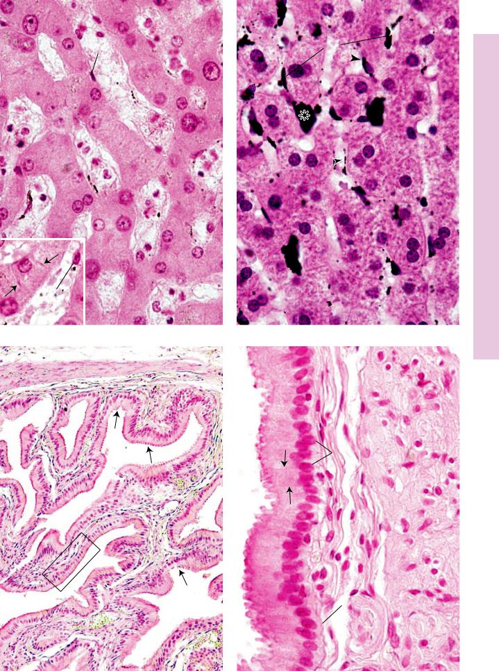
This photomicrograph is of a patient suffering from chronic pancreatitis. Observe that the connective tissue elements are highly exaggerated, the acini are greatly reduced in number, and the Islets of Langerhans are very close to each other because of the reduction in acinar population. (Reprinted with permission from Mills SE, Carter D, et al., eds. Sternberg’s Diagnostic Surgical Pathology, 5th ed., Philadelphia: Lippincott Williams & Wilkins, 2010, p. 1438.)
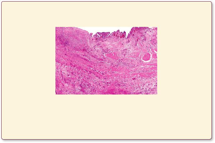
This photomicrograph is of a patient suffering from Kaposi’s sarcoma of the liver. Observe the presence of relatively normal hepatocytes in the upper left, whereas much of the right-hand side displays the presence of spindle-shaped cells, typical of Kaposi’s sarcoma cells. An additional typical feature of this disease is the presence of extravasated erythrocytes. (Reprinted with permission from Mills SE, Carter D, et al., eds. Sternberg’s Diagnostic Surgical Pathology, 5th ed., Philadelphia: Lippincott Williams & Wilkins, 2010, p. 1584.)

362 D I G E S T I V E S Y S T E M I I I
the patient population who succumbed to AIDS. Additionally, a Kaposi’s sarcoma–associated herpesvirus has also been determined to be a causative factor in this disease. The autopsied livers presented with numerous darkened nodules of a soft consistency most of which occupied expanded connective tissue of the intrahepatic biliary tract.
Type I Diabetes
Type I (insulin-dependent) diabetes is characterized by polyphagia (insatiable hunger), polydipsia (unquenchable thirst), and polyuria (excessive urination). It usually has a sudden onset before 20 years of age, is distinguished by damage to and destruction of beta cells, and results from a low level of plasma insulin.
Type II Diabetes Mellitus
Type II (non–insulin-dependent) diabetes mellitus commonly occurs in overweight individuals over 40 years of age. It does not result from low levels of plasma insulin and is insulin resistant, which is a major factor in its pathogenesis. The resistance to insulin is due to decreased binding of insulin to its plasmalemma receptors and to defects in postreceptor insulin action. Type II diabetes is usually controlled by diet.
Hepatitis
Hepatitis is inflammation of the liver, and although it could have various causes such as abuse of alcohol and certain drugs, its most common cause is one of the five types of hepatitis viruses, denoted by the first five letters of the alphabet, A through E. Hepatitis A is usually spread by poor hygiene (fecal-oral route and contaminated water) as well as by sexual contact. Usually, there are no symptoms; the patient recovers and does not become a carrier. Hepatitis B, a more serious condition than hepatitis A, is usually transmitted by body fluids and, in case of drug addicts, by the sharing of needles. Patients can become carriers of the virus, and in 10% of the patients, the condition may become chronic, leading to cirrhosis and cancer of the liver. In the past, hepatitis C was transmitted by blood transfusions, but screening has almost completely eradicated that route and now it is transmitted mostly by shared needles among drug addicts. About three-quar- ters of people who have the hepatitis C virus will reach the chronic stage, and of these, 20% to 25% will develop cirrhosis and then liver cancer. Hepatitis D is also transmitted by the sharing of needles and is always accompanied
by hepatitis B. The double infection is a more severe condition. Hepatitis E is spread by the fecal-oral route and is responsible for epidemics but mostly in underdeveloped countries. Neither chronic nor carrier states are present with this form of the hepatitis virus. Universal vaccination is recommended to protect the population from hepatitis B, and this has the added benefit of protection against hepatitis D; it is recommended that travelers to underdeveloped countries where hepatitis A is prevalent be vaccinated against hepatitis A. There are no vaccines currently available against hepatitis C or E.
This photomicrograph is of a patient suffering from acute alcoholinduced hepatitis. Observe that the specimen presents some of the earliest histopathological signs of alcohol-induced hepatitis, namely, macrovascular fatty changes, Mallory hyaline, and the infiltration by neutrophils. (Reprinted with permission from Mills SE, Carter D, et al., eds. Sternberg’s Diagnostic Surgical Pathology, 5th ed., Philadelphia: Lippincott Williams & Wilkins, 2010. p. 1513.)
Jaundice (Icterus)
Jaundice (icterus) is characterized by excess bilirubin in the blood and deposition of bile pigment in the skin and sclera of the eyes, resulting in a yellowish appearance. It may be hereditary or due to pathologic conditions such as excess destruction of red blood cells (hemolytic jaundice), liver dysfunction, and obstruction of the biliary passages (obstructive jaundice).
Gallstones (Biliary Calculi)
Gallstones (biliary calculi) are concretions, usually of fused crystals of cholesterol that form in gallbladder or bile duct. They may accumulate to such an extent that
D I G E S T I V E S Y S T E M I I I 363
the cystic duct is blocked, thus preventing emptying of the gallbladder, and they may require surgical removal if less invasive methods fail to dissolve or pulverize
them. If the obstruction occurs in an abrupt manner due to the gallstones, the gallbladder can rapidly become inflamed, a condition known as chronic cholecystitis.
This photomicrograph is from a gallbladder whose cystic duct was obstructed by the presence of gallstones resulting in acute cholecystitis. Observe that much of the luminal surface of the mucosa lacks an epithelial lining and that the lamina propria is edematous. Moreover, the adventitia is thicker than normal. (Reprinted with permission from Mills SE, Carter D, et al., eds. Sternberg’s Diagnostic Surgical Pathology, 5th ed., Philadelphia: Lippincott Williams & Wilkins, 2010. p. 1606.)

Pancreas • 1-15 GRAPHIC
364 D I G E S T I V E S Y S T E M I I I
Common bile duct
Main pancreatic duct
Accessory pancreatic duct
Capillary 
Islet of 
Langerhans
Pancreatic  acinar cell
Intercellular canaliculi
Centroacinar cell
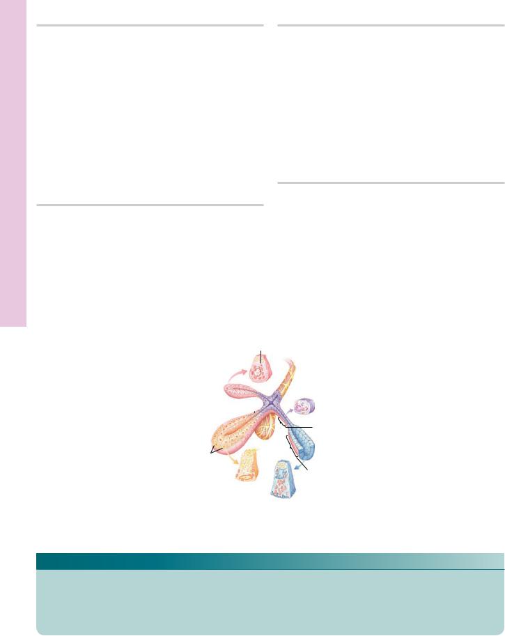
Exocrine function of the pancreas is served by its acinar cells, centroacinar cells, and intercalated ducts. The acinar cells secrete digestive enzymes, and the duct cells supply an alkaline buffer solution.
The endocrine portion is composed of the islets of Langerhans, richly vascularized spherical aggregates of cells encased by reticular fibers. The islets are composed of five types of cells, which can be differentiated from each other only with special stains.
Intralobular duct
Intercalated duct
Centroacinar cells
Pancreatic acinus
Zymogen granules
Golgi
Rough ER
Pancreatic acinar cell

D I G E S T I V E S Y S T E M I I I 365
Right lobe |
Left lobe |
PORTAL LOBULE: |
CLASSIC LOBULE: |
|
Bile drains to |
||
|
Falciform ligament |
||
|
Sinusoids drain |
||
|
bile duct. Portal |
||
|
|
to the central |
|
|
Hepatic artery |
area is center. |
|
|
vein. |
||
|
|
||
|
Vena cava |
|
|
|
Portal vein |
|
|
Liver• 2-15 GRAPHIC
HEPATIC LOBULE: Portal vein Central vein
PORTAL TRIAD:
Hepatic artery
Portal vein
Bile duct
Central vein
Bile canaliculus
Golgi
Sinusoid
Sinusoidal lining cell Space of Disse
Kupffer cell
PORTAL ACINUS:
Tissue supplied by terminal branches of the hepatic artery and portal vein. Cells nearest these vessels are first to receive oxygen and nutrients.
Sinusoids
PORTAL TRIAD:
Hepatic artery
Portal vein
Bile duct
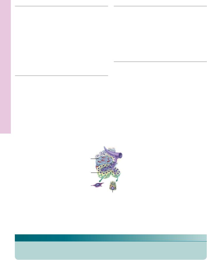
Hepatocytes, liver cells, deliver endocrine secretions into the vascular supply, and exocrine secretion, bile, into excretory ducts, the bile ducts. Each liver cell borders a vascular space, sinusoid, on at least one side and other hepatocytes on its remaining sides. Where two hepatocytes adjoin, they delimit a small intercellular space, bile canaliculus, into which the bile is delivered.
Since sinusoids are lined by endothelial cells (sinusoidal lining cells) and macrophages (Kupffer cells), hepatocytes do not come into contact with the bloodstream. The space of Disse intervenes between hepatocytes and sinusoidal lining cells. This space houses microvilli of hepatocytes, occasional fat-storing cells (Ito cells), and slender reticular fibers that help form the supporting framework of the liver.
Glands alivary • S1-15 PLATE
366 D I G E S T I V E S Y S T E M I I I
FIGURE 1. Parotid gland. Monkey. Plastic section. ×132.
The parotid gland is purely serous, with a connective tissue capsule sending trabeculae (T) into the substance of the gland, subdividing it into lobules (Lo). Slender connective tissue sheets penetrate the lobules, surrounding small blood vessels (BV) and intralobular ducts (iD). Interlobular ducts (ID) are surrounded by increased amounts of connective tissue (CT) and large blood vessels. Observe that the acini (Ac) are closely packed within each lobule. Inset. Parotid gland. Monkey. Plastic section. ×540. Note that the round nuclei (N) of these serous acini are basally located. The lateral cell membranes (arrows) are not clearly visible, nor are the lumina of the acini. Observe the slender sheets of connective tissue (arrowheads) investing each acinus.
FIGURE 3. Sublingual gland. Monkey. Plastic section. ×540.
This photomicrograph is a higher magnification of the boxed area of Figure 2. The flattened, dark nuclei (N) of the mucous acini are clearly evident as they appear to be pressed against the basal cell membrane. Observe that much of the cytoplasm is occupied by small, mucin-containing vesicles (arrows), that the lateral cell membrane (arrowheads) is evident, and that the lumen (L) is usually identifiable. Serous demilunes (SD) are composed of serousproducing cells whose nuclei (N) are round to oval in morphology. Note also that the lateral cell membranes are not distinguishable in serous cells.
FIGURE 2. Sublingual gland. Monkey. Plastic section. ×270.
The sublingual gland is a mixed gland in that it produces both serous and mucous secretory products. The mucous acini (MA) possess dark nuclei (N) that are flattened against the basal cell membrane. Moreover, the cytoplasm is filled with a frothy-appearing material, representing the viscous secretory product. Many of the mucous acini are capped by serous cells, forming a crescent-shaped cap, the serous demilune (SD). The sublingual gland is subdivided into lobes and lobules by connective tissue septa (CT) that act as the supporting network for the nerves, vessels, and ducts of the gland. The boxed area is presented at a higher magnification in Figure 3.
FIGURE 4. Submandibular gland. Monkey. Plastic section. ×132.
The submandibular gland also produces a mixed type of secretion; however, unlike in the sublingual gland, serous acini predominate. Serous (SA) and mucous acini (MA) are easily distinguishable from each other, but most mucous units display a cap of serous demilunes. Moreover, the submandibular gland is characterized by an extensive system of ducts (D), recognizable by their pale cytoplasm, comparatively large lumina (L), and round nuclei. This gland is also subdivided into lobes and lobules by connective tissue septa (CT). Inset. Submandibular gland. Monkey. Plastic section. ×540. Note the granular appearance of the cells comprising the serous demilune (SD) in contrast with the “frothy” appearing cytoplasm of the mucous acinus (MA).
Serous cell
Serous acinus |
|
|
|
|
|
|
|
|
|
|
Myoepithelial cell |
|||
|
|
|
|
|
|
|
|
|
|
|||||
|
|
|
|
|
|
|
|
|
Intercalated duct cell |
|||||
Mucous acinus |
|
|
|
|
|
|
|
|
|
|
|
|
|
|
|
|
|
|
|
|
|
|
|
|
|
|
|
||
|
|
|
|
|
|
|
Intercalated duct |
|||||||
|
|
|
|
|
||||||||||
|
|
|
|
|
|
|
|
|
|
|
||||
|
|
|
|
|
|
|
|
|
|
|
|
|
||
|
|
|
|
|
|
|
|
|
||||||
Serous demilunes |
|
|
|
|
|
|||||||||
|
|
|
|
|
|
|
|
|
|
|
|
Striated duct |
||
|
|
|
|
|
|
|
|
|
|
|
Striated duct cell |
|||
|
|
|
|
Mucous cell |
|
|||||||||
|
||||||||||||||
Salivary glands
KEY
Ac |
acinus |
ID |
interlobular duct |
SA |
serous acini |
BV |
blood vessel |
L |
lumen |
SD |
serous demilune |
CT |
connective tissue |
Lo |
lobule |
T |
trabeculae |
D |
duct |
MA |
mucous acini |
|
|
iD |
intralobular duct |
N |
nucleus |
|
|

T
BV
 CT iD
CT iD
ID
Ac |
N |
|
|
|
Lo |
FIGURE 1
N
L
N
SD 
FIGURE 3
N
SD
CT
MA
FIGURE 2
L
D
SA
D
 MA
MA
CT
SD
MA
FIGURE 4
Glands alivary • S1-15 PLATE
ancreas • P2-15 PLATE
368 D I G E S T I V E S Y S T E M I I I
FIGURE 1. Pancreas. Human. Paraffin section. ×132.
The pancreas is a complex gland since it has both exocrine and endocrine components. The exocrine portion comprises the bulk of the organ as a compound tubuloalveolar gland, secreting a serous fluid. The gland is subdivided into lobules by connective tissue septa (CT). Each acinus (Ac) is composed of several pyramid-shaped cells, possessing round nuclei. Cells located in the center of the acinus, centroacinar cells (CA), form the smallest ducts of the gland. The endocrine portion of the pancreas is composed of small, spherical clumps of cells, islets of Langerhans (IL), which are richly endowed by capillaries. These islets of Langerhans are haphazardly scattered among the serous acini of the pancreas. The boxed area is presented at a higher magnification in Figure 2.
FIGURE 3. Pancreas. Monkey. Plastic section. ×540.
With the use of plastic sections, the morphology of the pancreatic acinus is well defined. Observe that in fortuitous sections the acinus resembles a pie, with the individual cells clearly delineated (arrows). The nucleus (N) of each trapezoid-shaped cell is round and the basal cytoplasm (arrowhead) is relatively homogeneous, whereas the apical cytoplasm is packed with zymogen granules (ZG). Centroacinar cells (CA) may be recognized both by their locations as well as by the pale appearance of their nuclei. Inset.
Pancreas. Monkey. Plastic section. ×540. Observe the centroacinar cell (CA), whose pale nucleus is readily differentiated from the surrounding acinar cell nuclei.
FIGURE 2. Pancreas. Human. Paraffin section. ×270.
This photomicrograph is a higher magnification of the boxed area of Figure 1. Note that the connective tissue septa (CT), while fairly extensive in certain regions, are quite slender in the interlobular areas. The trapezoidal morphologies of individual cells of the serous acini are clearly evident in fortuitous sections (arrow). Observe also the centroacinar cells (CA), located in the center of acini, which represent the smallest units of the pancreatic duct system.
FIGURE 4. Islets of Langerhans. Monkey. Plastic section. ×270.
The islets of Langerhans (IL), the endocrine portion of the pancreas, are a more or less spherical configuration of cells randomly scattered throughout the exocrine portion of the gland. As such, each islet is surrounded by serous acini (Ac). The islets receive their rich blood supply (BV) from the connective tissue elements (CT) of the exocrine pancreas. Inset. Islets of Langerhans. Monkey. Plastic section. ×540. Observe the rich vascularity of the islets of Langerhans, as evidenced by the presence of erythrocyte (RBC)- engorged blood vessels. Although each islet is composed of A, B, C, and D cells, they can only be distinguished from each other by the use of special stains. However, it should be noted that, in the human, B cells are the most populous and are usually located in the center of the islet, whereas A cells are generally found at the periphery. This situation is reversed in the monkey.
Islet of
Langerhans
Pancreatic
acinar cell
 Zymogen granules

 Nucleus Centroacinar
cell Pancreatic acinar cell
Pancreatic cells
KEY
Ac |
acinus |
CT |
connective tissue septa |
RBC |
erythrocyte |
BV |
blood vessel |
IL |
islets of Langerhans |
ZG |
zymogen granule |
CA |
centroacinar cell |
N |
nucleus |
|
|

IL
CT
CA
|
|
AC |
CT |
CA |
|
|
CT |
|
|
||
|
|
|
CT
FIGURE 1 |
FIGURE 2 |
CT
CA
Ac
N
BV
ZG
IL
RBC
CA
FIGURE 3 |
FIGURE 4 |
ancreas • P2-15 PLATE
er •Liv3-15 PLATE
370 D I G E S T I V E S Y S T E M I I I
FIGURE 1. Liver. Pig. Paraffin section. ×14.
Note that the liver is invested by a connective tissue capsule, Glisson’s capsule (GC), from which, in the pig, septa (S) extend to subdivide the gland into more or less hexagon-shaped classical lobules (Lo). Blood vessels, lymph vessels, and bile ducts travel within the connective tissue septa to reach the apices of the classic lobules, which are known as the portal areas (PA). Bile reaches the portal areas from within the lobules, whereas blood enters the substance of the lobules from the portal areas. Within each lobule, the blood flows through tortuous channels, the liver sinusoids, to enter the central vein (CV) in the middle of the classical lobule.
FIGURE 3. Liver. Monkey. Plastic section. ×132.
The central vein (CV) of the liver lobule (a terminal radix of the hepatic vein) collects blood from the sinusoids (Si) and delivers it to sublobular veins. The plates of liver cells (PL) and hepatic sinusoids appear to radiate, as spokes of a wheel, from the central vein. The boxed area is presented at a higher magnification in Figure 4.
FIGURE 2. Liver. Dog. Paraffin section. ×132.
The portal area of the liver houses terminal branches of the hepatic artery (HA) and portal vein (PV). Note that the vein is much larger than the artery, and its wall is very thin in comparison to the size of its lumen. Branches of lymph vessels (LV) and bile ducts (BD) are also present in the portal area. Bile ducts may be recognized by their cuboidal-to-columnar epithelium. Observe that unlike in the pig, connective tissue septa do not demarcate the boundaries of classic liver lobules, although the various structures of the portal area are invested by connective tissue elements. Plates of liver cells (PL) and sinusoids (Si) extend from the portal areas.
FIGURE 4. Liver. Monkey. Plastic section. ×270.
This photomicrograph is a higher magnification of the boxed area of the previous figure. Note that the lumen of the central vein (CV) is lined by a simple squamous epithelium (Ep), which is continuous with the endothelial lining of the hepatic sinusoids (Si), tortuous vascular channels that freely communicate with each other. Observe also that the liver plates (LP) are composed of hepatocytes (H), one to two cell layers thick, and that each plate is bordered by sinusoids.
 Septum
Hepatic
lobule
Sinusoid
Hepatic
artery
Bile duct
Portal
vein
Liver
KEY
BD |
bile duct |
HA |
hepatic artery |
PL |
plates of liver cells |
CV |
central vein |
Lo |
lobule |
PV |
portal vein |
Ep |
epithelium |
LP |
liver plates |
S |
septa |
GC |
Glisson’s capsule |
LV |
lymph vessel |
Si |
sinusoid |
H |
hepatocyte |
PA |
portal area |
|
|
S |
GC |
|
Si |
PL |
|
PA
CV
BD HA
LV 
Lo
PV
CV
FIGURE 1 |
FIGURE 2 |
Si
H
Si
LP
CV |
|
CV |
|
|
|
|
|
|
H |
Ep |
Si |
|
|
||
PL |
|
|
|
|
|
|
|
|
|
|
LP |
FIGURE 3 |
FIGURE 4 |
er •Liv3-15 PLATE

Gallbladder er, •Liv4-15 PLATE
372 D I G E S T I V E S Y S T E M I I I
FIGURE 1. Liver. Monkey. Plastic section. ×540.
This photomicrograph is a high magnification of liver plates (LP). Observe that individual hepatocytes (H) are polygonal in shape. Each hepatocyte possesses one or two nuclei, although occasionally some have three nuclei. Plates of hepatocytes enclose hepatic sinusoids (Si) that are lined by sinusoidal lining cells (SC); therefore, hepatocytes do not come into direct contact with the bloodstream. The space between the sinusoidal lining cells and the hepatocytes, the space of Disse, is at the limit of resolution of the light microscope. Inset. Liver. Human. Paraffin section. ×540. The hepatocyte cell membranes are clearly evident in this photomicrograph. Note that in fortuitous sections, small intercellular spaces (arrows) are recognizable. These are bile canaliculi through which bile flows to the periphery of the lobule.
FIGURE 3. Gallbladder. Human. Paraffin section. ×132.
The gallbladder is a pear-shaped, hollow organ that functions in storing and concentrating bile. Its histologic structure is relatively simple, but its appearance may be deceiving. The mucosa of an empty gallbladder, as in this photomicrograph, is thrown into numerous folds (arrows), providing it with a glandular morphology. However, close observation of the epithelium (Ep) demonstrates that all of the simple columnar cells of the mucous membrane are identical. A loose connective tissue (CT), sometimes referred to as a lamina propria, lies deep to the epithelium. Observe that a muscularis mucosae is lacking, and the smooth muscle (SM) surrounding the connective tissue is the muscularis externa. The outermost coat of the gallbladder is a serosa or adventitia. A region similar to the boxed area is presented in Figure 4.
FIGURE 2. Liver. Paraffin section. ×540.
A system of macrophages known as Kupffer cells (KC) are found interspersed among the endothelial lining cells of liver sinusoids (Si). These macrophages are larger than the epithelial cells and may be recognized by the presence of phagocytosed material within them. Kupffer cells may be demonstrated by injecting an animal intravenously with india ink, as is the case in this specimen. Observe that some cells appear as large, black smudges since they are filled with phagocytosed ink (asterisk), whereas other cells possess only small quantities of the phagocytosed material (arrowheads). Note also that much of the sinusoidal lining is devoid of ink, indicating that the endothelial cells are probably not phagocytic.
FIGURE 4. Gallbladder. Human. Paraffin section. ×540.
This photomicrograph is a higher magnification of a region similar to the boxed area of Figure 3. Note that the epithelium (Ep) is composed of identical-appearing tall columnar cells, whose nuclei (N) are basally oriented. The lateral cell membranes are evident in certain regions (arrows), whereas the apical brush border is usually not visible in hematoxylin and eosin–stained specimens. Observe that a relatively thick basal membrane (BM) separates the epithelium from the underlying loose connective tissue (CT).
|
Sinusoids |
|||
Bile canaliculus |
|
|
PORTAL TRIAD: |
|
|
|
|
Hepatic artery |
|
|
|
|
|
|
Golgi |
|
|
|
Bile duct |
|
|
|
||
|
|
|
|
Portal vein |
|
|
|
|
|
Space of Disse |
|
|
|
Sinusoid |
|
|
|
|
|
Sinusoidal lining cell |
|
|
|
Kupffer cell |
|
|
|
||
|
|
|
||
|
|
|
|
|
|
|
|
|
Liver |
KEY
BM |
basal membrane |
KC |
Kupffer cell |
Si |
sinusoid |
CT |
connective tissue |
LP |
liver plate |
SM |
smooth muscle |
Ep |
epithelium |
N |
nucleus |
|
|
H |
hepatocyte |
SC |
sinusoidal lining cell |
|
|
SC |
KC |
LP
Si
Si
LP
H
SC
FIGURE 1 |
FIGURE 2 |
SM
Ep
Ep
N
CT
CT
Ep
BM
FIGURE 3 |
FIGURE 4 |
Gallbladder er, •Liv4-15 PLATE

Microscopy Electron Gland, alivary • S5-15 PLATE
374 D I G E S T I V E S Y S T E M I I I
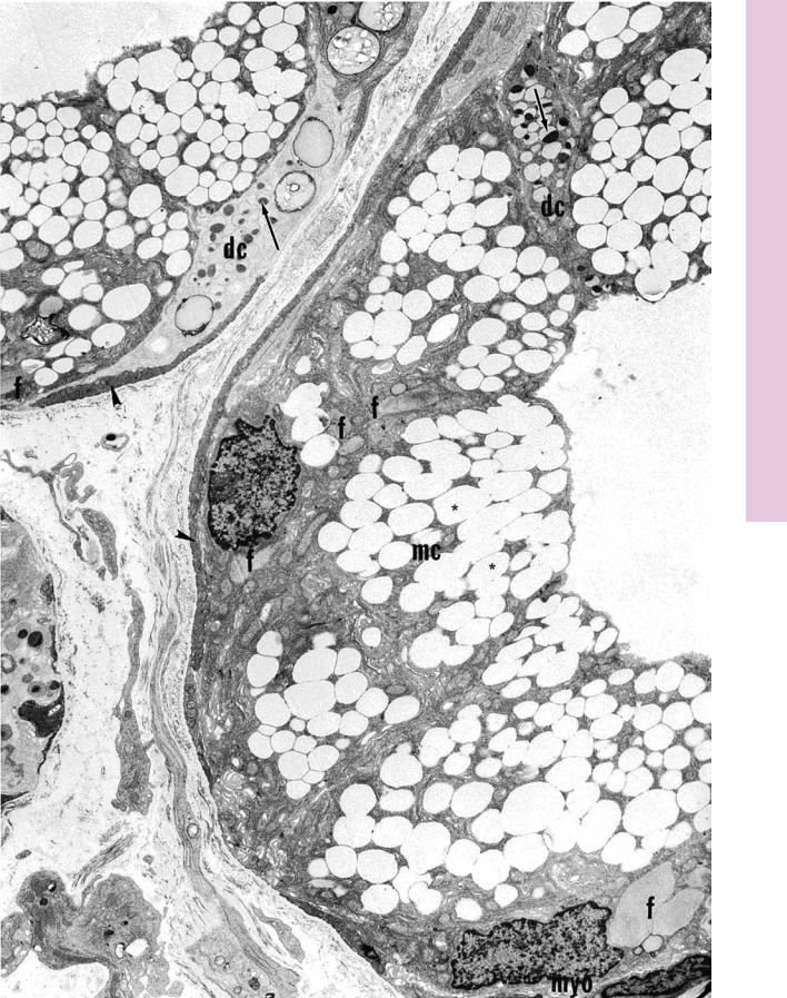
FIGURE 1. Sublingual gland. Human. Electron microscopy. ×4,050.
The human sublingual gland is composed mostly of mucous acini capped by serous demilunes. The mucous cells (mc) display numerous filamentous bodies (f) and secretory granules, which
appear to be empty (asterisks). The serous cells (dc) may be recognized by their paler cytoplasm and the presence of secretory granules (arrows) housing electron-dense materials. Note also the presence of myoepithelial cells (myo), whose processes (arrowheads) encircle the acinus. (Courtesy of Dr. A. Riva.)
Serous cell
Myoepithelial cell
Serous acinus
Intercalated duct cell
Mucous acinus
Intercalated
duct
Serous demilunes |
Striated duct |
|
|
|
Striated duct cell |
|
Mucous cell |
||||
|
||||
|
|
|||
Salivary glands
KEY
dc |
serous cells |
mc |
mucous cells |
f |
filamentous bodies |
myo |
myoepithelial cells |
D I G E S T I V E S Y S T E M I I I 375
Microscopy Electron Gland, alivary • S5-15 PLATE
FIGURE 1

376 D I G E S T I V E S Y S T E M I I I
Microscopy Electron er, •Liv6-15 PLATE
FIGURE 1
FIGURE 1. Liver. Mouse. Electron microscopy. ×11,255.
The hepatocytes of this electron micrograph display two of their surfaces, one bordering a sinusoid (Si) and the other where two parenchymal cells contact each other (arrows). The sinusoidal surface displays microvilli (mv) that extend into the space of Disse (sD). They almost contact sinusoidal lining cells (SC) that present numerous
fenestrae (arrowheads). The parenchymal contacts are characterized by the presence of bile canaliculi (BC), intercellular spaces that are isolated by the formation of occluding junctions (OC). The cytoplasm of hepatocytes houses the normal cellular complements, such as numerous mitochondria (m), elements of rough endoplasmic reticulum (rER), Golgi apparatus, smooth endoplasmic reticulum, lysosomes, and inclusions such as glycogen (g) and lipid droplets
(l). The nucleus (N) of one of the hepatocytes is evident.
Microscopy Electron , Langerhans of Islet• 7-15 PLATE
FIGURE 1
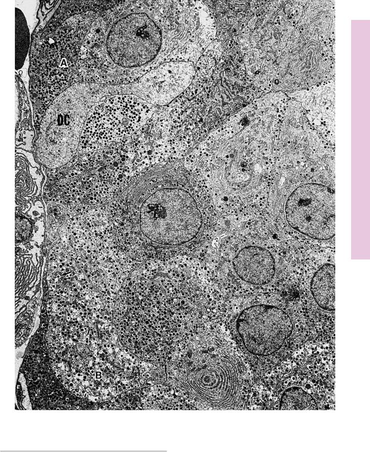
FIGURE 1. Islet of Langerhans. Rabbit. Electron microscopy. ×3,578.
The islets of Langerhans house four types of parenchymal cells, namely, A, B, C, and D cells. B cells (B) are the most numerous and may be recognized by the presence of secretory granules whose electron-dense core is surrounded by a clear zone (arrows).
A cells (A), the second most numerous secretory cell, also house many secretory granules; however, these lack an electron-lucent periphery. D cells (DC) are the least numerous and are characterized by secretory granules that are much less electron-dense than those of the other two cell types. (From Sato T, Herman L. Stereological analysis of normal rabbit pancreatic islets. Am J Anat 1981;161:71–84.)