

- •Preface
- •Acknowledgments
- •Reviewers
- •Contents
- •CHAPTER OUTLINE
- •CYTOPLASM
- •Plasmalemma
- •Mitochondria
- •Ribosomes
- •Endoplasmic Reticulum
- •Golgi Apparatus, cis-Golgi Network, and the trans-Golgi Network
- •Endosomes
- •Lysosomes
- •Peroxisomes
- •Proteasomes
- •Cytoskeleton
- •Inclusions
- •NUCLEUS
- •CELL CYCLE
- •CHAPTER OUTLINE
- •EPITHELIUM
- •Epithelial Membranes
- •GLANDS
- •Chapter Summary
- •CHAPTER OUTLINE
- •EXTRACELLULAR MATRIX
- •Fibers
- •Amorphous Ground Substance
- •Extracellular Fluid
- •CELLS
- •CONNECTIVE TISSUE TYPES
- •Chapter Summary
- •CHAPTER OUTLINE
- •CARTILAGE
- •BONE
- •Cells of Bone
- •Osteogenesis
- •Bone Remodeling
- •Chapter Summary
- •CHAPTER OUTLINE
- •FORMED ELEMENTS OF BLOOD
- •Lymphocytes
- •Neutrophils
- •PLASMA
- •COAGULATION
- •HEMOPOIESIS
- •Erythrocytic Series
- •Granulocytic Series
- •Chapter Summary
- •CHAPTER OUTLINE
- •SKELETAL MUSCLE
- •Sliding Filament Model of Muscle Contraction
- •CARDIAC MUSCLE
- •SMOOTH MUSCLE
- •Chapter Summary
- •CHAPTER OUTLINE
- •BLOOD-BRAIN BARRIER
- •NEURONS
- •Membrane Resting Potential
- •Action Potential
- •Myoneural Junctions
- •Neurotransmitter Substances
- •SUPPORTING CELLS
- •PERIPHERAL NERVES
- •Chapter Summary
- •CHAPTER OUTLINE
- •BLOOD VASCULAR SYSTEM
- •HEART
- •ARTERIES
- •Capillary Permeability
- •Endothelial Cell Functions
- •VEINS
- •LYMPH VASCULAR SYSTEM
- •Chapter Summary
- •CHAPTER OUTLINE
- •CELLS OF THE IMMUNE SYSTEM
- •Antigen-Presenting Cells
- •DIFFUSE LYMPHOID TISSUE
- •LYMPH NODES
- •TONSILS
- •SPLEEN
- •THYMUS
- •Chapter Summary
- •CHAPTER OUTLINE
- •PITUITARY GLAND
- •Pars Intermedia
- •Pars Nervosa and Infundibular Stalk
- •Pars Tuberalis
- •THYROID GLAND
- •Parathyroid Glands
- •Suprarenal Glands
- •Cortex
- •Medulla
- •Pineal Body
- •Chapter Summary
- •CHAPTER OUTLINE
- •SKIN
- •Epidermis of Thick Skin
- •Dermis
- •DERIVATIVES OF SKIN
- •Chapter Summary
- •CHAPTER OUTLINE
- •CONDUCTING PORTION OF THE RESPIRATORY SYSTEM
- •Extrapulmonary Region
- •Intrapulmonary Region
- •RESPIRATORY PORTION OF THE RESPIRATORY SYSTEM
- •MECHANISM OF RESPIRATION
- •Chapter Summary
- •CHAPTER OUTLINE
- •ORAL CAVITY AND ORAL MUCOSA
- •Oral Mucosa
- •Tongue
- •Teeth
- •Odontogenesis (See Graphic 13-2)
- •Chapter Summary
- •CHAPTER OUTLINE
- •REGIONS OF THE DIGESTIVE TRACT
- •Esophagus
- •Stomach
- •Small Intestine
- •Large Intestine
- •GUT-ASSOCIATED LYMPHOID TISSUE
- •DIGESTION AND ABSORPTION
- •Carbohydrates
- •Proteins
- •Lipids
- •Water and Ions
- •Chapter Summary
- •CHAPTER OUTLINE
- •MAJOR SALIVARY GLANDS
- •PANCREAS
- •LIVER
- •Exocrine Function of the Liver
- •Endocrine and Other Functions of the Liver
- •GALLBLADDER
- •Chapter Summary
- •CHAPTER OUTLINE
- •KIDNEY
- •Uriniferous Tubule
- •Nephron
- •Collecting Tubules
- •FORMATION OF URINE FROM ULTRAFILTRATE
- •EXTRARENAL EXCRETORY PASSAGES
- •Chapter Summary
- •CHAPTER OUTLINE
- •OVARY
- •Ovarian Follicles
- •Regulation of Follicle Maturation and Ovulation
- •Corpus Luteum and Corpus Albicans
- •GENITAL DUCTS
- •Oviduct
- •Uterus
- •FERTILIZATION, IMPLANTATION, AND THE PLACENTA
- •Fertilization and Implantation
- •Placenta
- •VAGINA
- •EXTERNAL GENITALIA
- •MAMMARY GLANDS
- •Chapter Summary
- •CHAPTER OUTLINE
- •TESTES
- •Spermatogenesis
- •GENITAL DUCTS
- •ACCESSORY GLANDS
- •PENIS
- •Erection and Ejaculation
- •Chapter Summary
- •CHAPTER OUTLINE
- •SENSORY ENDINGS
- •Chapter Summary
- •Terminology of Staining
- •Common Stains Used in Histology
- •Hematoxylin and Eosin
- •Wright Stain
- •Weigert Method for Elastic Fibers and Elastic van Gieson Stain
- •Silver Stain
- •Iron Hematoxylin
- •Bielschowsky Silver Stain
- •Masson Trichrome
- •Periodic Acid-Schiff Reaction (PAS)
- •Alcian Blue
- •von Kossa Stain
- •Sudan Red
- •Mucicarmine Stain
- •Safranin-O
- •Toluidine Blue

•The epithelial lining of the lumen and of the crypts is composed of goblet (and oligomucous) cells, surface absorptive cells, regenerative cells, and occasional DNES cells. There are no Paneth’s cells in the large intestine, with the possible exception of the appendix.
The large intestine functions in the absorption of the remaining amino acids, lipids, and carbohydrates, as well as fluids, electrolytes, and certain vitamins, and it also is responsible for the compaction of feces.
GUT-ASSOCIATED LYMPHOID TISSUE
The lumen of the digestive tract is rich in antigenic substances, bacteria, and toxins; in fact, it has been estimated that the intestinal tract houses several trillion microbes with a total weight of approximately 2 kg. Since only a thin, simple columnar epithelium separates the richly vascularized connective tissue from this threatening milieu, the lamina propria of the intestines is well endowed with lymphoid elements. These include
•scattered cells (B cells, T cells, plasma cells, mast cells, macrophages, etc.),
•individual lymphatic nodules, and,
•in the ileum, Peyer’s patches, clusters of lymphatic nodules.
Regions where lymphatic nodules come in contact with the epithelial lining of the intestines display flattened cells that form the interface between the lumen and the lymphatic nodule. These are M cells (microfold cells), which phagocytose antigens and transport them (without breaking them down into epitopes), via clathrincoated vesicles, to the basal aspect of the cell. The antigens are released into the lamina propria for uptake by antigen-presenting cells and dendritic cells.
It is interesting to note that the lamina propria of the colon has no lymph vessels, thus, generally, cancers of the colon metastasize at a much slower rate than do other cancers of the digestive tract.
DIGESTION AND ABSORPTION
Carbohydrates
•Amylases, present in the saliva and in the pancreatic secretion, hydrolyze carbohydrates to disaccharides.
•Oligo- and disaccharidases, present in the glycocalyx of surface absorptive cells, break down oligoand
D I G E S T I V E S Y S T E M I I 333
disaccharides into monosaccharides (glucose and galactose) that enter the surface absorptive cell, requiring active transport using sugar-glucose trans- porter-1. The cells then release the glucose and galactose into the lamina propria, where these sugars enter the circulatory system for transport to the liver.
Proteins
•Proteins, denatured by HCl in the lumen of the stomach, are hydrolyzed (by the enzyme pepsin) into polypeptides.
•These are further broken down into tri- and dipeptides by proteases of the pancreatic secretions.
•Tri- and dipeptidases of the glycocalyx hydrolyze dipeptides into individual amino acids, which enter the surface absorptive cells, involving active transport, and are transferred into the lamina propria, where they enter the capillary network to be transported to the liver.
Lipids
•Pancreatic lipase breaks lipids down into fatty acids, monoglycerides, and glycerol within the lumen of the duodenum and proximal jejunum.
•Bile salts, delivered from the gallbladder, emulsify the fatty acids and monoglycerides, forming micelles, which, along with glycerol, diffuse into the surface absorptive cells.
•Within these cells, they enter the smooth endoplasmic reticulum, are reesterified to triglycerides, and are covered by a coat of protein within the Golgi apparatus, forming lipoprotein droplets known as chylomicrons.
•Chylomicrons exit these cells at their basolateral membranes and enter the lacteals of the villi, contributing to the formation of chyle.
•Chyle enters the lymph vascular system, makes its way to the thoracic duct, and then into the venous system at the junction of the left internal jugular vein and left brachiocephalic vein.
•Fatty acids that are shorter than 12 carbon chains in length pass through the surface absorptive cells without being reesterified and gain entrance to the blood capillaries of the villi.
Water and Ions
Water and ions are absorbed through the surface absorptive cells of the small and the large intestines.
334 D I G E S T I V E S Y S T E M I I
CLINICAL CONSIDERATIONS
Crohn’s Disease
Crohn’s disease is a subcategory of inflammatory bowel disease, a condition of unknown etiology. It usually involves the small intestine or the colon but may affect any region of the digestive tract, from the esophagus to the anus, as well as extra-alimentary canal structures such as the skin, the kidney, and the larynx. It is characterized by patchy ulcers and deep fistulas in the intestinal wall. Clinical manifestations include abdominal pain, diarrhea, and fever, and these recur after various periods of ever shortening remission.
powerful vomiting or sometimes strenuous hiccuping. Frequently, the bleeding is self-limiting, but occasionally, it requires surgical intervention.
Peptic Ulcers
Peptic ulcers are areas of the stomach, but mostly of the duodenum, that are denuded of the epithelial lining due to the action of the acid chyme. Most commonly, the underlying reasons are Helicobacter pylori infections and the use of aspirin, corticosteroids, and nonsteroidal antiinflammatory drugs. The bacteria, H. pylori, are able to live in the mucous substance lining the gastric epithelium probably by forming a protective envelope of bicarbonate buffer around themselves that neutralizes the acidic milieu. It is now believed that strains of this bacterium that possess cagA gene are the causative agents of peptic ulcers. Interestingly, people who smoke and/ or drink alcoholic beverages develop peptic ulcers more frequently than do nonsmokers and nondrinkers. The symptoms involve mild to sharp pain in the midline of the lower thoracic and upper abdominal regions.
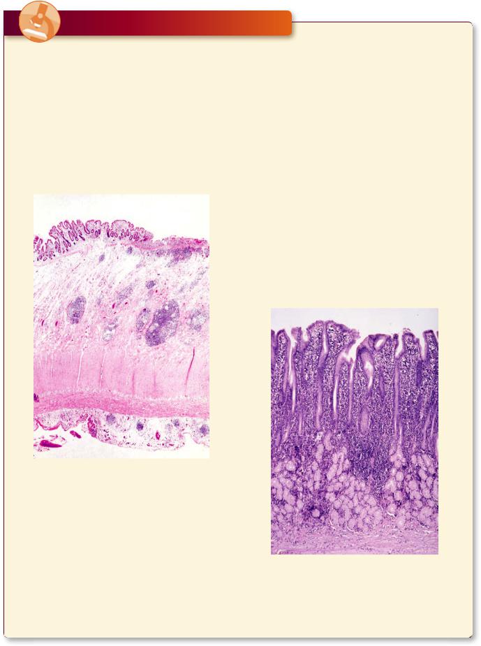
This figure is from the colon of a patient with Crohn’s disease displaying ulceration of the mucosa, a hypertrophied submucosa with clusters of lymphoid elements, as well as smaller aggregates of lymphoid elements in the subserosal connective tissue adjacent to the muscularis externa. (Reprinted with permission from Rubin R, Strayer D, et al., eds. Rubin’s Pathology. Clinicopathologic Foundations of Medicine, 5th ed. Baltimore: Lippincott Williams & Wilkins, 2008, p. 595.)
Mallory-Weiss Syndrome
Approximately 4% to 6% of the bleeding from the upper gastrointestinal tract is attributable to Mallory-Weiss syndrome. This is a laceration of the lower esophagus or the cardiac/fundic region of the stomach as a result of
A. This figure is from a patient with an active H. pylori infection that resulted in chronic gastritis, a condition that may progress to peptic ulcer disease. Observe that the lamina propria has a heavy infiltrate of lymphocytes and plasma cells.
D I G E S T I V E S Y S T E M I I 335
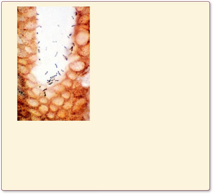
B. A higher magnification of the surface lining cells stained with silver display the presence of H. pylori as small, curved rods. (Reprinted with permission from Rubin R, Strayer D, et al., eds. Rubin’s Pathology. Clinicopathologic Foundations of Medicine, 5th ed. Baltimore, MD: Lippincott, Williams & Wilkins, 2008, p. 563.)
Zollinger-Ellison Syndrome
Zollinger-Ellison syndrome is a cancerous lesion of gastrin-producing cells in the stomach, duodenum, or the pancreas, resulting in the overproduction of HCl by
parietal cells of the stomach and the formation of numerous recurrent peptic ulcers. A high blood level of gastrin, especially after intravenous administration of secretin, usually is a strong indicator of this syndrome.
Antibiotic-Associated Colitis
Antibiotics such as ampicillin, cephalosporin, and clindamycin often cause an imbalance in the intestinal bacterial flora, permitting the vigorous proliferation of Clostridium difficile, resulting in infection by this organism. The two major toxins (Toxin A and Toxin B) produced by C. difficile frequently cause inflammation of the sigmoid colon. Depending on the severity of the infection, the patient will suffer from abdominal cramps, loose stool, bloody diarrhea, fever, and, in extreme cases, dehydration and perforation of the bowel.
Hiatal Hernia
Hiatal hernia is a condition in which a region of the stomach herniates through the esophageal hiatus of the diaphragm. It may be of two types, sliding and paraesophageal hiatal hernia. In the former condition, the cardioesophageal junction and the cardiac region of the stomach slides in and out of the thorax, whereas in the latter case the cardioesophageal junction remains in its normal place, below the diaphragm, but a part (or occasionally all) of the stomach pushes into the thorax and is positioned next to the esophagus. Usually, hiatal hernia is asymptomatic, although acid reflux disease is common in patients afflicted with this condition.
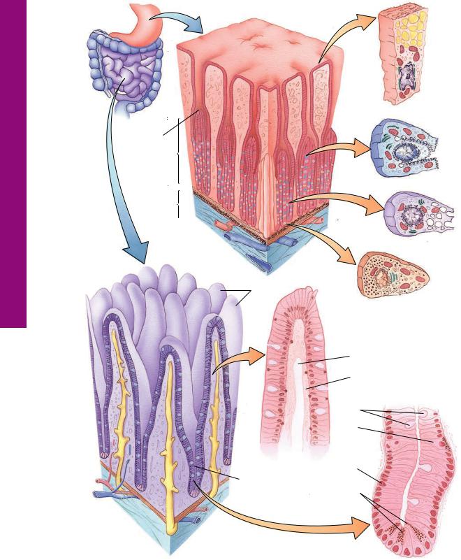
Intestine Small and Stomach • 1-14 GRAPHIC
336 D I G E S T I V E S Y S T E M I I
Mucosa
Gastric pit
pit
Gastric  gland proper
Submucosa
Stomach
Villi
 Lacteal
Gland (crypt
of Lieberkühn)
Small
Intestine
Surface lining cell
Oxyntic (parietal) cell
Zymogenic (chief) cell
Enteroendocrine cell (DNES cell)
 Goblet cells
Lymphatic (lacteal) capillary  Lymphocytes
Smooth muscle  Striated border
Goblet cells DNES cell
Regenerative cell
Paneth cells
D I G E S T I V E S Y S T E M I I 337
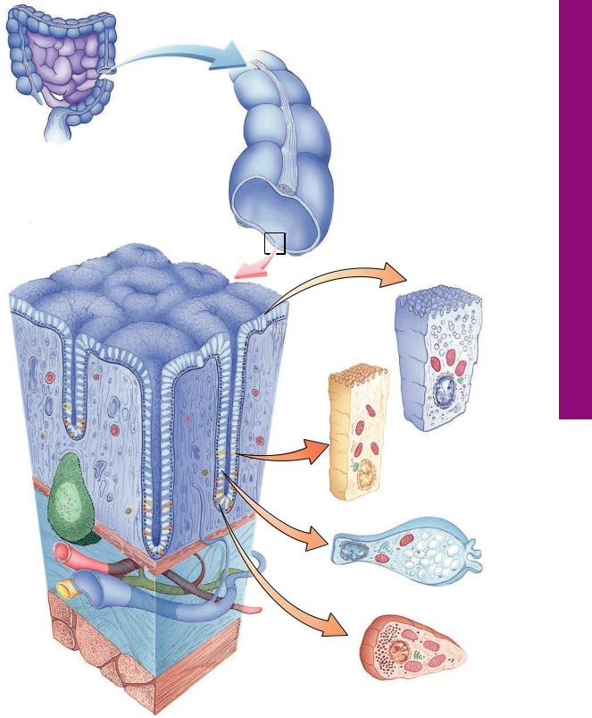
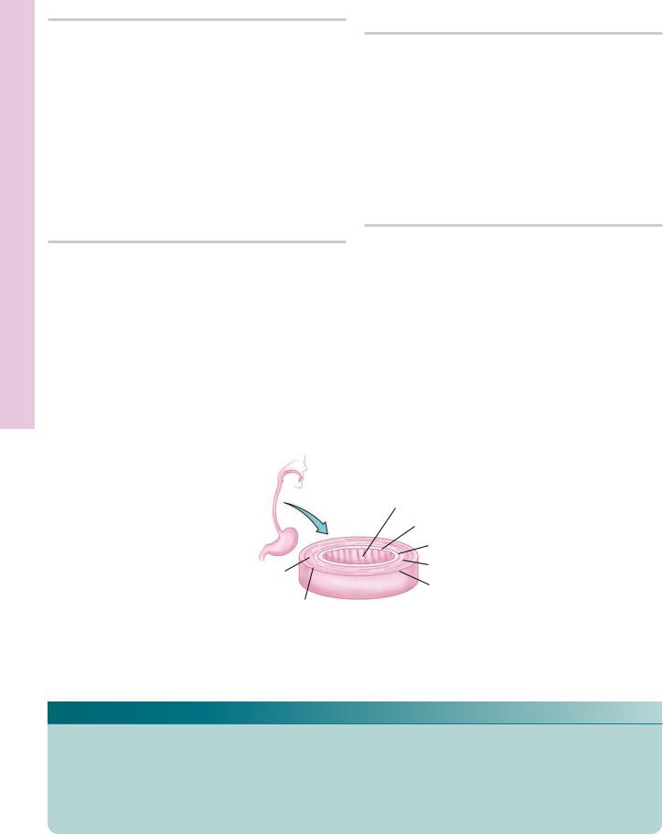
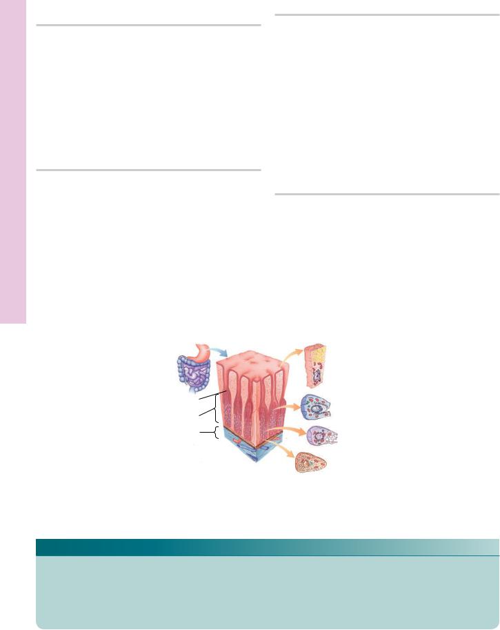
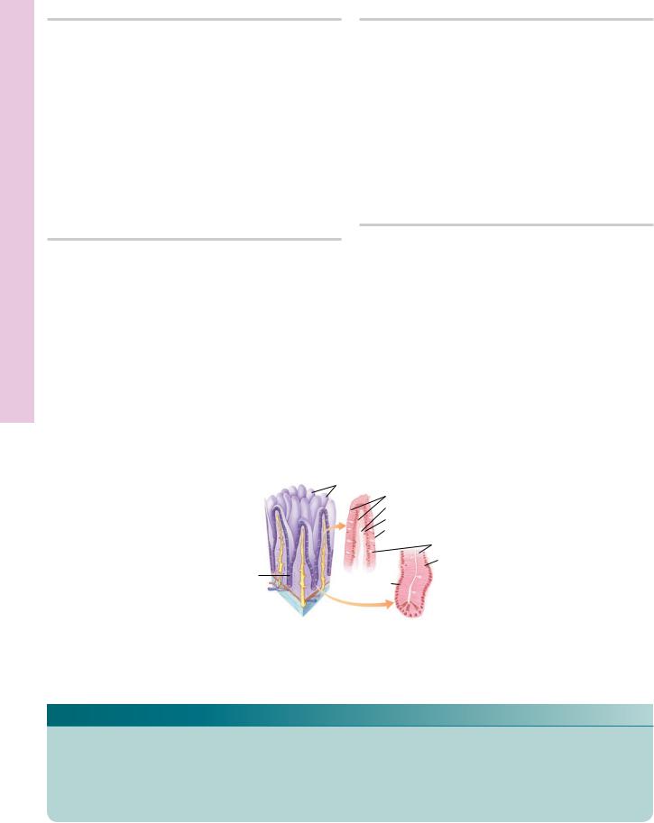
The large intestine has no villi, but it does possess crypts of Lieberkühn. The outer longitudinal layer of the muscularis externa is gathered into the teniae coli. Lymphatic nodules and lymphoid infiltration are frequently noted in the large and small intestines.
 Crypt
Crypt
of
Lieberkühn
The crypts of Lieberkühn are glands composed of a simple columnar type of epithelium. Four types of cells constitute this epithelium: mucus-producing goblet cells; absorptive cells that function in absorbing nutrients, electrolytes, and fluid; regenerative cells that proliferate and replace the other cells of the epithelium; and enteroendocrine cells that release paracrine hormones.
Absorptive cell
Regenerative cell
Goblet cell
Large
Intestine
Enteroendocrine cell (DNES cell)
Intestine Large• 2-14 GRAPHIC
Esophagus• 1-14 PLATE
338 D I G E S T I V E S Y S T E M I I
FIGURE 1. Esophagus. x.s. Paraffin section. ×14.
This photomicrograph of a cross section of the lower one-third of the esophagus displays the general structure of the digestive tract. The lumen (L) is lined by a stratified squamous nonkeratinized epithelium (EP) lying on a thin lamina propria (LP) that is surrounded by the muscularis mucosae (MM). The submucosa (Sm) contains glands and is surrounded by the muscularis externa (ME), composed of an inner circular (IC) and an outer longitudinal (OL) layer. The outermost tunic of the esophagus is the fibroelastic adventitia (Ad). A region similar to the boxed area is presented at a higher magnification in Figure 2.
FIGURE 3. Esophagus. Human. x.s. Paraffin section. ×132.
The lamina propria (LP) and submucosa (Sm) of the esophagus are separated from each other by the longitudinally oriented smooth muscle bundles, the muscularis mucosae (MM). Observe that the lamina propria is a very vascular connective tissue, housing numerous blood vessels (BV) and lymph vessels (LV), whose valves (arrow) indicate the direction of lymph flow. The submucosa also displays numerous blood vessels (BV) as well as the presence of the esophageal glands proper (EG), which produce a mucous secretion to lubricate the lining of the esophagus.
FIGURE 2. Esophagus. Human. x.s. Paraffin section. ×132.
This photomicrograph is a higher magnification of a region similar to the boxed area of the previous figure. The mucosa (M) of the esophagus consists of a stratified squamous nonkeratinized epithelium (EP); a loose collagenous connective tissue layer, the lamina propria (LP); and a longitudinally oriented smooth muscle layer, the muscularis mucosae (MM). The submucosa (Sm) is composed of a coarser collagenous connective tissue (CT), housing blood vessels (BV) and various connective tissue cells whose nuclei (N) are evident.
FIGURE 4. Esophagogastric junction. l.s. Dog. Paraffin section. ×14.
The junction of the esophagus (Es) and cardiac stomach (CS) is very abrupt, as evidenced by the sudden change of the stratified squamous epithelium (SE) to the simple columnar epithelium
(CE) of the stomach. Note that the esophageal glands proper (EG) continue for a short distance into the submucosa (Sm) of the stomach. Observe also the presence of gastric pits (arrows) and the increased thickness of the muscularis externa (ME) of the stomach compared with that of the esophagus. The outermost tunic of the esophagus inferior to the diaphragm is a serosa (Se) rather than an adventitia. The boxed area is presented at a higher magnification in Figure 1 of the next plate.
|
Lumen |
|
Lamina propria |
|
Muscularis mucosae |
Inner circular layer of |
Submucosae |
|
|
muscularis externa |
Adventitia |
Outer layer of muscularis externa
Esophagus
KEY
Ad |
adventitia |
Es |
esophagus |
MM |
muscularis mucosae |
BV |
blood vessels |
IC |
inner circular muscle |
N |
nucleus |
CE |
simple columnar epithelium |
L |
lumen |
OL |
outer longitudinal muscle |
CS |
cardiac stomach |
LP |
lamina propria |
SE |
stratified squamous epithelium |
CT |
connective tissue |
LV |
lymph vessels |
Se |
serosa |
EG |
esophageal glands proper |
M |
mucosa |
Sm |
submucosa |
EP |
epithelium |
ME |
muscularis externa |
|
|

EP
LP
M
MM
BV
N
CT
BV SM
FIGURE 1 |
FIGURE 2 |
LP
BV
LV
MM
SM
BV
EG
Esophagus• 1-14 PLATE
FIGURE 3 |
FIGURE 4 |
omach • St2-14 PLATE
340 D I G E S T I V E S Y S T E M I I
FIGURE 1. Esophagogastric junction. l.s. Dog. Paraffin section. ×132.
This photomicrograph is a higher magnification of the boxed region of Figure 4, Plate 14-1. The stratified squamous epithelium (SE) of the esophagus is replaced by the simple columnar epithelium (CE) of the stomach in a very abrupt fashion (arrow). The lamina propria (LP) displays gastric pits (GP), lined by the typical mucussecreting surface lining cells (SC), characteristic of the stomach. The structure labeled with an asterisk is not a lymphatic nodule but is a more or less tangential section through the esophageal epithelium. Note the presence of the muscularis mucosae (MM).
FIGURE 3. Fundic stomach. x.s. Dog. Paraffin section. ×132.
This photomicrograph presents a higher magnification of a region similar to the boxed area of Figure 2. The mucosa of the fundic stomach displays numerous gastric pits (GP) that are lined by a simple columnar epithelium, consisting mostly of mucus-produc- ing surface lining (surface mucous) cells (SC). The base of each pit accepts the isthmus of two to four fundic glands (FG). Although fundic glands are composed of several cell types, only two, parietal cells (PC) and chief cells (CC), are readily distinguishable in this preparation. The lamina propria (LP) is richly vascularized (BV). Note the muscularis mucosae (MM) beneath the lamina propria. A region similar to the boxed area is presented at a higher magnification (positioned at a 90 degree angle) in Figure 4.
FIGURE 2. Fundic stomach. l.s. Paraffin section. ×14.
The fundic region presents all of the characteristics of the stomach, as demonstrated by this low-power photomicrograph. The lumen (L) is lined by a simple columnar epithelium, deep to which is the lamina propria (LP), housing numerous gastric glands
(GG). Each gland opens into the base of a gastric pit (GP). The muscularis mucosae (MM) separates the lamina propria from the submucosa (Sm), a richly vascularized (BV) connective tissue, thrown into folds (rugae) in the empty stomach. The muscularis externa (ME) is composed of three poorly defined layers of smooth muscle: innermost oblique (IO), middle circular (MC), and outer longitudinal (OL). Serosa (arrow) forms the outermost tunic of the stomach. A region similar to the boxed area is presented at a higher magnification in Figure 3.
FIGURE 4. Fundic glands. x.s. Paraffin section. ×540.
This photomicrograph presents a higher magnification (positioned at a 90 degree angle) of a region similar to the boxed area of Figure 3. The lumina (L) of several glands can be recognized. Note that chief cells (CC) are granular in appearance and are much smaller than the round, plate-like parietal cells (PC). Parietal cells, as their name implies, are located at the periphery of the gland. Slender connective tissue (CT) elements, housing blood vessels, occupy the narrow spaces between the closely packed glands.
Surface lining cell
Gastric pit
Parietal cell
Gastric gland
Sumucosa
Chief cell
DNES cell
Stomach and cells
KEY
BV |
blood vessels |
GP |
gastric pits |
MM |
muscularis mucosae |
CC |
chief cells |
IO |
innermost oblique muscle |
OL |
outer longitudinal muscle |
CE |
columnar epithelium |
L |
lumen |
PC |
parietal cells |
CT |
connective tissue |
LP |
lamina propria |
SC |
surface lining cells |
FG |
fundic glands |
ME |
muscularis externa |
SE |
squamous epithelium |
GG |
gastric glands |
MC |
middle circular muscle |
Sm |
submucosa |
|
SE |
CE |
MM |
|
LP
SC
GP
|
FIGURE 1 |
FIGURE 2 |
|
GP |
SC |
||
CT |
|||
|
|
||
|
|
|
|
CC
PC
BV
|
FG |
|
|
|
PC |
|
|
LP |
PC |
|
|
L |
CC |
||
CC |
|||
|
CT
BV
MM
FIGURE 3 |
FIGURE 4 |
omach • St2-14 PLATE

omach • St3-14 PLATE
342 D I G E S T I V E S Y S T E M I I
FIGURE 1. Fundic stomach. x.s. Monkey. Plastic section. ×270.
The gastric pits (GP) of the fundic stomach are lined mostly by mucus-producing surface lining cells (SC). Each gastric pit receives two to four fundic glands, simple tubular structures that are subdivided into three regions: isthmus, neck, and base. The isthmus opens directly into the gastric pit and is composed of immature cells (Ic), which are responsible for the renewal of the lining of the gastric mucosa, surface lining cells (SC), and parietal cells (PC). The neck and base of these glands are presented in Figure 2.
FIGURE 2. Fundic gland. Stomach. x.s. Monkey. Plastic section. ×270.
The neck (n) and base (b) of the fundic gland both contain the large, plate-shaped parietal cells (PC). The neck also possesses a few immature cells as well as mucous neck cells (Mn), which manufacture a mucous substance. The base of the fundic glands contains numerous acid-manufacturing parietal cells (PC) and chief cells (CC), which produce digestive enzymes. Note that the lamina propria is tightly packed with glands and that the intervening connective tissue (CT) is flimsy. The bases of these glands extend to the muscularis mucosae (MM).
FIGURE 3. Pyloric gland. Stomach. x.s. Monkey. Plastic section. ×132.
The mucosa of the pyloric region of the stomach presents gastric pits (GP) that are deeper than those of the cardiac or fundic regions. The deep aspects of these pits are coiled (arrows). As in the other regions of the stomach, the epithelium (Ep) is simple columnar, consisting mainly of surface lining cells (SC). Note that the lamina propria (LP) is loosely packed with pyloric glands (PGs) and that considerable connective tissue (CT) is present. The pyloric glands are composed mainly of mucous cells (mc). Observe the two muscle layers of the muscularis mucosae (MM). A region similar to the boxed area is presented in Figure 4.
FIGURE 4. Pyloric gland. Stomach. x.s. Human. Paraffin section. ×270.
This is a photomicrograph of a region similar to the boxed area of Figure 3. The simple columnar epithelium (Ep) of the gastric pit is composed mostly of surface lining cells. These pits are not only much deeper than those of the fundic or cardiac regions but are also somewhat coiled (arrow), as are the pyloric glands (PG), which empty into the base of the pits. These glands are populated by mucus-secreting cells (mc) similar to mucous neck cells, whose nuclei (N) are flattened against the basal cell membrane. Note that the glands are not closely packed and that the lamina propria (LP) is very cellular and possesses a rich vascular supply (BV).
Surface lining cell
Gastric pit
Parietal cell
Gastric gland
Sumucosa
Chief cell
DNES cell
Stomach and cells
KEY
B |
base |
IC |
immature cells |
N |
neck |
BV |
blood vessels |
LP |
lamina propria |
PC |
parietal cells |
CC |
chief cells |
Mc |
mucous cells |
PG |
pyloric glands |
CT |
connective tissue |
MM |
muscularis mucosae |
SC |
surface lining cells |
EP |
epithelium |
Mn |
mucous neck cell |
|
|
GP |
gastric pits |
N |
nucleus |
|
|
SC |
|
PC |
|
|
|
|
|
GP |
CT |
n |
Mn |
|
|||
|
|
|
lc
b
CC
PC
MM
PC
FIGURE 1 |
FIGURE 2 |
EP
BV
LP
MM
mc
GP
CT
PG
N
GP
|
PG |
LP |
|
EP |
SC |
||
|
|||
|
BV |
|
 mc mc
mc mc
FIGURE 3 |
FIGURE 4 |
omach • St3-14 PLATE

Duodenum• 4-14 PLATE
344 D I G E S T I V E S Y S T E M I I
FIGURE 1a. Duodenum. l.s. Monkey. Plastic section. Montage. ×132.
The lamina propria of the duodenum possesses finger-like evaginations known as villi (V), which project into the lumen (L). The villi are covered by surface absorptive cells (SA), a simple columnar type of epithelium with a brush border. Interspersed among these surface absorptive cells are goblet cells (GC) as well as occasional APUD cells. The connective tissue (CT) core (lamina propria) of the villus is composed of lymphoid and other cellular elements whose nuclei stain very intensely. Blood vessels also abound in the lamina propria, as do large, blindly ending lymphatic channels known as lacteals (l), recognizable by their large size and lack of red blood cells. Frequently, these lacteals are collapsed. The deeper aspect of the lamina propria houses glands, the crypts of Lieberkühn (CL). These simple tubular glands deliver their secretions into the intervillar spaces. The bases of these crypts reach the muscularis mucosae (MM), composed of inner circular and outer longitudinal layers of smooth muscle. Deep to this muscle layer is the submucosa, which, in the duodenum, is occupied by compound tubular glands of Brunner (GB). These glands deliver their mucous secretion via ducts (D), which pierce the muscularis mucosae, into the crypts of Lieberkühn. A region similar to the boxed area is presented at a higher magnification in Figure 1b.
FIGURE 2. Duodenum. l.s. Monkey. Plastic section. ×132.
This photomicrograph is a continuation of the montage presented in Figure 1a (compare asterisks). Note that the submucosa (Sm), occupied by glands of Brunner (GB), is a vascular structure (BV) and also houses Meissner’s submucosal plexus. The submucosa extends to the muscularis externa (ME), composed of an inner circular (IC) and outer longitudinal (OL) smooth muscle layer. Note the presence of Auerbach’s myenteric plexus (AP) between these two muscle layers. The duodenum, in part, is covered by a serosa (Se), whose mesothelium provides this organ with a smooth, moist surface.
FIGURE 3a. Duodenum. x.s. Monkey. Plastic section. ×540.
The base of the crypt of Lieberkühn displays the several types of cells that compose this gland. Paneth’s cells (Pc) are readily recognizable due to the large granules in their apical cytoplasm. DNES cells (APD) are clear cells with fine granules usually located basally. Goblet cells (GC), columnar cells (Cc), and stem cells (Sc) constitute the remaining cell population.
FIGURE 1b. Epithelium and core of villus. Monkey. Plastic section. ×540.
This higher magnification of a region similar to the boxed area presents the epithelium and part of the connective tissue core of a villus. Note that the surface absorptive cells (SA) display a brush border (BB), terminal bars (arrow), and goblet cells (GC). Although APUD cells are also present, they constitute only a small percentage of the cell population. The lamina propria (LP) core of the villus is highly cellular, housing lymphoid cells (LC), smooth muscle cells (SM), mast cells, macrophages (Ma), and fibroblasts, among others.
FIGURE 3b. Duodenum. x.s. Monkey. Plastic section. ×540.
The submucosa of the intestinal tract displays small parasympathetic ganglia, Meissner’s submucosal plexus. Note the large postganglionic cell bodies (PB) surrounded by elements of connective tissue (CT).
KEY
AP |
Auerbach’s plexus |
GC |
goblet cell |
OL |
outer longitudinal muscle |
APD |
DNES cell |
IC |
inner circular muscle |
PB |
postganglionic cell body |
BB |
brush border |
I |
lacteal |
Pc |
Paneth’s cell |
BV |
blood vessels |
L |
lumen |
SA |
surface absorptive cell |
Cc |
columnar cell |
LC |
lymphoid cell |
Sc |
stem cell |
CL |
crypts of Lieberkühn |
LP |
lamina propria |
Se |
serosa |
CT |
connective tissue |
Ma |
macrophage |
Sm |
submucosa |
D |
duct |
ME |
muscularis externa |
SM |
smooth muscle cell |
GB |
glands of Brunner |
MM |
muscularis mucosae |
V |
villi |

a) |
L |
||
|
|
|
|
|
|
V |
GB |
GC |
|
|
BV |
|
|
|
|
|
|
|
Sm |
|
|
|
|
|
SA l |
|
|
BV |
CT |
IC |
|
|
|
|
|
|
|
|
|
|
|
|
|
|
|||
|
|
|
|
||||||
CL |
|
ME |
|||||||
|
AP |
|
|
|
|
||||
|
|
|
|
|
|||||
|
|
|
|
|
|
||||
|
|
OL |
|
|
|
|
|||
MM |
|
Se |
|
|
|
|
|
||
|
|
|
|
|
|
||||
|
FIGURE 2 |
||||||||
D |
a) |
APD |
|||||||
|
|||||||||
GB |
|
Cc |
|||||||
|
|
|
|
|
|
|
|
|
|
GC
Pc |
Sc |
|
|
FIGURE 1a |
FIGURE 3a |
|
b) |
|
|
b) |
|
|
LP |
|
LC |
CT |
|
|
|||
|
|
|
|
GC |
|
|
|
|
SA |
|
|
Ma |
PB |
|
SM |
BB |
|||
|
|
|
|
CT |
|
|
FIGURE 1b |
FIGURE 3b |
|
Duodenum• 4-14 PLATE
Ileum Jejunum,• 5-14 PLATE
346 D I G E S T I V E S Y S T E M I I
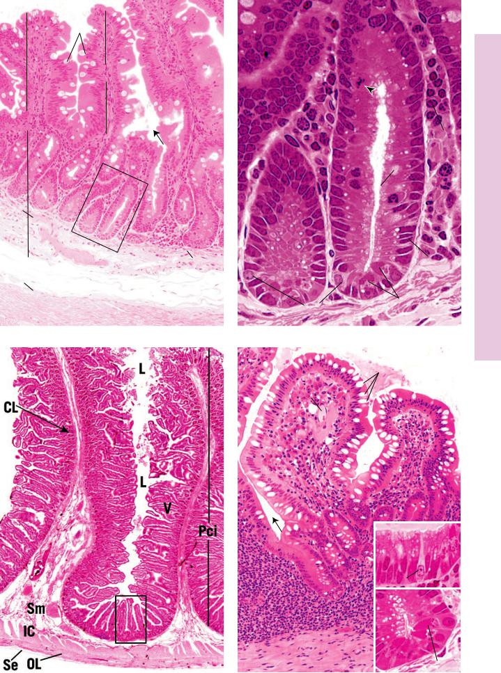
FIGURE 1. Jejunum. x.s. Monkey. Plastic section. ×132.
The mucosa (M) and submucosa (Sm) of the jejunum are presented in this photomicrograph. The villi (V) of this region possess more goblet cells (GC) than those of the duodenum. Observe that the crypts of Lieberkühn (CL) open into the intervillar spaces (arrow) and that the lamina propria displays numerous dense nuclei, evidence of lymphatic infiltration. The flimsy muscularis mucosae (MM) separates the lamina propria from the submucosa. Large blood vessels (BV) occupy the submucosa, which is composed of a loose type of collagenous connective tissue. The inner circular (IC) layer of the muscularis externa is evident at the bottom of the photomicrograph. The boxed region is presented at a higher magnification in Figure 2.
FIGURE 3. Ileum. l.s. Human. Paraffin section. ×14.
The entire wall of the ileum is presented, displaying spiral folds of the submucosa that partially encircle the lumen. These folds, known as plicae circulares (Pci), increase the surface area of the small intestines. Note that the lamina propria is clearly delineated from the submucosa (Sm) by the muscularis mucosae. The lamina propria forms numerous villi (V) that protrude into the lumen (L); glands known as crypts of Lieberkühn (CL) deliver their secretions into the intervillar spaces. The submucosa abuts the inner circular (IC) layer of smooth muscle that, in turn, is surrounded by the outer longitudinal (OL) smooth muscle layer of the muscularis externa. Observe the serosa (Se) investing the ileum. A region similar to the boxed area is presented at a higher magnification in Figure 4.
FIGURE 2. Jejunum. x.s. Monkey. Plastic section. ×540.
This photomicrograph is a higher magnification of the boxed area of Figure 1. The crypts of Lieberkühn are composed of several cell types, some of which are evident in this figure. Goblet cells (GC) that manufacture mucus may be noted in various degrees of mucus production. Narrow stem cells (Sc) undergo mitotic activity (arrowhead), and newly formed cells reconstitute the cell population of the crypt and villus. Paneth’s cells (PC) are located at the base of crypts and may be recognized by their large granules. DNES cells (APD) appear as clear cells, with fine granules usually basally located. The lamina propria displays numerous plasma cells (PlC).
FIGURE 4. Ileum. x.s. Monkey. Plastic section. ×132.
This is a higher magnification of a region similar to the boxed area of Figure 3. Note that the villi (V) are covered by a simple columnar epithelium, whose cellular constituents include numerous goblet cells (GC). The core of the villus displays blood vessels (BV) as well as a large lymphatic vessel known as a lacteal (l). The crypts of Lieberkühn (CL) open into the intervillar spaces (arrow). The group of lymphatic nodules of the ileum are known as Peyer’s patches (PP). Inset a. Crypt of Lieberkühn. l.s. Monkey. Plastic section. ×540. The crypts of Lieberkühn also possess DNES cells (APD), recognizable by their clear appearance and usually basally oriented fine granules. Inset b. Crypt of Lieberkühn. l.s. Monkey. Plastic section. ×540. The base of the crypt of Lieberkühn displays cells with large granules. These are Paneth’s cells (PC), which produce the bacteriocidal agent lysozyme and other substances.
Villi
Lymphocytes
Lymphatic (lacteal) capillary
Smooth muscle
Striated border
|
|
Goblet cells |
Lacteal |
|
DNES cell |
|
Gland (crypt
of Lieberkühn) Regenerative
cell
Paneth cells 
Small intestine
KEY
APD |
DNES cell |
L |
lumen |
PP |
Peyer’s patch |
BV |
blood vessels |
M |
mucosa |
OL |
outer longitudinal muscle |
CL |
crypts of Lieberkühn |
MM |
muscularis mucosae |
Sc |
stem cell |
GC |
goblet cell |
PC |
Paneth’s cell |
Se |
serosa |
IC |
inner circular muscle |
PCi |
plicae circulares |
Sm |
submucosa |
I |
lacteal |
PIC |
plasma cell |
V |
villi |
GC
V
M
PlC
CL |
GC |
|
Sm |
BV |
MM |
|
SC |
|
|
|
||||
|
|
IC |
|
APD |
PC |
|
|
|
|||
|
|
|
|
||
|
|
|
FIGURE 1 |
|
FIGURE 2 |
|
|
|
|
|
GC |
|
|
|
|
BV |
|
|
|
|
|
V |
l |
|
|
|
|
|
|
|
a) |
|
CL |
PP |
APD |
b) |
 PC
PC
FIGURE 3 |
FIGURE 4 |
Ileum Jejunum,• 5-14 PLATE
Appendix olon, • C6-14 PLATE
348 D I G E S T I V E S Y S T E M I I
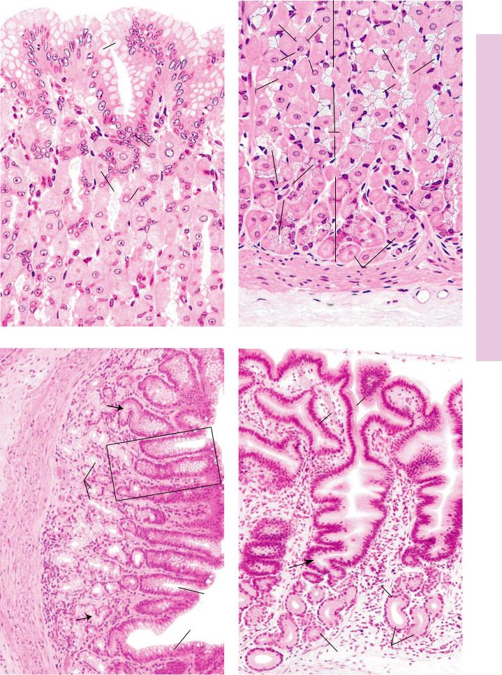
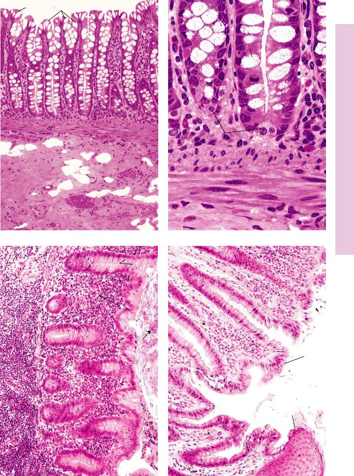
FIGURE 1. Colon. l.s. Monkey. Plastic section. ×132.
This photomicrograph depicts the mucosa and part of the submucosa of the colon. Note the absence of surface modifications such as pits and villi, which indicate that this section is not of the stomach or small intestines. The epithelium (Ep) lining the lumen (L) is simple columnar with numerous goblet cells (GC). The straight tubular glands are crypts of Lieberkühn (CL), which extend down to the muscularis mucosae (MM). The inner circular (IC) and outer longitudinal (OL) layers of smooth muscle comprising this region of the mucosa are evident. The submucosa (Sm) is very vascular (BV) and houses numerous fat cells (FC). The boxed area is presented at a higher magnification in Figure 2.
FIGURE 3. Appendix. x.s. Paraffin section. ×132.
The cross section of the appendix displays a lumen (L) that frequently contains debris (arrow). The lumen is lined by a simple columnar epithelium (Ep), consisting of many goblet cells (GC). Crypts of Lieberkühn (CL) are relatively shallow in comparison with those of the colon. The lamina propria (LP) is highly infiltrated with lymphoid cells (LC), derived from lymphatic nodules
(LN) of the submucosa (Sm) and lamina propria. The muscularis mucosae (MM) delineates the border between the lamina propria and the submucosa.
FIGURE 2. Colon. l.s. Monkey. Plastic section. ×540.
This photomicrograph is a higher magnification of the boxed area of Figure 1. The cell population of the crypts of Lieberkühn (CL) is composed of numerous goblet cells (GC), which deliver their mucus into the lumen (L) of the crypt. Surface epithelial cells
(SEC) as well as undifferentiated stem cells are also present. The latter undergo mitosis (arrow) to repopulate the epithelial lining. DNES cells (APD) constitute a small percentage of the cell population. Note that Paneth’s cells are not present in the colon. The lamina propria (LP) is very cellular, housing many lymphoid cells (LC). The inner circular (IC) and outer longitudinal
(OL) smooth muscle layers of the muscularis mucosae (MM) are evident.
FIGURE 4. Anorectal junction. l.s. Human. Paraffin section. ×132.
The anorectal junction presents a superficial similarity to the esophagogastric junction because of the abrupt epithelial transition. The simple columnar epithelium (CE) of the rectum is replaced by the stratified squamous epithelium of the anal canal (AC). The crypts of Lieberkühn (CL) of the anal canal are shorter than those of the colon. The lamina propria (LP) is infiltrated by lymphoid cells (LC).
|
|
Absorptive cell |
Crypt of |
|
Regenerative cell |
|
||
Lieberkühn |
|
|
|
|
|
|
|
Goblet cell |
|
|
DNES cell |
Large intestine
KEY
AC |
anal canal |
FC |
fat cell |
LP |
lamina propria |
APD |
DNES cell |
GC |
goblet cell |
MM |
muscularis mucosae |
BV |
blood vessels |
IC |
inner circular muscle |
OL |
outer longitudinal muscle |
CE |
simple columnar epithelium |
L |
lumen |
SE |
stratified squamous epithelium |
CL |
crypts of Lieberkühn |
LC |
lymphoid cell |
SEC |
surface epithelial cell |
EP |
epithelium |
LN |
lymphatic nodule |
Sm |
submucosa |
EP |
GC |
L |
CL |
|
|
|
|
|
GC |
|
|
|
|
GC |
|
|
|
|
L |
CL |
|
|
|
SEC |
|
|
|
||
|
|
|
LP |
|
|
|
|
|
LC |
MM |
|
|
IC |
|
|
|
OL |
|
|
|
|
|
APD |
|
|
|
|
||
|
|
|
|
|
IC
Sm FC MM
OL
BV
FIGURE 1 FIGURE 2
SM |
MM |
GC |
|
|
LP |
|
|
|
|
|
|
|
|
LC |
|
|
|
L |
|
|
CL |
CL |
LC |
CE
AC
LP
SE
EP
LN
FIGURE 3 |
FIGURE 4 |
Appendix olon, • C6-14 PLATE

350 D I G E S T I V E S Y S T E M I I
Microscopy Electron olon, • C7-14 PLATE
FIGURE 1
FIGURE 2
FIGURE 1. Colon. Rat. Electron microscopy. ×3,780. |
|
FIGURE 2. Colon. Rat. Electron microscopy. ×12,600. |
The deep aspect of the crypt of Lieberkühn presents columnar cells (c) and deep crypt cells that produce a mucous type of secretion that is delivered into the lumen (L) of the crypt. (From Altmann GG. Morphological observations on mucus-secreting nongoblet cells in the deep crypts of the rat ascending colon. Am J Anat 1983;167:95–117.)
At a higher magnification of the deep aspect of the crypt of Lieberkühn, the deep crypt cells present somewhat electrondense vacuoles (m). Note that many of these vacuoles coalesce, forming amorphous vacuolar profiles. The slender columnar cell
(C) displays no vacuoles but does possess numerous mitochondria and occasional profiles of rough endoplasmic reticulum. Observe the large, oval nucleus and clearly evident nucleolus. (From Altmann GG. Morphological observations on mucus-secret- ing nongoblet cells in the deep crypts of the rat ascending colon. Am J Anat 1983;167:95–117.)
D I G E S T I V E S Y S T E M I I 351
Microscopy Electron Scanning olon, • C8-14 PLATE
FIGURE 1
FIGURE 1. Colon. Monkey. Scanning electron microscopy. ×614.
This scanning electron micrograph displays the openings of the crypts of Lieberkühn (CL) as well as the cells lining the mucosal surface. (From Specian RD, Neutra MR. The surface topography of the colonic crypt in rabbit and monkey.
Am J Anat 1981;160:461–472.) Inset. Colon. Rabbit. Scanning electron microscopy. ×778. The openings of the crypts of Lieberkühn are not as regularly arranged in the rabbit as in the monkey. Observe the mucus arising from the crypt opening (arrow). (From Specian RD, Neutra MR. The surface topography of the colonic crypt in rabbit and monkey. Am J Anat 1981;160:461–472.)
Crypt of
Lieberkühn
Large intestine