

- •Preface
- •Acknowledgments
- •Reviewers
- •Contents
- •CHAPTER OUTLINE
- •CYTOPLASM
- •Plasmalemma
- •Mitochondria
- •Ribosomes
- •Endoplasmic Reticulum
- •Golgi Apparatus, cis-Golgi Network, and the trans-Golgi Network
- •Endosomes
- •Lysosomes
- •Peroxisomes
- •Proteasomes
- •Cytoskeleton
- •Inclusions
- •NUCLEUS
- •CELL CYCLE
- •CHAPTER OUTLINE
- •EPITHELIUM
- •Epithelial Membranes
- •GLANDS
- •Chapter Summary
- •CHAPTER OUTLINE
- •EXTRACELLULAR MATRIX
- •Fibers
- •Amorphous Ground Substance
- •Extracellular Fluid
- •CELLS
- •CONNECTIVE TISSUE TYPES
- •Chapter Summary
- •CHAPTER OUTLINE
- •CARTILAGE
- •BONE
- •Cells of Bone
- •Osteogenesis
- •Bone Remodeling
- •Chapter Summary
- •CHAPTER OUTLINE
- •FORMED ELEMENTS OF BLOOD
- •Lymphocytes
- •Neutrophils
- •PLASMA
- •COAGULATION
- •HEMOPOIESIS
- •Erythrocytic Series
- •Granulocytic Series
- •Chapter Summary
- •CHAPTER OUTLINE
- •SKELETAL MUSCLE
- •Sliding Filament Model of Muscle Contraction
- •CARDIAC MUSCLE
- •SMOOTH MUSCLE
- •Chapter Summary
- •CHAPTER OUTLINE
- •BLOOD-BRAIN BARRIER
- •NEURONS
- •Membrane Resting Potential
- •Action Potential
- •Myoneural Junctions
- •Neurotransmitter Substances
- •SUPPORTING CELLS
- •PERIPHERAL NERVES
- •Chapter Summary
- •CHAPTER OUTLINE
- •BLOOD VASCULAR SYSTEM
- •HEART
- •ARTERIES
- •Capillary Permeability
- •Endothelial Cell Functions
- •VEINS
- •LYMPH VASCULAR SYSTEM
- •Chapter Summary
- •CHAPTER OUTLINE
- •CELLS OF THE IMMUNE SYSTEM
- •Antigen-Presenting Cells
- •DIFFUSE LYMPHOID TISSUE
- •LYMPH NODES
- •TONSILS
- •SPLEEN
- •THYMUS
- •Chapter Summary
- •CHAPTER OUTLINE
- •PITUITARY GLAND
- •Pars Intermedia
- •Pars Nervosa and Infundibular Stalk
- •Pars Tuberalis
- •THYROID GLAND
- •Parathyroid Glands
- •Suprarenal Glands
- •Cortex
- •Medulla
- •Pineal Body
- •Chapter Summary
- •CHAPTER OUTLINE
- •SKIN
- •Epidermis of Thick Skin
- •Dermis
- •DERIVATIVES OF SKIN
- •Chapter Summary
- •CHAPTER OUTLINE
- •CONDUCTING PORTION OF THE RESPIRATORY SYSTEM
- •Extrapulmonary Region
- •Intrapulmonary Region
- •RESPIRATORY PORTION OF THE RESPIRATORY SYSTEM
- •MECHANISM OF RESPIRATION
- •Chapter Summary
- •CHAPTER OUTLINE
- •ORAL CAVITY AND ORAL MUCOSA
- •Oral Mucosa
- •Tongue
- •Teeth
- •Odontogenesis (See Graphic 13-2)
- •Chapter Summary
- •CHAPTER OUTLINE
- •REGIONS OF THE DIGESTIVE TRACT
- •Esophagus
- •Stomach
- •Small Intestine
- •Large Intestine
- •GUT-ASSOCIATED LYMPHOID TISSUE
- •DIGESTION AND ABSORPTION
- •Carbohydrates
- •Proteins
- •Lipids
- •Water and Ions
- •Chapter Summary
- •CHAPTER OUTLINE
- •MAJOR SALIVARY GLANDS
- •PANCREAS
- •LIVER
- •Exocrine Function of the Liver
- •Endocrine and Other Functions of the Liver
- •GALLBLADDER
- •Chapter Summary
- •CHAPTER OUTLINE
- •KIDNEY
- •Uriniferous Tubule
- •Nephron
- •Collecting Tubules
- •FORMATION OF URINE FROM ULTRAFILTRATE
- •EXTRARENAL EXCRETORY PASSAGES
- •Chapter Summary
- •CHAPTER OUTLINE
- •OVARY
- •Ovarian Follicles
- •Regulation of Follicle Maturation and Ovulation
- •Corpus Luteum and Corpus Albicans
- •GENITAL DUCTS
- •Oviduct
- •Uterus
- •FERTILIZATION, IMPLANTATION, AND THE PLACENTA
- •Fertilization and Implantation
- •Placenta
- •VAGINA
- •EXTERNAL GENITALIA
- •MAMMARY GLANDS
- •Chapter Summary
- •CHAPTER OUTLINE
- •TESTES
- •Spermatogenesis
- •GENITAL DUCTS
- •ACCESSORY GLANDS
- •PENIS
- •Erection and Ejaculation
- •Chapter Summary
- •CHAPTER OUTLINE
- •SENSORY ENDINGS
- •Chapter Summary
- •Terminology of Staining
- •Common Stains Used in Histology
- •Hematoxylin and Eosin
- •Wright Stain
- •Weigert Method for Elastic Fibers and Elastic van Gieson Stain
- •Silver Stain
- •Iron Hematoxylin
- •Bielschowsky Silver Stain
- •Masson Trichrome
- •Periodic Acid-Schiff Reaction (PAS)
- •Alcian Blue
- •von Kossa Stain
- •Sudan Red
- •Mucicarmine Stain
- •Safranin-O
- •Toluidine Blue

Larynx and Trachea
The conducting portion of the respiratory system is supported by a skeleton composed of bone and/or cartilage that assists in the maintenance of a patent lumen, whose diameters are controlled by smooth muscle cells located in their walls.
•The larynx, a region of the conducting portion, is designed for phonation and to prevent food, liquids, and other foreign objects from gaining access to its lumen.
It is composed of three paired and three unpaired cartilages, numerous extrinsic and intrinsic muscles, and several ligaments.
The actions of these muscles on the cartilages and ligaments modulate the tension and positioning of the vocal folds, thus permitting variations in the pitch of the sound being produced.
The lumen of the larynx is subdivided into three compartments: vestibule, ventricle, and infraglottic cavity.
•The trachea, continuous with the lumen of the infraglottic cavity, is supported by 15 to 20 C-rings, horseshoe-shaped segments of hyaline cartilage. The trachea has three layers, the mucosa, submucosa, and adventitia. It is the adventitia that houses the C-rings, whose open ends face posteriorly and are connected by a smooth muscle slip, the trachealis muscle. Contraction of this muscle reduces the lumen of the trachea, thus increasing the velocity of air flow.
The tracheal lumen is lined by a pseudostratified ciliated columnar epithelium, known as respiratory epithelium. This epithelium is composed of various cell types, namely, goblet cells, ciliated cells, basal cells, brush cells, serous cells, and hormone-produc- ing diffuse neuroendocrine system (DNES) cells.
Goblet cells constitute about 30% of the epithelial cells. Goblet cells are unicellular glands that produce mucinogen, a mucous substance that is released onto the wet epithelial surface where it becomes hydrated to form mucin. Once particular substances located in the tracheal lumen are intermixed with mucin, that viscous material becomes known as mucus.
Ciliated cells also compose about 30% of the cell population. They are tall, ciliated cells whose cilia sweep the mucus toward the larynx.
Basal cells also constitute approximately 30% of the epithelial cell population. They are regenerative cells that function in replacing the epithelial lining of the trachea.
Brush cells form only 3% of the cell population of the respiratory epithelium. They possess small mucinogen-containing granules in their
R E S P I R A T O R Y S Y S T E M 279
cytoplasm and long microvilli that reach into lumen of the trachea. Brush cells may have neurosensory functions or they may be defunct goblet cells that released their mucinogen.
Serous cells are tall, columnar cells whose cytoplasm houses small vesicles containing a serous secretion whose function is not understood.Serous cells form 3% of the epithelial cell population.
DNES cells constitute 3% to 4% of the epithelial cell population, and they form polypeptide hormones that they store in small granules localized in their basal cytoplams. When released, these hormones may act locally (paracrine hormones) or at a distance (hormones) to regulate respiratory functions. Nerve fibers often contact many of these DNES cells, to form structures, known as pulmonary neuroepithelial bodies, that by monitoring local hypoxic conditions can alert the brain’s respiratory center to increase respiration.
The trachea subdivides into the two primary bronchi that lead to the right and the left lungs.
Intrapulmonary Region
The intrapulmonary region is composed of intrapulmonary bronchi (secondary bronchi), whose walls are supported by irregular plates of hyaline cartilage.
•Each intrapulmonary bronchus gives rise to several bronchioles, tubes of decreasing diameter that do not possess a cartilaginous supporting skeleton.
The epithelial lining of the larger bronchioles is ciliated with a few goblet cells, but those of smaller bronchioles are simple columnar, with goblet cells being replaced by Clara cells. Moreover, the thickness of their walls also decreases, as does the luminal diameter.
The last region of the conduction portion is composed of terminal bronchioles, whose mucosa is further decreased in thickness and complexity. The patency of those airways whose walls do not possess a cartilaginous support is maintained by elastic fibers that radiate from their periphery and intermingle with elastic fibers emanating from nearby structures.
RESPIRATORY PORTION OF THE RESPIRATORY SYSTEM
The respiratory portion of the respiratory system begins with branches of the terminal bronchiole known as respiratory bronchioles (see Graphic 12-2).
•Respiratory bronchioles are very similar to terminal bronchioles except that they possess outpocketings

280 R E S P I R A T O R Y S Y S T E M
TABLE 12-2 • Components of the Blood-Air Barrier
Endothelial Component |
Epithelial and Pneumocyte Component |
Pneumocyte Component |
|
|
|
Attenuated endothelial cell |
Combined basal laminae |
Attenuated pneumocyte I |
|
|
Surfactant and fluid coating of the alveolus |
known as alveoli, structures whose thin walls permit gaseous exchange.
•Respiratory bronchioles lead to alveolar ducts, each of which ends in an expanded region known as an alveolar sac, with each sac being composed of a number of alveoli. The epithelium of alveolar sacs and alveoli is composed of two types of cells:
highly attenuated type I pneumocytes, which form much of the lining of the alveolus and alveolar sac, and
type II pneumocytes, are cells that
manufacture surfactant, a phospholipid that reduces surface tension of the alveolar surface
enter the cell cycle to form more type I and type II pneumocytes.
Associated with the respiratory portion of the lungs is an extremely rich capillary network, supplied by the pulmonary arteries and drained by the pulmonary veins.
•The capillaries invest each alveolus, and their highly attenuated nonfenestrated, continuous endothelial cells closely approximate the type I pneumocytes.
•In many areas, the basal laminae of the type I pneumocytes and endothelial cells fuse into a single basal lamina, providing for a minimal blood-air barrier, thus facilitating the exchange of gases (see Table 12-2).
Since the lung contains about 300 million alveoli with a total surface area of approximately 75 m2, these small spaces that crowd against each other are separated from one another by walls of various thicknesses known as interalveolar septa.
•The thinnest of these interalveolar septa often presents communicating alveolar pores, whereby air may pass between alveoli.
•A somewhat thicker septum may possess intervening connective tissue elements that may be as slender as a capillary with its attendant basal lamina, or it may have collagen and elastic fibers as well as smooth muscle fibers and connective tissue cells.
•Macrophages known as dust cells are often noted in interalveolar septa.
Dust cells are derived from monocytes and enter the lungs via the bloodstream.
Here, they mature and become extremely efficient scavengers. It is believed that dust cells are the most numerous of all cell types present in the lungs, even
though they are eliminated from the lungs at a rate of 50 million per day.
Although it is not known whether they actively migrate to the bronchioles or reach it via fluid flow, it is known that they are transported from there within the mucus layer, via ciliary action of the respiratory epithelium, into the pharynx.
Once they reach the pharynx, they are either expectorated or swallowed.
MECHANISM OF GASEOUS EXCHANGE
(See Graphic 12-2)
The partial pressures of O2 and CO2 are responsible for the uptake or release of these gases by red blood cells (RBC) within the bloodstream. Since cells convert O2 to CO2 during their metabolism, the partial pressure of CO2 is high in tissues, and every minute approximately 200 mL of this gas enters the bloodstream and is carried in the following manner:
•20 mL dissolves in its molecular form in the plasma
•40 mL forms a bond with the globin moiety of hemoglobin
•140 mL is taken up by the RBC; within the RBC cytosol
where carbonic anhydrase catalyzes the formation of H2CO3 from H2O and CO2
H2CO3 dissociates to form H+ and HCO3−.
HCO3− diffuses out of the erythrocyte cytosol into the plasma; in exchange
Cl− enters the erythrocyte cytosol from the plasma, a process known as the chloride shift.
The converse is true in the alveoli of the lungs, where O2 is taken up by RBC and CO2 is released in the following manner:
•HCO3− ions enter the RBC cytosol from the plasma, and in order to maintain electrical neutrality,
Cl− ions leave the RBC cytosol; therefore,
another chloride shift occurs but in a reverse direction
•HCO3− ions bind with H+ ions to form H2CO3
•O2 enters the RBC cytosol and binds to the heme moiety of hemoglobin
H2CO3 forms, with the assistance of the enzyme carbonic anhydrase, H2O and CO2
The CO2 leaves the erythrocyte, enters the blood stream, and from there enters the alveolar air spaces and is exhaled.
MECHANISM OF RESPIRATION
The process of inspiration requires energy, in that it depends on the contraction of the diaphragm and elevation of the ribs, increasing the size of the thoracic cavity.
•The visceral pleura adheres to the lungs and is separated from the parietal pleura by the pleural cavity,
R E S P I R A T O R Y S Y S T E M 281
that cavity is also enlarged, reducing the pressure within it.
•Pressure in the enlarged pleural cavities is less than the atmospheric pressure in the lungs, air enters the lungs (whose elastic fibers become stretched), and the volume of the pleural cavity is reduced.
Unlike inspiration, the process of expiration does not require energy, since it is dependent on relaxation of the muscles responsible for inspiration.
•As the muscles relax, the volume of the thoracic cage decreases, increasing the pressure inside the lung, which exceeds atmospheric pressure
•The stretched elastic fibers of the expanded lungs return to their resting length.
•These two forces drive the air out of the lungs.
CLINICAL CONSIDERATIONS
Hyaline Membrane Disease
Hyaline membrane disease is frequently observed in premature infants who lack adequate amounts of pulmonary surfactant. This disease is characterized by labored breathing, since a high alveolar surface tension, caused by inadequate levels of surfactant, makes it difficult to expand the alveoli. The administration of glucocorticoids prior to birth can induce synthesis of surfactant, thus circumventing the appearance of the disease.
Cystic Fibrosis
Although cystic fibrosis is viewed as a disease of the lungs, it is really a hereditary condition that alters the secretions of a number of glands, such as the liver, pancreas, salivary glands, sweat glands, and glands of the reproductive system. In the case of the lungs, liver, pancreas, and the intestines, the mucous secretions become abnormally thickened and block the lumina of these organs. In the respiratory system, the walls of the bronchioles thicken with the progression of the disease, areas of the lung become constricted, the thick secretions in the airways become infected, the lungs cease to function, and death ensues. In the most common type of cystic fibrosis, individuals possess two copies of the defective gene that code for altered ion channels, known as cystic fibrosis transmembrane conductance regulator (CFTR). In normal cells, the CFTR is embedded in the cell membrane and allows Cl− ions to leave the cell, which decreases the salt concentration inside the cell, causing water molecules to also
leave the cell. The water molecules then dilute the mucus that builds up outside the cell. The mucus can then be cleared from the extracellular space. In mutated cells the defective CFTR is either destroyed by the cell’s proteasome system or is embedded in the cell membrane but remains shut so that Cl− ions cannot leave the cell. Consequently, water does not leave the cell and the mucus becomes abnormally thick and viscous and cannot be cleared from the extracellular space. In the case of the small respiratory and terminal bronchioles as well as the larger elements of the conducting system of the respiratory system become clogged with mucus and the individual is unable to respire, succumbs to infections, and dies. Prior to the availability of antibiotics, most children with cystic fibrosis died in the first few years of life. However, with current treatment the median survival rate is 37 years of age.
Emphysema
Emphysema is a disease that results from destruction of alveolar walls with the consequent formation of large cyst-like sacs, reducing the surface available for gas exchange. Emphysema is marked by decreased elasticity of the lungs, which are unable to recoil adequately during expiration. It is associated with exposure to cigarette smoke and other substances that inhibit a1-antitrypsin, a protein that normally protects the lungs from the action of elastase produced by alveolar macrophages. Panacinar emphysema is a form of emphysema characterized by a uniform damage to the respiratory bronchiole, alveolar ducts, alveolar sacs, and alveoli. The

282 R E S P I R A T O R Y S Y S T E M
alveolar septa are almost completely destroyed and the lung tissue takes on a lacy appearance frequently referred to as “cotton candy lung.”
This figure is from the lung of a patient who had panacinar emphysema. Note the large air spaces and the absence of alveolar septa and the limited number of alveolar walls. (Reprinted with permission from Rubin R, Strayer D, et al., eds. Rubin’s Pathology. Clinicopathologic Foundations of Medicine, 5th ed. Baltimore: Lippincott Williams & Wilkins, 2008. p. 515.)
Bronchial Asthma
Bronchial asthma is a condition in which the bronchi become partially and reversibly obstructed by airway spasm (bronchoconstriction), mast cell–induced inflammatory response to allergens and/or other stimuli that would not affect a normal lung, and the formation of excess mucus. Some of the most characteristic alterations are the hypertrophy of the bronchial smooth muscle coat as well as the increase in the submucosal mucous glands. Moreover, the epithelium loses its pseudostratified ciliated characteristic and assumes a squamous metaplastic appearance with an increase in basal cell and goblet cell numbers. The basal lamina is also increased in thickness, and the submucosa is edematous and infiltrated by eosinophils and other leukocytes. Asthma attacks vary with the individual; in some it is hardly noticed, whereas in others shortness of breath is very evident and wheezing accompanies breathing out. Most individuals who suffer from asthmatic conditions use nebulizers containing bronchodilators, such as albuterol, to relieve the attack.
This figure is from the lung of a patient who died of asthma. Note that the lumen of the bronchus is obstructed by a mucous plug. The arrow indicates smooth muscle hyperplasia characteristic in advanced cases of asthma. (Reprinted with permission from Rubin R, Strayer D, et al., eds. Rubin’s Pathology. Clinicopathologic Foundations of Medicine, 5th ed. Baltimore: Lippincott Williams & Wilkins, 2008. p. 518.)
Pneumonia
Pneumonia is a possibly lethal infection of the alveoli and the connective tissue elements of the lungs. In the United States, of the 2 million people who contract pneumonia annually, approximately 40 to 70,000 succumb to this disease. The infection is more dangerous to patients who are immunocompromised and/or suffering from chronic diseases. In third world countries,

R E S P I R A T O R Y S Y S T E M 283
pneumonia and diarrhea-induced dehydration are the two most significant causes of death. There are numerous types of pneumonia depending on the causative agents, namely, bacterial, viral, or fungal, and the organism is either inhaled into the lungs or
enters the lungs via the circulatory system. The principal diagnostic features of pneumonia are productive coughs, fever, chills, shallow breathing, hearing rasping sounds amplified by stethoscopes, and the presence of white foci in the lung as observed on chest x-rays.
This figure is from the lung of a patient with adenovirus pneumonia. Note that the lumen of the alveolus houses cells with basophilic nuclear inclusions. These cells are referred to as “smudge cells” (arrow) and are characterized by a thin rim of cytoplasm surrounding the nucleus housing the basophilic inclusion. (Reprinted with permission from Rubin R, Strayer D, et al., eds. Rubin’s Pathology. Clinicopathologic Foundations of Medicine, 5th ed. Baltimore: Lippincott Williams & Wilkins, 2008. p. 502.)
System Respiratory of Portion Conducting • 1-12 GRAPHIC
284 R E S P I R A T O R Y S Y S T E M
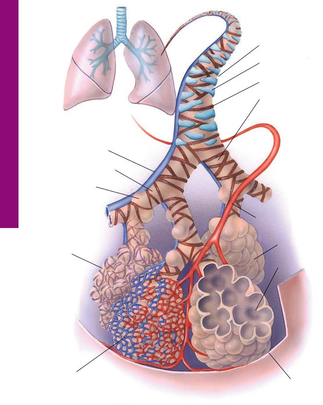
Pulmonary artery
(carrying deoxygenated blood)
Terminal bronchiole
Respiratory bronchiole
Alveolar elastin network 
Intrapulmonary bronchus
Cartilage plate
Smooth muscle fibers
Bronchiole
Pulmonary vein
(carrying oxygenated blood)
 Alveoli
Interalveolar septum
Alveolar capillary
network Visceral pleura

R E S P I R A T O R Y S Y S T E M 285
|
|
|
Respiratory |
|
|
|
|
|
|
|
|
|
|
|
|
|
|
|
|
|
|
|
|
|
|
|
|
|
|
|
|
|
|
|
||||||
|
|
|
|
|
|
|
|
|
|
|
|
|
|
|
|
|
|
|
|
|
|
|
|
|
|
|
|
|
|
|
|
|||||||||
|
|
|
|
|
|
|
|
|
|
|
|
|
|
|
|
|
|
|
|
|
|
|
|
|
|
|
|
|
|
|
|
|
||||||||
|
|
|
|
|
|
|
|
|
|
|
|
|
|
|
|
|
|
|
|
|
|
|
|
|
|
|
|
|
|
|
|
|
|
|
|
|
|
|
|
|
|
|
|
|
|
|
|
|
|
|
|
|
|
|
|
|
|
|
|
|
|
|
|
|
|
|
|
|
|
|
|
|
|
|
|
|
|
|
|||
|
|
|
|
|
|
|
|
|
|
|
|
|
|
|
|
|
|
|
|
|
|
|
|
|
|
|
|
|
|
|
|
|
|
|
|
|
|
|||
|
|
|
|
|
|
|
|
|
bronchiole |
|
|
|
|
|
|
|
|
|
|
|
|
|
|
|
|
|
|
|
|
|
|
|
|
|
||||||
|
|
|
|
|
|
|
|
|
|
|
|
|
|
|
|
|
|
|
|
|
|
|
|
|
|
|
|
|
|
|
|
|
||||||||
|
|
|
|
|
|
|
|
|
|
|
|
|
|
|
|
|
|
|
|
|
|
|
|
|
|
|
|
|
|
|
|
|
|
|||||||
|
|
|
|
|
|
|
|
|
|
|
|
|
|
|
|
|
|
|
|
|
|
|
|
|
|
|
|
|
|
|
|
|
|
|
|
|
|
|
|
|
|
|
|
|
|
|
|
|
|
|
|
|
|
|
|
|
|
|
|
|
|
|
|
|
|
|
|
|
|
|
|
|
|
|
|
|
|
|
|
||
|
|
|
|
|
|
|
|
|
|
|
|
|
|
|
|
|
|
|
|
|
|
|
|
|
|
|
|
|
|
|
|
|
|
|
|
|
|
|
||
|
|
|
|
|
|
|
|
|
|
|
|
|
|
|
|
|
|
|
|
|
|
|
|
|
|
|
|
|
|
|
|
|
|
|
|
|
|
|
||
|
|
|
|
|
|
|
|
|
|
|
|
|
|
|
|
|
|
|
|
|
|
|
|
|
|
|
|
|
|
|
|
|
|
|
|
|
|
|||
|
|
|
|
|
|
|
|
|
|
|
|
|
|
|
|
|
|
|
|
|
|
|
|
|
|
|
|
|
|
|
|
|
|
|
|
|
|
|||
|
|
|
|
|
|
|
|
|
|
|
|
|
|
|
|
|
|
|
|
|
|
|
|
|
|
|
|
|
|
|
|
|
|
|
|
|
|
|
||
|
|
|
|
|
|
|
|
|
|
|
|
|
|
|
|
|
|
|
|
|
|
|
|
|
|
|
|
|
|
|
|
|
|
|
|
|
|
|
||
|
|
|
|
|
|
|
|
|
|
|
|
|
|
|
|
|
|
|
|
|
|
|
|
|
|
|
|
|
|
|
|
|
|
|
|
|
|
|
||
|
|
|
|
|
|
|
|
|
|
|
|
|
|
|
|
|
|
|
|
|
|
|
|
|
|
|
|
|
|
|
|
|
|
|
|
|
|
|||
|
|
|
|
|
|
|
|
|
|
|
|
|
|
|
|
|
|
|
|
|
|
|
|
|
|
|
|
|
|
|
|
|
|
|
|
|
|
|||
|
|
|
|
|
|
|
|
|
|
|
|
|
|
|
|
|
|
|
|
|
|
|
Alveolar |
|
|
|
|
|
|
|
|
|
|
|||||||
|
|
|
|
|
|
|
|
|
|
|
|
|
|
|
|
|
|
|
|
|
|
|
|
|
|
|
|
|
|
|
|
|
||||||||
|
|
|
|
|
|
|
|
|
|
|
|
|
|
|
|
|
|
|
|
|
|
|
|
|
|
|
|
|
|
|
|
|
||||||||
|
|
|
|
|
|
|
|
|
|
|
|
|
|
|
|
|
|
|
|
|
|
|
|
|
|
|
|
|
|
|
|
|
|
|||||||
|
|
|
|
|
|
|
|
|
|
|
|
|
|
|
|
|
|
|
|
|
|
|
|
|
|
|
|
|
|
|
|
|
|
|
|
|
|
|
|
|
|
|
|
|
|
|
|
|
|
|
|
|
|
|
|
|
|
|
|
|
|
|
|
|
|
|
|
duct |
|
|
|
|
|
|
|
|
|||||
|
|
|
|
|
|
|
|
|
|
|
|
|
|
|
|
|
|
|
|
|
|
|
|
|
|
|
|
|
|
|
|
|
|
|
||||||
|
|
|
|
|
|
|
|
|
|
|
|
|
|
|
|
|
|
|
|
|
|
|
|
|
|
|
|
|
|
|
|
|
|
|
|
|
||||
|
|
|
|
|
|
|
|
|
|
|
|
|
|
|
|
|
|
|
|
|
|
|
|
|
|
|
|
|
|
|
|
|
|
|
|
|
|
|||
|
|
|
|
|
|
|
|
|
|
|
|
|
|
|
|
|
|
|
|
|
|
|
|
|
|
|
|
|
|
|
|
|
|
|
|
|
|
|
|
|
|
|
|
|
|
|
|
|
|
|
|
|
|
|
|
|
|
|
|
|
|
|
|
|
|
|
|
|
|
|
|
|
|
|
|
|
|
|
|||
|
|
|
|
|
|
|
|
|
|
|
|
|
|
|
|
|
|
|
|
|
|
|
|
|
|
|
|
|
|
|
|
|
|
|
|
|
|
|||
|
|
|
|
|
|
|
|
|
|
|
|
|
|
|
|
|
|
|
|
|
|
|
|
|
|
|
|
|
|
|
|
|
|
|
|
|
|
|||
|
|
|
|
|
|
|
|
|
|
|
|
|
|
|
|
|
|
|
|
|
|
|
|
|
|
|
|
|
|
|
|
|
|
|
|
|
|
|||
|
|
|
|
|
|
|
|
|
|
|
|
|
|
|
|
|
|
|
|
|
|
|
|
|
|
|
|
|
|
|
|
|
|
|
|
|
|
|||
|
|
|
|
|
|
|
|
|
|
|
|
|
|
|
|
|
|
|
|
|
|
|
|
|
|
|
|
|
|
|
|
|
|
|
|
|
|
|||
|
|
|
|
|
|
|
|
|
|
|
|
|
|
|
|
|
|
|
|
|
|
|
|
|
|
|
|
|
|
|
|
|
|
|
|
|
|
|||
|
|
|
|
|
|
|
|
|
|
|
|
|
|
|
|
|
|
|
|
|
|
|
|
|
|
|
|
|
|
|
|
|
|
|
|
|
|
|||
|
|
|
|
|
|
|
|
|
|
|
|
|
|
|
|
|
|
|
|
|
|
|
|
|
|
|
|
|
|
|
|
|
|
|
|
|
|
|
||
|
|
|
|
|
|
|
|
|
|
|
|
|
|
|
|
|
|
|
|
|
|
|
|
|
|
|
|
|
|
|
|
|
|
|
|
|
|
|
|
|
Alveolar pore
|
|
|
|
|
|
|
|
|
|
|
|
|
|
|
|
|
|
|
|
|
|
|
|
Alveolar |
|
|
|
|
|
|
|
|
sac |
|
|
|
|
|
|
|
|
|
|
|
|
|
|
|
|
|
|
|
|
|
|
|
|
Alveolus
Dust cell
(macrophage)
Lamellar bodies
Type II pneumocyte
Gas exchange occurring at the alveolar-capillary barrier
System Respiratory of Portion Respiratory • 2-12 GRAPHIC

Larynx Mucosa, tory Olfac• 1-12 PLATE
286 R E S P I R A T O R Y S Y S T E M
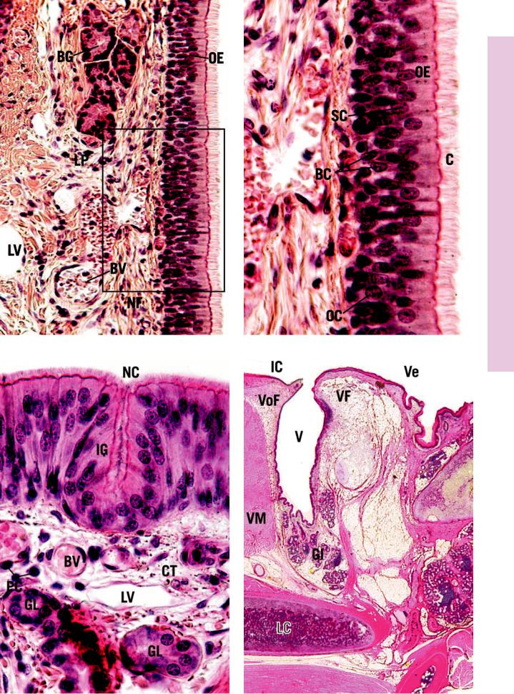
FIGURE 1. Olfactory area. Human. Paraffin section. ×270.
The olfactory mucosa of the nasal cavity is composed of a thick olfactory epithelium (OE) and a lamina propria (LP) richly endowed with blood vessels (BV), lymph vessels (LV), and nerve fibers (NF) frequently collected into bundles. The lamina propria also contains Bowman’s glands (BG), which produce a watery mucus that is delivered onto the ciliated surface by short ducts. The boxed area is presented at a higher magnification in Figure 2.
FIGURE 3. Intraepithelial gland. Human. Paraffin section. ×540.
The epithelium of the nasal cavity occasionally displays small, intraepithelial glands (IG). Note that these structures are clearly demarcated from the surrounding epithelium. The secretory product is released into the space (asterisk) that is continuous with the nasal cavity (NC). The subepithelial connective tissue (CT) is richly supplied with blood vessels (BV) and lymph vessels (LV). Observe the plasma cells (PC), characteristic of the subepithelial connective tissue of the respiratory system, which also displays the presence of glands (Gl).
FIGURE 2. Olfactory epithelium. Human. Paraffin section. ×540.
This is a higher magnification of the boxed area of the previous figure. The epithelium (OE) is pseudostratified ciliated columnar, whose cilia (C) are particularly evident. Although hematoxylin and eosin–stained tissue does not permit clear identification of the various cell types, the positions of the nuclei permit tentative identification. Basal cells (BC) are short, and their nuclei are near the basement membrane. Olfactory cell (OC) nuclei are centrally located, whereas nuclei of sustentacular cells (SC) are positioned near the apex of the cell.
FIGURE 4. Larynx. l.s. Human. Paraffin section. ×14.
The right half of the larynx, at the level of the ventricle (V), is presented in this survey photomicrograph. The ventricle is bounded superiorly by the ventricular folds (false vocal cords) (VF) and inferiorly by the vocal folds (VoF). The space above the ventricular fold is the beginning of the vestibule (Ve) and that below the vocal fold is the beginning of the infraglottic cavity (IC). The vocalis muscle (VM) regulates the vocal ligament present in the vocal fold. Acini of mucous and seromucous glands (GI) are scattered throughout the subepithelial connective tissue. The laryngeal cartilages (LC) are also shown to advantage.
KEY
BC |
basal cells |
LC |
laryngeal cartilages |
PC |
plasma cells |
BG |
Bowman’s glands |
LP |
lamina propria |
SC |
sustentacular cells |
BV |
blood vessels |
LV |
lymph vessels |
V |
ventricle |
C |
cilia |
NC |
nasal cavity |
Ve |
vestibule |
CT |
connective tissue |
NF |
nerve fibers |
VF |
ventricular folds |
GI |
glands |
OC |
olfactory cells |
VM |
vocalis muscle |
IC |
infraglottic cavity |
OE |
olfactory epithelium |
VoF |
vocal folds |
IG |
intraepithelial glands |
|
|
|
|
.p Larynx Mucosa, tory Olfac• 1-12 PLATE
FIGURE 1 |
FIGURE 2 |
FIGURE 3 |
FIGURE 4 |

rachea • T2-12 PLATE
288 R E S P I R A T O R Y S Y S T E M
FIGURE 1. Trachea. l.s. Monkey. Paraffin section. ×20.
This survey photomicrograph presents a longitudinal section of the trachea (Tr) and esophagus (Es). Observe that the lumen (LT) of the trachea is patent, due to the presence of discontinuous cartilaginous C-rings (CR) in its wall. The C-rings of the trachea are thicker anteriorly than posteriorly and are separated from each other by thick, fibrous connective tissue (arrows) that is continuous with the perichondrium of the C-rings. The adventitia of the trachea is adhered to the esophagus via a loose type of connective tissue (CT), which frequently contains adipose tissue. Note that the lumen (LE) of the esophagus is normally collapsed. A region similar to the boxed area is presented at a higher magnification in Figure 3.
FIGURE 3. Trachea. l.s. Monkey. Paraffin section. ×200.
This photomicrograph is a higher magnification of a region similar to the boxed area of Figure 1. The pseudostratified, ciliated columnar epithelium (E) lies on a basement membrane that separates it from the underlying lamina propria. The outer extent of the lamina propria is demarcated by an elastic lamina (arrows), deep to which is the submucosa (SM), containing a rich vascular supply (BV). The C-ring (CR), with its attendant perichondrium (Pc), constitutes the most substantive layer of the tracheal wall. The adventitia of the trachea, which some consider to include the C-ring, is composed of a loose type of connective tissue, housing some adipose cells (AC), nerves (N), and blood vessels (BV). Collagen fiber bundles of the adventitia secure the trachea to the surrounding structures.
FIGURE 2. Trachea. l.s. Monkey. Plastic section. ×270.
The trachea is lined by a pseudostratified ciliated columnar epithelium (E), which houses numerous goblet cells (GC) that actively secrete a mucous substance. The lamina propria (LP) is relatively thin, whereas the submucosa (SM) is thick and contains mucous and seromucous glands (GI), whose secretory product is delivered to the epithelial surface via ducts that pierce the lamina propria. The perichondrium (Pc) of the hyaline cartilage C-rings (CR) merges with the submucosal connective tissue. Note a longitudinal section of a blood vessel (BV), indicative of the presence of a rich vascular supply.
C-ring
Epithelium
C-ring
Perichondrium
Lamina propria
Trachea
KEY
AC |
adipose cells |
Es |
esophagus |
LT |
lumen—trachea |
BV |
blood vessels |
GC |
goblet cells |
N |
nerves |
CR |
C-rings |
GI |
mucous/seromucous glands |
Pc |
perichondrium |
CT |
connective tissue |
LE |
lumen—esophagus |
SM |
submucosa |
E |
epithelium |
LP |
lamina propria |
Tr |
trachea |

rachea • T2-12 PLATE
FIGURE 1 |
FIGURE 2 |
FIGURE 3

Microscopy Electron Cilia, and Epithelium tory Respira• 3-12 PLATE
290 R E S P I R A T O R Y S Y S T E M
FIGURE 1. Tracheal epithelium. Hamster. Electron microscopy. ×7,782.
The tracheal epithelium of the hamster presents mucus-producing goblet cells (GC) as well as ciliated columnar cells (CC), whose cilia (arrows) project into the lumen. Note that both cell types
are well endowed with Golgi apparatus (GA), whereas goblet cells are particularly rich in rough endoplasmic reticulum (rER). (Courtesy of Dr. E. McDowell.) Inset. Bronchus. Human. Electron microscopy. ×7,782. The apical region of a ciliated epithelial cell presents both cilia (C) and microvilli (arrow). (Courtesy of Dr. E. McDowell.)
KEY
C |
cilia |
GA |
Golgi apparatus |
rER |
rough endoplasmic |
CC |
ciliated columnar cell |
GC |
goblet cell |
|
reticulum |
PLATE 2-3Respira• |
tory Epithelium and Cilia, Electron Microscopy |
|
|
FIGURE 1
Bronchioles onchi, • Br4-12 PLATE
292 R E S P I R A T O R Y S Y S T E M
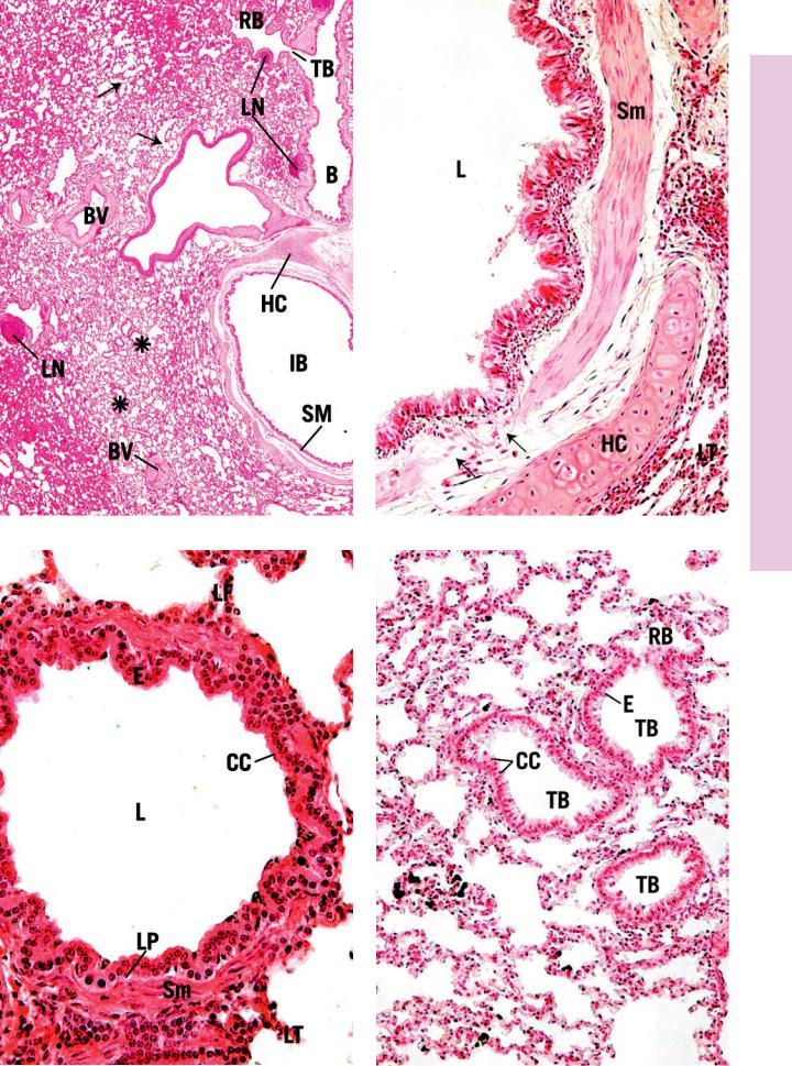
FIGURE 1. Lung. Paraffin section. ×14.
This survey photomicrograph presents a section of a lung that permits the observation of the various conduits that conduct air and blood to and from the lung. The intrapulmonary bronchus (IB) is recognizable by its thick wall, containing plates of hyaline cartilage (HC) and smooth muscle (Sm). Longitudinal sections of a bronchiole (B), terminal bronchiole (TB), and respiratory bronchiole (RB) are also evident. Smaller bronchioles (asterisks) may also be recognized, but their identification cannot be ascertained. Arrows point to structures that are probably alveolar ducts leading into alveolar sacs. Several blood vessels (BV), branches of the pulmonary circulatory system, may be noted. Observe that lymphatic nodules (LN) are also present along the bronchial tree.
FIGURE 3. Bronchiole. x.s. Paraffin section. ×270.
Bronchioles maintain their patent lumen (L) without the requirement of a cartilaginous support, since they are attached to surrounding lung tissue by elastic fibers radiating from their circumference. The lumina of bronchioles are lined by simple columnar to simple cuboidal epithelium (E), interspersed with Clara cells (CC), depending on the diameter of the bronchiole. The lamina propria (LP) is thin and is surrounded by smooth muscle (Sm), which encircles the lumen. Bronchioles have no glands in their walls and are surrounded by lung tissue (LT).
FIGURE 2. Intrapulmonary bronchus. x.s. Paraffin section. ×132.
Intrapulmonary bronchi are relatively large conduits for air, whose lumina (L) are lined by a typical respiratory epithelium (E). The smooth muscle (Sm) is found beneath the mucous membrane, and it encircles the entire lumen. Note that gaps (arrows) appear in the muscle layer, indicating that two ribbons of smooth muscle wind around the lumen in a helical arrangement. Plates of hyaline cartilage (HC) act as the skeletal support, maintaining the patency of the bronchus. The entire structure is surrounded by lung tissue (LT).
FIGURE 4. Terminal bronchioles. x.s. Paraffin section. ×132.
The smallest conducting bronchioles are referred to as terminal bronchioles (TB). These have very small diameters, and their lumina are lined with a simple cuboidal epithelium (E) interspersed with Clara cells (CC). The connective tissue is much reduced, and the smooth muscle layers are incomplete and difficult to recognize at this magnification. Terminal bronchioles give rise to respiratory bronchioles (RB), whose walls resemble those of the terminal bronchioles except that the presence of alveoli permits the exchange of gases to occur. Observe the alveolar duct (not labeled) in the lower right-hand corner.
Terminal |
|
Hyaline cartilage |
|
||
|
|
|
bronchiole |
|
Bronchiole |
|
|
|
Respiratory |
|
|
bronchiole |
|
|
Bronchial system and lung
KEY
B |
bronchiole |
IB |
intrapulmonary bronchus |
RB |
respiratory bronchiole |
BV |
blood vessels |
L |
lumen |
Sm |
smooth muscle |
CC |
Clara cells |
LN |
lymphatic nodule |
TB |
terminal bronchiole |
E |
epithelium |
LP |
lamina propria |
|
|
HC |
hyaline cartilage |
LT |
lung tissue |
|
|
Bronchioles onchi, • Br4-12 PLATE
FIGURE 1 |
FIGURE 2 |
FIGURE 3 |
FIGURE 4 |

Tissue ung • 5L -12 PLATE
294 R E S P I R A T O R Y S Y S T E M
FIGURE 1. Respiratory bronchiole. Paraffin section. ×270.
The respiratory bronchiole whose lumen (L) occupies the lower half of this photomicrograph presents an apparently thick wall with small outpocketings of alveoli (A). It is in these alveoli that gaseous exchanges first occur. The wall of the respiratory bronchiole is composed of a simple cuboidal epithelium consisting of some ciliated cells and Clara cells (CC). The remainder of the wall presents an incomplete layer of smooth muscle cells surrounded by fibroelastic connective tissue. Careful examination of this photomicrograph reveals that the wall of the respiratory bronchiole is folded upon itself, thus giving a misleading appearance of thick walls.
FIGURE 2. Alveolar duct. l.s. Human. Paraffin section. ×132.
Alveolar ducts (AD), unlike respiratory bronchioles, do not possess a wall of their own. These structures are lined by a simple squamous epithelium (E), composed of highly attenuated cells. Alveolar ducts present numerous outpocketings of alveoli (A), and they end in alveolar sacs (AS), consisting of groups of alveoli clustered around a common air space. Individual alveoli possess small smooth muscle cells that, acting like a purse string, control the opening into the alveolus. These appear as small knobs (arrow). A region similar to the boxed area is presented at a higher magnification in Figure 3.
FIGURE 3. Interalveolar septum. Monkey. Plastic section. ×540.
This photomicrograph is a higher magnification of a region similar to the boxed area of Figure 2. Two alveoli (A) are presented, recognizable as empty spaces separated from each other by an interalveolar septum (IS). The septum is composed of a capillary (Ca), the nucleus (asterisk) of whose endothelial lining bulges into the lumen containing red blood cells (RBC). The interalveolar septum as well as the entire alveolus is lined by type I pneumocytes (P1), which are highly attenuated squamous epithelial cells, interspersed with type II pneumocytes (P2). Thicker interalveolar septa house blood vessels (BV) and connective tissue elements including macrophages known as dust cells (DC). Note the presence of smooth muscle cells (Sm) and connective tissue elements that appear as knobs at the entrance into the alveolus.
FIGURE 4. Lung. Dust cells. Paraffin section. ×270.
The highly vascular nature of the lung is evident in this photomicrograph, since the blood vessels (BV) and the capillaries (Ca) of the interalveolar septa are filled with red blood cells. The dark blotches that appear to be scattered throughout the lung tissue represent dust cells (DC), macrophages that have phagocytosed particulate matter. Inset. Lung. Dust cell. Monkey. Plastic section. ×540. The nucleus (N) of a dust cell (DC) is surrounded by phagosomes containing particulate matter that was probably phagocytosed from an alveolus of the lung.
Respiratory portion of |
|
|
|
Respiratory bronchiole |
|||||||
|
|||||||||||
|
|
|
|
|
|
|
|
|
|
|
|
|
|
|
|
|
|
|
|
|
|
|
|
|
|
|
|
|
|
|
|
|
|
|
|
respiratory system |
|
|
|
|
|
|
|
|
|
Alveolar duct |
|
|
|
|
|
|
|
|
|
||||
|
|
|
|
|
|
|
|||||
|
|
|
|
|
|
|
|
|
|
||
|
|
|
|
|
|
|
|
|
|
|
|
|
|
|
|
|
|
|
|
|
|
|
|
|
|
|
|
|
|
|
|
|
|
|
|
|
|
|
|
|
|
|
|
|
|
|
|
|
|
|
|
|
|
|
|
|
|
|
|
|
|
|
|
|
|
|
|
|
|
|
|
Alveolar pore
Dust cell
Lamellar  bodies
Type II pneumocyte
|
|
|
|
|
|
|
Alveolar |
|
|
|
|
|
|
|
|
|
|
|
|
|
|
|
|
|
|
|
|
|
|
|
|
|
|
|
|
|
|
|
|
|
|
|
sac |
|
|
|
|
|
|
|
|
|
|
|
|
|
|
|
Alveolus |
Type I pneumocyte
Gas exchange occuring at the alveolar-capillary barrier
KEY
A |
alveolus |
CC |
Clara cell |
N |
nucleus |
AD |
alveolar duct |
DC |
dust cell |
P1 |
type I pneumocytes |
AS |
alveolar sac |
E |
epithelium |
P2 |
type II pneumocytes |
BV |
blood vessel |
IS |
interalveolar septum |
RBC |
red blood cells |
Ca |
capillary |
L |
lumen |
Sm |
smooth muscle |

Tissue ung • 5L -12 PLATE
FIGURE 1 |
FIGURE 2 |
FIGURE 3 |
FIGURE 4 |
Microscopy Electron , Barrier Air-Blood• 6-12 PLATE
FIGURE 1
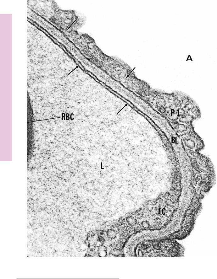
FIGURE 1. Blood-air barrier. Dog. Electron microscopy. ×85,500.
The blood-air barrier is composed of highly attenuated endothelial cells (EC), type I pneumocytes (P1), and an intervening basal lamina (BL). Note that the cytoplasm (arrows) of both cell types is greatly reduced, as evidenced by the close proximity of the
plasmalemma on either side of the cytoplasm. The air space of the alveolus (A) is empty, whereas the capillary lumen (L) presents a part of a red blood cell (RBC). (From DeFouw D. Vesicle numerical densities and cellular attenuation: comparisons between endothelium and epithelium of the alveolar septa in normal dog lungs. Anat Rec 1984;209:77–84.)