

- •Preface
- •Acknowledgments
- •Reviewers
- •Contents
- •CHAPTER OUTLINE
- •CYTOPLASM
- •Plasmalemma
- •Mitochondria
- •Ribosomes
- •Endoplasmic Reticulum
- •Golgi Apparatus, cis-Golgi Network, and the trans-Golgi Network
- •Endosomes
- •Lysosomes
- •Peroxisomes
- •Proteasomes
- •Cytoskeleton
- •Inclusions
- •NUCLEUS
- •CELL CYCLE
- •CHAPTER OUTLINE
- •EPITHELIUM
- •Epithelial Membranes
- •GLANDS
- •Chapter Summary
- •CHAPTER OUTLINE
- •EXTRACELLULAR MATRIX
- •Fibers
- •Amorphous Ground Substance
- •Extracellular Fluid
- •CELLS
- •CONNECTIVE TISSUE TYPES
- •Chapter Summary
- •CHAPTER OUTLINE
- •CARTILAGE
- •BONE
- •Cells of Bone
- •Osteogenesis
- •Bone Remodeling
- •Chapter Summary
- •CHAPTER OUTLINE
- •FORMED ELEMENTS OF BLOOD
- •Lymphocytes
- •Neutrophils
- •PLASMA
- •COAGULATION
- •HEMOPOIESIS
- •Erythrocytic Series
- •Granulocytic Series
- •Chapter Summary
- •CHAPTER OUTLINE
- •SKELETAL MUSCLE
- •Sliding Filament Model of Muscle Contraction
- •CARDIAC MUSCLE
- •SMOOTH MUSCLE
- •Chapter Summary
- •CHAPTER OUTLINE
- •BLOOD-BRAIN BARRIER
- •NEURONS
- •Membrane Resting Potential
- •Action Potential
- •Myoneural Junctions
- •Neurotransmitter Substances
- •SUPPORTING CELLS
- •PERIPHERAL NERVES
- •Chapter Summary
- •CHAPTER OUTLINE
- •BLOOD VASCULAR SYSTEM
- •HEART
- •ARTERIES
- •Capillary Permeability
- •Endothelial Cell Functions
- •VEINS
- •LYMPH VASCULAR SYSTEM
- •Chapter Summary
- •CHAPTER OUTLINE
- •CELLS OF THE IMMUNE SYSTEM
- •Antigen-Presenting Cells
- •DIFFUSE LYMPHOID TISSUE
- •LYMPH NODES
- •TONSILS
- •SPLEEN
- •THYMUS
- •Chapter Summary
- •CHAPTER OUTLINE
- •PITUITARY GLAND
- •Pars Intermedia
- •Pars Nervosa and Infundibular Stalk
- •Pars Tuberalis
- •THYROID GLAND
- •Parathyroid Glands
- •Suprarenal Glands
- •Cortex
- •Medulla
- •Pineal Body
- •Chapter Summary
- •CHAPTER OUTLINE
- •SKIN
- •Epidermis of Thick Skin
- •Dermis
- •DERIVATIVES OF SKIN
- •Chapter Summary
- •CHAPTER OUTLINE
- •CONDUCTING PORTION OF THE RESPIRATORY SYSTEM
- •Extrapulmonary Region
- •Intrapulmonary Region
- •RESPIRATORY PORTION OF THE RESPIRATORY SYSTEM
- •MECHANISM OF RESPIRATION
- •Chapter Summary
- •CHAPTER OUTLINE
- •ORAL CAVITY AND ORAL MUCOSA
- •Oral Mucosa
- •Tongue
- •Teeth
- •Odontogenesis (See Graphic 13-2)
- •Chapter Summary
- •CHAPTER OUTLINE
- •REGIONS OF THE DIGESTIVE TRACT
- •Esophagus
- •Stomach
- •Small Intestine
- •Large Intestine
- •GUT-ASSOCIATED LYMPHOID TISSUE
- •DIGESTION AND ABSORPTION
- •Carbohydrates
- •Proteins
- •Lipids
- •Water and Ions
- •Chapter Summary
- •CHAPTER OUTLINE
- •MAJOR SALIVARY GLANDS
- •PANCREAS
- •LIVER
- •Exocrine Function of the Liver
- •Endocrine and Other Functions of the Liver
- •GALLBLADDER
- •Chapter Summary
- •CHAPTER OUTLINE
- •KIDNEY
- •Uriniferous Tubule
- •Nephron
- •Collecting Tubules
- •FORMATION OF URINE FROM ULTRAFILTRATE
- •EXTRARENAL EXCRETORY PASSAGES
- •Chapter Summary
- •CHAPTER OUTLINE
- •OVARY
- •Ovarian Follicles
- •Regulation of Follicle Maturation and Ovulation
- •Corpus Luteum and Corpus Albicans
- •GENITAL DUCTS
- •Oviduct
- •Uterus
- •FERTILIZATION, IMPLANTATION, AND THE PLACENTA
- •Fertilization and Implantation
- •Placenta
- •VAGINA
- •EXTERNAL GENITALIA
- •MAMMARY GLANDS
- •Chapter Summary
- •CHAPTER OUTLINE
- •TESTES
- •Spermatogenesis
- •GENITAL DUCTS
- •ACCESSORY GLANDS
- •PENIS
- •Erection and Ejaculation
- •Chapter Summary
- •CHAPTER OUTLINE
- •SENSORY ENDINGS
- •Chapter Summary
- •Terminology of Staining
- •Common Stains Used in Histology
- •Hematoxylin and Eosin
- •Wright Stain
- •Weigert Method for Elastic Fibers and Elastic van Gieson Stain
- •Silver Stain
- •Iron Hematoxylin
- •Bielschowsky Silver Stain
- •Masson Trichrome
- •Periodic Acid-Schiff Reaction (PAS)
- •Alcian Blue
- •von Kossa Stain
- •Sudan Red
- •Mucicarmine Stain
- •Safranin-O
- •Toluidine Blue
The digestive system functions in the ingestion, digestion, and absorption of food as well as in the elimination of its unusable portions. To accomplish
these functions, the digestive system is organized into three major components:
•the oral cavity, where food is reduced in size, is moistened, begins to be digested, and is introduced as small spherical portions, each known as a bolus, into the alimentary canal;
•a muscular alimentary canal, along whose lumen the ingested foods are converted, both physically and chemically, into absorbable substances; and
•an extramural glandular portion, which provides fluids, enzymes, and emulsifying agents necessary so that the alimentary canal can perform its various functions.
ORAL CAVITY AND ORAL MUCOSA
The oral cavity may be subdivided into two smaller cavities: the externally positioned vestibule and the internally placed oral cavity proper.
•The vestibule is the space bounded by the lips and cheeks anteriorly and laterally, whereas its internal boundary is formed by the dental arches. The ducts of the parotid glands deliver their secretory products into the vestibule (see Graphics 13-1 and 13-2).
•The oral cavity proper is bounded by the teeth externally, the floor of the mouth inferiorly, and the hard and soft palates superiorly.
At its posterior extent, the oral cavity proper is separated from the oral pharynx by an imaginary plane drawn between the palatoglossal folds just anterior to the palatine tonsils.
Both the oral cavity proper and the vestibule are lined by stratified squamous epithelium, which, in regions that are subject to abrasive forces, is modified into stratified squamous keratinized (or parakeratinized) epithelium
(see Table 13-1).
Oral Mucosa
The epithelium and underlining connective tissue constitute the oral mucosa. If the epithelium is keratinized (or parakeratinized), the mucosa is said to be masticatory mucosa, and if the epithelium is not keratinized, the mucosa is referred to as lining mucosa.
•Most of the oral cavity possesses lining mucosa, with the exception of the gingiva, hard palate, and the dorsal surface of the tongue, which are covered by masticatory mucosa.
DIGESTIVE SYSTEM I |
301 |
•The oral cavity has areas of specialized mucosa, located mostly on the dorsal surface of the tongue, though present also on the soft palate and pharynx, where barrel-shaped intraepithelial structures known as taste buds function in taste perception.
SALIVARY GLANDS, PALATE,
AND TONSILS
The three pairs of major salivary glands—parotid, sublingual, and submandibular—deliver their secretions into the oral cavity. The hard palate assists the tongue in the preparation of the bolus, and the soft palate, a moveable structure, seals the communication between the oral and nasal pharynges, thus preventing passage of food and fluids from the former into the latter.
•The connective tissue underlying the epithelium of the oral cavity is richly endowed with minor salivary glands that, secreting saliva in a continuous fashion, contribute to the maintenance of a moist environment.
Saliva functions also in assisting in the process of deglutition by acting as a lubricant for dry foods and for holding the bolus together in a semisolid mass.
Moreover, enzymes present in saliva initiate digestion of carbohydrates, whereas secretory antibodies protect the body against antigenic substances.
The entrance to the pharynx is guarded against bacterial invasion by the tonsillar ring, composed of the lingual, pharyngeal, and palatine tonsils.
TONGUE, TEETH, AND
ODONTOGENESIS
The contents of the oral cavity are the tongue, a muscular structure that functions in the preparation of the bolus, tasting of the food, and beginning of deglutition (swallowing), and teeth, utilized in biting and mastication of food.
Tongue
The tongue is a mucosa-invested moveable muscular structure that has two regions, the root (base) and the body (see Graphic 13-2).
•The root anchors the tongue into the hyoid bone, the posterior aspect of the oral cavity and the pharynx.

302 DIGESTIVE SYSTEM I
TABLE 13-1 • Summary of the Oral Mucosa
Summary of the Oral Mucosa
|
|
Height of Connective |
|
Mucosal Region |
Type of Epithelium |
Tissue Papillae |
Special Comments |
|
|
|
|
Lip |
|
|
|
|
|
|
|
Skin aspect |
Stratified squamous |
Medium |
Hair, sebaceous glands, and sweat glands |
|
keratinized |
|
|
|
|
|
|
Vermilion zone |
Stratified squamous |
High |
Few sebaceous glands? The vermilion zone |
|
keratinized |
|
must be moistened by tongue |
|
|
|
|
Vestibular aspect |
Lining mucosa |
Medium |
Mucous (mixed?) salivary glands |
|
|
|
|
Cheek |
|
|
|
|
|
|
|
Skin aspect |
Stratified squamous |
Medium |
Hair, sebaceous glands, and sweat glands |
|
keratinized |
|
|
|
|
|
|
Vestibular aspect |
Lining mucosa |
Medium |
Mucous (mixed?) salivary glands; Fordyce’s |
|
|
|
granules |
|
|
|
|
Gingiva |
|
|
|
|
|
|
|
Free and attached |
Masticatory mucosa |
High |
Tightly bound to periosteum |
|
|
|
|
Sulcular |
Lining mucosa |
Low |
|
|
|
|
|
Junctional epithe- |
Lining mucosa |
None |
Attached to tooth surface by |
lium |
|
|
hemidesmosomes |
|
|
|
|
Col |
Lining mucosa |
Low to none |
|
|
(junctional epithelium?) |
|
|
|
|
|
|
Alveolar Mucosa |
|
|
|
|
|
|
|
|
Lining mucosa |
Low |
Some minor salivary glands |
|
|
|
|
Hard Palate |
|
|
|
|
|
|
|
Anterior lateral |
Masticatory mucosa |
High |
Fat globules |
|
|
|
|
Posterior lateral |
Masticatory mucosa |
High |
Mucous salivary glands |
|
|
|
|
Raphe |
Masticatory mucosa |
High |
Tightly bound to periosteum |
|
|
|
|
Soft Palate |
|
|
|
|
|
|
|
|
Lining mucosa |
Low |
Elastic lamina; mucous salivary glands |
|
|
|
|
Uvula |
Lining mucosa |
Low |
Mucous salivary glands |
|
|
|
|
Floor of Mouth |
|
|
|
|
|
|
|
|
Lining mucosa |
Low |
Mucous salivary glands |
|
|
|
|
Tongue |
|
|
|
|
|
|
|
Dorsal surface |
Specialized mucosa |
|
Taste buds; lingual papillae, serous, mucous, |
|
|
|
and mixed salivary glands; lingual tonsils |
|
|
|
|
Ventral surface |
Lining mucosa |
Low |
Plica fimbriata |
(Reprinted with permission from Leslie P. Gartner Essentials of Oral Histology and Embryology, 3rd ed. P. 118, Jen House Publishing Company, Baltimore,
MD 1999.)
•The body is freely moving in the oral cavity and its dorsal surface (facing the palate) is divided into an anterior two-thirds and a posterior one-third by a shallow, posteriorly directed V-shaped groove, the sulcus terminalis, whose apex is a shallow depression, the foramen cecum. The dorsum of the posterior onethird of the tongue has crypts that burrow into the submucosal lymphoid tissue, the lingual tonsil.
During embryogenesis, the thyroglossal duct, which will form the thyroid gland, originates from the foramen cecum.
The dorsum of the tongue is covered by masticatory mucosa sporting lingual papillae, and the ventral surface is covered by lining mucosa. The core of the tongue is composed of two groups of skeletal muscle, the intrinsic group and the extrinsic group, interspersed with connective tissue and three pairs of minor salivary glands, posterior mucous glands, glands of von Ebner (purely serous glands), and Blandin-Nuhn glands (mixed glands).
Lingual Papillae
The four types of lingual papillae are outgrowths of the mucosa of the dorsal surface:
•Filiform papillae are the most numerous, and they are conical in shape, have no taste buds, and their stratified squamous epithelium is highly keratinized.
•Fungiform papillae are mushroom-shaped and possess a few taste buds on their free surface. The epithelium of fungiform papillae is stratified squamous nonkeratinized.
•Foliate papillae are located on the posterolateral aspects of the anterior two-thirds of the tongue. They present as shallow furrows that possess taste buds for the first 2 years of life after which the taste buds degenerate.Glands of von Ebner release their secretion into the furrows.
•The twelve or so circumvallate papillae, located just anterior to the sulcus terminalis, possess numerous taste buds and are surrounded by a deep, moat-like furrow. Glands of von Ebner release their serous secretion into the bottom of the moat-like depression.
Taste Buds
Each taste bud is barrel-shaped, is completely intraepithelial, and is composed of 60 to 80 spindle-shaped neuroepithelial cells that are of four types:
•basal cells (type IV), which act as regenerative cells;
•dark cells (type I cells), which probably arise directly from basal cells and mature into
•light cells (type II); and
•intermediate cells (type III cells), which will undergo apoptosis and die.
DIGESTIVE SYSTEM I |
303 |
The complete life cycle of these cells is about 10 days to 2 weeks, and they are continuously replaced by basal cell derivatives. The cells are compacted together and form an opening known as a taste pore at the epithelial surface (see Graphic 13-2).
Basally, cell types I, II, and III form synaptic contacts with nerve fibers;
apically, they possess long microvilli known as taste hairs, which pass through the taste pore and are exposed to the moist environment of the oral cavity.
The taste hairs have two types of taste receptors (TR1 and TR2) that bind dissolved chemicals from the food, known as tastants, resulting in the activation of G proteins and/or direct opening of ion channels.
The end result is that the neuroepithelial cells become activated and release neurotransmitter substances at their synaptic junctions with the nerve fibers.
The central nervous system then registers the signal and interprets the taste that was sensed by the taste bud.
Each taste bud recognizes one or more of the five taste sensations: sour, sweet, salty, umami (savory), or bitter.
Teeth
Humans have two sets of dentition, there are 20 deciduous teeth in the mouth of a child, and as they are exfoliated, they are replaced by the permanent dentition, composed of 20 succedaneous teeth and an additional 12 accessional teeth for a total of 32 permanent teeth. At approximately 6 to 13 years of age, the dentition is mixed in that both deciduous and permanent teeth are present in the mouth at the same time. The increase in the number of teeth is probably a function of the greater space availability in the adult mouth. Each tooth is composed of a crown and a root and the cervix, where the crown and root contact each other. Three calcified substances, enamel, dentin, and cementum form the substance of each tooth. Dentin is located both in the crown (coronal dentin) and in the root (radicular dentin) and surrounds the pulp, a very vascularized and highly ordered connective tissue. Enamel covers coronal dentin, cementum covers radicular dentin, and the two meet at the cervix.
•Enamel is the hardest tissue in the body; it is
96% inorganic matrix composed of calcium hydroxyapatite crystals and
4% organic matrix consisting mostly of the protein enamelin
304DIGESTIVE SYSTEM I
Manufactured by cells known as ameloblasts, which are not present after the tooth erupts into the oral cavity; therefore, enamel is acellular posteruption and cannot repair itself.
•Dentin is the second hardest tissue in the body, it is
65% to 70% inorganic matrix composed of calcium hydroxyapatite crystals and
30% to 35% type I collagen fibers
elaborated by cells known as odontoblasts that remain in their position in the pulp and continue to form dentin throughout the tooth’s life
•Cementum is
45% to 50% inorganic matrix composed of calcium hydroxyapatite crystals
50% to 55% type I collagen fibers, glycosaminoglycans, and proteoglycans
Formed by cementoblasts that continue to manufacture cementum throughout the life of the tooth because the addition of cementum compensates for the erosion of enamel, thus maintaining the length of the tooth for proper occlusion.
•Pulp is a gelatinous, highly vascularized connective tissue that fills the pulp cavity, known as the pulp chamber in the crown of the tooth and root canal in the root of the tooth.
The peripheral layer of the pulp is composed of odontoblasts.
Deep to the odontoblasts is an acellular layer (the cell-free zone) and deep to that is a layer of fibroblasts and mesenchymal cells (the cell-rich zone)
core of the pulp has normal connective tissue cells and also houses blood vessels, lymph vessels, and nerve fibers.
The nerve fibers are of two types: autonomic that serve blood vessels and sensory fibers that conduct pain information from the pulp.
The root of each tooth is suspended in its bony housing, the alveolus, by a dense collagenous connective tissue ligament, the periodontal ligament. The cervix of each tooth is surrounded by gingiva whose epithelium forms a collar, the junctional epithelium, whose attachment to the cervical enamel creates occluding junctions, thus isolating the connective tissue of the gingiva from the oral cavity.
Odontogenesis (See Graphic 13-2)
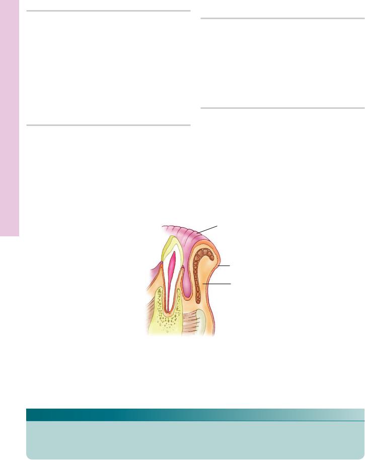
Odontogenesis, tooth formation, begins at 6 1/2 weeks of development as a horseshoe-shaped epithelial band, known as the dental lamina, arises from the oral epithelium of both the maxillary and the mandibular processes. Ten epithelial swellings, known as tooth buds, form on the lingual aspect of each dental lamina and press into the
surrounding ectomesenchyme. It should be noted that cells of the ectomesenchyme are neural crest derivatives.
•Each tooth bud develops at a different rate to form a three-dimensional, three-layered epithelial structure, the cap stage of tooth development, composed of the enamel organ whose indentation is filled with ectomesenchymal cells, known as the dental papilla. The enamel organ and dental papilla together form the tooth germ.
The enamel organ’s three layers are the outer enamel epithelium and inner enamel epithelium that form a rim, the cervical loop, at their junction, and the intervening space between the two epithelial layers are filled with cells known as stellate reticulum.
The concavity of the inner enamel epithelial layer is filled with ectomesenchymal cells, the dental papilla, which is responsible for the formation of dentin and the pulp.
Ectomesenchymal cells surrounding the tooth germ condense to form a connective tissue capsule, the dental sac, around the developing tooth germ. The dental sac is responsible for the formation of cementum, the periodontal ligament, and the bony alveolus.
A new epithelial growth develops from the dental lamina just lingually directed from the cap, known as the succedaneous lamina. This lamina grows deep into the ectomesenchyme; its distal terminus will form a tooth bud that will give rise to the permanent replacement of the forming deciduous tooth.
A group of cells derived from the stellate reticulum form a cluster against the inner enamel epithelium known as the enamel knot. These cells will either undergo apoptosis during the cap stage or they will survive into the next stage of tooth development.
The inner enamel epithelial cells will differentiate into ameloblasts and will form the enamel of the tooth.
•As the cap enlarges and forms a fourth layer of cells, the stratum intermedium, located between the stellate reticulum and the inner enamel epithelium, the tooth germ is in the bell stage of odontogenesis.
If the enamel knot survives into the bell stage, the enamel organ rearranges itself to form a premolar or a molar tooth. If the enamel knot undergoes apoptosis during the cap stage, the developing tooth will be an incisor or a canine tooth.
During the late bell stage, the peripheral-most cells of the dental papilla begin to differentiate into odontoblasts to start forming dentin.
In response to the formation of the odontoblasts, the cells of the inner enamel epithelium differentiate into ameloblasts to start forming enamel.
•Once the tooth germ forms dentin as well as enamel, odontogenesis has progressed into a new stage known as apposition.
The appositional stage of tooth development is responsible for the formation of the crown of the tooth.
•After the enamel of the crown is completely formed, odontogenesis enters its new phase, namely, root formation.
This process occurs simultaneously with eruption, in that as the root(s) of the tooth increase(s) in length the tooth moves toward the oral cavity and will erupt through the connective tissue and eventually the oral epithelium.
Once the tooth reaches the oral cavity, it will continue to erupt at a rapid pace until it contacts its opposing tooth in the opposite arch.
It is important to understand that the root does not push the tooth into its position in the oral cavity, instead, modified fibroblasts, myofibroblasts, of the forming periodontal ligament pull on the collagen fibers attached to the cementum of the root and “drag” the forming tooth into its proper position.
Molecular Mechanisms of Odontogenesis
Odontogenesis is induced by the ectodermally derived cells of the dental lamina that express lymphoid enhancer factor-1 (Lef-1), a transcription factor.
DIGESTIVE SYSTEM I |
305 |
•Lef-1 induces the epithelial cells to synthesize and release bone morphogenic protein-4 (BMP-4), sonic hedgehog (Shh), and fibroblast growth factor-8 (FGF-8).
•These signaling molecules act on the underlying ectomesenchymal cells to differentiate into odontogenic tissues.These neural crest–derived cells begin to express activin bA, BMP-4, the adhesive glycoprotein tenascin, and the membrane-bounded proteoglycan syndecan.
•Moreover, they also express several transcription factors, namely, Egr-1 (early growth response-1), Msx-1 (homeobox-containing genes), and Msx-2.
•This activation of the ectomesenchyme elicits their role in the induction of the tooth morphology, so that it is the ectomesenchyme that will determine, for instance, whether the developing tooth will become a molar or an incisor.
•Signaling molecules from the ectomesenchyme induce the formation of the enamel knot, which synthesizes and releases its own signaling molecules, namely, FGF- 4, BMP-2, BMP-4, BMP-7, and Shh.
•These signaling molecules promote the differentiation of the inner enamel epithelial cells into ameloblasts and those of the peripheral-most layer of the dental papilla into odontoblasts.
•As mentioned above, continued maintenance of the enamel knot is responsible for the remodeling of the inner enamel epithelium, resulting in the morphodifferentiation of the enamel organ into a template that is the prototype of a molar tooth, whereas if the enamel knot undergoes apoptosis, morphodifferentiation is constrained and an incisor is formed.
306 DIGESTIVE SYSTEM I
CLINICAL CONSIDERATIONS
Herpetic Stomatitis
Herpetic stomatitis, a relatively common disease caused by the herpes simplex virus type I, is distinguished by painful fever blisters appearing on or in the vicinity of the lips. This is a recurring disease since the virus, in its dormant phase, inhabits the trigeminal ganglion. It travels along the axon to cause the appearance of the blisters. During the active stage, the patient is highly contagious, since the virus is shed via the seeping clear exudate.
Caries
Caries, or cavities, are formed by the action of acid-secret- ing bacteria that adhere to very small defects or irregularities of the enamel surface. The acids formed by the bacteria decalcify the enamel, providing larger defects that can house a much larger number of the proliferating bacteria with the formation of more acid and decalcification of more of the enamel. The carious lesion is painfree until it reaches the underlying dentin. Since the most sensitive region of dentin is at the dentinoenamel junction, the tooth is sensitive to heat, cold, mechanical contact, and sweets. Continued bacterial activity, without the intervention of a dental health professional, could cause eventual loss of the tooth and perhaps even more serious sequelae.
Hemorrhage of the Pulp
Darkening of a tooth may be due to hemorrhage of the pulp. Although frequently the pulp is damaged severely enough that it can no longer be saved, a dental professional should be consulted because tooth discoloration does not necessarily require root canal therapy.
Necrotizing Ulcerative Gingivitis
Necrotizing ulcerative gingivitis is an acute ulcerative condition of the gingiva with accompanying necrosis, halitosis, erythematous appearance, and moderate to severe pain. Fever and regional lymphadenopathy may also be evident. This is usually a disease of the young
adult who is experiencing stress and is not particularly attentive to dental hygiene. Frequently, Treponema vincentii and fusiform bacillus are present in large numbers, and they are also believed to be causative agents of the condition.
Spindle Cell Carcinoma
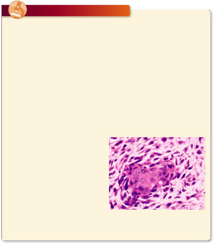
Spindle cell carcinoma is a modified type of squamous cell carcinoma where the histologic appearance of the malignant epithelial cells is spindle-shaped, resembling fibroblasts. It is highly aggressive, resulting in a survival rate of only 40% after 2 years. Spindle cell carcinoma is more commonly present in males 60 years of age or older, and in the oral region, this tumor is usually restricted to the gingiva, tongue, and lower lip. The most common causative agents of spindle cell carcinoma are alcoholism, tobacco use, and poor oral hygiene. Diagnostic features include painful inflammation, ulcers that do not heal readily, and growths that may be as large as 10 cm in diameter.
This light microscopic image from a patient with spindle cell carcinoma displays both epithelioidand spindle-shaped malignant cells. (Reprinted with permission from Mills SE, Carter, D, et al., eds. Sternberg’s Diagnostic Surgical Pathology, 5th ed., Philadelphia: Lippincott Williams & Wilkins, 2010. p. 794.)
DIGESTIVE SYSTEM I |
307 |
Odontomas
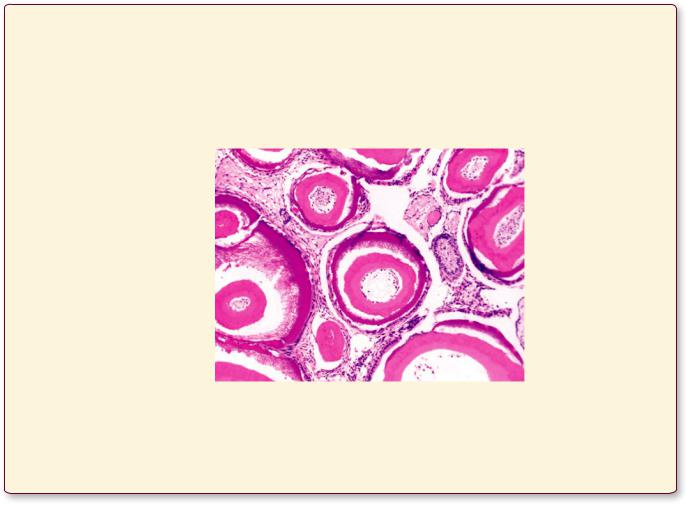
Odontomas are hamartomatous anomalies (developmental malformations) that appear to be malignant, but fortunately, they are benign. These are the most frequent tumor-like structures of the maxillary and mandibular arches, and they arise from remnants of embryonic
odontogenic tissues, forming tooth-like structures that are frequently calcified and display a haphazard arrangement. They are usually asymptomatic and are discovered on radiographs taken during routine dental examinations. Complex odontomas do not pose a significant health risk.
This light microscopic image from a patient with complex odontoma displays the presence of dentin, enamel, and pulp-like tissues scattered in a haphazard manner. (Reprinted with permission from Mills SE, Carter D, et al., eds. Sternberg’s Diagnostic Surgical Pathology, 5th ed., Philadelphia: Lippincott Williams & Wilkins, 2010, p. 807.)
Development Tooth and Tooth • 1-13 GRAPHIC
308 |
DIGESTIVE SYSTEM I |
|
|
||
|
|
|
|
|
Enamel |
|
Gingival sulcus |
|
Pulp |
||
|
|
||||
|
Epithelium of gingiva |
|
|
|
Dentin |
|
|
|
|||
|
Gingiva |
|
|||
|
|
|
|||
|
Alveolar bone |
|
Cementum |
||
Root canal
Periodontal ligament
Apical foramen
Tooth
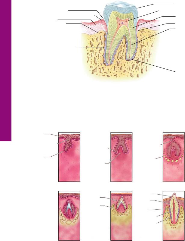
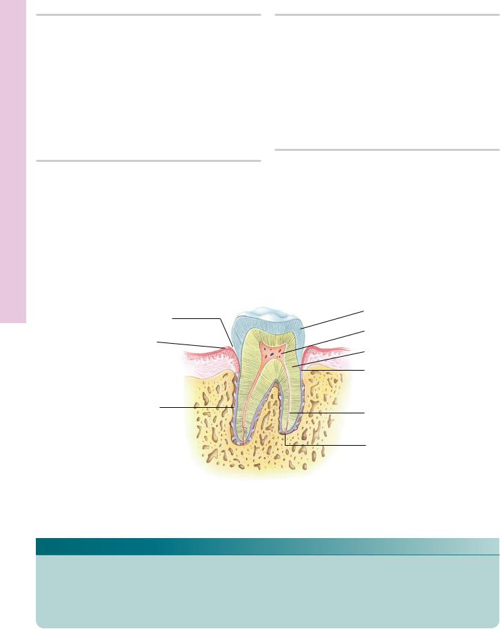
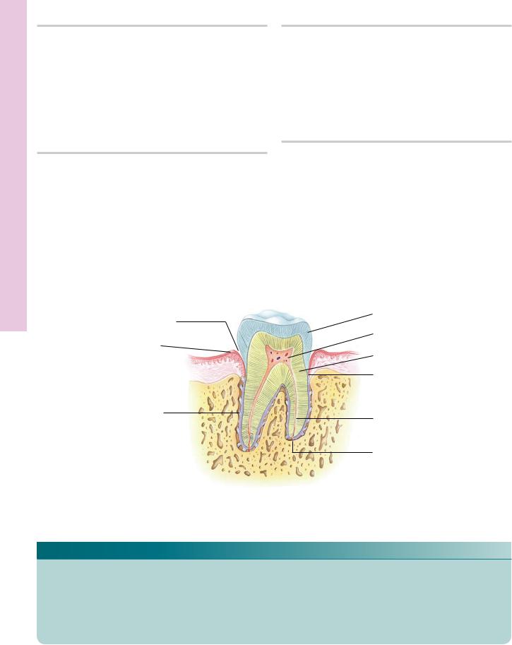
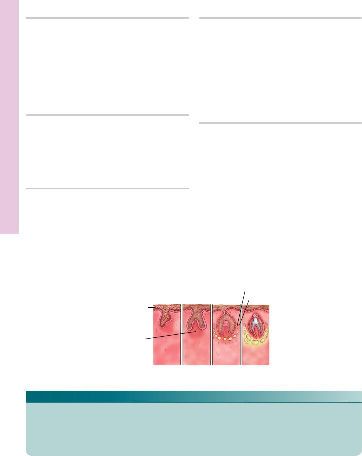
The tooth, composed of a crown and root, is suspended in its bony socket, the alveolus, by a dense, collagenous connective tissue, the periodental ligament. The crown of the tooth consists of two calcified tissues, dentin and enamel, whereas the root is composed of dentin and cementum. The pulp chamber of the crown and the root canal of the root are continuous with one another. They are occupied by a gelatinous connective tissue, the pulp, which houses blood and lymph vessels, nerve fibers, connective tissue elements, as well as odontoblasts, the cells responsible for the maintenance and repair of dentin. Vessels and nerves serving the pulp enter the root canal via the apical foramen, a small opening at the apex of the root.
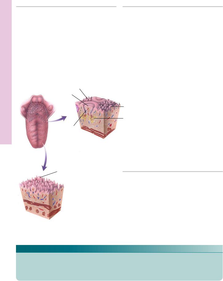
(A) Bud stage |
(B) Cap stage |
(C) Bell stage |
Oral |
|
|
epithelium |
|
|
Dental lamina |
Enamel organ |
Dental lamina |
|
|
|
Bud |
|
Bony crypt |
|
|
|
|
Condensed |
|
|
mesenchyme |
|
(D) Apposition |
(E) Beginning of eruption |
(F) Eruption into oral cavity |
|
|
Enamel |
|
Enamel |
Dentin |
|
|
|
|
Alveolar |
|
|
bone |
Pulp |
|
|
|
|
|
Cementum |
|
|
Periodontal |
|
|
ligament |
DIGESTIVE SYSTEM I |
309 |
Epiglottis
Filiform papilla
Lingual tonsil
Palatine tonsil
Salivary glands
Circumvallate papilla
Taste bud
Intrinsic muscle
 Median sulcus
 Fungiform papilla
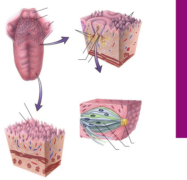
The core of the tongue is composed of skeletal muscle fibers that interlace with one another, connective tissue, and minor salivary glands.
Section of taste bud
Fungiform papilla
Filiform papilla
Pore
Taste cell
Sustentacular cell
Taste buds are small, intraepithelial structures composed of a total of 40–70 cells, basal cells, neurepithelial (taste) cells, and sustentacular (supporting) cells. They function in the perception of the five primary taste sensations, salt, sweet, bitter, sour, and umami.
The dorsal surface of the tongue is subdivided into an anterior two-thirds, populated by the four types of lingual papillae, and a posterior one-third housing the lingual tonsils. The two regions are separated from one another by a "V-shaped" depression, the sulcus terminalis. Filiform papillae are short, conical, and highly keratinized. Fungiform papillae are mushroom-shaped, and the dorsal aspect of their epithelia houses three to five taste buds. Circumvallate papillae, the largest of the lingual papillae, are six to twelve in number. Each circumvallate papilla is depressed into the surface of the tongue and is surrounded by a moat-like trough. The lateral aspect of the papilla as well as the lining of the trough houses numerous taste buds. Foliate papillae are located on the lateral aspect of the tongue.
Bud Taste and Tongue• 2-13 GRAPHIC
•Lip1-13 PLATE
310 DIGESTIVE SYSTEM I
FIGURE 1. Lip. Human. Paraffin section. ×14.
The human lip presents three surfaces and a core (C). The external surface is covered by skin, composed of epidermis (E) and dermis
(D). Associated hair follicles (arrow) and glands are evident. The vermillion (red) zone (VZ) is only found in humans. The high dermal papillae (arrowheads) carry blood vessels close to the surface, accounting for the pinkish coloration of this region. The internal aspect is lined by a wet, stratified, squamous, nonkeratinized epithelium (Ep), and the underlying connective tissue houses minor salivary glands. The core of the lip is composed of skeletal muscle interspersed with fibroelastic connective tissue.
FIGURE 3. Lip. Human. External aspect. Paraffin section. ×132.
The external aspect of the lip is covered by thin skin. Neither the epidermis (E) nor the dermis (D) presents any unusual features. Numerous hair follicles (HF) populate this aspect of the lip, and sebaceous glands (Sg) as well as sweat glands are noted in abundance.
FIGURE 2. Lip. Human. Internal aspect. Paraffin section. ×270.
The internal aspect of the lip is lined by a mucous membrane that is continuously kept moist by saliva secreted by the three major and numerous minor salivary glands. The thick epithelium (Ep) is a stratified squamous nonkeratinized type, which presents deep rete ridges (RR) that interdigitate with the connective tissue papillae (CP). The connective tissue is fibroelastic in nature, displaying a rich vascular supply (BV).
FIGURE 4. Lip. Human. Vermilion zone. Paraffin section. ×132.
The vermilion zone of the lip is covered by a modified skin composed of stratified squamous keratinized epithelium (Ep) that forms extensive interdigitations with the underlying dermis (D). Neither hair follicles nor sweat glands populate this area (though occasional sebaceous glands may be present). Note the cross-sec- tional profiles of skeletal muscle fibers (SM) and the rich vascular supply (BV) of the lip.
Vermilion zone
Epidermis
Dermis
Lip
KEY
BV |
vascular supply |
E |
epidermis |
Sg |
sebaceous glands |
C |
core |
Ep |
epithelium |
SM |
skeletal muscle |
CP |
connective tissue papillae |
HF |
hair follicles |
VZ |
vermilion (red) zone |
D |
dermis |
RR |
rete ridges |
|
|
•Lip1-13 PLATE
Ep
Cp
RR
CP
BV
FIGURE 1 |
FIGURE 2 |
E
BV
D |
SM |
|
D
Sg |
Ep |
HF
FIGURE 3 |
FIGURE 4 |
Pulp and ooth • T2-13 PLATE
312 DIGESTIVE SYSTEM I
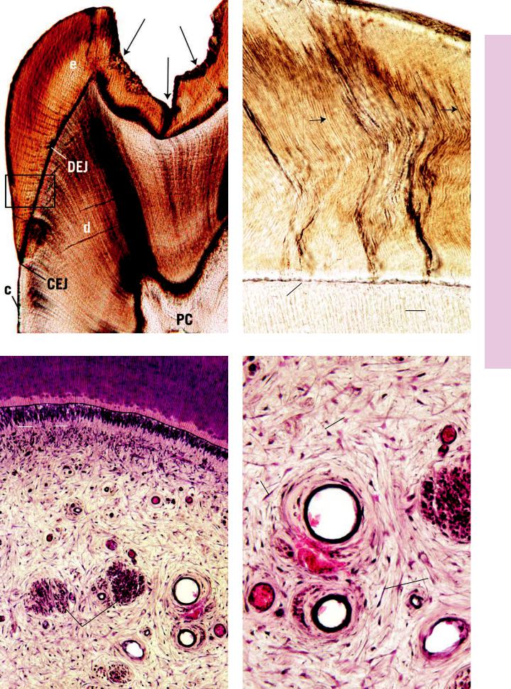
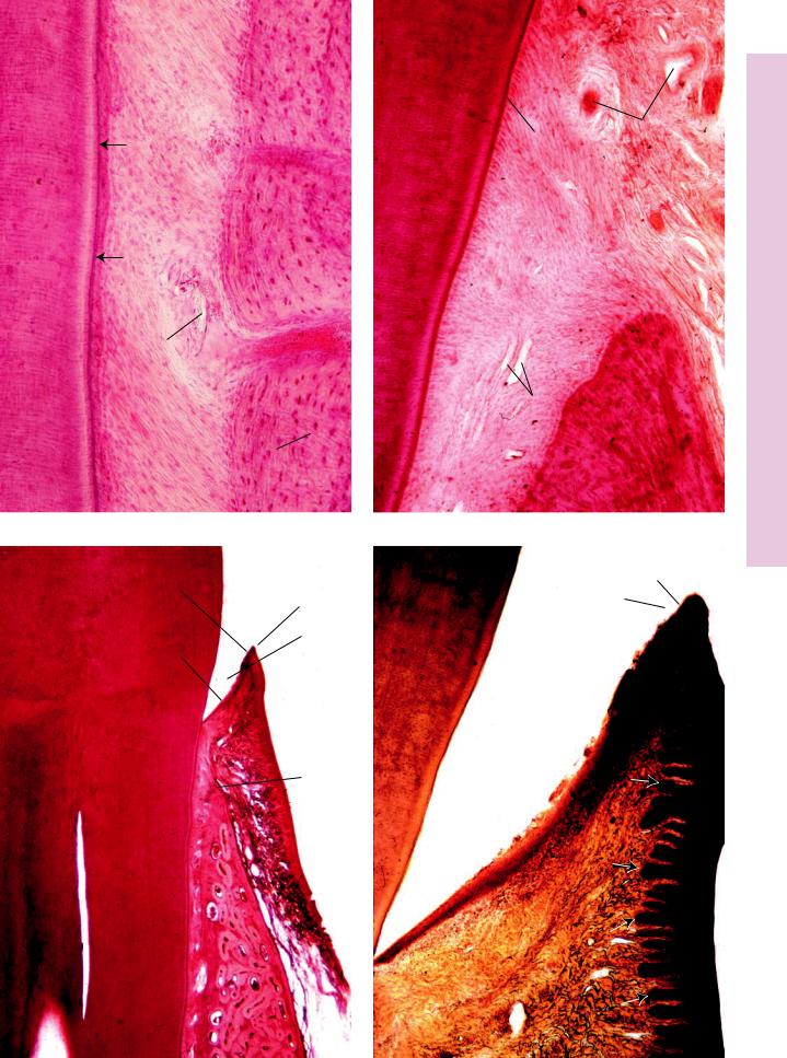
FIGURE 1. Tooth. Human. Ground section. ×14.
The tooth consists of a crown, neck, and root, composed of calcified tissue surrounding a chamber housing a soft, gelatinous pulp. In ground section, only the hard tissues remain. The crown is composed of enamel (e) and dentin (d), whose interface is known as the dentinoenamel junction (DEJ). At the neck of the tooth, enamel meets cementum (c), forming the cementoenamel junction (CEJ). The pulp chamber (PC) is reduced in size as the individual ages. The gap in the enamel (arrows) is due to the presence of a carious lesion (cavity). A region similar to the boxed area is presented at a higher magnification in Figure 2.
FIGURE 3. Pulp. Human. Paraffin section. ×132.
The pulp is surrounded by dentin (d) from which it is separated by a noncalcified dentin matrix (DM). The pulp is said to possess four regions: the odontoblastic layer (OL), the cell-free zone (CZ), the cell-rich zone (CR), and the core (C). The core of the pulp is composed of fibroblasts (F), delicate collagen fibers, numerous nerve bundles (NB), and blood vessels (BV). Branches of these neurovascular structures reach the periphery of the pulp, where they supply the cell-rich zone and the odontoblasts with capillaries and fine nerve fibers.
Gingival sulcus
Epithelium of gingiva
Gingiva 
Alveolar bone 
Periodontal ligament
FIGURE 2. Tooth. Human. Ground section. ×132.
This photomicrograph is a higher magnification of a region similar to the boxed area of the previous figure. The enamel (e) is composed of enamel rods (arrows), each surrounded by a rod sheath. Hypomineralized regions of enamel present the appearance of tufts of grass, enamel tufts (ET), which extend from the dentinoenamel junction (DEJ) partway into the enamel. Dentin (d), not as highly calcified as enamel, presents long, narrow canals, dentinal tubules (DT), which in the living tooth house processes of odontoblasts, cells responsible for the formation of dentin.
FIGURE 4. Pulp. Human. Paraffin section. ×270.
This is a higher magnification of the lower right corner of the previous figure. Note the presence of blood vessels (BV) and nerve fibers (NF), as well as the numerous fibroblasts (F) of this gelatinous connective tissue.
Enamel
Pulp
Dentin
Cementum
Root canal
Apical foramen
Tooth
KEY
BV |
blood vessel |
d |
dentin |
F |
fibroblasts |
C |
core |
DEJ |
dentinoenamel junction |
NB |
nerve bundles |
c |
cementum |
DM |
dentin matrix |
OL |
odontoblastic layer |
CEJ |
cementoenamel junction |
DT |
dentinal tubule |
PC |
pulp chamber |
CR |
cell-rich zone |
e |
enamel |
|
|
CZ |
cell-free zone |
ET |
enamel tufts |
|
|
e
ET
|
DEJ |
d |
DT |
|
|
||
|
|
|
|
FIGURE 1 |
|
|
FIGURE 2 |
d
DM
F
CZ
CR OL
|
F |
|
C |
NF |
|
BV |
||
|
F
F
BV
NB |
BV |
FIGURE 3 |
FIGURE 4 |
Pulp and ooth • T2-13 PLATE
Gingiva and Ligament eriodontal • P3-13 PLATE
314 DIGESTIVE SYSTEM I
FIGURE 1. Periodontal ligament. Human. Paraffin section. ×132.
The root of the tooth, composed of dentin (d) and cementum (c), is suspended in its alveolus (A) by a collagenous tissue, the periodontal ligament (PL). The strong bands of collagen fibers
(CF) are embedded in the bone via Sharpey’s fibers (SF). Blood vessels (BV) from the bone enter and supply the periodontal ligament. The dentinocemental junction (arrows) is clearly evident. Near the apex of the root, the cementum becomes thicker and houses cementocytes.
FIGURE 3. Gingiva. Human. Paraffin section. ×14.
This is a decalcified longitudinal section of an incisor tooth; thus, all of the calcium hydroxyapatite crystals have been extracted from the tooth and from its bony alveolus (A). Since enamel is composed almost completely of calcium hydroxyapatite crystals, only the space where enamel used to be, the enamel space (ES), is represented in this photomicrograph. The crest (cr) of the alveolus is evident, as are the periodontal ligament (PL) and the gingiva (G). The gingival margin (GM), free gingiva (FG), attached gingiva (AG), sulcular epithelium (SE), junctional epithelium
(JE), and alveolar mucosa (AM) are also identified.
Gingival sulcus
Epithelium of gingiva
Gingiva 
Alveolar bone 
Periodontal ligament
FIGURE 2. Periodontal ligament. Human. Paraffin section. ×270.
The root of the tooth, composed of dentin (d) and cementum (c), is suspended in its bony alveolus (A) by fibers of the periodontal ligament (PL). Note that this photomicrograph is taken in the region of the crest (cr) of the alveolus, above which the periodontal ligament is continuous with the connective tissue of the gingiva (G). Note that both the gingiva and the periodontal ligament are highly vascular, as evident from the abundance of blood vessels (BV).
FIGURE 4. Gingiva. Human. Paraffin section. ×132.
This photomicrograph is a higher magnification of the gingival margin region of the previous figure. Note that the enamel space (ES) is located between the dentin (d) of the incisor tooth’s crown and the junctional epithelium (JE). The sulcular epithelium (SE) of the free gingiva (FG) borders a space known as the gingival sulcus (GS), which would be clearly evident if the enamel were still present in this photomicrograph. Observe the well-developed interdigitations of the epithelium and connective tissue, known as the rete apparatus (arrows) of the free gingiva (FG) and attached gingiva, indicative of the presence of abrasive forces that act on these regions of the oral cavity.
Enamel
Pulp
Dentin
Cementum
Root canal
Apical foramen
Tooth
KEY
A |
alveolus |
CF |
collagen fibers |
G |
gingiva |
AM |
alveolar mucosa |
d |
dentin |
GM |
gingival margin |
AT |
attached gingiva |
DEJ |
dentinoenamel junction |
GS |
gingival sulcus |
BV |
blood vessel |
DT |
dentinal tubule |
JE |
junctional epithelium |
c |
cementum |
ES |
enamel space |
PC |
pulp chamber |
cr |
crest of alveolus |
ET |
enamel tufts |
PL |
periodontal ligament |
CEJ |
cementoenamel junction |
FG |
free gingiva |
SE |
sulcular epithelium |
d c
PL
A
BV
CF
SF 
FIGURE 1
SE
GM
ES
JE
 FG
FG
 G
G
 AG
AG
cr
PL
AM
A
FIGURE 3
d |
G |
|
|
|
|
C |
BV |
P3-13 PLATE |
|
• |
|
|
cr |
Gingiva and Ligament eriodontal |
|
|
BV
PL
A
FIGURE 2
SE
GS
FG
ES
JE
FIGURE 4
Development ooth • T4-13 PLATE
316 DIGESTIVE SYSTEM I
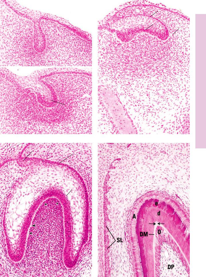
FIGURE 1a. Tooth development. Dental lamina. Frontal section. Pig. Paraffin section. ×132.
The dental lamina (DL) is a horseshoe-shaped band of epithelial tissue that arises from the oral epithelium (OE) and is surrounded by mesenchymal cells (MC). A frontal section of the dental lamina is characterized by the club-shaped appearance in this photomicrograph. The mesenchymal cells in discrete regions at the distal aspect of the dental lamina become rounded and congregate to form the precursor of the dental papilla responsible for the formation of the pulp and dentin of the tooth.
FIGURE 1b. Tooth development. Bud stage. Frontal section. Pig. Paraffin section. ×132.
At various discrete locations along the dental lamina (DL), an epithelial thickening, the bud (B), makes its appearance. Each bud will provide the cells necessary for enamel formation for a single tooth. The dental papilla (DP) forms a crescent-shaped area at the distal aspect of the bud.
FIGURE 3. Tooth development. Bell stage. Frontal section. Pig. Paraffin section. ×132.
As the enamel organ expands in size, it resembles a bell, hence the bell stage of tooth development. This stage is characterized by four cellular layers: outer enamel epithelium (OEE), stellate reticulum (SR), inner enamel epithelium (IEE), and stratum intermedium (SI). Observe that the enamel organ is still connected to the dental lamina (DL). The dental papilla (DP) is composed of rounded mesenchymal cells, whose peripheral-most layer (arrows) will differentiate to form odontoblasts. Note the wide basement membrane (arrowheads) between the future odontoblasts and inner enamel epithelium (the future ameloblasts). Observe also the spindle-shaped cells of the dental sac (DS).
Oral epithelium
Dental lamina 
Condensed mesenchyme
FIGURE 2. Tooth development. Cap stage. Frontal section. Pig. Paraffin section. ×132.
Increased mitotic activity transforms the bud into a cap-shaped structure. Observe that three epithelial layers of the enamel organ may be recognized: the outer enamel epithelium (OEE), the inner enamel epithelium (IEE), and the intervening stellate reticulum
(SR). The inner enamel epithelium has begun to enclose the dental papilla (DP). Note that mesenchymal cells become elongated, forming the dental sac (DS), which will envelop the enamel organ and dental papilla. Moreover, a bony crypt (BC) will enclose the dental sac.
FIGURE 4. Tooth development. Apposition. Frontal section. Pig. Paraffin section. ×132.
The elaboration of dentin (d) and enamel (e) is indicative of apposition. Dentin is manufactured by odontoblasts (O), the peripheral-most cell layer of the dental papilla (DP). The odontoblastic processes (arrows) are visible in this photomicrograph as they traverse the dentin matrix (DM). Ameloblasts (A) are highly elongated, columnar cells that manufacture enamel. The long, epithelial structure located to the left is the succedaneous lamina (SL), which is responsible for the development of the permanent tooth.
Dental sac
Bony crypt
Bud |
Cap |
Bell Apposition |
stage |
stage |
stage |
KEY
A |
ameloblast |
DP |
dental papilla |
OE |
oral epithelium |
B |
bud |
DS |
dental sac |
OEE |
outer enamel epithelium |
BC |
bony crypt |
e |
enamel |
SI |
stratum intermedium |
d |
dentin |
IEE |
inner enamel epithelium |
SL |
succedaneous lamina |
DL |
dental lamina |
MC |
mesenchymal cell |
SR |
stellate reticulum |
DM |
dentin matrix |
O |
odontoblast |
|
|
OE
 DL
DL
MC
(a)
DL
DP
(b)
FIGURE 1
DL
SR
DP
|
OEE |
|
SR |
IEE |
DS |
|
||
|
DP |
|
B
BC
FIGURE 2
OEE
SI
 IEE
IEE
DS
FIGURE 3 |
FIGURE 4 |
Development ooth • T4-13 PLATE
ongue • T5-13 PLATE
318 DIGESTIVE SYSTEM I
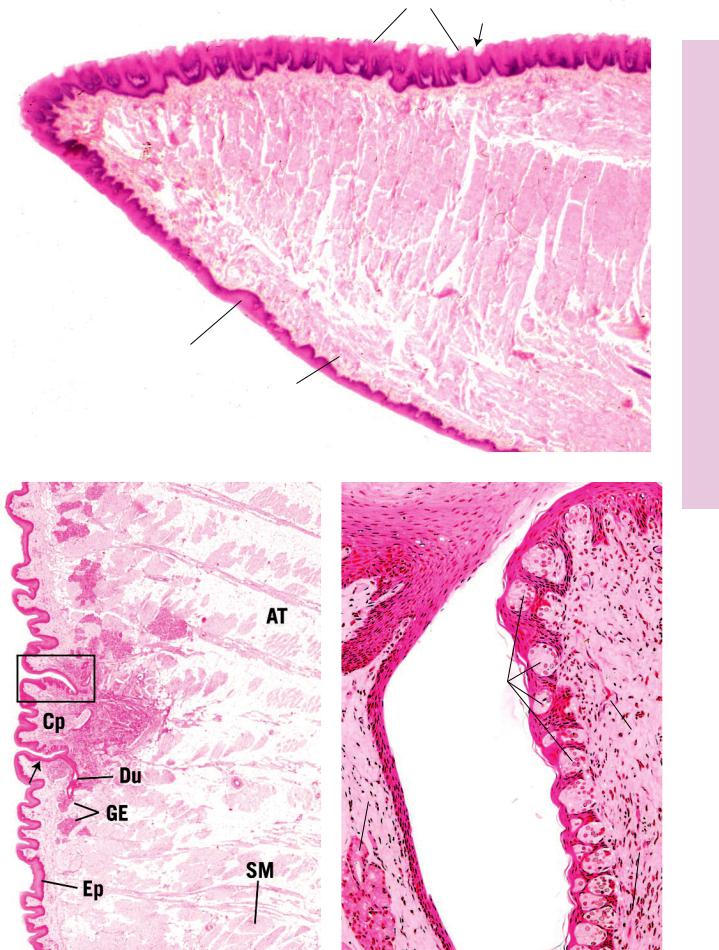
FIGURE 1. Tongue. Human. l.s. Paraffin section. ×20.
Part of the anterior two-thirds of the tongue is presented in this photomicrograph. This muscular organ bears numerous filiform papillae (FP) on its dorsal surface, whose stratified squamous epithelium is keratinized (arrow). The ventral surface of the tongue is lined by stratified squamous nonkeratinized epithelium (Ep). The intrinsic muscles of the tongue are arranged in four layers: superior longitudinal (SL), vertical (V), inferior longitudinal (IL), and horizontal (not shown here). The mucosa of the tongue tightly adheres to the perimysium of the intrinsic tongue muscles by the subepithelial connective tissue (CT).
FIGURE 2. Tongue. Human. l.s. Paraffin section. ×14.
The posterior aspect of the anterior two-thirds of the tongue presents circumvallate papillae (Cp). These papillae are surrounded by a deep groove (arrow), the base of which accepts a serous secretion via the ducts (Du) of the glands of von Ebner (GE). The epithelium (Ep) of the papilla houses taste buds along its lateral aspects but not on its superior surface. The core of the tongue contains skeletal muscle (SM) fibers of the extrinsic and intrinsic lingual muscles as well as glands and adipose tissue (AT). A region similar to the boxed area is presented at a higher magnification in Figure 3.
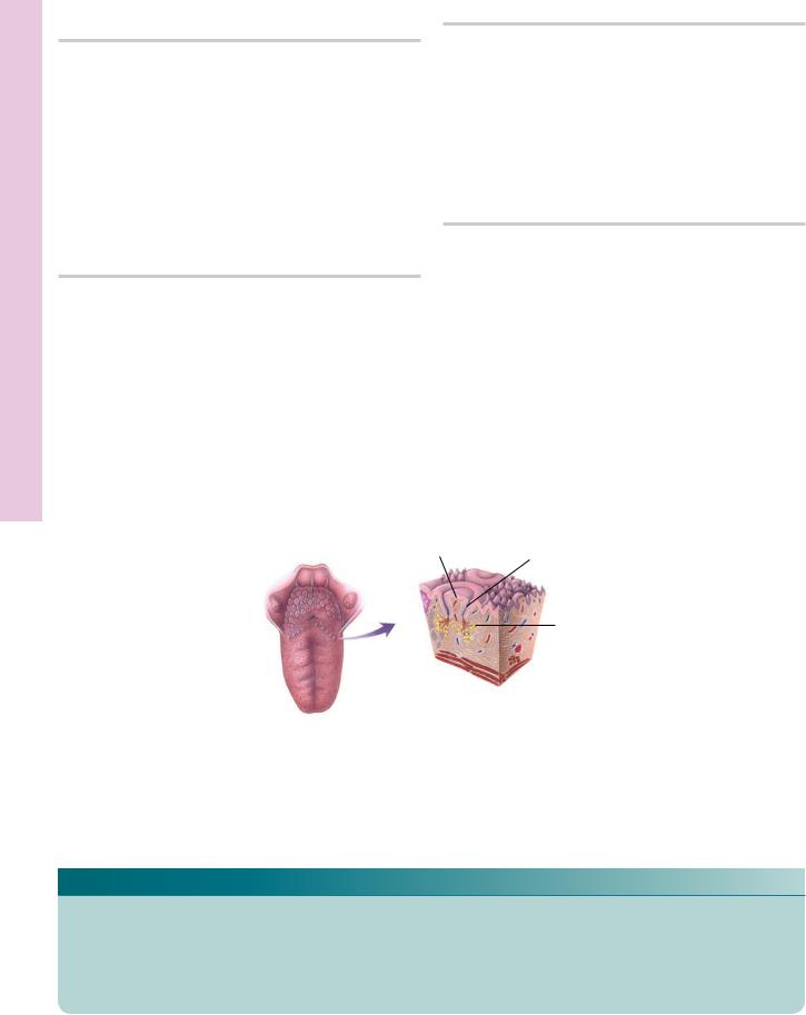
Filiform papilla
Circumvallate papilla
Taste bud
Fungiform papilla
 Filiform papillae
Tongue
Fungiform papilla
Glands of von Ebner
FIGURE 3. Circumvallate papilla. Monkey. x.s. Plastic section. ×132.
This photomicrograph is a higher magnification of a region similar to the boxed area of the previous figure, rotated 90 degrees. Note the presence of the groove (G) separating the circumvallate papilla (Cp) from the wall of the groove. Glands of von Ebner (GE) deliver a serous secretion into this groove, whose contents are monitored by numerous intraepithelial taste buds (TB). Observe that taste buds are not found on the superior surface of the circumvallate papilla, only on its lateral aspect. The connective tissue core of the papilla is richly endowed by blood vessels (BV) and nerves (N).
KEY
AT |
adipose tissue |
Ep |
epithelium |
N |
nerves |
BV |
blood vessels |
FP |
filiform papillae |
SL |
superior longitudinal muscle |
Cp |
circumvallate papillae |
G |
groove |
SM |
skeletal muscle |
CT |
connective tissue |
GE |
glands of von Ebner |
TB |
taste buds |
Du |
ducts |
IL |
inferior longitudinal muscle |
V |
vertical muscle |
FP
SL
V
Ep
CT |
IL |
|
FIGURE 1 |
ongue • T5-13 PLATE
 BV
BV
TB |
Cp |
|
|
|
BV |
G
GE
N
FIGURE 2 |
FIGURE 3 |
Palate and ongue • T6-13 PLATE
320 DIGESTIVE SYSTEM I
FIGURE 1. Circumvallate papilla. Monkey. Paraffin section. ×132.
The base of the circumvallate papilla (Cp), the surrounding groove (G), and the wall of the groove are evident in this photomicrograph. The glands of von Ebner (GE) deliver their serous secretions via short ducts (Du) into the base of the groove. Observe the rich vascular (BV) and nerve (N) supply to this region. Numerous taste buds (TB) populate the epithelium of the lateral aspect of the circumvallate papilla. Each taste bud possesses a taste pore (arrows) through which taste hairs (microvilli) protrude into the groove. A region similar to the boxed area is presented at a higher magnification in Figure 2.
FIGURE 3. Hard palate. Human. Paraffin section. ×132.
The hard palate possesses a nasal and an oral surface. The stratified squamous parakeratinized epithelium (Ep) of the oral surface forms deep invaginations, rete ridges (RR), which interdigitate with the subepithelial connective tissue (CT). The thick collagen fiber bundles (CF) firmly bind the palatal mucosa to the periosteum of the underlying bone. The hard palate also houses large deposits of adipose tissue and mucous glands.
FIGURE 2. Taste bud. Monkey. Paraffin section. ×540.
This is a higher magnification of a region similar to the boxed area of Figure 1. Note that the stratified squamous parakeratinized epithelium (Ep) displays squames in the process of desquamation (arrowheads). The taste buds (TB) are composed of four cell types. Basal (lateral) cells (BC) are believed to be regenerative in nature, whereas light cells (LC), intermediate cells, and dark cells (DC) are gustatory. Observe the presence of blood vessels (BV) in the subepithelial connective tissue (CT).
FIGURE 4. Soft palate. Human. Paraffin section. ×132.
The oral surface of the soft palate is lined by a stratified squamous nonkeratinized epithelium (Ep), which interdigitates with the lamina propria (LP) by the formation of shallow rete ridges (RR). The soft palate is a moveable structure, as attested by the presence of skeletal muscle fibers (SM). The core of the soft palate also houses numerous mucous glands (MG) that deliver their secretory products into the oral cavity via short, straight ducts.
Circumvallate papilla
Taste bud
Glands of von Ebner
Tongue
KEY
BC |
basal cells |
Du |
ducts |
MG |
mucous glands |
BV |
blood vessels |
Ep |
epithelium |
N |
nerve |
CF |
collagen fiber bundles |
G |
groove |
RR |
rete ridges |
Cp |
circumvallate papilla |
GE |
glands of von Ebner |
SM |
skeletal muscle |
CT |
connective tissue |
LC |
light cells |
TB |
taste buds |
DC |
dark cells |
LP |
lamina propria |
|
|
|
|
|
|
|
|
BC |
|
|
|
|
|
|
|
|
TB |
|
G |
|||
Cp |
|
|
|
|
|
TB |
|
|
|
|
|||
|
|
|
|
|
|
|
TB |
|
|
|
|
LC |
|
|
|
|
|
|
||
|
|
|
|
|
|
DC |
|
|
|||||
|
|
|
|
|
|
Ep |
GE
Du
N
BV
BV
N |
CT |
FIGURE 1 |
FIGURE 2 |
Ep
RR
Lp RR
SM
Ep
CT
MG
CF
RR 
FIGURE 3 |
FIGURE 4 |
Palate and ongue • T6-13 PLATE

Palate Hard the of Aspect Nasal and eeth • T7-13 PLATE
322 DIGESTIVE SYSTEM I
FIGURE 1. Human central incisor roots. Paraffin section. ×132.
The roots of two human central incisors and their supporting tissues are noted in this composite photomicrograph. Note that the root of one incisor, Root 1, is at the top of the figure, and progressing down the page, the hyaline layer of Hopewell-Smith
(HL) separates the dentin (d) of the root from the cementum (c). The periodontal ligament (PL1), with its attendant blood vessels
(BV), of this tooth suspends tooth 1 in its alveolus. The interdental septum (IS), positioned between the two incisors and composed of woven bone, is formed by the fusion of the alveolar bones proper (ABP 1 and 2) of each root. Note the presence of osteons (Os) in the woven bone; the center of these osteons approximates the line of fusion between the two alveolar bones proper. The periodontal ligament of the other incisor (PL 2) is located between the alveolar bone proper (ABP 2) and the cementum of this tooth. Its dentin (d) and hyaline layer of Hopewell-Smith
(HL) of root 2 are evident.
FIGURE 2. Hard palate. Human. Paraffin section. ×132.
The hard palate possesses a nasal and an oral surface. Note that the pseudostratified ciliated columnar epithelium (Ep) displays cilia and an intraepithelial gland (IeGL). Observe the presence of glands (Gl) and blood vessels (BV) in the subepithelial connective tissue (CT). The epithelium and the subepithelial connective tissue are collectively referred to as the mucoperiosteum (MP), which is firmly attached to the bony shelf (B) of the palate. A higher magnification of the boxed area is presented in Figure 3.
FIGURE 3. Hard palate. Human. Paraffin section. ×132.
This is a higher magnification of a region similar to the boxed area of Figure 2. Note the presence of glands (Gl), blood vessels (BV), and lymph vessels (LV) within the subepithelial connective tissue (CT). The thick collagen fiber bundles (CF) firmly bind the palatal mucosa to the periosteum of the underlying bone. Observe the clearly visible cilia (c) of the pseudostratified ciliated columnar epithelium (Ep) covering the nasal surface of the hard palate.
KEY
ABP |
alveolar bone proper |
d |
dentin |
IeGL |
intraepithelial gland |
B |
bony shelf |
Ep |
epithelium |
IS |
interdental septum |
BV |
blood vessel |
GL |
gland |
MP |
palatal mucosa |
C |
cementum |
HL |
hyaline layer of |
Os |
osteon |
CT |
connective tissue |
|
Hopewell-Smith |
PL |
periodontal ligament |

Root 1 |
|
|
|
|
IeGl |
||
|
|
|
|
|
|
||
|
|
|
d |
CT |
Ep |
||
c |
|
|
B |
Gl |
|||
|
|
|
|
||||
|
|
HL |
|
|
|
||
|
|
|
|
|
|
|
|
|
|
|
|
BV |
|
||
|
|
|
|
|
|
|
|
|
|
|
|
|
|
|
|
|
BV |
PL 1 |
|
|
BV |
|
|
|
|
|
|
||||
|
|
|
|
|
|
|
|
|
|
|
|
|
|
|
|
|
|
|
|
|
|
|
|
ABP 1 |
|
MP |
|
Os
IS |
FIGURE 2 |
|
Gl |
Ep |
ABP 2 |
|
|
|
|
|
|
|
BV |
|
|
|
CT
c L 
PL 2
|
|
BV |
c |
|
|
|
|
|
d |
Root 2 |
HL |
|
FIGURE 1 |
FIGURE 3 |
Palate Hard the of Aspect Nasal and eeth • T7-13 PLATE
324 DIGESTIVE SYSTEM I
Enamel of Micrograph Electron Scanning eeth • T8-13 PLATE
FIGURE 1
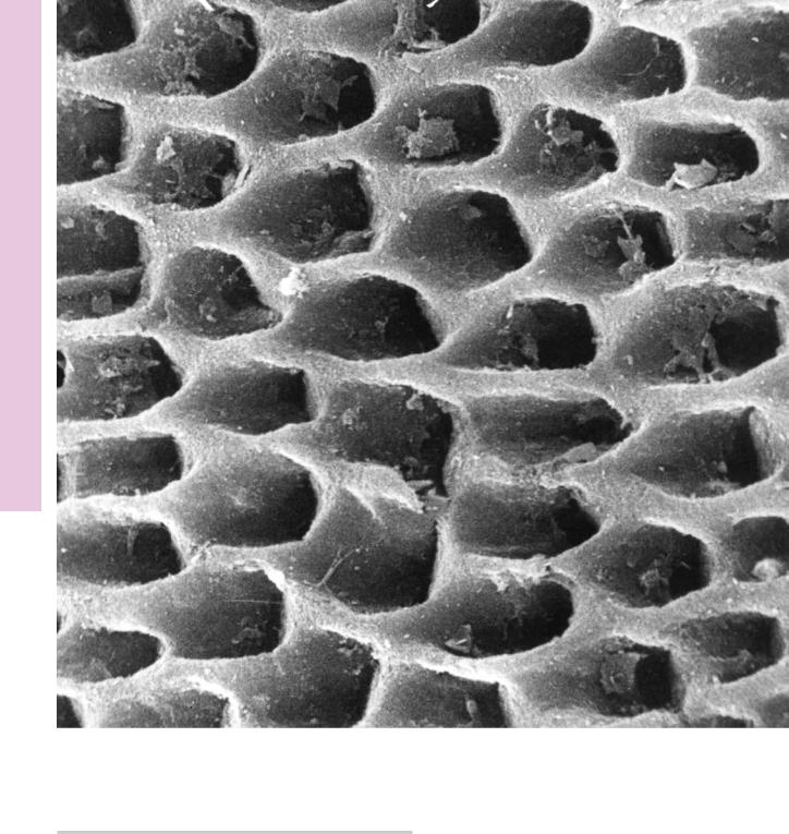
FIGURE 1. Human enamel. Scanning electron microscopy. ×3,150.
This three-dimensional view of the forming mineralized human enamel displays rod spaces (the recesses) surrounded by the interrod enamel. The rod spaces were occupied by Tomes’ processes of the ameloblasts, and as the ameloblasts recede, rod
spaces are filled in by the secretory mechanism, and the spaces are filled by enamel known as rod segments. The arched aspects of the rod spaces are directed occlusally. As rod segments are positioned on top of each other, they form an enamel rod whose shape resembles a keyhole. (From Fejerskov O. Human dentition and experimental animals. J Dent Res 1979;58(Special Issue B):725–734.)
Dentin of Micrograph Electron Scanning eeth • T9-13 PLATE
FIGURE 1
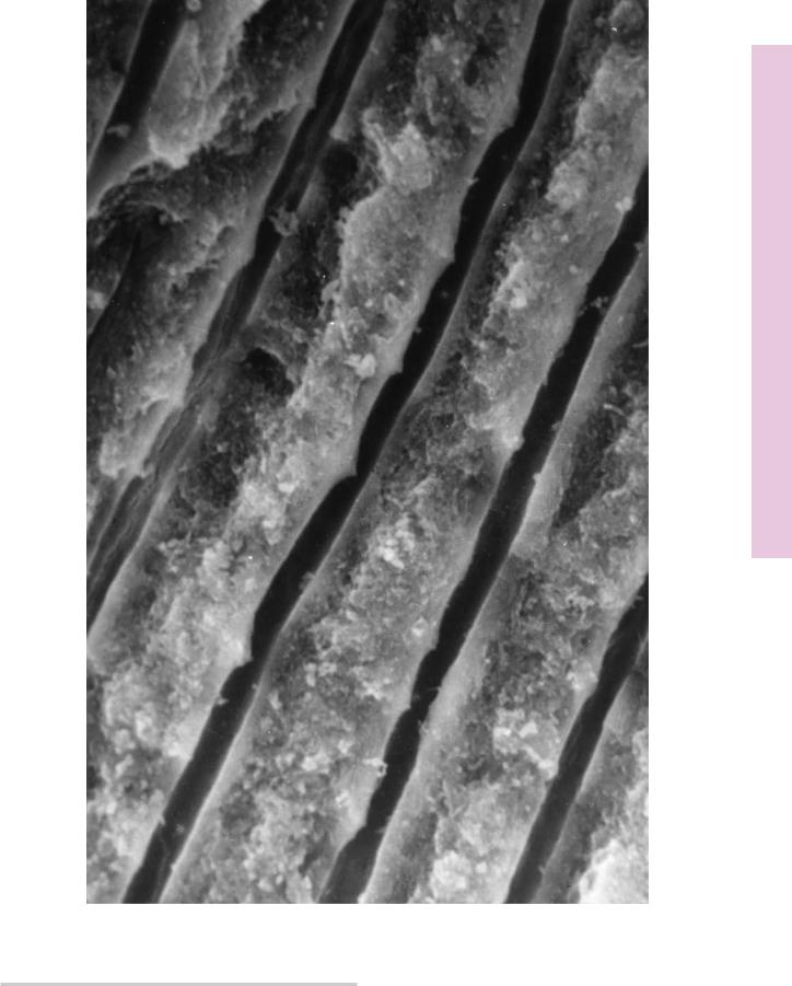
FIGURE 1. Human dentin. Scanning electron microscopy. ×3,800.
This three-dimensional view of mineralized human dentin displays a longitudinal section of dentinal tubules. In a healthy, living dentin, the tubules house the processes of odontoblasts that extend
at least 1 mm into the dentinal tubule. Additionally, some of the tubules also house nerve fibers, and all of the tubules are filled completely with an extracellular fluid that originates in the pulp of the tooth. (From Thomas H. The dentin-predentin complex and its permeability: anatomical overview. J Dent Res 1985;64(Special Issue B):607–612.)