

KAPLAN_USMLE_STEP_1_LECTURE_NOTES_2018_BIOCHEMISTRY_and_GENETICS
.pdfPart I ● Biochemistry
AMINO ACID ACTIVATION AND CODON TRANSLATION BY tRNAs
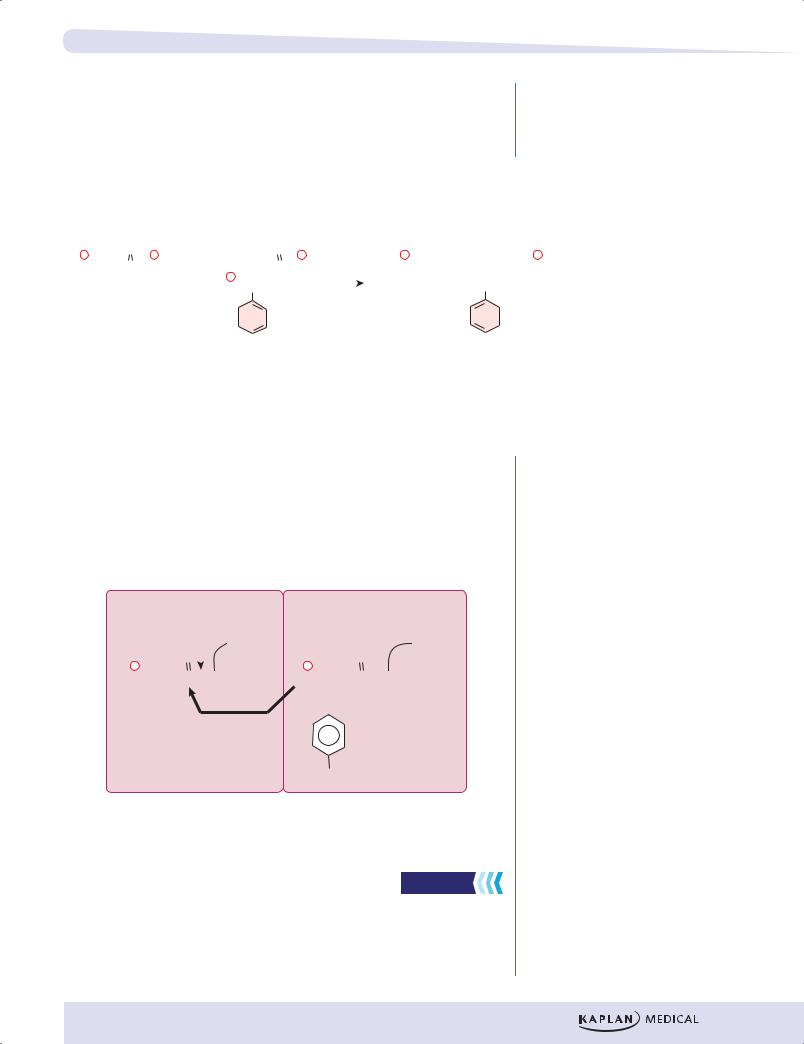
Inasmuch as amino acids have no direct affinity for mRNA, an adapter molecule, which recognizes an amino acid on one end and its corresponding codon on the other, is required for translation. This adapter molecule is tRNA.
Amino Acid Activation
As tRNAs enter the cytoplasm, each combines with its cognate amino acid in a process called amino acid activation.
|
|
|
+ |
R |
O |
|
|
|
H3N |
C C |
O- |
OH |
+ |
R |
O |
H |
|
P 3´ end |
H3N |
C |
C |
P |
3´ end |
5´ end |
|
H |
O- |
5´ end |
|
|
|
Amino |
ATP AMP + PPi |
|
|
|
|
acid |
|
|
|
|
|
aminoacyl-tRNA |
|
|
|
|
|
synthetase |
|
|
|
anticodon |
|
|
|
anticodon |
|
tRNA |
|
|
Aminoacyl-tRNA |
||
Figure I-4-3. Activation of Amino Acid for Protein Synthesis
•Each type of amino acid is activated by a different aminoacyl tRNA synthetase.
•Two high-energy bonds from an ATP are required.
•The aminoacyl tRNA synthetase transfers the activated amino acid to the 3´ end of the correct tRNA.
•The amino acid is linked to its cognate tRNA with an energy-rich bond.
•This bond will later supply energy to make a peptide bond linking the amino acid into a protein.
Aminoacyl tRNA synthetases have self-checking functions to prevent incorrectly paired aminoacyl tRNAs from forming. If, however, an aminoacyl tRNA synthetase does release an incorrectly paired product (ala-tRNAser), there is no mechanism during translation to detect the error and an incorrect amino acid will be introduced into some protein.
Each tRNA has an anticodon sequence that allows it to pair with the codon for its cognate amino acid in the mRNA. Because base pairing is involved, the orientation of this interaction will be complementary and antiparallel. For example, the amino acyl tRNA arg-tRNAarg has an anticodon sequence, UCG, allowing it to pair with the arginine codon CGA.
54
Chapter 4 ● The Genetic Code, Mutations, and Translation
TRANSLATION (PROTEIN SYNTHESIS)
Protein synthesis occurs by peptide bond formation between successive amino acids whose order is specified by a gene and thus by an mRNA. The formation of a peptide bond between the carboxyl group on one amino acid and the amino group of another is illustrated below.
|
|
|
|
|
|
|
|
|
|
|
|
|
|
|
|
|
|
|
|
|
|
Peptide Bond |
|
|
|||||||||||||
|
|
|
|
|
O |
|
|
|
|
|
|
|
|
|
O |
|
|
|
|
|
|
|
|
|
|
|
|
|
|
|
|
|
|
|
|
|
|
|
|
|
|
|
|
|
|
|
|
|
|
|
|
|
|
|
|
|
|
|
|
|
|
|
|
|
|
|
|
|
|
|
|
|
|
||
+ |
|
|
|
|
|
– |
|
|
|
|
|
|
|
|
– |
+ |
|
|
|
|
O |
|
|
|
|
|
|
|
O |
|
– |
||||||
|
|
|
|
|
+ |
|
|
|
|
|
|
|
|
|
|
|
|
|
|
|
|
|
|
|
|
|
|
|
|||||||||
H3N |
|
|
CH |
|
C |
|
O |
H3N |
|
|
CH |
|
C |
|
O |
|
H3N |
|
|
CH |
|
C |
|
N |
|
|
CH |
|
C |
|
O |
||||||
|
|
|
|
|
|
|
|
|
|
|
|
|
|
||||||||||||||||||||||||
|
|
|
|
|
|
|
|
|
+ |
|
|
|
|
|
|
|
|
|
|
|
|
|
|
|
|
|
H |
|
|
|
|
|
|
|
|||
|
|
|
CH2 |
|
|
|
|
|
CH2 |
|
|
|
|
|
|
CH2 |
|
|
CH2 |
|
|
||||||||||||||||
|
|
|
|
|
|
|
|
|
|
|
|
|
|
|
|
|
|
||||||||||||||||||||
|
|
|
|
|
|
|
|
|
|
|
|
|
|
|
|
||||||||||||||||||||||
|
|
|
CH2 |
|
|
|
|
|
|
|
|
|
|
|
|
|
|
|
|
CH2 |
|
|
|
|
|
|
|
|
|
|
|
||||||
|
|
|
|
|
|
|
|
|
|
|
|
|
|
|
|
|
|
|
|
|
|
|
|
|
|
|
|
|
|
|
|
|
|
|
|
|
|
|
|
S |
|
|
|
|
|
|
|
|
|
|
|
|
|
|
|
S |
|
|
|
|
|
|
|
|
|
|
|
||||||||
|
|
|
|
|
|
|
|
|
|
|
|
|
|
|
|
|
|
|
|
|
|
|
|
|
|
|
|
|
|
|
|
|
|||||
|
CH3 |
|
|
|
|
|
OH |
|
|
|
|
|
|
CH3 |
|
|
|
OH |
|
|
|||||||||||||||||
|
|
Met |
|
|
|
|
|
|
Tyr |
|
|
|
|
|
|
|
Met-Tyr |
|
|
||||||||||||||||||
Figure I-4-4. Peptide Bond Formation
During translation, the amino acids are attached to the 3′ ends of their respective tRNAs. The aminoacyl–tRNAs are situated in the P and A sites of the ribosome as shown in the figure below. Notice that the peptide bond forms between the carboxyl group of the amino acid (or growing peptide) in the P site and the amino group of the next amino acid in the A site. Proteins are synthesized from the amino to the carboxyl terminus.
P |
Bond breaks |
A |
|
|
|
|
|
|
|
|||||||||
|
|
|
|
|
|
|
|
|
||||||||||
|
|
|
|
|
|
|
|
|
|
tRNAmet |
|
|
|
|
|
|
|
tRNAtyr |
+ |
|
|
|
|
|
O |
|
|
+ |
|
|
|
|
O |
||||
|
|
|
|
|
|
|
|
|
|
|||||||||
H3N |
|
|
|
CH |
|
C |
|
|
|
O |
H3N |
|
|
CH |
|
C |
|
O |
|
|
|
|
|
|
|
|
|
||||||||||
|
|
|
|
|
|
|
|
|
||||||||||
|
|
|
|
CH2 |
|
|
|
New |
|
|
|
CH2 |
||||||
|
|
|
|
|
|
|
|
|
|
|||||||||
|
|
|
|
CH2 |
|
|
|
|
|
|
|
|
|
|
|
|||
|
|
|
|
|
|
|
|
|
|
peptide |
|
|
|
|
|
|
|
|
|
|
|
|
|
|
|
|
|
|
|
|
|
|
|||||
|
|
S |
|
|
|
|
|
|
|
|
||||||||
|
|
|
|
|
|
bond |
|
|
|
|
|
|
|
|
||||
|
|
|
|
CH3 |
|
|
|
|
|
|
OH |
|||||||
|
|
|
|
|
|
|
|
|
|
|
||||||||
Figure I-4-5. Formation of a Peptide Bond by a Ribosome During Translation
Steps of Translation
High-Yield
Translation occurs in the cytoplasm of both prokaryotic (Pr) and eukaryotic (Eu) cells. In prokaryotes, ribosomes can begin translating the mRNA even before RNA polymerase completes its transcription. In eukaryotes, translation and transcription are completely separated in time and space with transcription
55

Part I ● Biochemistry
in the nucleus and translation in the cytoplasm. The process of protein synthesis occurs in 3 stages: initiation, elongation, and termination.
Special protein factors for initiation (IF), elongation (EF), and termination (release factors), as well as GTP, are required for each stage.
Small ribosomal subunit
5' |
A |
|
AAA |
|
U |
G |
|
5' cap |
or Shine- |
G CU |
|
|
|
||
(Eu) |
Dalgarno |
|
|
|
(Pr) |
|
|
INITIATION
UAC
3' met |
|
met-tRNA; (Eu) |
Large |
or |
subunit |
fmet-tRNA; (Pr) |
|
5' IFs met
P |
|
|
G |
AU |
|
|
C |
A |
|
U |
|
A CUG
|
3' |
|
A |
A |
|
A |
|
5' P
|
G |
AU |
|
|
C |
A |
|
U |
|
met
A
CUG GAC
Leu
ELONGATION
|
3' |
5' |
P |
|
|
A |
|
|
A |
|
|
G |
C |
|
|
A |
|
|
|
||||
A |
|
|
U |
|
UG |
||
|
|
A |
|
G |
|||
|
|
|
|
C |
A |
C |
|
|
|
|
A |
|
|
||
|
|
|
U |
|
|
|
|
5'
3'
|
A |
A |
|
A |
|
A
UG
P
CUG GAC
A
A
A
3'
A
Aminoacyl-tRNA binds to A site |
Peptide bond forms. Peptidyl |
GTP |
transferase in large subunit |
EF-TU and EFTS (Pr) |
|
eEF-1(Eu) |
|
Shiga toxin inhibits |
|
(cuts 28S rRNA) |
|
Translocation of ribosome 3 nucleotides along the mRNA GTP
EF-G(Pr) eEF-2(Eu)
Pseudomonas and diphtheria toxin inhibit (ADP-ribosylation) eEF-2
Elongation cycle repeats for each amino acid added
TERMINATION
5' |
P |
|
|
G |
|
|
|
|
|
C |
|
|
G |
C |
|
G |
|
|
C |
|
A
UAG
3'
STOP CODON
in A Site
Completed protein released from ribosome Ribosomal subunits separate
mRNA Released
Ala |
Ala COOH |
H2N-metLeuLys |
H2N-metLeuLys |
Figure I-4-6. Steps in Translation
56

Chapter 4 ● The Genetic Code, Mutations, and Translation
Initiation
The small ribosomal subunit binds to the mRNA. In prokaryotes, the 16S rRNA of the small subunit binds to the Shine-Dalgarno sequence in the 5′ untranslated region of the mRNA. In eukaryotes, the small subunit binds to the 5′ cap structure and slides down the message to the first AUG.
The charged initiator tRNA becomes bound to the AUG start codon on the message through base pairing with its anticodon. The initiator tRNA in prokaryotes carries fMet, whereas the initiator tRNA in eukaryotes carries Met.
The large subunit binds to the small subunit, forming the completed initiation complex.
There are 2 important binding sites on the ribosome called the P site and the A site.
•The peptidyl site (P site) is the site on the ribosome where fMet–tRNAi initially binds. After formation of the first peptide bond, the P site is a binding site for the growing peptide chain.
•The aminoacyl site (A site) binds each new incoming tRNA molecule carrying an activated amino acid.
Elongation
Elongation is a 3-step cycle that is repeated for each amino acid added to the protein after the initiator methionine. Each cycle uses 4 high-energy bonds (2 from the ATP used in amino acid activation to charge the tRNA, and 2 from GTP). During elongation, the ribosome moves in the 5′ to 3′ direction along the mRNA, synthesizing the protein from amino to carboxyl terminus. The 3 steps are:
•A charged tRNA binds in the A site. The particular aminoacyl–tRNA is determined by the mRNA codon aligned with the A site.
•Peptidyl transferase, an enzyme that is part of the large subunit, forms the peptide bond between the new amino acid and the carboxyl end of the growing polypeptide chain. The bond linking the growing peptide to the tRNA in the P site is broken, and the growing peptide attaches to the tRNA located in the A site.
•In the translocation step, the ribosome moves exactly 3 nucleotides (one codon) along the message. This moves the growing peptidyl– tRNA into the P site and aligns the next codon to be translated with the empty A site.
In eukaryotic cells, elongation factor-2 (eEF-2) used in translocation is inactivated through ADP-ribosylation by Pseudomonas and Diphtheria toxins.
Shiga and Shiga-like toxins clip an adenine residue from the 28S rRNA in the 60S subunit stopping protein synthesis in eukaryotic cells.
Termination
When any of the 3 stop (termination or nonsense) codons moves into the A site, peptidyl transferase (with the help of release factor) hydrolyzes the completed protein from the final tRNA in the P site. The mRNA, ribosome, tRNA, and factors can all be reused for additional protein synthesis.
57

Part I ● Biochemistry
Clinical Correlate
Gray baby syndrome is a dangerous condition that occurs in newborns (especially premature babies) who are given the drug chloramphenicol. Chloramphenicol is a drug used to fight bacterial infections, including meningitis. If given to a newborn, however, this drug can trigger a potentially deadly reaction. Babies do not have sufficient UDP-glucuronyl transferase activity needed to allow excretion of this drug. The drug builds up in the baby’s bloodstream and can lead to:
•Blue lips, nail beds, and skin (cyanosis)
•Death
•Low blood pressure
Recall Question
Which of the following terms is used to describe a disease that, from generation to generation, shows a decrease in the age of onset and an increase in the severity of symptoms?
A.Acception
B.Assumption
C.Anticipation
D.Mutation
Answer: C
POLYSOMES
Messenger RNA molecules are very long compared with the size of a ribosome, allowing room for several ribosomes to translate a message at the same time. Because ribosomes translate mRNA in the 5′ to 3′ direction, the ribosome closest to the 3′ end has the longest nascent peptide. Polysomes are found free in the cytoplasm or attached to the rough endoplasmic reticulum (RER), depending on the protein being translated.
INHIBITORS OF PROTEIN SYNTHESIS
Some well-known inhibitors of prokaryotic translation include streptomycin, erythromycin, tetracycline, and chloramphenicol. Inhibitors of eukaryotic translation include cycloheximide and Diphtheria and Pseudomonas toxins.
Certain antibiotics (for example, chloramphenicol) inhibit mitochondrial protein synthesis, but not cytoplasmic protein synthesis, because mitochondrial ribosomes are similar to prokaryotic ribosomes.
PROTEIN FOLDING AND SUBUNIT ASSEMBLY
As proteins emerge from ribosomes, they fold into 3-dimensional conformations that are essential for their subsequent biologic activity. Generally, 4 levels of protein shape are distinguished:
Primary—sequence of amino acids specified in the gene.
Secondary—folding of the amino acid chain into an energetically stable structure. Two common examples are the α-helix and the β-pleated sheet. These shapes are reinforced by hydrogen bonds. An individual protein may contain both types of secondary structures. Some proteins, like collagen, contain neither but have their own more characteristic secondary structures.
58

Chapter 4 ● The Genetic Code, Mutations, and Translation
Tertiary—positioning of the secondary structures in relation to each other to generate higher-order 3-dimensional shapes (the domains of the IgG molecule are examples). Tertiary structure also includes the shape of the protein as a whole (globular, fibrous). Tertiary structures are stabilized by weak bonds (hydrogen,hydrophobic, ionic) and, in some proteins, strong, covalent disulfide bonds. Agents such as heat or urea disrupt tertiary structure to denature proteins, causing loss of function.
Quaternary—in proteins such as hemoglobin that have multiple subunits, quaternary structure describes the interactions among subunits.
TRANSLATION OCCURS ON FREE RIBOSOMES AND ON THE ROUGH ENDOPLASMIC RETICULUM
Although all translation of eukaryotic nuclear genes begins on ribosomes free in the cytoplasm, the proteins being translated may belong in other locations. For example, certain proteins are translated on ribosomes associated with the rough endoplasmic reticulum (RER), including:
•Secreted proteins
•Proteins inserted into the cell membrane
•Lysosomal enzymes
Proteins translated on free cytoplasmic ribosomes include:
•Cytoplasmic proteins
•Mitochondrial proteins (encoded by nuclear genes)
Molecular Chaperones and Proteasomes
High-Yield
Protein folding is an essential step in the final synthesis of any protein. There is a class of specialized proteins, chaperones, whose function is to assist in this process. Molecular chaperones function in many cell compartments, including the endoplasmic reticulum, where extensive protein synthesis occurs. Failure to fold correctly usually results in eventual destruction of the protein.
59
Part I ● Biochemistry
Clinical Correlate
Cystic Fibrosis
The majority of cases of cystic fibrosis result from the deletion of phenylalanine at position 508 (∆F508), which interferes with proper protein folding and the posttranslational processing of oligosaccharide side chains. The abnormal chloride channel protein (CFTR) is degraded by the cytosolic proteasome complex rather than being translocated to the cell membrane. Other functional defects in CFTR protein reaching the cell membrane may also contribute to the pathogenesis of cystic fibrosis.
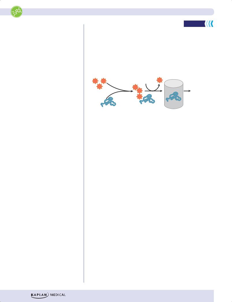
Proteasomes and Ubiquitin |
High-Yield |
|
Whenever protein synthesis occurs in a cell, a few copies of a particular protein may not fold correctly. These defective copies are covalently marked for destruction by the addition of multiple copies of ubiquitin. Polyubiquinated proteins are directed to proteasomes for destruction. Proteasomes are large, cytoplasmic complexes that have multiple protease activities capable of digesting damaged proteins to peptides. Proteasomes also play a role in producing antigenic peptides for presentation by class I MHC molecules.
Ubiquitin |
|
|
U U |
Proteasome |
U |
U |
U U |
Peptide |
|
||
|
U |
fragments |
|
|
Misfolded
protein
Figure I-4-7. Degradation of Misfolded Proteins by Proteasomes
Many proteins require signals to ensure delivery to the appropriate organelles. Especially important among these signals are:
•The N-terminal hydrophobic signal sequence used to ensure translation on the RER
•Phosphorylation of mannose residues important for directing an enzyme to a lysosome
The targeting process for these proteins is illustrated below.
60
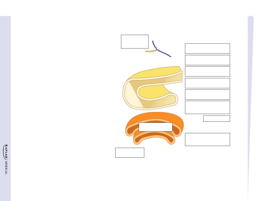
Rough Peroxisomes endoplasmic
reticulum (RER)
Mitochondrion
Lysosome
Golgi apparatus
Nucleus 
Smooth  endoplasmic reticulum (SER)
Microtubule & filaments
N-Terminal |
|
|
5' |
hydrophobic |
|
|
|
signal sequence |
|
|
|
N |
|
|
|
Involves signal |
|
|
3' |
recognition |
|
|
Cytoplasm |
particle (SRP) |
|
|
|
5' |
5' |
|
3' |
3' |
|
|
|
|
|
ER lumen |
|
3' |
|
N |
|
|
|
||
N |
|
N-Glycosylation |
|
|
|
||
5' |
|
|
(Dolichol-P) |
N
Golgi
O-linked glycosylation in the Golgi
To cell membrane 
 or secretion
Figure I-4-8. Synthesis of Secretory, Membrane, and Lysosomal Proteins
61
Translation begins in cytoplasm
Signal sequence causes ribosomes to attach to ER
Signal peptidase removes the signal sequence
Translation continues on RER
Glycosylation in ER
(continues in Golgi)
Proper folding is required in ER for transfer of protein to Golgi
To lysosomes
Phosphorylation of mannose by phosphotransferase signals to lysosome
Translation and Mutations, Code, Genetic The ● 4 Chapter

Part I ● Biochemistry
N-Terminal Hydrophobic Signal Sequence
High-Yield
This sequence is found on proteins destined to be secreted (insulin), placed in the cell membrane (Na+-K+ ATPase), or ultimately directed to the lysosome (sphingomyelinase). These proteins all require N-terminal hydrophobic signal sequences as part of their primary structure. Translation begins on free cytoplasmic ribosomes, but after translation of the signal sequence, the ribosome is positioned on the ER (now RER) with the help of a signal recognition particle. During translation, the nascent protein is fed through the membrane of the RER and captured in the lumen. The signal sequence is cleaved off in the ER, and then the protein passes into the Golgi for further modification and sorting.
In transit through the ER and Golgi, most proteins acquire oligosaccharide side chains, becoming glycoproteins. N-glycosylation refers to the addition of sugar chains to the nitrogen of asparagine residues (N-linked). The attachment of sugars in N-glycosylation begins in the ER (cotranslational modification) and requires the participation of a special lipid called dolichol phosphate. The N-linked sugar chain can further be modified upon entry in the Golgi (posttranslational modification).
O-glycosylation refers to the addition of sugar chains to the hydroxyl group of either serine or threonine residues of the protein, and it occurs exclusively in the Golgi (posttranslational modification). Depending of the particular glycoprotein, some proteins are solely N-glycosylated (for example, transferrin); some are solely O-glycosylated (for example, heparin); and some are both N- and O-glycosylated (for example, LDL receptor). Significantly, the structure and sequence of the oligosaccharide chains on proteins and lipids (glycolipids) are the basis of the A, B, O blood groups.
Accumulation or ineffective targeting of misfolded proteins
Proteins synthesized in the endoplasmic reticulum must fold correctly for transport to the Golgi and then to their final destinations. In certain genetic diseases, the mutation may cause all copies of the protein to fold incorrectly. The result is loss of protein function and, in some cases, accumulation of the misfolded protein in the endoplasmic reticulum.
α1-Antitrypsin Deficiency
A 70-year-old woman with elevated liver function tests was being evaluated for cirrhosis. Her serum α1-antitrypsin level was 25 mg/dL (normal 90–225 mg/dL). A liver biopsy showed micronodular cirrhosis and prominent fibrosis. Immunohistochemical studies showed intense staining with α1-antitrypsin antibody. The patient was tested for likely mutations in the α1-antitrypsin gene and found to be homozygous for the Z mutation (ZZ). This mutation causes the α1-antitrypsin protein to misfold and aggregate in the endoplastic reticulum, where it damages cells, eventually leading to cirrhosis. She had no evidence of pulmonary disease.
α1-antitrypsin is a protein synthesized primarily by the liver and secreted in the bloodstream. Its function is to protect cells by serving as an inhibitor of proteases released during a normal inflammatory response. Among the more than 90 allelic variants of the α1-antitrypsin gene, the Z and S variants are most often encountered with α1-antitrypsin deficiency. Both are the result of point mutations, which can be detected with the polymerase chain reaction (PCR) technique.
62

Chapter 4 ● The Genetic Code, Mutations, and Translation
Lysosomal Enzymes and |
High-Yield |
|
|
Phosphorylation of Mannose |
|
Lysosomal enzymes are glycosylated and modified in a characteristic way. Most importantly, when they arrive in the Golgi apparatus, specific mannose residues located in their N-linked oligosaccharide chains are phosphorylated by N-acetylglucosamine-1 phosphotransferase, forming a critical mannose-6-phosphate in the oligosaccharide chain. This phosphorylation is the critical event that removes them from the secretion pathway and directs them to lysosomes. Genetic defects affecting this phosphorylation produce I-cell disease in which lysosomal enzymes are released into the extracellular space, and inclusion bodies accumulate in the cell, compromising its function.
Major Symptoms of I-Cell Disease
A child aged 5 months was referred to a specialist. The child had been born with dislocated hips and a coarse featured face. He had been suffering repeated upper respiratory tract infections and did not seem to be developing his motor abilities, Clinical examination revealed hyperplasia of the gums, restriction of joint mobility and hepatosplenomegaly. On listening to the heart a mitral valve murmur could be detected. Further investigation involved cell culture of the child’s fibroblasts obtained from a skin biopsy. Examination of the fibroblasts under the microscope revealed the presence of numerous intracellular inclusions, which on electron microscopy were revealed to be large lysosomes. Biochemical analysis showed decreased levels of the lysosomal hydrolase ß-glucuronidase within the fibroblasts, but elevated levels of this enzyme within the culture medium. A diagnosis of I-cell disease was made.
•Coarse facial features, gingival hyperplasia, macroglossia
•Craniofacial abnormalities, joint immobility, clubfoot, claw-hand, scoliosis
•Psychomotor retardation, growth retardation
•Cardiorespiratory failure, death in first decade
•Bone fracture and deformities
•Mitral valve defect
•Secretion of active lysosomal enzymes into blood and extracellular fluid
COAND POSTTRANSLATIONAL COVALENT MODIFICATIONS
In addition to disulfide bond formation while proteins are folding, other covalent modifications include:
•Glycosylation: addition of oligosaccharide as proteins pass through the ER and Golgi apparatus
•Proteolysis: cleavage of peptide bonds to remodel proteins and activate them (proinsulin, trypsinogen, prothrombin)
•Phosphorylation: addition of phosphate by protein kinases
Bridge to Anatomy
Lysosomes
•Organelles whose major function is to digest materials that the cell has ingested by endocytosis.
•Contain multiple enzymes that, collectively, digest carbohydrates (glycosylases), lipids (lipases), and proteins (proteases).
•Especially prominent in cells such as neutrophils and macrophages, though they serve this essential role in almost all cells.
When a lysosomal enzyme is missing (for instance in a genetic disease such as Tay-Sachs), the undigested substrate accumulates in the cell, often leading to serious consequences.
63