

Книги по МРТ КТ на английском языке / MRI for Orthopaedic Surgeons Khanna ed 2010
.pdf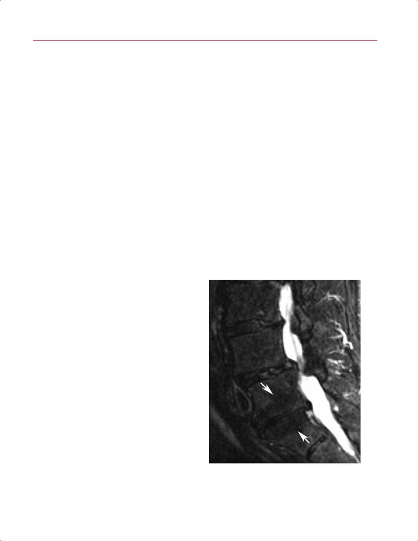
 B
B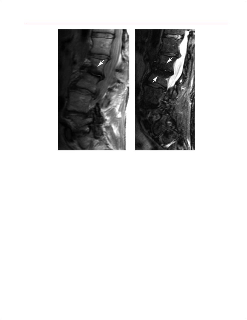
282 IV Spine
A |
B |
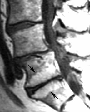
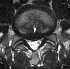
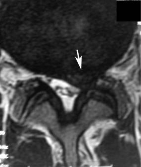
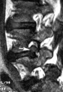
Fig. 11.17 Modic type 3 (sclerotic) changes. Sagittal T1-weighted |
level that is seen with Modic type 3 end-plate changes. Note that |
(A) and fat-suppressed T2-weighted (B) images showing the typical |
degenerative changes are seen at other levels and that there is also |
pattern (arrow[s] on each) of decreased signal intensity at the L2-L3 |
evidence of lumbar scoliosis. |
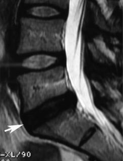
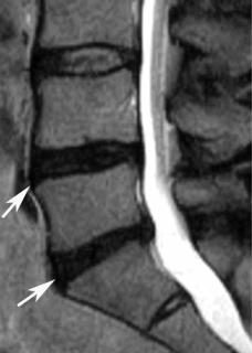
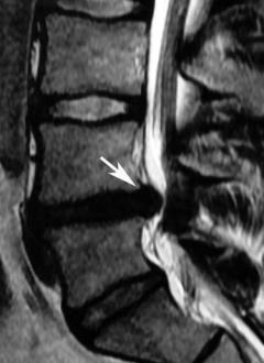
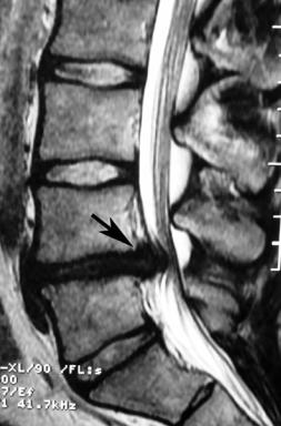
Annular Tears |
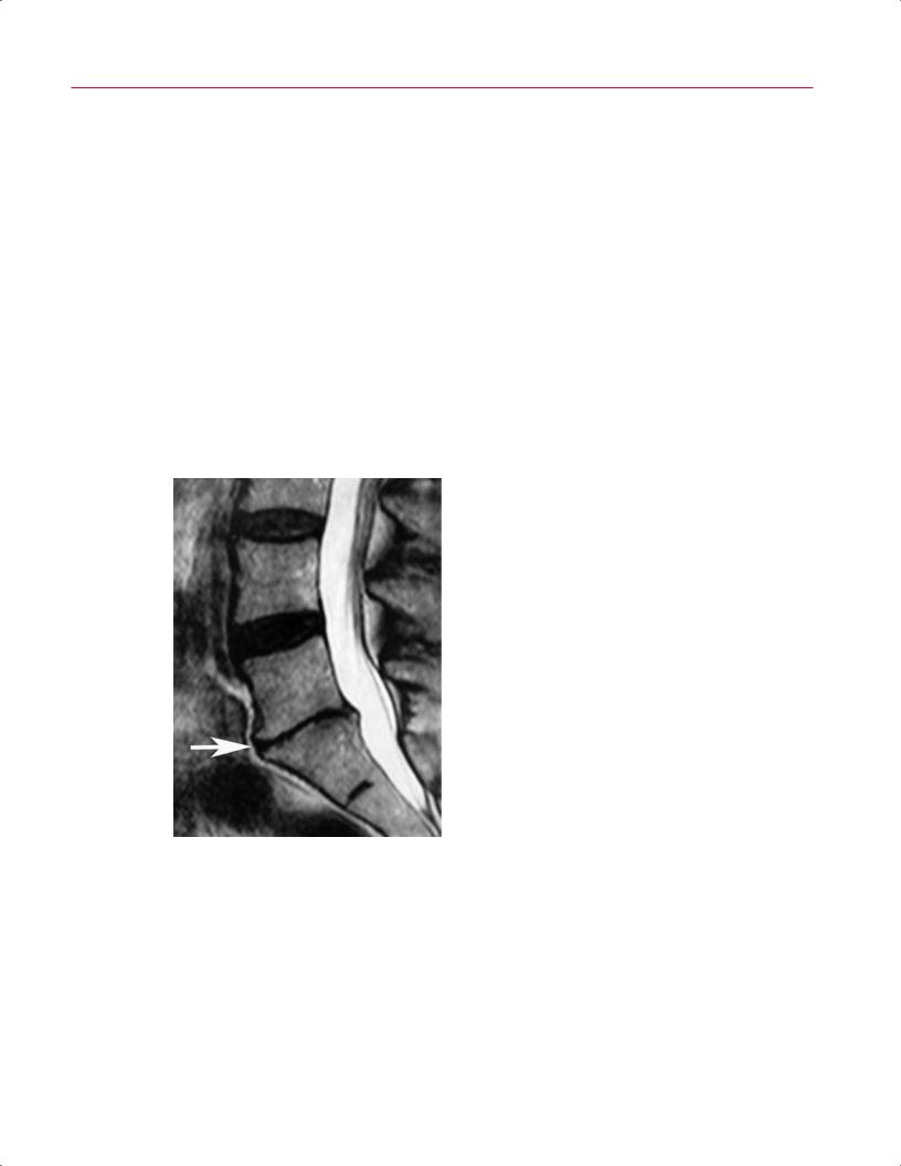
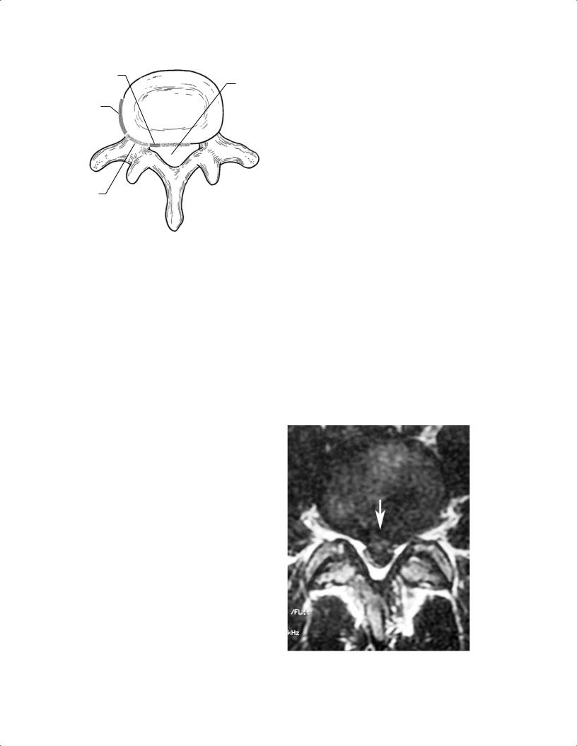
(Fig. 11.8). The high-intensity zone is defined as a focal area |
|
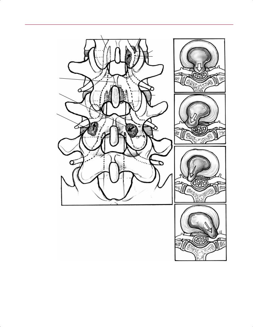
Annular tears on MRI have a variable appearance, ranging |
of high signal intensity within the posterior annulus of the |
|
degenerating disc, separate from the nucleus. These high- |
||
from intermediate to high signal intensity on T2-weighted |
||
intensity zones may also enhance after intravenous gado- |
||
images. Studies have shown a correlation between high |
||
linium administration.33 |
||
signal intensity annular tears in the lumbar spine and |
||
Discography can be used to further evaluate patients |
||
painful concordant annular tears seen at provocative |
||
with annular tears. In addition to the morphologic in- |
||
discography.37,38 Some investigators have suggested that |
||
formation provided on fluoroscopic images and on post- |
||
the inflammation associated with these annular tears |
||
discography CT, the patient’s pain response can be used |
||
results in irritation of the adjacent nerve root, poten- |
||
to help predict whether an annular tear or other degen- |
||
tially leading to radiculopathy without overt mechanical |
||
erative pathology is the patient’s pain generator.39,40 It is |
||
nerve root compression.38 T2-weighted sequences have |
||
important to keep in mind, however, that the use of dis- |
||
been used to show the following three types of annular |
||
cography in the diagnosis of discogenic low back pain con- |
||
tears: |
||
tinues to be debated and is not uniformly accepted at all |
||
• Concentric |
||
centers. |
||
• Radial |
|
|
• Transverse |
Lumbar Herniated Nucleus Pulposus |
|
Concentric tears involve the entire extent of the annulus. |
||
The terms used to describe the progressive states of herni- |
||
Transverse tears occur at the periphery of the disc as a result |
||
of disruption of Sharpey’s fibers. Radial tears extend from |
ated nucleus pulposus have been addressed above (see No- |
|
the nucleus through the annulus and may extend into the |
menclature and Classification of Lumbar Disc Pathology). |
|
outer annulus, manifested on MRI as a high-intensity zone |
Shown here are the MRI appearances of each: |
 B
B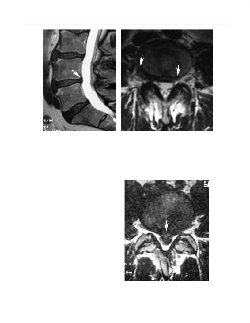
 B
B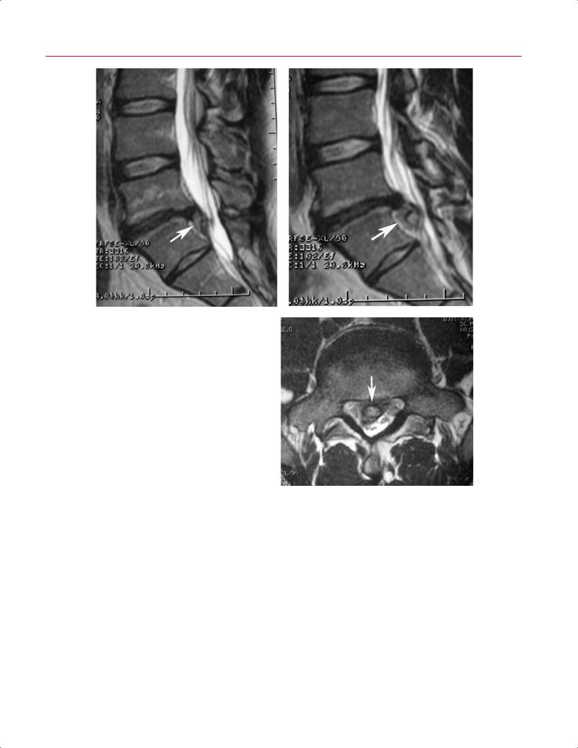
 B
B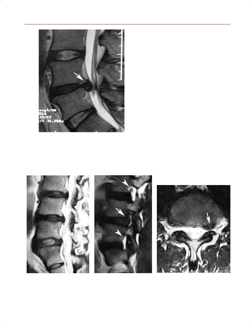
 B
B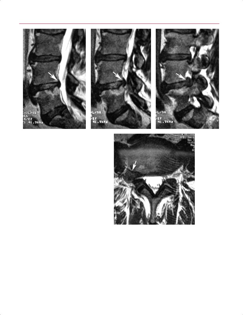
 E
E