

Erik_Kandel_-_V_poiskakh_pamyati_angl
.pdfavailable). I also learned how much a person who had recovered from such a devastating disorder could accomplish. Many young people who went on to have superb careers in science and I myself owe our start and much of our later success to the personal and professional example of Wade Marshall. Despite my obvious inexperience, he did not insist that I work only on problems that interested him. Rather, he allowed me to think about what I wanted to do— which was to study how learning and memory are achieved in the cells of the brain. Science gives one a structured opportunity to try out ideas—and, if one is not afraid of falling on one's face, to try out ideas that are raw, important, and bold. Marshall gave me the freedom to try to think creatively.
Grundfest, Purpura, Crain, Marshall, and later Steve Kuffler influenced me greatly. They transformed my life. They and Mr. Campagna, who paved the way for me to go to Harvard, illustrate the importance of student-teacher relationships in one's intellectual development. Equally, they underscore the role of chance influences and of the generosity of spirit that encourages young people to thrive. Young people, for their part, must strive to have an open mind and seek out places where they will be surrounded by first-rate intellects.
8
DIFFERENT MEMORIES, DIFFERENT BRAIN REGIONS
By the time I arrived at Wade Marshall's laboratory, I had progressed from the naive notion of trying to find the ego, id, and superego in the brain to the slightly less vague idea that finding the biological basis of memory might be an effective approach to understanding higher mental processes. It was clear to me that learning and memory are central to psychoanalysis and psychotherapy After all, aspects of many psychological problems are learned, and psychoanalysis is based on the principle that what is learned can be unlearned. In a large sense, learning and memory are central to our very identity. They make us who we are.
At that time, however, the biology of learning and memory was in a muddle. It was dominated by the thinking of Karl Lashley a professor of psychology at Harvard who had convinced many scientists that there were no specific areas within the cerebral cortex that were specialized for memory.
Shortly after I arrived at NIMH, two research scientists changed all that. In a newly published research paper, Brenda Milner, a psychologist at the Montreal Neurological Institute of McGill University, and William Scoville, a neurosurgeon in Hartford, Connecticut, reported that they had tracked memory to specific regions in the brain. This news had a
powerful impact on me and on many others, for it meant that perhaps there was at last a decisive end to a very old controversy about the human mind.
UNTIL THE MIDDLE OF THE TWENTIETH CENTURY, THE SEARCH
for where memory resides in the brain was shaped by two competing views of how the brain—and especially the cerebral cortex—works. One held that the cerebral cortex is composed of discrete regions with specific functions—one representing language, another vision, and so on. The other view was that mental capacities are products of the combined activity of the entire cerebral cortex.
The first person to champion the idea that particular mental abilities are located in specific regions of the cortex was Franz Joseph Gall, a German physician and neuroanatomist who taught at the
University of Vienna from 1781 to 1802. Gall made two enduring conceptual contributions to the science of mind. First, he argued that all mental processes are biological, and so arise from the brain. Second, he proposed that the cerebral cortex has many distinct regions that govern specific mental functions.
Gall's theory that all mental processes are biological put him at odds with dualism, the dominant theory of his time. Promulgated in 1632 by René Descartes, a mathematician and the father of modern philosophy, this theory holds that human beings have a dual nature: the body, which is material, and the soul, which resides outside the body and is immaterial and indestructible. This dual nature reflects two types of substances. The res externa—the physical substance that fills the body, including the brain—runs through the nerves and imbues the muscles with animal spirits. The res cogitans—the nonphysical stuff of thinking—is uniquely human. It gives rise to rational thought and consciousness, and it reflects in its nonphysical character the spiritual nature of the soul. Reflex actions and many other physical behaviors are carried out by the brain, while mental processes are carried out by the soul. Descartes believed these two agents interacted through the pineal gland, a small structure located deep in the middle of the brain.
The Roman Catholic Church, feeling its authority threatened by new discoveries in anatomy, embraced dualism because it separated
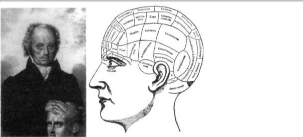
8-1 Phrenology. Franz Joseph Gall (1758-1828) assigned different mental functions to specific regions of the brain, based on his observations. Gall later developed phrenology, a system that related personality to bumps on the skull. (Gall image courtesy of Anthony A. Walsh.)
the realms of science and religion. Gall's radical argument for a materialist view of mind appealed to the scientific community because it ended the concept of the nonbiological soul, but it threatened the powerful conservative elements of society. Indeed, Emperor Francis I forbade Gall from lecturing in public and expelled him from Austria.
Gall also speculated on which areas of the cortex do different things. Academic psychology at the time had settled on twenty-seven mental faculties. Gall assigned these faculties to twenty-seven different regions of the cortex, each of which he called a "mental organ." (Additional regions were added later by Gall and others.) These mental faculties—such as factual memory, cautiousness, secretiveness, hope, belief in God, sublimity, parental love, and romantic love—were both abstract and complex, yet Gall insisted that each was controlled by a single, distinct region of the brain. This theory of localized function opened a debate that persisted through the next century.

Although correct in principle, Gall's theory was flawed in its details. First, most of the "faculties" considered discrete mental functions in
Gall's time are far too complex to arise from single regions of the cerebral cortex. Second, Gall's method of assigning functions to specific areas of the brain was misguided.
Gall distrusted studies of the behavior of people who had lost parts of their brain, so he ignored clinical findings. Instead, he developed an approach based on studies of the skull. He believed that each area of the cerebral cortex grew with usage and that this growth caused the overlying skull to protrude (figure 8-1).
Gall developed his idea in stages, beginning when he was young. In school, he had formed the impression that his most intelligent classmates had prominent foreheads and eyes. In contrast, a very romantic and enchanting widow he encountered had a prominent back of the head. Thus, Gall came to believe that great intelligence creates greater mass in the front of the brain, whereas great romantic passion produces greater mass in the back. In each case the overlying skull was enlarged by growth of the brain. Gall believed that by examining the bumps and ridges on the skulls of people well endowed with specific faculties, he could identify the centers of those faculties.
He systematized his thinking further when, as a young physician, he was put in charge of an insane asylum in Vienna. There he examined the skulls of criminals and found a bump above the ear that was remarkably similar to one found in carnivorous animals; Gall associated the bump with a part of the brain he believed was responsible for sadistic and destructive behavior. This approach to identifying the sites of mental faculties led to phrenology, a discipline that correlated personality and character with the shape of the skull.
By the late 1820s Gall's ideas and the discipline of phrenology had become extremely popular, even among the public. Pierre Flourens, a French experimental neurologist, decided to put them to the test. Using various animals for his experiments, Flourens removed, one by one, the areas of the cerebral cortex that Gall had associated with specific mental functions, but he failed to find any of the behavioral deficits Gall had predicted. In fact, Flourens was unable to associate any deficits in behavior with specific regions of the cortex. It was only the size of the area removed, not its location or the complexity of behavior involved, that mattered.
Flourens therefore concluded that all regions of the cerebral hemispheres were equally important. The cortex is equipotential, he argued, meaning that every region is able to perform any of the brain's functions. Thus, an injury to a particular region of the cerebral cortex would not affect one capacity more than another. 'All perceptions, all volitions occupy the same seat in these [cerebral] organs; the faculty of perceiving, of conceiving, of willing merely constitutes thereby a faculty which is essentially one," Flourens wrote.
Flourens's views spread rapidly. Their swift acceptance was certainly due in part to the credibility of his experimental work, but it also represented a religious and political reaction against Gall's materialistic view of the brain. If the materialistic view was correct, there was no need to postulate a soul as a necessary mediator of human cognitive functions.
THE DEBATE BETWEEN THE FOLLOWERS OF GALL AND THE
followers of Flourens colored thinking about the brain over the next several decades. It was not resolved until the second half of the nineteenth century, when the question attracted the attention of two neurologists, Pierre-Paul Broca in Paris and Carl Wernicke in Breslau, Germany. In the course of their studies of patients with specific language deficits, or aphasias, Broca and Wernicke made several important discoveries. Taken together, these discoveries form one of the most exciting chapters in the study of human behavior—namely, the first insight into the biological basis of a complex cognitive ability, language.
Rather than exploring the normal brain to test Gall's ideas, as Flourens had done, Broca and Wernicke studied disease states—what physicians at that time referred to as nature's experiments. They succeeded in associating specific disorders of language with damage to particular areas of the cerebral cortex, thus providing convincing evidence that at least some higher mental functions arise there.
The cerebral cortex has two important characteristics. First, although its two hemispheres appear to be mirror images of each other, they differ in both structure and function. Second, each hemisphere is concerned primarily with sensing and moving the opposite side of the body. Thus, sensory information that arrives at the spinal
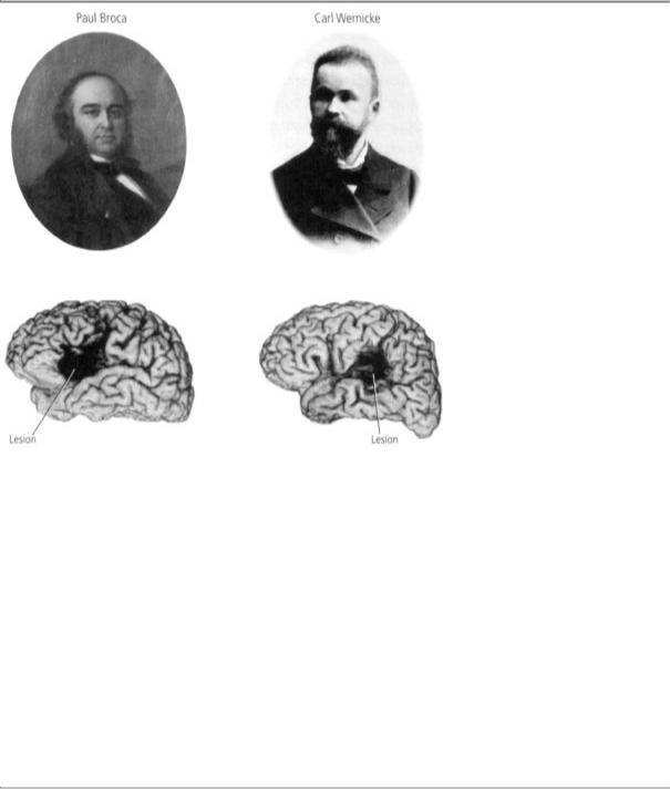
Broca (1824-1880) discovered that damage to the left frontal lobe of the cerebral hemisphere makes it impossible for a person to speak.
Wernicke (1848-1905) discovered that damage to the posterior part of the left cerebral hemisphere makes it impossible for a person to understand speech.
8-2 Two pioneers in the study of brain function in language. (Portraits reprinted from Essentials of Neural Science and Behavior, Kandel, Schwartz, and Jessell, McGraw-Hill, 1995. Brain images courtesy of Hanna Damasio.)
cord from the left side of the body—from the left hand, say—crosses over to the right side of the nervous system on its way to the cerebral cortex. Similarly, motor areas in the right hemisphere control movements of the left half of the body.
Broca (figure 8-2), who was also a surgeon and anthropologist, founded what we now call neuropsychology, a science that examines alterations in mental processes produced by brain damage. In 1861 he described a fifty-one-year-old Parisian shoemaker named Leborgne who had suffered a stroke twenty-one years earlier. As a result of his stroke,

Leborgne had lost the ability to speak fluently, although he indicated with facial expressions and actions that he understood the spoken language quite well. Leborgne had none of the conventional motor deficits that would affect speech. He had no difficulty moving his tongue, mouth, or vocal cords. In fact, he could utter isolated words, whistle, and sing a melody without difficulty, but he could not speak grammatically or create complete sentences. Moreover, his difficulty was not limited to the spoken language; Leborgne could not express his ideas in writing either.
Leborgne died one week after Broca first examined him. At the postmortem examination Broca discovered a damaged area, or lesion, in a region of the frontal lobe now called Broca's area (figure 8-2). He went on to study, after their deaths, the brains of eight other patients who had been unable to speak. Each had a similar lesion in the frontal lobe of the left cerebral hemisphere. Broca's findings provided the first empirical evidence that a welldefined mental capacity could be assigned to a specific region of the cortex. Since all the patients' lesions were in the left hemisphere, Broca established that the two hemispheres, though apparently symmetrical, have different roles. This discovery led him to announce, in 1864, one of the most famous principles of brain function: "Nous parlons avec l'hémisphère gauche!" (We speak with the left hemisphere!)
Broca's discovery stimulated a search for the location of other behavioral functions in the cortex. Nine years later two German physiologists, Gustav Theodor Fritsch and Eduard Hitzig, galvanized the scientific community when they showed that dogs would move their limbs in predictable ways when a specific region of the cerebral cortex was stimulated electrically. Moreover, Fritsch and Hitzig identified the small areas of the cortex that controlled the individual muscle groups responsible for the movements.
In 1879 Carl Wernicke (figure 8-2) described a second type of aphasia. This disorder is not an impairment in the production of speech, but a disruption in the comprehension of spoken or written language. Moreover, while people with Wernicke's aphasia can speak, what they say is completely incoherent to anyone else. Like Broca's aphasia, this aphasia is caused by a lesion in the left side of the brain, but in this
case the damage is in the back of the brain, in a region now called Wernicke's area (figure 8-2).
Based on his own and Broca's work, Wernicke put forward a theory of how the cortex is wired for language. That theory, although simpler than our current understanding of language, is nevertheless consistent with how we view the brain today. As a first principle, Wernicke proposed that any complex behavior is the product not of a single region but of several specialized, interconnected areas of the brain. For language, these are Wernicke's area (comprehension) and Broca's area (expression). The two areas, as Wernicke knew, are connected by a neural pathway (figure 8-3). He also understood that large, interconnected networks of specialized regions, such as those governing language, enable people to experience mental activity as seamless.
The idea that different regions of the brain are specialized for different purposes is central to modern brain science, and Wernicke's model of a network of interconnected, specialized regions is a dominant theme in the study of the brain. One reason this conclusion eluded investigators
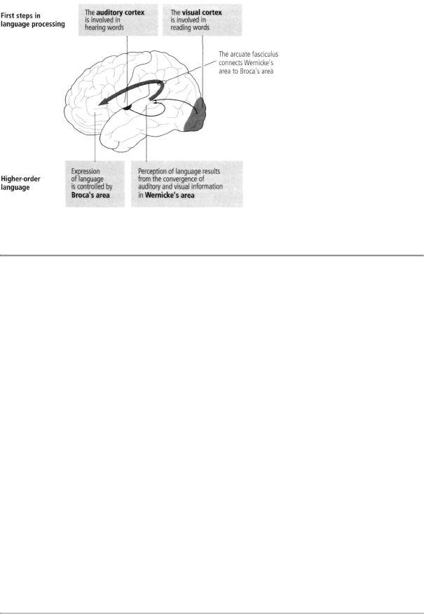
8-3 Complex behavior, such as language, involves several interconnected areas of the brain.
for so many years can be found in another organizational principle of the nervous system: brain circuitry has a built-in redundancy. Many sensory, motor, and cognitive functions are served by more than one neural pathway—the same information is processed simultaneously and in parallel in différent regions of the brain. When one region or pathway is damaged, others may be able to compensate, at least partially, for the loss. When compensation occurs and no behavioral deficits are obvious, researchers have difficulty linking a damaged site in the brain to a behavior.
Once it was known that language is produced and understood in specific regions of the brain, regions governing each of the senses were identified, providing a foundation for Wade Marshall's later discoveries of sensory maps for touch, vision, and hearing. It was only a matter of time before these searches turned to memory. Indeed, the fundamental question of whether memory is a unique neural process or is associated with motor and sensory processes remained open.
INITIAL ATTEMPTS TO PINPOINT A REGION OF THE BRAIN
responsible for memory, or even to delineate memory as a unique mental process, failed. In a series of famous experiments in the 1920s, Karl Lashley trained rats to run through a simple maze. He then removed different areas of the cerebral cortex and retested the rats twenty days later to see how much training they had retained.
Based on these experiments, Lashley formulated the law of mass action, which holds that the severity of memory impairment is correlated with the size of the cortical area removed, not with its specific location. Thus Lashley wrote, echoing Flourens a century earlier, "It is certain that the maze habit, when formed, is not localized in any single area of the cerebrum [the cerebral cortex] and that its performance is somehow conditioned by the quantity of tissue which is intact."
Many years later Lashley's results were reinterpreted by Wilder Penfield and Brenda Milner at the Montreal Neurological Institute. As more scientists experimented with rats, it became clear that mazes were not suitable for studying the location of memory function. Maze learning is an activity that involves many different sensory and motor capabilities. When an animal is deprived of one kind of sensory cue (for example, touch), it can still recognize a place reasonably well
using other senses (such as vision or smell). In addition, Lashley focused his efforts on the cerebral cortex, the outer layer of the brain; he did not explore the structures that lie deeper in the brain. Subsequent research has shown that many forms of memory require one or more of these deeper regions.
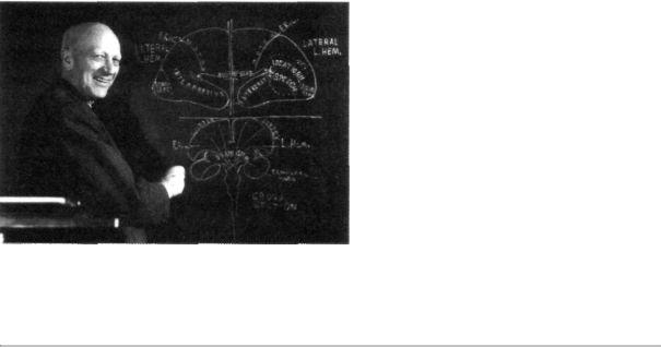
The first suggestion that some aspects of human memory can be stored in specific regions of the brain arose in 1948 from Penfield's neurosurgical work (figure 8-4). Penfield had trained in physiology with Charles Sherrington while on a Rhodes scholarship. He began to use surgery to treat focal epilepsy, a disorder that produces seizures in limited regions of the cortex. He developed a technique, still used today, of removing epileptic tissue while avoiding or minimizing damage to areas involved in the patient's mental processes.
Because the brain contains no pain receptors, surgery can be carried out with a local anesthetic. Thus Penfield's patients remained fully conscious during surgery and were able to report their experiences. (In describing this to Sherrington, who had spent his career working on cats and monkeys, Penfield could not resist adding, "Imagine having an experimental preparation that can talk back to you.") Penfield applied
8-4 Wilder Penfield (1891-1976) exposed the surface of the brain in conscious patients during surgery for epilepsy. He then stimulated different parts of the cortex and through the patients' responses identified the temporal lobe as a potential site for memory storage. (Courtesy of the Penfield Archive and the Montreal Neurological Institute.)
weak electrical stimulation to various areas of his patients' cerebral cortex during surgery and determined the effects of this stimulation on their ability to speak and comprehend language. Through his patients' responses, he could pinpoint Broca's and Wernicke's areas and try to avoid damage to them when removing epileptic tissue.
Over the years, Penfield explored much of the surface of the cerebral cortex in more than a thousand persons. On occasion, in response to electrical stimulation, a patient would describe complex perceptions or experiences: "It sounded like a voice saying words, but it was so faint that I couldn't get it." Or, "I am seeing a picture of a dog and cat. . . the dog is chasing the cat." Such responses were rare (occurring in only about 8 percent of cases) and were invariably elicited only from the temporal lobes of the brain, never from other areas. The responses suggested to Penfield that the experiences elicited by electrical stimulation of the temporal lobes are snippets of memory, of the stream of experience in a person's life.
Lawrence Kubie, the psychoanalyst whom I knew through Ernst Kris, traveled to Montreal and used a tape recorder to monitor Penfield's patients' utterances. Kubie became convinced that the temporal lobe stored a particular type of unconscious information called the preconscious unconscious. I read an important paper by Kubie when I was in medical school and heard him lecture several times while I was working in Grundfest's lab, and I was influenced by his enthusiasm for the temporal lobe.

In time, Penfield's view that the temporal lobes store memory was called into question. First, all of his patients had abnormal brains because of their epilepsy; moreover, in almost half of the cases the mental experience evoked by stimulation was identical to the hallucinatory mental experiences that often accompanied seizures. These findings convinced most brain scientists that Penfield was eliciting seizurelike phenomena with his electrical stimulation—specifically that he might be eliciting the auras (hallucinatory experiences) characteristic of the early phase of an epileptic attack. Second, the reports of mental experiences included elements of fantasy as well as improbable or impossible situations; they were more like dreams than memo-
ries. Finally, removing the brain tissue under the stimulating electrode did not erase the patient's memory.
Nevertheless, a number of neurosurgeons were inspired by Penfield's work, among them William Scoville, who obtained direct evidence that the temporal lobes are critical to human memory. In the paper that I had read on arriving at NIH, Scoville and Brenda Milner reported the extraordinary story of a patient known to science only by his initials, H.M.
AT THE AGE OF NINE, H.M. WAS KNOCKED DOWN BY SOMEONE
riding a bicycle. He sustained a head injury that led eventually to epilepsy. Over the years, his seizures worsened, until he was having as many as ten blackouts and one major seizure a week. By age twentyseven, he was severely incapacitated.
Because H.M.'s epilepsy was thought to have originated within the temporal lobe (specifically, the medial temporal lobe), Scoville decided, as a last resort, to remove the inner surface of that lobe on both sides of the brain, as well as the hippocampus, which lies deep within the temporal lobe. The surgery succeeded in relieving H.M.'s seizures, but it left him with a devastating memory loss from which he never recovered. After his operation in 1953, H.M. remained the same intelligent, kind, and amusing man he had always been, but he was unable to convert any new memories into permanent memory.
In a series of studies, Milner (figure 8-5) documented in exquisite detail the memory ability H.M. had lost, the memory ability he retained, and the areas of the brain responsible for each. She found that what H.M. retained was remarkably specific. To begin with, he had perfectly good short-term memory, lasting for minutes. He could readily remember a multidigit number or a visual image for a short period after learning it, and he could carry on a normal conversation, provided it did not last too long or move among too many topics. This short-term memory function was later called working memory and shown to involve an area known as the prefrontal cortex, which had not been removed from H.M. Second, H.M. had perfectly good long-term memory for events that had occurred before his surgery. He
8-5 Brenda Milner (b. 1918), whose studies of H.M. opened up the modern study of memory storage by localizing memory to a particular site in the brain. Milner identified the roles of the hippocampus and the medial temporal lobe in explicit memory and provided the first evidence of implicit memory storage. (Reprinted from Essentials of Neural Science and Behavior, Kandel, Schwartz, and Jessell, McGraw-Hill, 1995.)
could remember the English language, his IQ was good, and he recalled vividly many events from his childhood.
What H.M. lacked, and lacked to the most profound degree, was the ability to convert new short-term memory into new long-term memory. Without this ability he forgot events shortly after they happened. He could retain new information as long as his attention was not diverted from it, but a minute or two after his attention was directed to something else, he could not remember the previous subject or anything he thought about it. Less than an hour after eating he could not remember anything he had eaten or even the fact that he had had a meal. Brenda Milner studied H.M. monthly for almost thirty years, and each time she entered the room and greeted him he failed to recognize her. He did not recognize himself in recent photographs or in the mirror because he remembered himself only as he was prior to surgery. He had no memory of his changed appearance: his identity has been frozen for over fifty years, from the time of his surgery until today. Milner was to say of H.M., "He couldn't acquire the slightest new piece of knowledge. He lives today chained to the past, in a sort of childlike world. You can say his personal history stopped with the operation."
From her systematic studies of H.M., Milner extracted three important principles of the biological basis of complex memory. First, memory is a distinct mental function, clearly separate from other perceptual, motor, and cognitive abilities. Second, short-term memory and long-term memory can be stored separately. Loss of medial temporal lobe structures, particularly loss of the hippocampus, destroys the ability to convert new short-term memory to new long-term memory. Third, Milner showed that at least one type of memory can be traced to specific places in the brain. Loss of brain substance in the medial temporal lobe and the hippocampus profoundly disrupts the ability to lay down new long-term memories, whereas losses in certain other regions of the brain do not affect memory.
Milner thus disproved Lashley's theory of mass action. It is only in the hippocampus that the various strands of sensory information necessary for forming long-term memory come together. Lashley never went below the surface of the cortex in his experiments. Moreover, Milner's finding that H.M. had good long-term memory for events that happened prior to the surgery showed clearly that the medial temporal lobe and the hippocampus are not the permanent storage sites of memory that has been in long-term storage for some time.
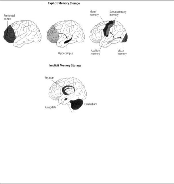
We now have reason to believe that long-term memory is stored in the cerebral cortex. Moreover, it is stored in the same area of the cerebral cortex that originally processed the information—that is, memories of visual images are stored in various areas of the visual cortex, and memories of tactile experiences are stored in the somatosensory cortex (figure 8-6). That explains why Lashley, who used complex tasks based on several different sensory modalities, could not erase his rats' memories fully by removing selected sections of the cortex.
For many years Milner thought H.M.'s memory defect was complete, that he was unable to convert any shortterm memory to long-term memory. But in 1962 she demonstrated another principle of the biological basis of memory—the existence of more than one kind of memory. Specifically, Milner found that in addition to conscious memory, which requires the hippocampus, there is an unconscious memory that resides outside the hippocampus and the medial temporal lobe.
8-6 Explicit and implicit memories are processed and stored in different regions in the brain. In the short term, explicit memory for people, objects, places, facts, and events is stored in the prefrontal cortex. These memories are converted to longterm memories in the hippocampus and then stored in the parts of the cortex that correspond to the senses involved—that is, in the same areas that originally processed the information. Implicit memories of skills, habits, and conditioning are stored in the cerebellum, striatum, and amygdala.
(This distinction had been proposed on behavioral grounds in the 1950s by Jerome Bruner at Harvard, one of the fathers of cognitive psychology.)
Milner thus demonstrated this distinction by showing that the two forms of memory require different anatomical systems (figure 8-6). She found that H.M. could learn and remember some things over the long term—that is, he had a kind of long-term memory that does not
depend on the medial temporal lobe or the hippocampus. He learned to trace the outline of a star in a mirror and his skill at tracing improved from day to day, just as it would in a person without brain damage (figure 8-7). Yet, even though his performance improved at the beginning of each day's test, H.M. could never remember having performed the task on an earlier day.