- •Operative Cranial Neurosurgical Anatomy
- •Contents
- •Foreword
- •Preface
- •Contributors
- •1 Training Models in Neurosurgery
- •2 Assessment of Surgical Exposure
- •3 Anatomical Landmarks and Cranial Anthropometry
- •4 Presurgical Planning By Images
- •5 Patient Positioning
- •6 Fundamentals of Cranial Neurosurgery
- •7 Skin Incisions, Head and Neck Soft-Tissue Dissection
- •8 Techniques of Temporal Muscle Dissection
- •9 Intraoperative Imaging
- •10 Precaruncular Approach to the Medial Orbit and Central Skull Base
- •11 Supraorbital Approach
- •12 Trans-Ciliar Approach
- •13 Lateral Orbitotomy
- •14 Frontal and Bifrontal Approach
- •15 Frontotemporal and Pterional Approach
- •16 Mini-Pterional Approach
- •17 Combined Orbito-Zygomatic Approaches
- •18 Midline Interhemispheric Approach
- •19 Temporal Approach and Variants
- •20 Intradural Subtemporal Approach
- •21 Extradural Subtemporal Transzygomatic Approach
- •22 Occipital Approach
- •23 Supracerebellar Infratentorial Approach
- •24 Endoscopic Approach to Pineal Region
- •25 Midline Suboccipital Approach
- •26 Retrosigmoid Approach
- •27 Endoscopic Retrosigmoid Approach
- •29 Trans-Frontal-Sinus Subcranial Approach
- •30 Transbasal and Extended Subfrontal Bilateral Approach
- •32 Surgical Anatomy of the Petrous Bone
- •33 Anterior Petrosectomy
- •34 Presigmoid Retrolabyrinthine Approach
- •36 Nasal Surgical Anatomy
- •37 Microscopic Endonasal and Sublabial Approach
- •38 Endoscopic Endonasal Transphenoidal Approach
- •39 Expanded Endoscopic Endonasal Approach
- •41 Endoscopic Endonasal Odontoidectomy
- •42 Endoscopic Transoral Approach
- •43 Transmaxillary Approaches
- •44 Transmaxillary Transpterygoid Approach
- •45 Endoscopic Endonasal Transclival Approach with Transcondylar Extension
- •46 Endoscopic Endonasal Transmaxillary Approach to the Vidian Canal and Meckel’s Cave
- •48 High Flow Bypass (Common Carotid Artery – Middle Cerebral Artery)
- •50 Anthropometry for Ventricular Puncture
- •51 Ventricular-Peritoneal Shunt
- •52 Endoscopic Septostomy
- •Index

17 Combined Orbito-Zygomatic Approaches
Pietro Mortini, Alfo Spina, Michele Bailo, Anthony J. Caputy, Cristian Gragnaniello, and Filippo Gagliardi
17.1 Introduction
The Fronto-orbito-zygomatic (FOZ) approach represents one of the workhorses of skull base surgery. Through this approach, it is possible to provide a wide exposure and a direct access to diferent tumoral and cerebrovascular pathology of the anterior and middle cranial fossa, sellar and suprasellar area, and anterior upper brainstem. In addition, the orbitozygomatic osteotomy provides the beneft of the extradural unroofng of the optic canal.
17.2 Indications
•Sellar, suprasellar, presellar and parasellar lesions.
•Lesions of the third ventricle.
•Lesions of the anterior surface of the upper brainstem.
•Aneurisms of circle of Willis.
17.3 Patient Positioning
•Position: The patient is positioned supine with head fxed with a Mayfeld head holder.
•Body: The chest should be elevated to facilitate venous return.
•Head: The head is rotated 30° to the contralateral side, and extended about 20°.
•The zygomatic process must represent the highest point of the surgical feld.
•Pin holders must be positioned as distant as possible to the planned skin incision.
17.4 Skin Incision
17.4.1 Extended Coronal Skin Incision (Figs. 17.1)
•Starting point: Incision starts less than 1 cm anterior to the tragus on the side of the approach.
•Course: Incision runs toward the midline and to the contralateral side, just behind the hairline.
•Ending point: It ends at the junction between the lateral and middle third of the coronal line.
17.4.2 Critical Structures
•Superfcial temporal artery and its distal branches.
•Peripheral branches of the facial nerve.
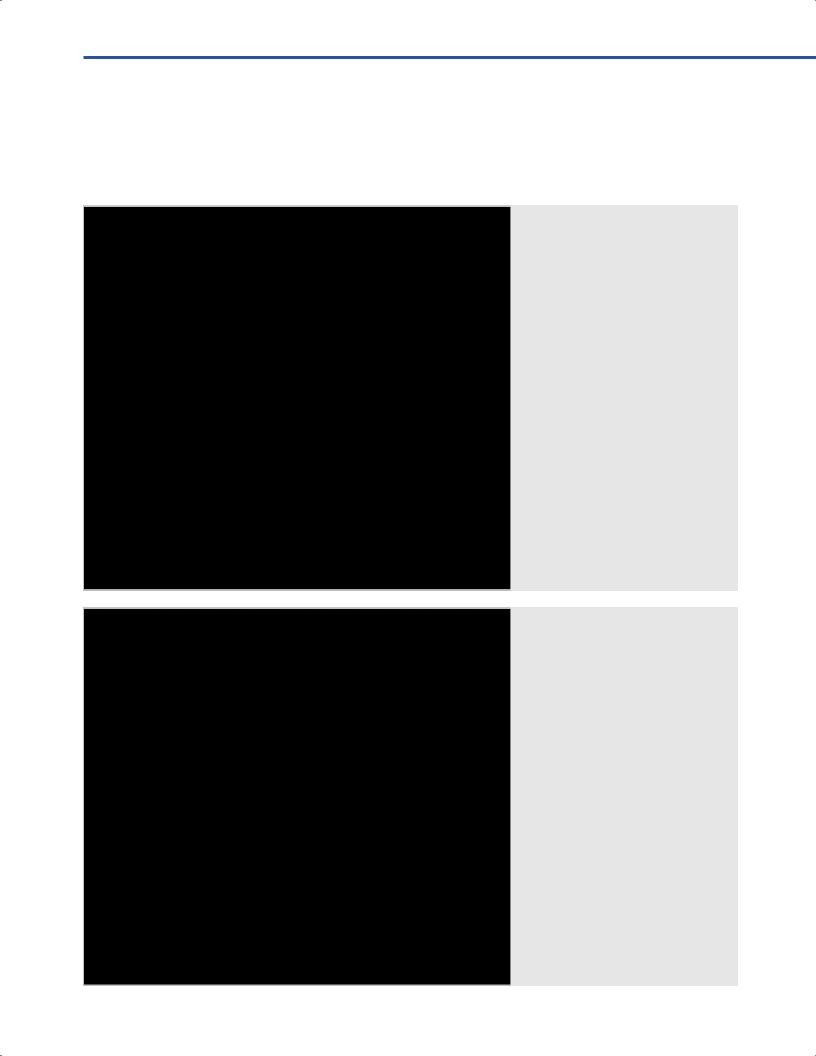
Fig. 17.1 Bicoronal skin incision.
105

III Cranial Approaches
17.5 Soft Tissue Dissection
•Pericranial layer
○The pericranial fap is refected together with the skin fap, up to the orbital rim.
•Muscle (Figs. 17.2, 17.3)
○Interfascial dissection of the temporal muscle is carried out
(see Chapters 6 and 8).
○The subperiosteal dissection of the temporal muscles is performed to the frontal process of the zygoma; the body of the zygomatic bone and the temporal fossa are exposed.
○Temporal muscle is refected inferiorly.
•Bone exposure (Fig. 17.4)
○Zygomatic process of the frontal bone, frontal process of the zygomatic bone, temporal fossa (greater wing of the sphenoid, temporal squama).
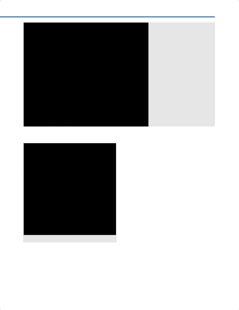
Fig. 17.2 al soft tissue dissection.
Abbreviations: CS = coronal suture; E = ear;
FB = frontal bone; FP = fat pad; SON =
supraorbital nerve; ST |
al tempo - |
ral artery; TF = temporal fascia. |
|
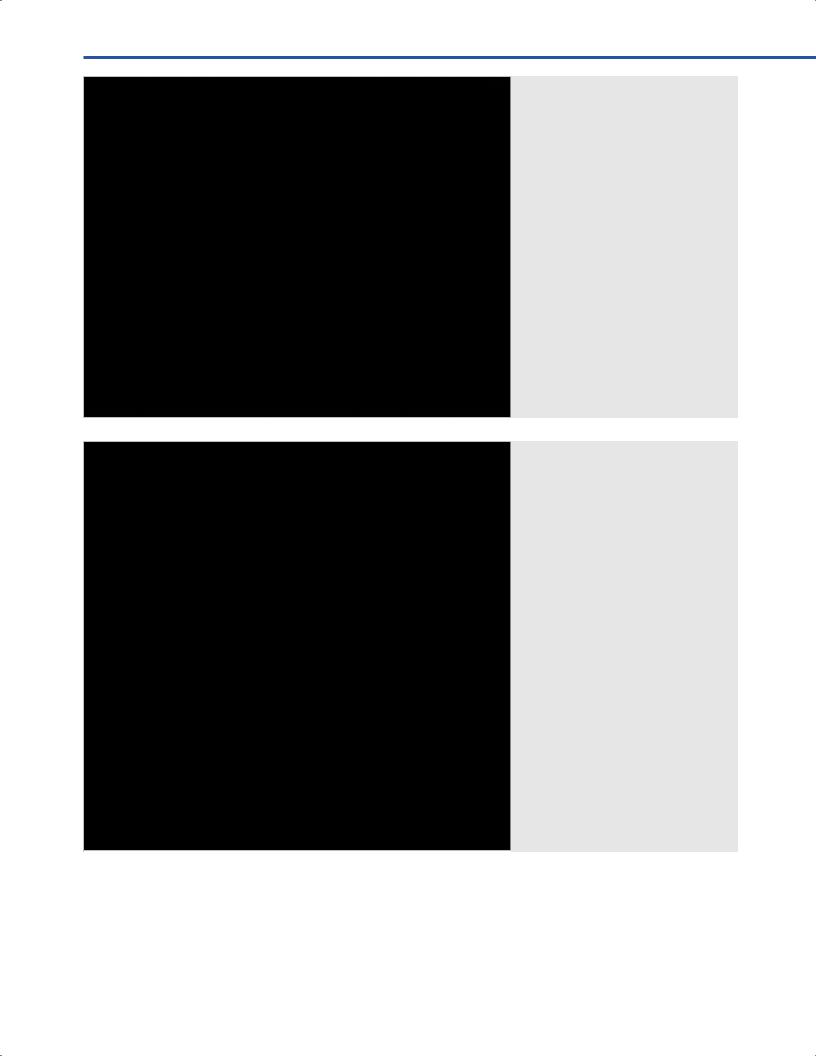
Fig. 17.3 al soft tissue dissection: Interfascial dissection.
Abbreviations: CS = coronal suture; E = ear; FB = frontal bone; FP = fat pad; SON = supraorbital nerve; ST al temporal artery; TF = temporal fascia; Z = zygomatic process of the frontal bone.
106

17 Combined Orbito-Zygomatic Approaches
○Supraorbital foramen is opened avoiding injury to the supraorbital nerve, artery and vein (see Chapter 6).
•Orbital contents
○Periorbit is subperiostally dissected from the roof and the lateral wall of the orbit, 3 cm deep from the orbital rim.
17.5.1 Critical Structures
•Frontal branch of the facial nerve.
•Supraorbital nerve, artery and vein.
17.6 Craniotomy
17.6.1 Frontotemporal Craniotomy (Figs. 17.5, 17.6)
•Burr holes
○I: The frst burr hole is made at the keyhole, just posterior to the frontozygomatic suture.
○II: The second burr hole is placed behind the sphenosquamous suture of the temporal squama.
•Craniotomy landmarks
○Anteriorly: A basal exposure is performed from the keyhole to the medial third of the orbital rim.
○Laterally: The burr holes are connected by drilling the lesser sphenoid wing with a high-speed drill.
○Medially: Craniotomy runs parallel to the superior sagittal sinus.
○Posteriorly: The posterior cut is made from the second burr hole to the medial limit of the craniotomy in the frontal bone.
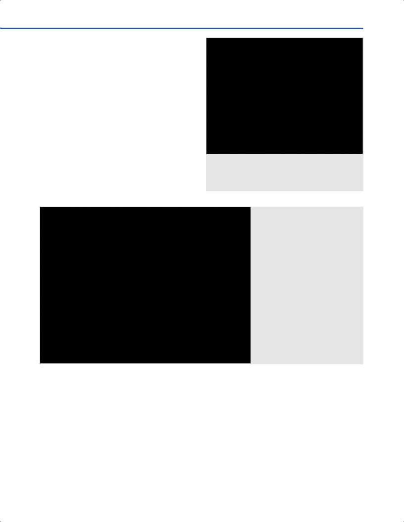
Fig. 17.4 Bone exposure. Abbreviations: CS = coronal suture; FB =
frontal bone; FP = fat pad; TM = temporal muscle; Tsq = temporal squama; Z = zygomatic process of the frontal bone.
17.6.2 Variants
•One and half craniotomy
○The craniotomy crosses the midline toward the contralateral frontal bone.
○One and half craniotomy provides the exposure of the superior sagittal sinus, the interhemispheric fssure, and the contralateral frontal lobe.
17.6.3 Critical Structures
•Middle meningeal artery.
•Superior sagittal sinus.
17.7 Orbitozygomatic Osteotomy (Figs. 17.7, 17.8)
The roof and the lateral wall and the orbit are exposed by elevating the dura from the frontal and the temporal bone.
17.7.1 Osteotomies cuts
•I: The frst cut is made at the medial third of the superior orbital rim.
•II: The second cut is made at the lateral orbital rim, just superiorly to the body of the zygomatic bone at the fronto-zygomatic suture, and it is directed toward the inferior orbital fssure.
•III: The two cuts are connected starting from the inferior orbital fssure, always keeping the saw anterior to the superior orbital fssure.
•IV: The remaining part of the orbital roof, located close to the orbital apex, is removed.
107

III Cranial Approaches
Fig. 17.5 Craniotomy. Abbreviations: CS = coronal suture; FB = frontal bone; FP = fat pad; SON =
supraorbital nerve; TM = temporal muscle; Tsq = temporal squama; Z = zygomatic process of the frontal bone.
Fig. 17.6 Dural exposure. Abbreviations: CS = coronal suture; FB =
frontal bone; FD = frontal dura; FP = fat pad; OR = orbital ridge; SON = supraorbital nerve; TM = temporal muscle.
108

17 Combined Orbito-Zygomatic Approaches
Fig. 17.7 Exposure of the orbital roof. Red dotted lines depict the cuts of orbitotomy. Abbreviations: CS = coronal suture; FB = frontal bone; FD = frontal dura; FP = fat pad; OR = orbital roof; SON = supraorbital nerve; TM = temporal muscle.
17.7.2 Variants
• Zygomatic osteotomies (Fig. 17.9)
○ I: The posterior cut can be made across the root of the zygomatic process.
○ II: The anterior cut can be made obliquely at the level of the maxillary process of the zygomatic bone.
• Unroofng of the optic canal
○ Unroofng of the optic canal is required in case of preoperative compressive optic neuropathy.
○ The technique consists in drilling of the superior, lateral and medial surface of the optic canal.
17.7.3 Critical Structures
• Supraorbital neurovascular bundle.
• Superior orbital fssure.
• Optic nerve.
Fig. 17.8 Orbitotomy.
109

III Cranial Approaches
17.8 Dural Opening (Fig. 17.10)
•Dura mater is incised in a C-shape.
•Dural fap is refected anteriorly above the periorbit and suspended to the skin fap.
Fig. 17.9 Main variants of orbitotomies. Abbreviations: FO = fronto-orbital approach; FOZ = fronto-orbito-zygomatic approach; FTOZ = complete fronto-temporal-orbito- zygomatic approach.
Fig. 17.10 Dural opening. Abbreviations: CS = coronal suture; FB =
frontal bone; FD = frontal dura; FP = fat pad; PO = periorbit; SON = supraorbital nerve; TM = temporal muscle.
•After dural opening the lateral surface of the frontal and temporal lobe as well as the Sylvian fssure come into view.
110

17 Combined Orbito-Zygomatic Approaches
17.9 Intradural Exposure (Figs. 17.11–17.14)
•Parenchymal structures: Orbital and Sylvian surfaces of the frontal and temporal lobes.
•Arachnoidal layer: Sylvian fssure, carotid and opticchiasmatic cisterns.
•Cranial nerves: Olfactory and optic nerves.
•Arteries: Internal carotid, posterior communicating, anterior choroidal, anterior cerebral, anterior communicating, middle cerebral, posterior cerebral, and superior cerebellar arteries.
•Veins: Sylvian, frontal and temporal cortical veins.
•Intra-cavernous space: Internal carotid artery, oculomotor, trochlear, abducens, frst and second branch of the trigeminal nerve.
Fig. 17.11 Intradural exposure.
Abbreviations: CS = coronal suture; FB = frontal bone; FP = fat pad; IFG = inferior frontal gyrus; MFG = middle frontal gyrus; PO = periorbit; SF = Sylvi sure; SFG = superior frontal gyrus; SON = supraorbital nerve; TM = temporal muscle.
Fig. 17.12 Intradural clinoidectomy. Abbreviations: D = dura; FL = frontal lobe; ICA = internal carotid artery; MCA = middle cerebral artery; ON = optic nerve; RAC = resected anterior clinoid; TL = temporal lobe.
111

III Cranial Approaches
Fig. 17.13 Intradural view. Abbreviations: ACA = anterior cerebral ar-
tery; ICA = internal carotid artery; III = third cranial nerve; MCA = middle cerebral artery; ON = optic nerve.
References
1. Fossett D, Caputy AJ. Operative neurosurgical anatomy. New York, NY: Thieme Medical Publisher; 2002
2. Mortini P, Gagliardi F, Boari N, Roberti F, Caputy AJ. The combined interhemispheric subcommissural translaminaterminalis approach for large craniopharyngiomas. World Neurosurg 2013;80(1–2):160–166
3. Mortini P, Losa M, Pozzobon G, et al. Neurosurgical treatment of craniopharyngioma in adults and children: early and long-term results in a large case series. J Neurosurg 2011;114(5):1350–1359
Fig. 17.14 Intradural view.
Abbreviations: BA = basilar artery; ICA = internal carotid artery; III = third cranial nerve; MCA = middle cerebral artery; ON = optic nerve.
112
