- •Operative Cranial Neurosurgical Anatomy
- •Contents
- •Foreword
- •Preface
- •Contributors
- •1 Training Models in Neurosurgery
- •2 Assessment of Surgical Exposure
- •3 Anatomical Landmarks and Cranial Anthropometry
- •4 Presurgical Planning By Images
- •5 Patient Positioning
- •6 Fundamentals of Cranial Neurosurgery
- •7 Skin Incisions, Head and Neck Soft-Tissue Dissection
- •8 Techniques of Temporal Muscle Dissection
- •9 Intraoperative Imaging
- •10 Precaruncular Approach to the Medial Orbit and Central Skull Base
- •11 Supraorbital Approach
- •12 Trans-Ciliar Approach
- •13 Lateral Orbitotomy
- •14 Frontal and Bifrontal Approach
- •15 Frontotemporal and Pterional Approach
- •16 Mini-Pterional Approach
- •17 Combined Orbito-Zygomatic Approaches
- •18 Midline Interhemispheric Approach
- •19 Temporal Approach and Variants
- •20 Intradural Subtemporal Approach
- •21 Extradural Subtemporal Transzygomatic Approach
- •22 Occipital Approach
- •23 Supracerebellar Infratentorial Approach
- •24 Endoscopic Approach to Pineal Region
- •25 Midline Suboccipital Approach
- •26 Retrosigmoid Approach
- •27 Endoscopic Retrosigmoid Approach
- •29 Trans-Frontal-Sinus Subcranial Approach
- •30 Transbasal and Extended Subfrontal Bilateral Approach
- •32 Surgical Anatomy of the Petrous Bone
- •33 Anterior Petrosectomy
- •34 Presigmoid Retrolabyrinthine Approach
- •36 Nasal Surgical Anatomy
- •37 Microscopic Endonasal and Sublabial Approach
- •38 Endoscopic Endonasal Transphenoidal Approach
- •39 Expanded Endoscopic Endonasal Approach
- •41 Endoscopic Endonasal Odontoidectomy
- •42 Endoscopic Transoral Approach
- •43 Transmaxillary Approaches
- •44 Transmaxillary Transpterygoid Approach
- •45 Endoscopic Endonasal Transclival Approach with Transcondylar Extension
- •46 Endoscopic Endonasal Transmaxillary Approach to the Vidian Canal and Meckel’s Cave
- •48 High Flow Bypass (Common Carotid Artery – Middle Cerebral Artery)
- •50 Anthropometry for Ventricular Puncture
- •51 Ventricular-Peritoneal Shunt
- •52 Endoscopic Septostomy
- •Index

14 Frontal and Bifrontal Approach
Filippo Gagliardi, Alfo Spina, Michele Bailo, Martina Piloni, Cristian Gragnaniello, Anthony J. Caputy, and Pietro Mortini
14.1 Introduction
The unilateral frontal craniotomy and its further extension, the bilateral frontal approach, are extremely versatile procedures, which can be easily tailored according to the existing pathology. The approaches are suitable for the unilateral/bilateral exposure of the lateral and anterior surfaces of the frontal lobe, as well as the most anterior aspect of the interhemispheric fssure.
It is possible to extend the bilateral approach by performing a bilateral orbitotomy to take advantage of the subfrontal anatomical corridor to reach the anterior skull base, as described in detail in Chapter 30. The approach is indicated for intra-axial lesions of the frontal lobe, as well as extra-axial tumors of the frontal convexity, of the anterior aspect of the falx and anterior cranial fossa. The approach is also suitable to treat vascular lesions of A2 as well as distal frontal branches of the anterior and middle cerebral artery.
14.2 Indications
•Intra-axial lesions of the frontal lobes.
•Extra-axial lesions of the frontal convexity and anterior third of the falx.
•Vascular lesions of the second segment (A2) and distal frontal branches of the anterior and middle cerebral artery.
14.3 Patient Positioning
•Position: The patient is positioned supine with the head fxed in a Mayfeld head holder.
•Body: The trunk and the head are slightly elevated to facilitate the venous backfow.
•Head: The head is placed in neutral position in case of bilateral approach. Alternatively, it can be slightly turned to the opposite side (about 15-20°) in case of unilateral approach.
•Neck: The neck is slightly extended (about 20°), to facilitate further brain relaxation after dural opening.
Fig. 14.1 Alternative skin incisions.
○Course: The incision line runs slightly curved backward until reaching the coronal suture and then turns toward the contralateral side (2 cm beyond the midline).
○Ending point: It ends 2 cm beyond the midline, on the contralateral side just behind the hairline.
14.4.1 Critical Structures
•Superfcial temporal artery and its branches.
•Frontal and temporal branches of the facial nerve.
14.4 Skin Incision (Fig. 14.1)
•Bicoronal skin incision (suggested for the frontal bilateral craniotomy):
○Starting point: Incision starts 1 cm anterior to the ipsilateral tragus, just above the zygoma.
○Course: The incision line runs to the contralateral side, just behind the hairline over the coronal suture.
○Ending point: It ends 1 cm anterior to the tragus on the contralateral side. Alternatively, it can be taken to the contralateral superior temporal line.
•Frontotemporal skin incision (suggested for the frontal unilateral craniotomy):
○Starting point: Incision starts 1 cm anterior to the ipsilateral tragus, just above the zygoma.
14.5 Soft Tissue Dissection (Figs. 14.2, 14.3)
•Pericranial layer
○Pericranium is smoothly dissected from the bone and the superfcial temporal fascia, taking care to preserve its anatomical integrity.
○It is refected anteriorly together with the skin fap.
○Pericranium must be preserved as it may be needed for further reconstruction.
•Muscle
○Frontal unilateral craniotomy
Ipsilateral temporal muscle inter-fascial dissection is carried out according to the technique described in
Chapters 6 and 8.
86

14 Frontal and Bifrontal Approach
The superfcial layer of the temporal fascia together with the fat pad is refected anteriorly.
The deep layer is dissected from the temporal squama in a subperiosteal fashion and refected inferiorly, in order to expose the part of temporal squama, located just below the superior temporal line, where burr holes will be made.
Fig. 14.2 Soft tissue dissection. Abbreviations: CS = coronal suture; FB = frontal bone; PC = pericranium; TF = temporal fascia.
Fig. 14.3 Soft tissue dissection, lateral view. Abbreviations: CS = coronal suture; FB = frontal bone; FP = fat pad; PC = pericranium; STL = superior temporal line; TF = temporal fascia; TM = temporal muscle.
○Frontal bilateral craniotomy
The procedure described for the unilateral variant has to be carried out also on the contralateral side.
•Bone exposure
The bone exposure is completed, when the following structures come into view:
87

III Cranial Approaches
○Ipsilateral pterion and superior temporal line (bilateral exposure in case of frontal bilateral approach).
○Anterior aspect of the sagittal suture.
○Ipsilateral coronal suture (bilateral exposure in case of frontal bilateral approach).
○Ipsilateral frontal bone (bilateral exposure in case of frontal bilateral approach).
○Orbital rim (bilateral exposure in case of frontal bilateral approach).
14.5.1 Critical Structures
•Frontal branch of the facial nerve.
•Supraorbital nerve, artery and vein.
14.6 Craniotomy (Figs. 14.4–14.7)
14.6.1 Frontal Unilateral Craniotomy,
Landmarks (Figs. 14.4, 14.5)
•Burr holes
○I: The frst burr hole is made at the keyhole on the ipsilateral side.
○II: The second one is placed 4-5 cm posteriorly to the keyhole, just below the superior temporal line for cosmetic reasons to allow it to be covered by temporal muscle.
○III: The third one is made either 1 cm lateral to the midline (in case of a paramedian frontal unilateral craniotomy), or over the superior sagittal sinus (in case of a midline frontal unilateral craniotomy). Posterior extension of the frontal craniotomy has to be tailored according to the pathology, and the planned approach.
Fig. 14.4 Frontal unilateral approach. Craniotomy landmarks (superior view). Abbreviations: BH = burr hole; CS = coronal suture; FB = frontal bone; PC = pericranium; TF = temporal fascia; TM = temporal muscle.
•Cuts
○I: The frst cut is made between the frst and second burr hole, taking care not to damage the middle meningeal artery.
○II: The second cut is made from the keyhole toward the midline, taking care not to tear the dura around the area of the pterion.
○III: The third cut is made from median/paramedian hole to the II hole. Craniotome should be directed away from the sinus to minimize the risk of dural tearing and bridging veins damage.
○IV: The fourth cut is made over the sinus.
•Craniotomy landmarks
Anatomical landmarks which have to be taken into consideration in designing the craniotomy are as follows:
○Anteriorly: Ipsilateral orbital rim.
○Laterally: Ipsilateral superior temporal line.
○Medially: Anterior aspect of the sagittal suture.
○Posteriorly: Ipsilateral coronal suture.
14.6.2 Frontal Bilateral Craniotomy,
Landmarks (Figs. 14.6, 14.7)
•Burr holes
○I: The frst burr hole is made at the keyhole on the ipsilateral side.
○II: The second one is made at the keyhole on the contralateral side.
○III: The third one is placed on the midline over the superior sagittal sinus.
•Cuts
○I: The frst cut is made between the frst two holes.
○II/III: The second and third cuts are made from the median hole to the keyholes bilaterally.
88

14 Frontal and Bifrontal Approach
Fig. 14.5 Frontal unilateral approach. Craniotomy landmarks (lateral view). Abbreviations: BH = burr hole; CS = coronal suture; FB = frontal bone; PC = pericranium; TF = temporal fascia; TM = temporal muscle.
Fig. 14.6 Frontal bilateral approach. Craniotomy landmarks.
Abbreviations: BH = burr hole; CS = coronal suture; FB = frontal bone; FP = fat pad; SN = supraorbital nerve (outlined in green); TM = temporal muscle.
89

III Cranial Approaches
•Craniotomy landmarks
Anatomical landmarks that have to be taken into consideration in designing the craniotomy are as follows:
○Anteriorly: Orbital rims on both sides.
○Laterally: Superior temporal lines on both sides.
○Posteriorly: Coronal suture on both sides.
14.6.3 Variants
•Inter-hemispheric approach (See Chapter 18)
○The craniotomy might be extended contralaterally using an additional burr hole, which has to be placed on the contralateral side of the sinus, in order to completely expose the superior sagittal sinus.
•Subfrontal approach (See Chapter 30)
○A monolateral or bilateral orbitotomy might be performed to allow for a more basal exposure, opening the subfrontal and interhemispheric corridors. The orbitotomy optimizes the surgical access to the foor of the anterior cranial fossa and to the suprasellar area.
Fig. 14.7 Frontal bilateral approach, lateral view.
Abbreviations: BH = burr hole; FB = frontal bone; FP = fat pad; SN = supraorbital nerve; STL = superior temporal line; TM = temporal muscle; TS = temporal squama.
○The closure of the frontal ostium is obtained by harvesting and positioning a vascularized pericranial fap.
14.7 Dural Opening
•Frontal unilateral approach
○Dura is opened in a C-shaped fashion.
○The dural fap is based on the superior sagittal sinus and refected medially.
•Frontal bilateral approach
○Dura is opened into two single faps, both of them in a
C-shaped fashion.
○Dural faps are based on the ipsilateral orbital rim, sparing the integrity of the superior sagittal sinus.
14.7.1 Critical Structures
•Cortical draining veins.
•Superior sagittal sinus.
14.6.4 Critical Structures
•Superior sagittal sinus.
•In case of frontal sinus opening a sinus cranialization is suggested according to the following technique
(See Chapter 6).
○Mucosa must be removed from the walls of the sinus cavity.
○The posterior wall of the sinus has to be completely removed by using a drill and a rongeur (cranialization).
14.8 Intradural Exposure (Figs. 14.8–14.11)
14.8.1 Frontal Unilateral Approach (Figs. 14.8, 14.9)
•Parenchymal structures: Anterior and lateral surface of the ipsilateral frontal lobe; in particular middle
90

14 Frontal and Bifrontal Approach
and lower frontal gyrus in case of the paramedian frontal unilateral approach, together with the upper frontal gyrus in case of the median frontal unilateral approach.
•Arachnoidal layer: Sylvian fssure in case of the paramedian frontal unilateral approach, together with the anterior aspect of the interhemispheric fssure in case of the median frontal unilateral approach.
Fig. 14.8 Frontal unilateral approach. Intradural view.
Abbreviations: CS = coronal suture;
FB = frontal bone; FP = fat pad; IFG = inferior frontal gyrus; MFG = middle frontal gyrus; PC = pericranium; TF = temporal fascia;
TM = temporal muscle.
Fig. 14.9 Frontal unilateral approach. Interhemispheric variant. Intradural view. Abbreviations: DV = draining vein; FB = frontal bone; FP = fat pad; IF = interhemispheric sure; IFG = inferior frontal gyrus; MFG = middle frontal gyrus; PC = pericranium;
PCG = precentral gyrus; TF = temporal fascia; TM = temporal muscle; SFG = superior frontal gyrus; SSS = superior sagittal sinus.
•Cranial nerves: Ipsilateral olfactory and optic nerves if orbitotomy is performed.
•Arteries: Frontal branches of the middle cerebral artery in case of the paramedian frontal unilateral approach, together with frontal branches of the anterior cerebral artery in case of the median frontal unilateral approach.
•Veins: Interhemispheric vein, frontal cortical draining veins, frontal branches of the superfcial middle cerebral vein.
91

III Cranial Approaches
14.8.2 Frontal Bilateral Approach (Figs. 14.10, 14.11)
•Parenchymal structures: Anterior, medial, basal and lateral surfaces of both the frontal lobes.
•Arachnoidal layer: Bilateral exposure of the Sylvian fissure and the anterior aspect of the interhemispheric fissure.
Fig. 14.10 Frontal bilateral approach. Intradural view.
Abbreviations: DV = draining vein; F = falx; FB = frontal bone; IFG = inferior frontal gyrus; MFG = middle frontal gyrus;
OG = orbital gyrus; OR = orbital roof; PF = pericrani G = superior frontal gyrus; SON = supraorbital nerve;
SSS = superior sagittal sinus.
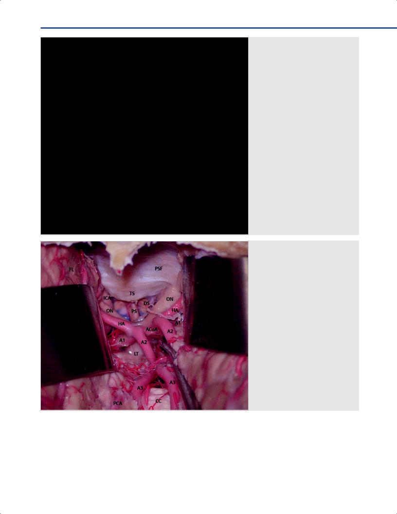
Fig. 14.11 Frontal bilateral approach. Interhemispheric intradural view. Abbreviations: A1 = A1 segment of the anterior cerebral artery; A2 = A2 segment of the anterior cerebral artery; A3 = A3 segment of the anterior cerebral artery; ACoA = anterior communicating artery; CC = corpus callosum; DS = diaphragma sellae; FL = frontal lobe; HA = Heubner’s artery; ICA = internal carotid artery;
LT = lamina terminalis; ON = optic nerve; PCA = pericallosal artery; PS = pituitary stalk; PSF = planum sphenoidalis; TS = tuberculum sellae.
•Cranial nerves: Bilateral exposure of olfactory and optic nerves if orbitotomy is performed.
•Arteries: Bilateral exposure of both anterior cerebral arteries, of frontal branches of the middle cerebral artery and anterior cerebral artery.
•Veins: Bilateral exposure of interhemispheric veins, frontal cortical draining veins, frontal branches of the superfcial middle cerebral vein.
92

14 Frontal and Bifrontal Approach
References
1.Connolly ES, McKhann GM II, Huang J. Fundamentals of operative techniques in neurosurgery. New York, NY:
Thieme Medical Publishers; 2011
2.Fossett D, Caputy AJ. Operative neurosurgical anatomy. New York, NY: Thieme Medical Publisher; 2002
3.Kobayashi S. Neurosurgery of complex vascular lesions and tumors. New York, NY: Thieme Medical Publishers; 2005
4.Sekhar LN, Fessler RG. Atlas of neurosurgical techniques. Brain. Volume 1. New York, NY: Thieme Medical Publishers; 2016
5.Wanibuchi M, Friedman AH, Fukushima T. Photo Atlas of
Skull Base Dissection: Techniques and Operative Approaches.
New York, NY: Thieme Medical Publishers; 2009
93
