

Cardiology / Электрокардиография Ослопова 2005 года
.pdf60
ventricle is depolarized early. These short-circuiting fibers are known as accessory atrioventricular, nodoventricular and fasciculoventricular pathways - the atriofascicular bypass tract and the intranodal bypass tract, depending on their location (Fig. 39).
Figure 39. Schematic diagram of possible accessory conduction pathways (old eponymic nomenclature in parentheses). A - atriofascicular (atrio-Hisian) bundles; K -
accessory atrioventricular (Kent) bundles; J - intranodal bypass (James) tracts; M - Mahaim fibers: M1 - accessory nodoventricular; M2 - accessory fasciculoventricular; M3 - nodofascicular fibers. Dual AV node pathways are represented by the fast and slow symbols [from Wellens HJJ, Brugada P, Penn OC. The management of preexcitation syndromes. JAMA 257:2325, 1987]
Two pre-excitation syndromes exist:
1)the Wolff-Parkinson-White (WPW)syndrome,
2)the Lown-Ganong-Levine (LGL) syndrome.
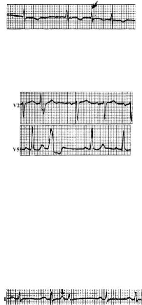
The classic example of pre-excitation is WPW syndrome, which refers to patients with symptomatic accessory AV connections. This syndrome is characterized electrocardiographically by a short PR-inter- val followed by a wide QRS complex, which is a fusion beat between the ventricular myocardium that is pre-excited and that which is excited via normal conduction pathways (Fig. 40). The portion of the complex
61
caused by pre-excitation is called the delta wave because of its resemblance to the Greek capital letter delta ( ). If the accessory bundle connects the atria with the left ventricle, the electrocardiographic pattern resembles RBBB (type A WPW). On the other hand, if the connection is with the right ventricle, the pattern resembles LBBB (type B WPW); the negative -wave in lead II in this situation may be taken for a Q wave, and the mistaken diagnosis of remote myocardial infarction can be made.
Figure 40. The Wolff-Parkinson-White syndrome.
If the atrial fibers insert into the bundle of His and short-circuit the AV node, the P-R interval is short, but no -wave is seen because below the AV node conduction occurs along the usual pathways. This syndrome is known as the Lown-Ganong-Levine syndrome (LGL syndrome). A number of other variants of pre-excitation syndrome have been described but are much rarer than these two common disorders.
The EGG manifestations of pre-excitation may vary from time to time in a given patient because, if conduction occurs through the normal anatomic pathways rather than through accessory fibers, no pre-- excitation is seen on the EGG. When pre-excitation is facilitated because of disease in the AV node or because of drugs that suppress conduction through the AV node (e.g., digitalis, calcium channel blockers, or -blockers), abnormalities on the EGG are seen.
Re-entrant supraventricular arrhythmias are common in patients with pre-excitation; the estimates vary from 13 to 60%, usually paroxysmal supraventricular tachycardia, but atrial fibrillation and flutter also occur. The morphology of the QRS complex during the tachyarrhythmia depends on the direction in which the re-entrant tachycardia occurs. If re-entry occurs antegradly through the AV conducting system and retrogradly through an accessory pathway, then the QRS duration during tachyarrhythmia may be normal because

62
the ventricle is depolarized in a normal direction through its normal specialized conducting tissue. If the circuit is established in the opposite direction, the QRS complex is wide, with a bundle branch block pattern, because most or all of the ventricle is depolarized by the way of the accessory pathway, and arrhythmia can easily be confused with ventricular tachycardia.
REMEMBER:
WPW syndrome
Patients with WPW possess an accessory pathway of depolarization, the bundle of Kent. Three electrocardiographic criteria for WPW are: 1) short PRinterval, 2) wide QRS complex, 3) -wave. The QRS complex is widened by the -wave in exactly the same amount as the P-R interval is shortened. The -wave is a slurring of the initial portion of the QRS complex produced by the early depolarization. The major clinical manifestation of WPW is recurrent tachycardia.
LGL Syndrome
LGL is a result of some of the internodal fibers' (James fibers) bypassing the major portion of the atrioventricular node and terminating in the bundle of His. Three electrocardiographic criteria for LGL are: 1) short PR-interval without -wave, 2) normal QRS, 3) recurrent paroxysmal tachycardia. It should be noted that, unlike in WPW, episodes of tachycardia are required for the diagnosis of LGL.
CONTROL TASKS AND QUESTIONS
1.How is the impulse conduction from the sinus node to the different parts of the heart performed?
2.What degrees of SA block is impossible to register on the ECG and why?
3.Name the ECG signs of the SA block of the II degree.
4.Name the ECG signs of the complete heart block.
5.Name the signs of the AV block of the II degree with SamoilovWenckebach periodics.
6.Name the signs of the AV block of the II degree, Mobitz I.
7.What does Pre-excitation Syndrome mean?
8.What is the difference between WPW & LGL syndromes?
63
9. Define the rhythm disturbance on the ECG given below (paper speed 25 mm/sec).
№1.
№2.
№3.
№4.
№5.
№6.
64
UNIT 2
Theme 2. SUPRAVENTRICULAR ARRHYTHMIAS (PREMATURE ATRIAL AND JUNCTIONAL CONTRACTIONS,
PAROXYSMAL SUPRAVENTRICULAR TACHYCARDIA, ATRIAL FIBRILLATION AND FLUTTER). VENTRICULAR ARRHYTHMIAS (VENTRICULAR PREMATURE BEATS, VENTRICULAR TACHYCARDIA, TORSADES DE POINTES AND THE LONG QT INTERVAL SYNDROME, AND VENTRICULAR FIBRILLATION).
Goal:
to get a notion about supraventricular and ventricular arrhythmias; to master skills.
Knowledge objectives:
to know the criteria of premature atrial and junctional contractions, paroxysmal supraventricular tachycardia, atrial fibrillation and flutter, ventricular premature beats, ventricular tachycardia, ventricular fibrillation, and the long QT interval syndrome; approach to ECGrhythm analysis.
Skill objectives:
to develop practical skills in analyzing electrocardiogram with premature atrial and junctional contractions, paroxysmal supraventricular tachycardia, atrial fibrillation and flutter, ventricular premature beats, ventricular tachycardia, ventricular fibrillation, and the long QT interval syndrome.
EDUCATIONAL MATERIAL
SUPRAVENTRICULAR ARRHYTHMIAS
Premature Atrial and Junctional Contractions
Premature or Ectopic beats (contractions) arise in some focus other than the sino-atrial node and interrupt the regular sequence of normal sinus rhythm. The term extrasystole, often used synonymously, is only strictly accurate in relatively rare instances where the ectopic beat is literally interpolated between successive sinus beats (Fig. 41).
65
Fig. 41. True ―extrasystole‖.
More often, the following sinus impulse finds the myocardium still refractory after its response to the ectopic stimulus, and the next beat is deferred for an additional cycle length - the so-called compensatory pause (Fig. 42). The total number of beats is unchanged.
Fig. 42. The ―Compensatory pause‖.
Premature Atrial and Junctional Contractions (PACs and PJCs) are commonly seen in patients who are otherwise well. They often are induced by the same stimuli that produce sinus tachycardia, especially caffeine or nicotine. However, in patients with congestive heart failure or chronic pulmonary disease, PACs or PJCs may progress to atrial fibrillation or flutter.
Supraventricular foci characteristically produce a normal QRS-T pattern, since they excite the ventricles via the normal conduction system. Atrial beats are recognised by their initial P wave; depending on their site of origin within the atrial myocardium, this may be deformed (Fig. 43) or may precede the QRS complex by a longer or shorter interval than in the normal beats (Fig. 44).
Fig. 43. Atrial ectopic beat with deformed P-wave.

66
Fig. 44. Atrial ectopic beat with prolonged PR-interval (paper speed is 25 mm/sec).
Because of its prematurity, an atrial ectopic impulse sometimes finds the A-V conducting system refractory and fails to excite a ventricular response (Fig. 45).
Fig. 45. Atrial ectopic beat with functional AV-block.
Another possibility is that only one or other bundle branch proves refractory, producing a functional bundle branch block - the atrial ectopic beat with ―aberration‖ (Fig. 46). Only the premature occurrence of P distinguishes this from a ventricular focus.
Fig. 46. Atrial ectopic beat with functional right bundle branch block.
In beats of A-V nodal origin, P is either missing altogether, or appears, usually inverted, in close association with QRS due to retrograde atrial excitation (Fig. 47).
Fig. 47. Junctional ectopic beat with retrograde P-wave.
Another fundamental sign of supraventricular premature beat is that the compensatory pause is not complete, i.e. the interval between the preceding and following beat is less than twice the normal cycle length
67
(because an ectopic beat which causes atrial excitation discharges the impulse accumulating within the sino-atrial node).
REMEMBER:
Premature Atrial Contractions are reflected in the EGG by a premature morphologically abnormal P wave followed by a premature morphologically normal QRS complex.
Premature Junctional contractions are reflected in the EGG by a retrograde P wave (negatively deflected in leads II, III, and aVF) that may follow, be hidden in, or precede a morphologically normal but premature QRS complex.
Compensatory pause is not complete.
PAROXYSMAL SUPRAVENTRICULAR TACHYCARDIA
The term paroxysmal supraventricular tachycardia (PSVT) refers to a group of supraventricular arrhythmias that start and terminate abruptly and generally result from reentry.
PSVT causes:
1. Atrioventricular nodal reentrant tachycardia (AVNRT).
It is the most common cause of PSVT (accounting for two-thirds of all cases of PSVT). AVNRT occurs in the setting of two functionally distinct conduction pathways in the region of the AV node (called the fast and slow pathways).
2.Orthodromic AV reciprocating tachycardia (accessory pathwaymediated tachycardia).
This type of tachycardia, accounting for approximately one-third of all cases of PSVT, results from the electrical impulse travelling from the atria to the ventricles via the AV node and returning to the atria via an accessory pathway that connects the atrium and ventricle.
3.Re-entrant or triggered atrial tachycardia.
It accounts for less than 5% of all cases of PSVT.
PSVT is defined as three or more consecutive PACs. The heart rate during episodes of PSVT may vary most commonly from 100 to 200 (240) beats/minute (Fig. 48). In general, PSVT involving an accessory pathway tends to be more rapid and PSVT caused by an atrial

68
tachycardia tends to be slower. However, because of a tremendous amount of overlap, the rate of tachycardia is usually not helpful in establishing a diagnosis.
Fig. 48. Paroxysmal atrial tachycardia with rate 168/minute (paper speed 25 mm/sec).
There is a fixed relationship of the P wave to the QRS complex. If the impulse is generated in the AV node (as in AVNRT), the P wave may be buried in the QRS complex, but the process can be identified by the normal appearance of the QRS complex and the regularity of the rate. When the P wave is visible, it may follow the QRS complex (some nodal reentry rhythms, all accessory pathway reentry rhythms). It may also precede the QRS complex and may appear morphologically normal (atrial reentry or ectopic rhythm), in which case the diagnosis can be made (by EGG) only if the rate is high enough to make sinus tachycardia unlikely. The P wave also may be hidden in the T wave, but again, the regularity of the rate and the usually normal duration of the QRS complex establish the diagnosis.
Multi-Focal Atrial Tachycardia (MFAT) is a chaotic supraventricular arrhythmia. It results from the presence of multiple, different atrial pacemaker foci. This rhythm disturbance is characterized by a tachycardia with beat-to-beat variation of the P wave morphology. QRS morphology is normal and every QRS complex is preceded by a P wave (Fig. 49).
Fig. 49. Multifocal atrial tachycardia
(note the variation in the morphology of the P-waves and the duration of the PRintervals).
69
ATRIAL FIBRILLATION
Atrial fibrillation is defined electrophysiologically as the generation of multiple reentry wave fronts by the atria. It is usually triggered by a PAC that triggers the development of these wave fronts and results in an atrial rate in excess of 300 beats/minute. These impulses enter the AV node randomly. Because of the unique conduction properties of the AV node, including slow conduction and decremental conduction, only a small proportion of the impulses are conducted to the ventricle. This results in a much slower and irregular ventricular rate.
The prevalence of atrial fibrillation increases with age and the development of structural heart disease. When atrial fibrillation occurs in the absence of any evidence of structural heart disease in patients less than 50 years of age, it is called lone atrial fibrillation. In some patients, factors that trigger episodes of atrial fibrillation can be identified such as physical or emotional stress, alcohol, nicotine, or caffeine. Soaking in a hot tub while imbibing alcoholic beverages has become a recognized precipitating cause of atrial fibrillation. The major noncardiac illness associated with atrial fibrillation is hyperthyroidism; in the presence of a fast ventricular response refractory to digitalis, atrial fibrillation may be the first clue to the diagnosis.
The incidence of atrial fibrillation increases greatly with age, and often appears shortly after the onset of symptomatic coronary heart disease. Hypertensive and rheumatic heart disease (especially when it involves the mitral valve) also predispose to the development of atrial fibrillation, but almost every kind of myocardial disorder has been associated with it. Also, the tachyarrhythmic component of the sick sinus syndrome may be atrial fibrillation.
The EGG shows rapid irregular fibrillatory atrial activity at rates of 300 to 500/minute. No coordinated atrial excitation is possible, and in leads favourable to atrial activity P waves are replaced by a rapid irregular series of oscillation known as f-waves (Fig. 50, leadsV1-2); in other leads no atrial deflections can be distinguished (Fig. 50, lead V6).

70
Fig. 50. Atrial fibrillation, ventricular response 60/minute (paper speed 25 mm/sec), note f-waves in V1-2; no P waves are present.
Ventricular response to this bombardment is partial and irregular, being limited by the capacity of the AV conducting system. In cases uncontrolled by digitalis, it may be as high, as 200 /minute (Fig. 51).
Fig. 51. Uncontrolled atrial fibrillation, ventricular response 190/minute (paper speed 25 mm/sec).
Thus, the ventricular rhythm is irregularly irregular, at rates that at onset are usually 150 to 200/minute, unless there is a coexistent disease in the AV node, in which case slower rates are possible (Fig. 50).
Depending on the number of ventricular complexes the following 3 forms of cardiac fibrillation are distinguished:
-bradycardiac (less than 60 per minute),
-normocardiac (60-90 (100) per minute),
-tachycardiac (more than 90 (100) per minute).
71
The QRS complex is usually morphologically normal. Occasionally there is an aberrant conduction of an impulse in the ventricles, after a beat that has been preceded by a long pause. The aberrant beat usually has a right bundle branch block (RBBB) configuration. This so-called Ashman phenomenon is caused by prolonged refractoriness of the (usually) right bundle branch after the long pause (Fig. 51). These aberrant beats must be distinguished from ventricular premature beats. Apart from their typical relationship to a preceding long R-R interval, aberrant beats are often triphasic (RSR') in lead V1 and their initial vector is the same as that of the normally conducted beats; there is no compensatory pause after aberrant beat. Neither of these features is characteristic of ventricular premature beats.
Fig. 51. Ashman phenomenon.
In some cases atrial fibrillation is associated with AV complete heart block, then the ventricles function rhythmically and rarely (Fig. 52), while fibrillation concerns only the atria. This syndrome was described by Fredericke and is named after him.
Fig. 51. Fredericke syndrome
ATRIAL FLUTTER
Atrial flutter is a reentrant arrhythmia that is confined to the right atrium and results in an atrial rate of about 300 beats/minute. Usually there is a 2:1 AV conduction block so that the ventricular response is about 150/minute, and unlike atrial fibrillation, both atrial and ventricular responses are regular. Atrial flutter is almost always seen in

72
patients who have underlying disease: ischemic heart disease, rheumatic heart disease, congestive cardiomyopathy, atrial septal defect, mitral valve disease, chronic obstructive pulmonary disease, and thyrotoxicosis (the same diseases often associated with atrial fibrillation). In contrast to atrial fibrillation, however, atrial flutter is rarely seen in patients who are otherwise healthy.
In atrial flutter, the EGG commonly shows rapid regular saw-tooth flutter waves at about 300/minute; P waves are absent. The ventricular response is regular, usually at about 150/minute, and the QRS complex is ordinarily morphologically normal (Fig. 52).
Fig. 52. Atrial flutter with 2:1 AV ratio and ventricular rate of 150/minute (paper speed 25 mm/sec).
If the AV node is diseased (e.g. conductivity is impaired by myocardial disease or digitalis therapy) higher degrees of AV block may be seen, usually a multiple of 2 (4:1, 8:1, etc.) (Fig. 53). Aberrant conduction is unusual.
Fig. 53. Atrial flutter with 4:1 AV ratio and ventricular rate of 75/minute (paper speed 25 mm/sec).
73
VENTRICULAR ARRHYTHMIAS
Ventricular tissue is capable of spontaneous depolarization. When this occurs, a premature ventricular contraction (PVC) is initiated. Because the depolarization wave arises in the myocardium, it usually does not follow the normal path of ventricular depolarization. Therefore, the QRS complex is prolonged and bizarre in shape. In addition to PVCs, ectopic ventricular beats produce ventricular tachycardia and sometimes ventricular fibrillation. Ventricular escape rhythms also occur.
Ventricular Premature Beats
Ventricular premature beats (VPBs) are impulses generated in the ventricles, usually as a result of re-entry of an impulse conducted down from the atria through the AV node, but sometimes as the result of the firing of an ectopic (parasystolic) focus.
Occasional VPBs occur in many healthy people sporadically during their lives, more often in older people. However, often VPBs are associated with underlying organic heart disease (e.g., hypertensive heart disease, ischemic heart disease, cardiomyopathy, or mitral valve prolapse). The frequency of VPBs may be increased in people both with and without heart disease by caffeine, alcohol, sympathomimetic drugs, tricyclic antidepressants, phenothiazines, hypokalemia, hypomagnesemia, hypoxia, and emotional stress. VPBs are a common manifestation of digitalis intoxication. Exercise usually abolishes VPBs in people without structural heart disease; conversely, an increase in the number of VPBs after exertion is highly suggestive of structural heart disease.
The EGG shows a premature ventricular response with a morphologically abnormal, often bizarre, wide QRS complex. No P wave precedes a VPB. The ST segment and the T wave have an opposite vector from the QRS complex. Typically, a VPB is followed by a compensatory pause; that is, the R-R interval between two normal beats separated by a VPB is the same as that between two normal beats separated by another normal beat (Fig. 54), because of retrograde conduction of the premature beat to the AV node, thereby blocking the succeeding sinus beat.

74
When VPBs are caused by reentry, they have a fixed temporal (coupled) relationship to the preceding normal beats. When they are caused by the firing of an ectopic (parasystolic) focus, they have no fixed relationship to the preceding normal beats but do have a regular pattern (i.e., the ectopic intervals are constant or are multiples of a constant).
Fig. 54. Premature ventricular beat
(note that the R-R interval between the two normal beats separated by the PVB is the same as that between two normal beats separated by another normal beat).
In general, ventricular foci cause widened, distorted ventricular complexes of the bundle branch block pattern, indicating asynchronous ventricular excitation (one ventricle being excited from the other by relatively slow muscle-to-muscle conduction). The site within the ventricular mass can usually be inferred: an LV ectopic excites the LV first and the RV late, the complex resembling right bundle branch block (Fig. 55); conversely, an RV focus resembles LBBB (Fig. 56).
Fig. 55. Ectopic focus in the left ventricular.
Fig. 56. Ectopic focus in the right ventricular.
75
Very frequent ventricular ectopic beats, particularly when multifocal, suggest a dangerous degree of ventricular ―auto-maticity‖ - due, for example, to ischaemic myocardial disease or to digitalis overdosage. In the latter case, the ectopic beats may alternate with those of normal origin, causing coupling (or bigeminy) of the pulse (Fig. 57). More rarely, they form a trigeminal pattern (Fig. 58).
Fig. 57. Alternating ventricular ectopic beats (bigeminy).
Fig. 58. Trigeminal ventricular ectopic beats.
Ventricular Tachycardia
Ventricular tachycardia is defined as a run of 3 or more PVCs and may occur in bursts or paroxysmally. They may be persistent until stopped by intervention. The heart rate is usually 100 to 200 beats per minute. Ventricular tachycardia is a life-threatening arrhythmia.
Ventricular tachycardia results in a rapid regular series of QRS-T complexes of widened and distorted ―idioventricular‖ form (Fig. 59).
Fig. 59. Ventricular tachycardia 214/min (paper speed 25 mm/sec).

76
Certain diagnosis of ventricular tachycardia requires identification of independent atrial activity - if necessary by means of a special oesophageal electrode. Otherwise it is difficult to exclude the alternative possibility of a supra-ventricular rhythm with coincident bundle branch block - either pre-existing, or the temporary, functional result of refractoriness of one bundle branch at a rapid rate of stimulation (Fig. 60).
Fig. 60. Atrial tachycardia with LBBB, mimicking appearance of ventricular tachycardia.
In Fig. 61, regular atrial deflections are unmasked during the brief cessation of ventricular contractions.
Fig. 61. Ventricular tachycardia; short period of ventricular asystole reveals regular P waves.
Torsades de Pointes and the Long QT Interval Syndrome
One of the variants of ventricular paroxysmal tachycardia is ventricular tachycardia of the ―pirouette‖ type - two-directed, spindleshaped, ventricular tachycardia (torsade de pointes – ―pirouette‖).
Torsades de pointes (literally ―twisting of the spikes‖ [QRS complexes]) is a potentially life-threatening arrhythmia characterized by a form of polymorphic ventricular tachycardia, in which the QRS axis seemingly twists about the isoelectric line (Fig. 62). Torsades de pointes is most often seen in the setting of acquired Q-T prolongation caused by drug interactions, but may be seen with the syndrome of idiopathic or
77
congenital Q-T prolongation, or without Q-T prolongation in the setting of ischemia.
Antiarrhythmic drugs that prolong the Q-T interval, such as quinidine or sotalol, are the most common cause of torsades de pointes. In most patients, predisposing factors such as diuretic use with associated hypokalemia or hypomagnesemia are present. Severe bradycardia is another important predisposing factor, as is female gender, which is associated with 70% of cases of torsades de pointes in the setting of acquired Q—T prolongation. Besides antiarrhythmic drugs, a variety of other drugs may cause Q-T prolongation and have been associated with torsades de pointes. These include antidepressants; antihistamines of the Ha-blocking type (astemizole and terfenadine); some antibiotics, particularly erythromycin; and high dosages of cisapride, a gastric motility agent. Although the incidence of torsades de pointes in patients taking these drugs is rare, these drugs should be used with caution in women, or in patients taking several of these drugs in combination, especially in the setting of hypokalemia or hypomagnesemia, and in patients with the long Q-T syndrome.
In contrast to the acquired long Q-T syndrome, idiopathic or congenital long Q-T syndrome is a congenital disorder in which delayed repolarization is expressed as a long Q-T interval (more than 0.45 second when corrected for heart rate). In some families the inheritance is autosomal recessive and is associated with nerve deafness; in others, the inheritance is autosomal dominant and hearing is normal. The long Q-T interval predisposes to torsades de pointes, which often causes syncope and may cause sudden death, especially in the setting of acute stress. The Q-T interval should be measured routinely in the ECG of patients who complain of syncope for which there is no explanation.
Fig. 62. Two-directed form of the ventricular paroxysmal tachycardia (rhythm rate 170/min).

78
VENTRICULAR FLUTTER
Ventricular Flutter (VF) is an extremal, critical situation foe a patient demanding an immediate therapeutic approach. Not rarely it is a condition of clinical death. Some authors consider VF to be one of the variants of ventricular tachycardia.
Ventricular Flutter is called the regular waves of contraction of ventricular separate fibres of the small amplitude; their frequency, as a rule, is equal to 150-300 per minute. These contractions are hemodynamically noneffective. At the same time in ventricular flutter some minimal blood flow can remain for a short period of time.
Flutter waves with frequency about 200 per minute represent broad, rather high monophasic curves, when it’s impossible to distinguish the waves of QRS ventricular complex, or ST segment, or T wave, i.e. it’s impossible to reveal differentiated elements of the ECG.
The uninterrupted wavy line in VF hac the form of the so-called sinusoid curve (Fig. 63).
Fig. 63. Ventricular Flutter.
VENTRICULAR FIBRILLATION
Ventricular fibrillation is often the continuation and consequence of Ventricular Flutter. In ventricular fibrillation, the ECG reflects the chaotic electromechanical activity of the ventricular myocardium (Fig. 64). Excitation frequency of ventricular separate fibres is 150 – 500 per minute.
Fig. 65. Ventricular fibrillation.
79
CONTROL TASKS AND QUESTIONS
1.Characterize the notion of ―paroxysmal‖ tachycardia.
2.What is the difference between supraventricular and ventricular paroxysmal tachycardia?
3.What signs are characteristic of ventricular flutter?
4.Why are different RR-intervals recorded in atrial fibrillation?
5.What is the difference between atrial fibrillation and atrial
flutter?
6.How does the ventricular flutter look on the ECG?
7.Define the kind of the rhythm disturbance on the ECG listed below (paper speed 25 mm/sec).
№1. 
№2. 
№3. 
№4.