Severity of Stenosis:
•Normal aortic valve area 2.5-3.5 cm2
•Mild stenosis 1.5-2.5 cm2
•Moderate stenosis 1.0-1.5 cm2
•Severe stenosis < 1.0 cm2
•Critical stenosis < 0.7 cm2
•Onset of symptoms
~0.9 cm2 with CAD
~0.7 cm2 without CAD

Symptoms:
•Cardinal Symptoms:
–Chest pain (angina).
•Reduced coronary flow reserve.
•Increased demand-high afterload:
–Syncope/Dizziness (exertional pre-syncope).
•Fixed cardiac output.
•Vasodepressor response:
–Dyspnea on exertion & rest;
–Impaired exercise tolerance.
•Other signs of LV failure.
–Diastolic & systolic dysfunction.

Inspection, palpation, percussion:
1)pallor of the skin and mucous membranes
2)lifting apical impulse,
shifted down and to the left, spilled, high, resistant
3) systolic tremor in the I m / p to the right of sternum and behind the breastbone ("cat purr")

Auscultation of the heart:
1)Weakening 1 tone at the top:
2)1 tone weakens or disappears due to wrinkling of the aortic valve cusps.
3)Diastolic murmur, in the aorta, including Botkin and
even, sluggish, at the top, due
a backward wave of blood from the aorta to the LV. Noise
occurs immediately after the I tone, gradually decreases in intensity towards the end diastole., differs in soft blowing
character.
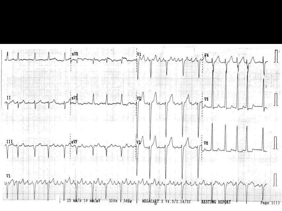
EKG:
1. LVH.
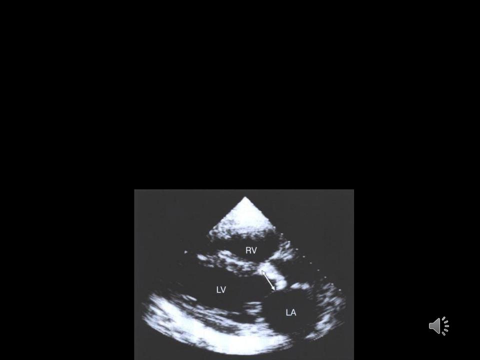
Echocardiography:
Etiology
Valve gradient and areaLVH
Systolic LV functionDiastolic LV functionLA size
Concomitant regional wall motion abnormalitiesCoarctation associated with bicuspid AV
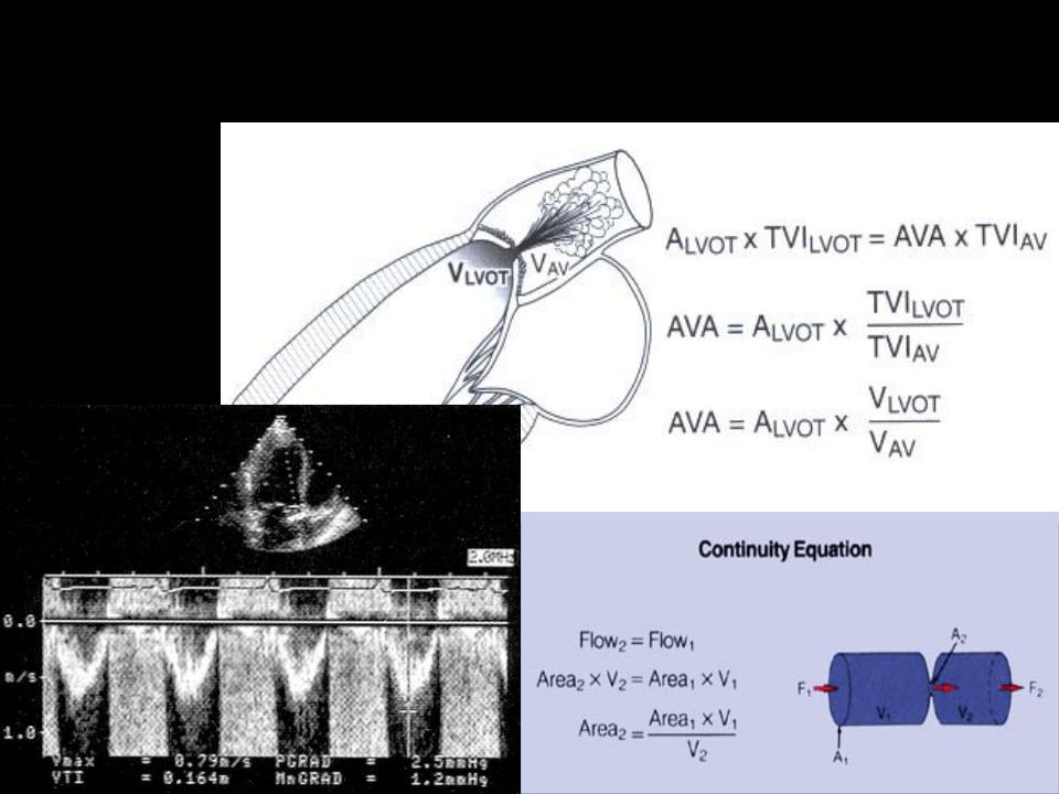
Doppler estimation of AVA:

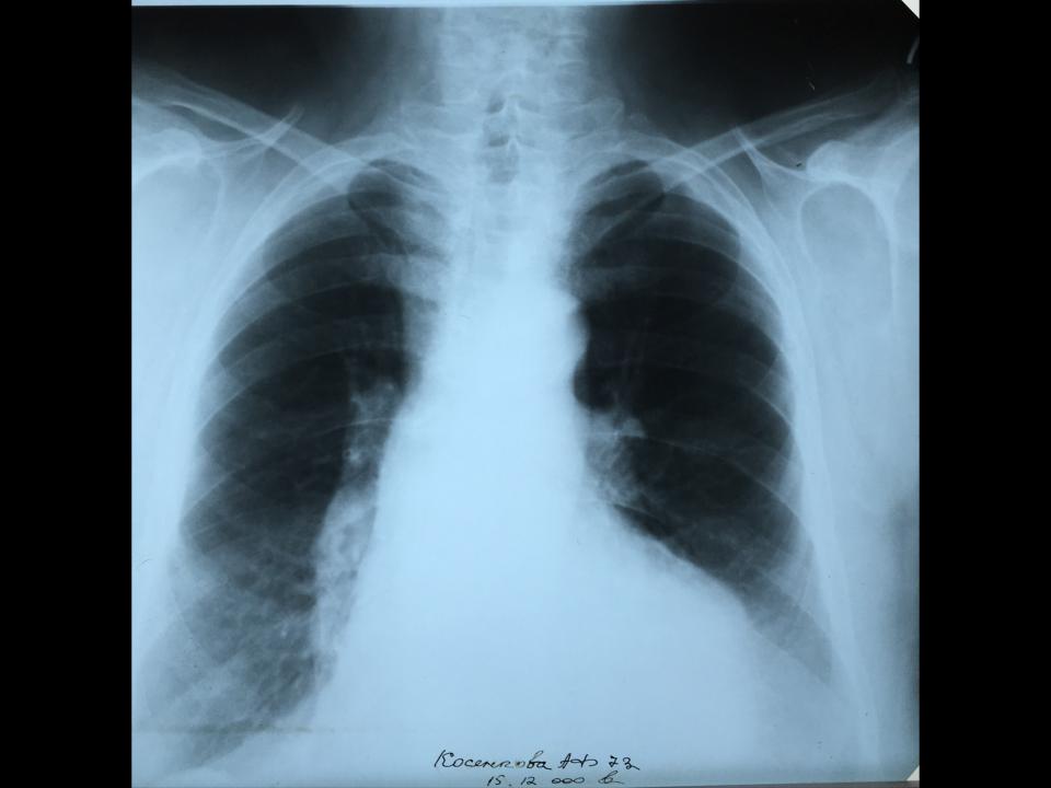
X-ray diagnostics:
1.LV hypertrophy.
2.Aortic configuration of the heart.
3.Deviation of the esophagus along a large radius.