

6) On the femoral arteries, a double tone of
Traube is sometimes heard (systolic and diastolic blood movement) and double murmur Vinogradov-Du Rozier, who appears if the phonendoscope to press on the listener artery, thereby creating conditions for stenosis.
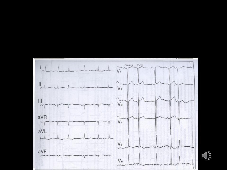
ECG:
1.ECG - signs of hypertrophy left ventricle.
2.The negative tooth
3.TV5-6, I, aV1 appears only during development
heart

Echocardiographic signs:
1. Changes aortic valve:
-bicuspid aortic valve, prolapse flaps,
-thickening calcification,
-vegetation, fusion of valves along commissures. 2. Non-closure of the valve leaflets in diastole.
3. Increased left ventricular cavity and AT sequelae of the atrium.
4. Regurgitation stream on the aortic valve.
5. Turbulence transaortic diastolic flow.

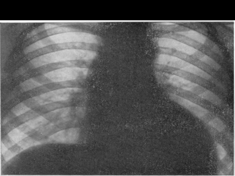
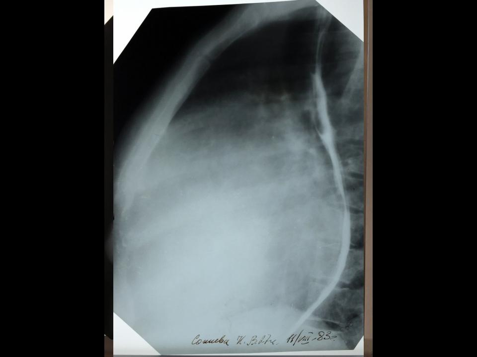
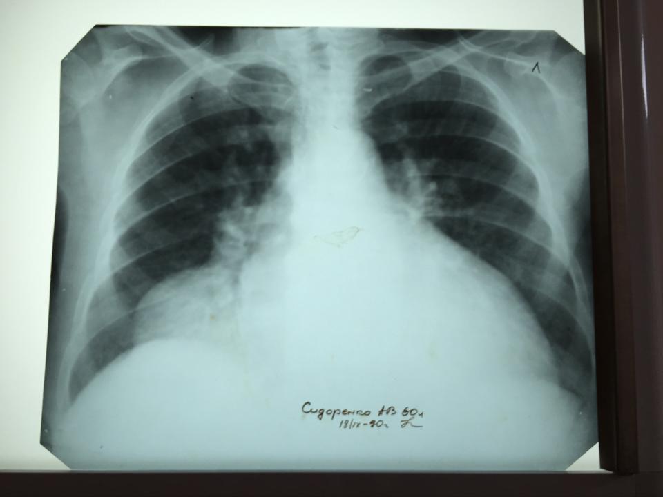
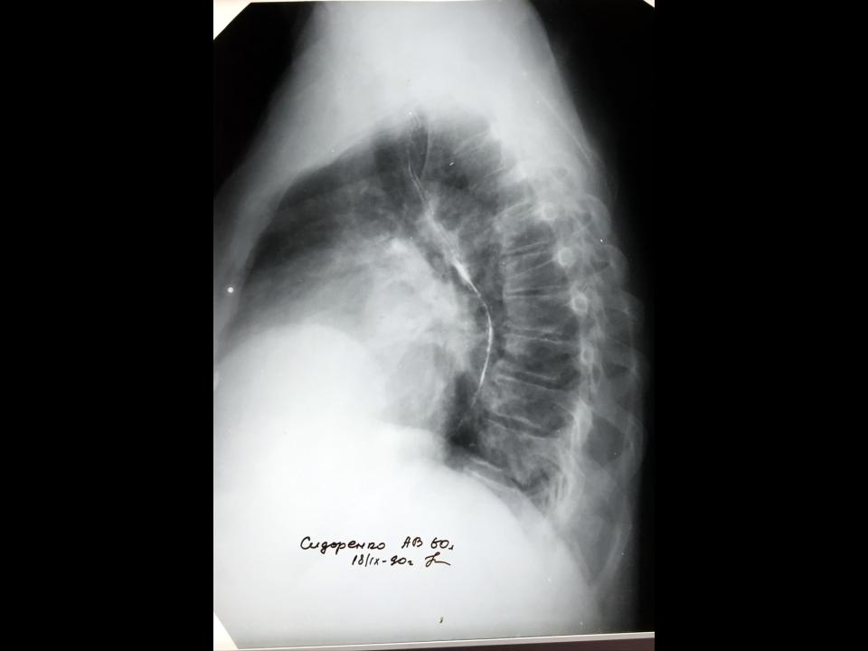
X-ray research
1.Identifies an increase in the left ventricle with an accentuated heart waist, expansion of the aorta.
2.Deviation of the esophagus on a large radius.

Aortic Stenosis
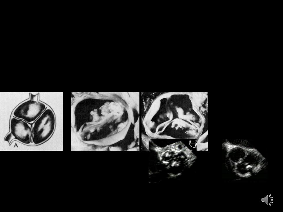
Etiology:
•Congenital bicuspid valve is the most common abnormality
•Rheumatic heart disease and degeneration with calcification are found as well
Normal |
Bicuspid Ao V “Normal” geriatric |
Rheumatic |
calcific valve |