

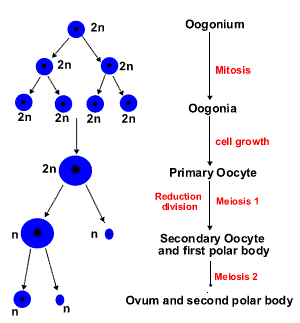
Oogenesis
.The process of oogenesis occurs in the ovary. The oogenesis includes 3 phases (Fig.4):
prolif
growth
maturation
The phase of proliferation. Once primordial germ cells have arrived in the gonad of a genetic female, they differentiate into oogonia. Oogonia divide by mitosis only during embrional life. Beginning in the 3-rd fetal month, some oogonia enter the prophase of the 1st meiotic division and become much larger primary oocytes. In the human, this process is completed by the end of the seventh month of gestation. During this time, many primary oocytes are lost as a result of a degenerative process called atresia. So oocytes are formed during intrauterine life, and their number does not increase after birth.
The phase of growth. This phase lasts a very long time (from 12 up to 50 years) and subdivided onto small and large growth.
Small growth period. At birth all egg cells in the ovary are primary oocytes. Their total number varies from 700,000 to 2 million.
The primary oocytes become surrounded by a layer of flattened follicular cells and become known as primordial follicles. The process of encapsulation of the primary oocyte blocks the first meiotic division in the prophase. Primary oocytes do not finish their 1st meiotic division but remain in the diplotene stage until puberty.
Large growth period. With the onset of puberty a number of primordial follicles begin to grow and mature with each ovarian cycle under influence of follicle-stimulating hormone (FSH). Under normal conditions, only one of these follicles reaches full maturity, the others degenerate and become atretic. Only approximately 400,000 primary oocytes are present by the beginning of puberty, and fewer than 500 will be ovulated in the reproductive lifetime of the individual.
The phase of maturation. The first meiotic division is completed just before ovulation. The chromosomes are equally divided between the daughter cells, but one cell, the secondary oocyte, retains almost all of the cytoplasm. The other becomes the first polar body, a very small cell containing the nucleus and a minimal amount of cytoplasm. Immediately after expulsion of the first polar body the secondary oocyte starts the 2nd meiotic division, which stops in metaphase. The 2nd meiotic (maturation) division will be completed only if the oocyte is fertilized. The secondary oocyte, in turn, gives rise to a mature oocyte plus another polar body. Hence, a primary oocyte develops into one mature oocyte and three polar bodies.
Зрелая яйцеклетка млекопитающих – крупная, в сравнении со сперматозоидом, неподвижная клетка. Особенность строения яйцеклетки – отсутствие центриолей и наличие характерных трофических включений – желточных гранул. По периферии яйцеклетки, под клеточной мембраной, равномерно распределены кортикальные гранулы, содержащие ферменты (гидролазы), необходимые для формирования оболочки оплодотворения после проникновения сперматозоида.
При овуляции яйцеклетка окружена (рис.6):
прозрачной оболочкой (zona pellucida), содержащей густую сеть тонких нитей, состоящих из гликопротеинов (фракции ZP1, ZP2, ZP3);
фолликулярными клетками, образующими лучистый венец
The phase of formation in oogenesis is lacking.
Table 1
Comparison of spermatogenesis and oogenesis |
||
Similarities |
Differences |
|
Spermatogenesis |
Oogenesis |
|
Both processes begin with mitosis (phase of proliferation) Both processes have meiosis |
|
|