

- •Preface
- •Contents
- •List of Abbreviations
- •6. Adverse Drug Effects
- •7b. Cholinergic System and Drugs
- •9. Adrenergic System and Drugs
- •11. Histamine and Antihistaminics
- •12. 5-Hydroxytryptamine, its Antagonists and Drug Therapy of Migraine
- •16. Drugs for Cough and Bronchial Asthma
- •17a. Introduction
- •17b. Anterior Pituitary Hormones
- •20. Corticosteroids
- •21. Androgens and Drugs for Erectile Dysfunction
- •24. Drugs Affecting Calcium Balance
- •25. Skeletal Muscle Relaxants
- •26. Local Anaesthetics
- •27. General Anaesthetics
- •28. Ethyl and Methyl Alcohols
- •29 Sedative-Hypnotics
- •30. Antiepileptic Drugs
- •31. Antiparkinsonian Drugs
- •32. Drugs Used in Mental Illness: Antipsychotic and Antimanic Drugs
- •38. Antiarrhythmic Drugs
- •40. Antihypertensive Drugs
- •41b. Diuretics
- •42. Antidiuretics
- •46. Drugs for Peptic Ulcer and Gastroesophageal Reflux Disease
- •48. Drugs for Constipation and Diarrhoea
- •51. Beta-Lactam Antibiotics
- •53. Aminoglycoside Antibiotics
- •55. Antitubercular Drugs
- •56. Antileprotic Drugs
- •57. Antifungal Drugs
- •58. Antiviral Drugs
- •59. Antimalarial Drugs
- •61. Anthelmintic Drugs
- •62. Anticancer Drugs
- •63. Immunosuppressant Drugs
- •64. Drugs Acting on Skin and Mucous Membranes
- •66. Chelating Agents
- •67. Vitamins
- •68. Vaccines and Sera
- •69. Drug Interactions
- •Appendices
- •Selected References for Further Reading
- •Index
SECTION 5
HORMONES AND RELATED DRUGS
Chapter 17a Introduction
Hormone (Greek hormaein—to stir up) is a substance of intense biological activity that is produced by specific cells in the body and is transported through circulation to act on its target cells.
Hormones regulate body functions to bring about a programmed pattern of life events and maintain homeostasis in the face of markedly variable external/internal environment.
Body function |
Major regulator |
|
|
|
hormone(s) |
|
|
|
|
|
|
1. |
Availability of fuel |
: Insulin, Glucagon, |
|
|
Growth hormone |
2. |
Metabolic rate |
: Triiodothyronine, Thyroxine |
3. |
Somatic growth |
: Growth hormone, |
|
|
Insulin-like growth factors |
4. |
Sex and |
: Gonadotropins, Androgens, |
|
reproduction |
Estrogens, Progestins |
5. |
Circulating |
: Aldosterone, |
|
volume |
Antidiuretic hormone |
6. |
Adaptation to |
: Glucocorticoids, |
|
stress |
Adrenaline |
7. |
Calcium balance |
: Parathormone, Calcitonin, |
|
|
Vitamin D |
Hormones are secreted by the endocrine or ductless glands. These are:
1. Pituitary
(a)Anterior Growth hormone (GH), Prolactin (Prl),
Adrenocorticotropic hormone (ACTH, Corticotropin),
Thyroid stimulating hormone (TSH, Thyrotropin),
Gonadotropins—Follicle stimulating hormone (FSH) and Luteinizing hormone (LH).
(b)Posterior—Oxytocin,
Antidiuretic hormone (ADH, Vasopressin).
2.Thyroid Thyroxine (T4), Triiodothyronine (T3), Calcitonin.
3.Parathyroid Parathormone (PTH).
4.Pancreas (Islets of Langerhans) Insulin, Glucagon.
5.Adrenals
(a) Cortex Glucocorticoids (hydrocortisone) Mineralocorticoids (aldosterone) Sex steroids (dehydroepiandrosterone)
(b) Medulla Adrenaline, Noradrenaline
6. Gonads Androgens (testosterone) Estrogens (estradiol) Progestins (progesterone)
In addition, hypothalamus, which is a part of the CNS and not a gland, produces many releasing

HORMONES AND RELATED DRUGS |
235 |
|
|
|
Hypothalamic |
Chemical |
|
hormone/factor |
nature |
|
|
|
1. |
Thyrotropin releasing |
Tripeptide |
|
hormone (TRH) |
|
2. |
Corticotropin releasing |
Peptide |
|
hormone (CRH) |
(41 AAs) |
3. |
Gonadotropin releasing |
Decapeptide |
|
hormone (GnRH, |
|
|
LH-RH/FSH-RH), |
|
|
Gonadorelin |
|
4. |
Prolactin release inhibi- |
Dopamine |
|
tory hormone (PRIH) |
|
5. |
Growth hormone |
Peptide |
|
releasing hormone |
(40, 44 AAs) |
|
(GHRH) |
|
6. |
Somatostatin (Growth |
Peptide (14 AA) |
|
hormone release |
|
|
inhibitory hormone) |
|
and inhibitory hormones which control the secretion of anterior pituitary hormones. Some important ones of these are given in the box.
Placenta also secretes many hormones:
Chorionic gonadotropin |
Prolactin |
Estrogens |
Progesterone |
Placental lactogen |
Chorionic |
|
thyrotropin |
The natural hormones and in many cases their synthetic analogues which may be more suitable therapeutically, are used as drugs for substitution therapy as well as for pharmacotherapy. In addition, hormone antagonists and synthesis/release inhibitors are of therapeutic importance.
Sites and mechanisms of hormone action
The hormones act on their specific receptors located on or within their target cells. Receptor activation by the hormones is translated into response in a variety of ways.
1. At cell membrane receptors
a. Through alteration |
Adrenaline, Glucagon, |
||||
of intracellular |
TSH, |
FSH, LH, |
|||
cAMP concentra- |
PTH, |
Calcitonin, |
|||
tion alteration of |
ACTH, some |
||||
protein kinase A |
hypothalamic |
||||
regulation of |
cell |
releasing hormones, |
|||
function: Ca2+ |
acting |
Vasopressin (V2) |
|||
as |
third |
messenger |
|
|
|
in |
some |
situations. |
|
|
|
b. Through IP3/DAG |
Vasopressin (V1), |
||||
generation: release |
Oxytocin |
||||
of |
intracellular Ca2+ |
|
|
||
and protein kinase C |
|
|
|||
activation. |
|
|
|
||
c. Direct transmembrane |
Insulin, |
||||
activation of tyrosine |
Growth hormone |
||||
protein kinase |
Prolactin |
||||
phosphorylation |
|
|
|||
cascade regulation |
|
|
|||
of various enzymes. |
|
|
|||
2. At cytoplasmic receptors
Penetrating cell |
Steroidal hormones: |
membrane, hormone |
Glucocorticoids |
combines with a |
Mineralocorticoid |
cytoplasmic receptor |
Androgens |
exposes its DNA |
Estrogens |
binding domain |
Progestins; |
migrates to nucleus |
Calcitriol |
and binds to specific genes DNA mediated mRNA synthesissynthesis of functional proteins.
3. At nuclear receptor |
|
The hormone pene- |
Thyroid hormones: |
trates the nucleus |
Thyroxine, |
combines with its |
Triiodothyronine |
receptor alters |
|
DNARNA mediated |
|
protein synthesis. |
|
5 SECTION

Chapter 17b Anterior Pituitary Hormones
Anterior pituitary (adenohypophysis), the master endocrine gland, elaborates a number of important regulatory hormones. All of these are peptide in nature and act at extracellular receptors located on their target cells. Their secretion is controlled by the hypothalamus through releasing and release-inhibitory hormones that are transported via hypothalamohypophyseal portal system, and is subjected to feedback inhibition by the hormones of their target glands. Each anterior pituitary hormone is produced by a separate group of cells, which according to their staining characteristic are either acidophilic or basophilic.
The acidophils are either somatotropes GH; or lactotropes Prolactin.
The basophils are gonadotropes FSH and LH; thyrotropes TSH; and corticotrope-lipo- tropes ACTH. The latter in addition to ACTH also produce two melanocyte stimulating hormones (MSHs) and two lipotropins, but these are probably not important in man.
GROWTH HORMONE (GH)
It is a 191 amino acid, single chain peptide of MW 22000.
Physiological functions GH promotes growth of bones and all other organs by inducing hyperplasia. In general, there is a proportionate increase in the size and mass of all parts, but in the absence of gonadotropins, sexual maturation does not take place. The growth of brain and eye is independent of GH. It promotes retention of nitrogen, calcium and other tissue constituents: more protoplasm is formed. The positive nitrogen balance results from increased uptake of amino acids by tissues and their synthesis into proteins. GH promotes utilization of fat and spares carbohydrates: uptake of glucose by muscles is reduced while its output from liver is enhanced; fat is broken down.
GH acts on cell surface JAK-STAT binding protein kinase receptors (see p. 50) which are present on practically all cells. Binding of one GH molecule to the extracellular domain of a GH-receptor diamer results in the formation of a ternary complex which undergoes a conformational change and activates the intracellular domain to associate with cytoplasmic JAK-STAT tyrosine-protein kinase resulting in metabolic effects as well as regulation of gene expression.
Fig. 17.1: Action of growth hormone (GH) and regulation of its secretion
GHRH—Growth hormone releasing hormone; IGF-1: Insulin like growth factor-1; Stimulation (—— ); Inhibition (- - - - )

ANTERIOR PITUITARY HORMONES |
237 |
|
|
The growth promoting, nitrogen retaining and certain metabolic actions of GH are exerted indirectly through the elaboration of peptides called Somatomedins or Insulin-like growth factors (mainly IGF-1, also IGF-2) which are extracellular mediators of GH response (Fig. 17.1). Liver is the major source of circulating IGF-1, while IGF- 1 produced by other target cells acts locally in a paracrine manner. Like insulin, IGF-1 promotes lipogenesis and glucose uptake by muscles. The IGF-1 receptor also is structurally and functionally analogous to the insulin receptor (see p. 261).
GH acts directly as well to induce lipolysis in adipose tissue, gluconeogenesis and glycogenolysis in liver and decreased glucose utilization by muscles. These effects are opposite to those of IGF-1 and insulin. As such, GH accentuates the metabolic derangement in diabetes.
Regulation of secretion The hypothalamus produces GH releasing (GHRH) as well as release inhibitory (somatostatin) hormones. Both are peptides. Somatostatin is also produced by D cells of islets of Langerhans in the pancreas and by few other tissues. Receptors for GHRH and somatostatin are G protein coupled receptors (GPCRs) which enhance or inhibit GH secretion by increasing or decreasing cAMP formation respectively in pituitary somatotropes. Somatostatin has also been shown to inhibit Ca2+ channels and open K+ channels.
Stimuli that cause GH release are—fasting, hypoglycaemia, exercise, stress and i.v. infusion of arginine. GH secretion is inhibited by rise in plasma free fatty acid levels and by high doses of glucocorticoids. Dopaminergic agents cause a brief increase in GH release in normal subjects but paradoxically depress it in acromegalics. IGF-1 causes feedback inhibition of GH secretion. Short-loop feedback inhibition of secretion by GH itself has also been described.
Pathological involvements Excess production of GH is responsible for gigantism in childhood and acromegaly in adults. Hyposecretion of GH in children results in pituitary dwarfism. Adult GH deficiency is rare, but when it occurs, it results in low muscle and bone mass, lethargy, decreased work capacity, hyperlipidaemia and increased cardiovascular risk.
Preparations and use The primary indication for GH is pituitary dwarfism—0.03–0.06 mg/kg daily in the evening or on alternate days, upto the age of 20 years or more. Human GH produced by recombinant DNA technique (rhGH) somatropin (191AA) is available for clinical use. Somatropin causes IGF-1 to appear in plasma after a delay of several hours. IGF-1 then remains detectable for upto 48 hours. Early diagnosis and institution of GH therapy restores stature to near normal. rhGH can also be used in Turner’s syndrome and in children with renal failure.
Somatropin has been tried in children with constitutional short stature (only if epiphyses are open) with encouraging results. Commercial interests are promoting it for accelerating growth in children without GH deficiency, but medical, ethical, cost-benefit and social objections have been raised.
In adult GH deficient patients, rHGH 150–300 g/day s.c. adjusted later according to response increases lean body mass, decreases body fat, improves energy and mentation and may reudce excess morbidity and mortality, but stature is unaffected. Benefits of rHGH therapy in GH deficient adults are now well recognized. Unlimited availability of recombinant GH has provided opportunity for its trial in catabolic states like severe burns, bedridden patients, chronic renal failure, osteoporosis, etc. It is now approved for AIDSrelated wasting: higher dose (0.05–0.1 mg/kg/day) is needed. However, it should not be given to postoperative, trauma, cancer and other critically ill patients. Somatropin is also being promoted for ageing, but benefits are uncertain. Its abuse by athletes is banned, and it is one of the drugs included in ‘dope testing’.
Somatropin: NORDITROPIN 5, 10, 15 mg inj, HUMATROPE 6 mg, 12 mg cartridges, 1.33 and 5.33 mg vials.
Adverse effects Somatropin has low immunogenicity; allergic reactions or resistance to treatment are not a problem. Pain at injection site, lipodystrophy, glucose intolerance, hypothyroidism (due to unmasking of TSH deficiency), salt and water retention, hand stiffness, myalgia, headache are the possible adverse effects. Rise in intracranial tension occurs in few cases.
GH Inhibitors
Somatostatin
This 14 amino acid peptide inhibits the secretion of GH, prolactin, and TSH by pituitary; insulin and glucagon by pancreas, and of almost all gastrointestinal secretions including that of gastrin and HCl. The g.i. action produces steatorrhoea, diarrhoea, hypochlorhydria, dyspepsia and nausea as side effect. Somatostatin constricts splanchnic, hepatic and renal blood vessels. The decreased g.i. mucosal blood flow can be utilized for controlling bleeding esophageal varices and bleeding peptic ulcer, but octreotide is preffered now due to longer duration of action. Its antisecretory action is beneficial in pancreatic, biliary or intestinal fistulae; can also be used to reduce complications after pancreatic surgery. It also has adjuvant value in diabetic ketoacidosis (by inhibiting glucagon and GH secretion).
Use of somatostatin in acromegaly is limited by its short duration of action (t½ 2–3 min), lack of specificity for inhibiting only GH secretion and GH rebound on discontinuation. Surgical removal of pituitary adenomas is the preferred treatment modality, but somatostatin analogues are being increasingly used.
Dose: (for upper g.i.bleeding) 250 µg slow i.v. injection over 3 min followed by 3 mg i.v. infusion over 12 hours.
17 CHAPTER

238 |
HORMONES AND RELATED DRUGS |
|
|
SECTION 5
STILMEN, SOMATOSAN, SOMASTAT 250 µg and 3 mg amps.
Octreotide This synthetic octapeptide surrogate of somatostatin is 40 times more potent in suppressing GH secretion and longer acting (t½ ~90 min), but only a weak inhibitor of insulin secretion. It is preferred over somatostatin for acromegaly and secretory diarrhoeas associated with carcinoid, AIDS, cancer chemotherapy or diabetes. Control of diarrhoea is due to suppression of hormones which enhance intestinal mucosal secretion.
Dose: Initially 50–100 µg s.c. twice daily, increased upto 200 µg TDS; for acromegaly maintain with 10-30 mg i.m. of microsphere formulation every 4 weeks.
Adverse effects are abdominal pain, nausea, steatorrhoea, diarrhoea, and gall stones (due to biliary stasis).
Octreotide injected i.v. (100 µg followed by 25–50 µg/hr) reduces hepatic blood flow and helps stop esophageal variceal bleeding.
SANDOSTATIN, OCTRIDE 50 µg, 100 µg in 1 ml amps. SANDOSTATIN LAR (microsphere formulation) 20 mg/5 ml inj.
Lanreotide Another long-acting analogue of somatostatin, very similar in actions and specificity to octreotide, which on i.m. injection acts for 10–15 days. It is indicated for pharmacotherapy of acromegaly.
Pegvisomant This polyethylene glycol complexed mutant GH binds to the GH receptor but does not trigger signal transduction: acts as a GH antagonist. It is approved for treatment of acromegaly due to small pituitary adenomas.
PROLACTIN
It is a 199 amino acid, single chain peptide of MW 23000; quite similar chemically to GH. It was originally described as the hormone which causes secretion of milk from crop glands of pigeon and later found to be of considerable importance in human beings as well.
Physiological function Prolactin is the primary stimulus which in conjunction with estrogens, progesterone and several other hormones, causes growth and development of breast during pregnancy. It promotes proliferation of ductal as well as acinar cells in the breast and induces synthesis of milk proteins and lactose.
After parturition, prolactin induces milk secretion, since the inhibitory influence of high estrogen and progesterone levels is withdrawn.
Prolactin suppresses hypothalamo-pituitary- gonadalaxisbyinhibitingGnRHrelease.Continued high level of prolactin during breastfeeding is responsible for lactational amenorrhoea, inhibition of ovulation and infertility for several months postpartum. Prolactin may affect immune response through action on T-lymphocytes.
A specific prolactin receptor is expressed on the surface of target cells, which is structurally and functionally analogous to GH receptor: action is exerted by transmembrane activation of JAK—cytoplasmic tyrosine protein kinases and STAT. Placental lactogen and GH also bind to prolactin receptor and exert similar effects, but prolactin does not bind to GH receptor.
Regulation of secretion Prolactin is under predominant inhibitory control of hypothalamus through PRIH which is dopamine that acts on pituitary lactotrope D2 receptor. Dopaminergic agonists (DA, bromocriptine, cabergoline) decrease plasma prolactin levels, while dopaminergic antagonists (chlorpromazine, haloperidol, metoclopramide) and DA depleter (reserpine) cause hyperprolactinemia.
Though TRH, prolactin releasing peptide and VIP can stimulate prolactin secretion, no specific prolactin releasing factor has been identified. Endogenous opioid peptides may also be involved in regulating prolactin secretion, but no feedback regulation by any peripheral hormone is known. Prolactin levels in blood are low in childhood, increase in girls at puberty and are higher in adult females than in males. A progressive increase occurs during pregnancy, peaking at term. Subsequently, high prolactin secretion is maintained by suckling: it falls if breast feeding is discontinued. Stress, exertion and hypoglycaemia also stimulate prolactin release.
Physio-pathological involvement Hyperprolactinaemia is responsible for the galactorrhoea– amenorrhoea–infertility syndrome in women. In males it causes loss of libido and depressed fertility. The causes of hyperprolactinaemia are:
(i)Disorders of hypothalamus removing the inhibitory control over pituitary.
(ii)Antidopaminergic and DA depleting drugs —these are a frequent cause now.
(iii)Prolactin secreting tumours—these may be microprolactinomas or macroprolactinomas.
(iv)Hypothyroidism with high TRH levels—also increases prolactin secretion.
Use Therearenoclinicalindications for prolactin.

ANTERIOR PITUITARY HORMONES |
239 |
|
|
Prolactin inhibitors Bromocriptine
This synthetic ergot derivative 2-bromo- - ergocryptine is a potent dopamine agonist; most of its actions are based on this property. It has greater action on D2 receptors, while at certain dopamine sites in the brain it acts as a partial agonist or antagonist of D1 receptor. It is also a weak adrenergic blocker but not an oxytocic.
Actions
1.Decreases prolactin release from pituitary by activating dopaminergic receptors on lactotrope cells: is a strong antigalactopoietic.
2.Increases GH release in normal individuals, but decreases the same from pituitary tumours that cause acromegaly.
3.Has levodopa like actions in CNS—anti- parkinsonian and behavioral effects.
4.Produces nausea and vomiting by stimulating dopaminergic receptors in the CTZ.
5.Hypotension—due to central suppression of postural reflexes and weak peripheral a adrenergic blockade.
6.Decreases gastrointestinal motility.
Pharmacokinetics Only 1/3 of an oral dose of bromocriptine is absorbed; bioavailability is further lowered by high first pass metabolism in liver. Even then, it has higher oral: parenteral activity ratio than ergotamine. Metabolites are excreted mainly in bile. Its plasma t½ is 3–6 hours.
PROCTINAL, PARLODEL, SICRIPTIN, BROMOGEN 1.25 mg, 2.5 mg tabs.
Uses Bromocriptine should always be started at a low dose, 1.25 mg BD and then gradually increased till response occurs otherwise side effects become limiting.
1. Hyperprolactinemia due to microprolactinomas causing galactorrhoea, amenorrhoea and infertility in women; gynaecomastia, impotence and sterility in men. Bromocriptine and cabergoline are the first line drug for most cases. Relatively lower doses (bromocriptine 2.5–10 mg/day or cabergoline 0.25–1.0 mg twice weekly) are effective. Response occurs in a few weeks
and serum prolactin levels fall to the normal range; many women conceive. Bromocriptine should be stopped when pregnancy occurs, though no teratogenic effect is reported. Most (60–75%) tumours show regression during therapy and neurological symptoms (visual field defects, etc.) due to pressure on optic chiasma ease. However, response is maintained only till the drug is given— recurrences occur in many, but not all patients.
2.Acromegaly due to small pituitary tumours and inoperable cases. Relatively higher doses are required (5–20 mg/day) and it is less effective than octreotide/lanreotide. Oral administration and lower cost are the advantages..
3.Parkinsonism Bromocriptine, if used alone, is effective only at high doses (20–80 mg/day) which produce marked side effects. However, response is similar to that of levodopa. It is now recommended in low dose only, as an adjunct to levodopa in patients not adequately benefited and in those showing marked ‘on-off’ effect.
4.Diabetes mellitus (DM) A new use of bromocriptine
based on its dopamine D2 agonistic action in the hypothalamus has been found in type 2 DM, and it has been approved by US-FDA as an adjunctive drug.
5.Hepatic coma: Bromocriptine may cause arousal.
6.Bromocriptine suppresses lactation and breast engorgement in case of neonatal death, but is not recommended due to unfavourable risk: benefit ratio.
Side effects: Side effects are frequent and dose related.
Early: Nausea, vomiting, constipation, nasal blockage. Postural hypotension may be marked at initiation of therapy—syncope may occur if starting dose is high. Hypotension is more likely in patients taking antihypertensives.
Late: Behavioral alterations, mental confusion, hallucinations, psychosis—are more prominent than with levodopa.
Abnormal movements, livedo reticularis.
Cabergoline
It is a newer D2 agonist; more potent; more D2 selective and longer acting (t½ > 60 hours) than
17 CHAPTER

240 |
HORMONES AND RELATED DRUGS |
|
|
SECTION 5
bromocriptine; needs to be given only twice weekly. Incidence of nausea and vomiting is also lower; some patients not tolerating or not responding to bromocriptine have been successfully treated with cabergoline. It is preferred for treatment of hyperprolactinemia and acromegaly. Some patients who achieve total regression of prolactinoma and normalization of prolactin levels can stop cabergoline without recurrence.
Dose: Start with 0.25 mg twice weekly; if needed increase after every 4–8 weeks to max. of 1 mg twice weekly.
CABERLIN 0.5 mg tab, CAMFORTE 0.5, 1 mg tabs.
GONADOTROPINS (Gns)
The anterior pituitary secretes two Gns viz. FSH and LH. Both are glycoproteins containing 23–28% sugar and consist of two peptide chains. The -chain (92AA) is common between FSH and LH, but their -chains are different: FSH (111 AA), LH (121 AA). Paradoxically the MW of FSH (~33KD) is greater than that of LH (~30 KD), because of the sugar moieties.
Physiological functions FSH and LH act in concert to promote gametogenesis and secretion of gonadal hormones.
FSH In the female it induces follicular growth, development of ovum and secretion of estrogens. In the male it supports spermatogenesis and has a trophic influence on seminiferous tubules. Ovarian and testicular atrophy occurs in the absence of FSH.
LH It induces preovulatory swelling of the ripe graafian follicle and triggers ovulation followed by luteinization of the ruptured follicle and sustains corpus luteum till the next menstrual cycle. It is also probably responsible for atresia of the remaining follicles. Progesterone secretion occurs only under the influence of LH. In the male LH stimulates testosterone secretion by the interstitial cells and is designated interstitial cell stimulating hormone (ICSH).
Distinct LH and FSH receptors are expressed on the target cells. Both are G protein coupled receptors which on activation increase cAMP production. This in turn stimulates gametogenesis and conversion of cholesterol to
pregnenolone—the first step in progesterone, testosterone and estrogen synthesis. In the testes FSH receptor is expressed on seminiferous (Sertoli) cells while LH receptor is expressed on interstitial (Leydig) cells. In the ovaries FSH receptors are present only on granulosa cells, while LH receptors are widely distributed on interstitial cells, theca cells, preovulatory granulosa cells and luteal cells.
Regulation of secretion A single releasing factor (decapeptide designated GnRH) is produced by the hypothalamus which stimulates synthesis and release of both FSH and LH from pituitary. It is, therefore, also referred to as FSH/LH-RH or simply LHRH or gonadorelin. It has been difficult to explain how hypothalamus achieves a divergent pattern of FSH and LH secretion in menstruating women through a single releasing hormone. Since GnRH is secreted in pulses and the frequency as well as amplitude of the pulses differs during follicular (high frequency, low amplitude) and luteal (lower frequency, higher amplitude) phases, it is considered that frequency and amplitude of GnRH pulses determines whether FSH or LH or both will be secreted, as well as the amount of each. Further, the feedback regulation of FSH and LH may be different. In general, feedback inhibition of LH is more marked than that of FSH. In females estradiol and progesterone inhibit both FSH and LH secretion mainly through hypothalamus, but also by direct action on pituitary. However, the marked and sustained preovulatory rise in estrogen level paradoxically stimulates LH and FSH secretion. In addition there are other regulatory substances, e.g. Inhibin—a peptide from ovaries and testes, selectively inhibits FSH release, but not LH release. Dopamine inhibits only LH release. Testosterone is weaker than estrogens in inhibiting Gn secretion, but has effect on both FSH and LH. GnRH acts on gonadotropes through a G-protein coupled receptor which acts by increasing intracellular Ca2+ through PIP2 hydrolysis.
The Gn secretion increases at puberty and is higher in women than in men. In men, the levels of FSH and LH remain practically constant (LH > FSH) while in menstruating women they fluctuate cyclically. During the follicular phase, moderate levels of FSH and low levels of LH prevail. There is a midcycle surge of both, but more of LH, just before ovulation, followed by progressive fall during the luteal phase. Gn levels are high in menopausal women due to loss of feedback inhibition by sex steroids and inhibin.
Pathological involvement Disturbances of Gn secretion from pituitary may be responsible for delayed puberty or precocious puberty both in girls and boys.
Inadequate Gn secretion results in amenorrhoea and sterility in women; oligozoospermia, impotence and infertility in men. Excess production of Gn in adult women causes polycystic ovaries.
Preparations
All earlier gonadotropin preparations were administered by i.m. route. The newer more purified preparations can be

ANTERIOR PITUITARY HORMONES |
241 |
|
|
given s.c. They are partly metabolized, but mainly excreted unchanged in urine: t½ 2–6 hours.
1.Menotropins (FSH + LH): is a preparation obtained from urine of menopausal women:
PREGNORM, PERGONAL, GYNOGEN 75/150; 75 IU FSH + 75 IU LH activity per amp, also 150 IU FSH + 150 IU LH per amp.
2.Urofollitropin or Menotropin (pure FSH): METRODIN, FOLGEST, FOLICULIN, PUREGON 75 IU and 150 IU per amp. This preparation has been preferred over the combined FSH + LH preparation for induction of ovulation in women with polycystic ovarian disease: these patients have elevated LH/FSH ratio; use of FSH alone is considered advantageous. It is also claimed to improve chances of obtaining good quality ova for in vitro fertilization.
endogenous FSH/LH secretion either by continuous pretreatment with a superactive GnRH agonist or by a GnRH antagonist.
2. Hypogonadotrophic hypogonadism in males manifesting as delayed puberty or defective spermatogenesis oligozoospermia, male sterility. Generally, sexual maturation is induced by androgens and therapy with HCG is started when fertility is desired. Start with 1000–4000 IU of HCG i.m. 2–3 times a week (to stimulate testosterone secretion), add FSH 75 IU + LH 75 IU after 3–4 months (to stimulate spermato-
3.Human chorionic gonadotropin (HCG): is derived from genesis) and reduce dose of HCG; continue
urine of pregnant women.
CORION, PROFASI, PUBERGEN 1000 IU, 2000 IU, 5000 IU, 10,000 IU, all as dry powder with separate solvent for injection. The foetal placenta secretes HCG which is absorbed in maternal circulation and maintains corpus luteum of pregnancy. It is a glycoprotein with 33% sugar and 237 amino acids in two chains, MW 38000. It is excreted in urine by the mother from which it is commercially obtained. HCG binds to LH receptor with equal avidity; action of HCG is indistinguishable from that of LH.
Recombinant human FSH (rFSH: Follitropin and follitropin ) and recombinant human LH (rLH: Lutropin) as well as recombinant HCG (rHCG: Choriogonadotropin ) have become available. These are more purified and have vertually replaced the urine derived preparations in the developed countries. They are more expensive.
Recombinant human LH (rhLH) is marketed as LUVERIS 75 IU inj.; indications and use is similar to HCG.
Uses
1. Amenorrhoea and infertility When it is due to deficient production of Gns by pituitary. Gns are generally tried when attempts to induce ovulation with clomiphene have failed or when nonovulation is due to polycystic ovaries. The procedure is to give 1 injection of menotropins (75 IU FSH + 75 IU LH or 75 IU pure FSH)) i.m. daily for 10 days followed the next day by 10,000 IU of HCG. Ovulation occurs within the next 24–48 hours in upto 75% cases and the woman may conceive. However, rates of abortion and multiple pregnancy are high, but not of teratogenesis.
To improve predictability of time of ovulation (controlled ovarian hyperstimulation) most experts now concurrently suppress
treatment for 6–12 months for optimum results, which nevertheless are not always impressive.
3.Cryptorchidism Since undescended testes can cause infertility and predispose to testicular cancer, medical/surgical treatment is imperative. Descent of testes can be induced by androgens whose production is stimulated by LH. Treatment with HCG can be tried at the earliest after the age of 1 year, preferably before 2 years if there is no anatomical obstruction; 1000–2000 IU is given i.m. 2–3 times a week till the testes descend. If 2–6 week treatment does not induce descent, surgery should be performed.
4.To aid in vitro fertilization Menotropins (FSH + LH or pure FSH) have been used to induce simultaneous maturation of several ova and to precisely time ovulation so as to facilitate their harvesting for in vitro fertilization.
Adverse effects and precautions
Ovarian hyperstimulation—polycystic ovary, pain in lower abdomen and even ovarian bleeding and shock can occur in females.
Precocious puberty is a risk when given to children.
Allergic reactions have occurred and skin tests are advised. Hormone dependent malignancies (prostate, breast) must be excluded.
Other side effects are edema, headache, mood changes.
17 CHAPTER

242 |
HORMONES AND RELATED DRUGS |
|
|
SECTION 5
GONADOTROPIN RELEASING HORMONE (GnRH):
GONADORELIN
Synthetic GnRH injected i.v. (100 µg) induces prompt release of LH and FSH followed by elevation of gonadal steroid levels. It has a short plasma t½ (4–8 min) due to rapid enzymatic degradation; has been used for testing pituitarygonadal axis in male as well as female hypogonadism.
Since only pulsatile exposure to GnRH induces FSH/LH secretion, while continuous exposure desensitizes pituitary gonadotropes resulting in loss of Gn release, therapy with GnRH or its analogues is not useful in the treatment of hypogonadism.
Superactive / long-acting GnRH agonists
Many analogues of GnRH, e.g. Goserelin, Leuprolide, Nafarelin, Triptorelin, have been developed which are 15-150 times more potent than natural GnRH and longer acting (t½ 2–6 hours) because of high affinity for GnRH receptor and resistance to enzymatic hydrolysis. Because physiological release of GnRH is in pulses, whereas these agonists act continuously; they only initially increase Gn secretion. After 1–2 weeks they cause desensitization and down regulation of GnRH receptors inhibition of FSH and LH secretionsuppression of gonadal function. Spermatogenesis or ovulation cease and testosterone or estradiol levels fall to castration levels. Recovery occurs within 2 months of stopping treatment.
The superactive GnRH agonists are used as nasal spray or injected s.c. Long-acting preparations for once a month s.c. injection have been produced (triptorelin, goserelin depot). The resulting reversible pharmacological oophorectomy/ orchidectomy is being used in precocious puberty, prostatic carcinoma, endometriosis, premenopausal breast cancer, uterine leiomyoma, polycystic ovarian disease and to assist induced ovulation. They also have potential to be used as contraceptive for both males and females.
Nafarelin This long-acting GnRH agonist is 150 times more potent than native GnRH. It is used as intranasal spray from which bioavailability is only 4–5%.
NASAREL 2 mg/ml soln for nasal spray; 200 µg per actuation.
Down regulation of pituitary GnRH receptors occurs in10 days but peak inhibition of Gn release occurs at one month. It is broken down in the body to shorter peptide segments; plasma t½ is 2–3 hours. Uses are:
Assisted reproduction: Endogenous LH surge needs to be suppressed when controlled ovarian hyperstimulation is attempted by exogenous FSH and LH injection, so that precisely timed mature oocytes can be harvested. This is achieved by 400 g BD intranasal nafarelin, reduced to 200 g BD when menstrual bleeding occurs.
Uterine fibroids: Nafarelin 200 g BD intranasal for 3–6 months can reduce the size of leiomyoma and afford symptomatic relief.
Endometriosis: 200 µg in alternate nostril BD for upto 6 months. As effective as danazol, but second course cannot be given due to risk of osteoporosis.
Central precocious puberty: 800 µg BD by nasal spray; breast and genital development is arrested in girls and boys. The effect is reversible; pubertal changes resume when therapy is discontinued.
Adverse effects: Hot flashes, loss of libido, vaginal dryness, osteoporosis, emotional lability.
Goserelin Another long-acting GnRH agonist available as a depot s.c./i.m. injection to be used both for endogenous Gn suppression before ovulation induction, as well as for endometriosis, carcinoma prostate, etc. To achieve pituitary desensitization before ovulation induction with exogenous Gns: 3.6 mg of the depot injection is given once in the anterior abdominal wall 1–3 weeks earlier.
For endometriosis and carcinoma prostate 3.6 mg is injected in the same way every 4 weeks or 10.8 mg is injected every 3 months. An androgen antagonist (bicalutamide) is given concurrently for 3–4 weeks when goserelin is used for carcinoma prostate.
ZOLADEX 3.6 mg prefilled syringe, ZOLDEX L-A 10.8 mg vial depot injection.
Triptorelin: This long acting GnRH agonist is formulated as a regular release daily s.c. injection for short term indications, such as female infertility, and as a depot i.m. monthly injection for long-term Gn suppression in the treatment of carcinoma prostate, endometriosis, precocious puberty and uterine leiomyoma. For prostate cancer, it is combined with an androgen antagonist flutamide or bicalutamide to prevent the initial flare up of the tumour that occurs due to increase in Gn secretion for the first 1–2 weeks.
Continuous treatment with any GnRH agonist is not advised beyond 6 months due to risk of osteoporosis and other complications.

ANTERIOR PITUITARY HORMONES |
243 |
|
|
Fibroids, endometriosis, carcinoma prostate: 3.75–7.5 mg i.m. every 4 weeks.
Precocious puberty: 50 g/kg i.m. of depot inj. every 4 weeks. Assisted reproduction: 0.1 mg s.c. daily for 10 days from 2nd day of cycle.
DECAPEPTYL DAILY 0.1 mg inj., DECAPEPTYL DEPOT 3.75 mg inj.
Leuprolide This long acting GnRH agonist is injected s.c./i.m. daily or as a depot injection once a month for palliation of carcinoma prostate alongwith an androgen antagonist, as well as for other conditions needing long term Gn suppression.
LUPRIDE 1 mg inj., 3.75 mg depot inj., PROGTASE 1 mg/ml inj.
GnRH antagonists Some more extensively substituted GnRH analogues act as GnRH receptor antagonists. They inhibit Gn secretion without causing initial stimulation. The early GnRH antagonists had the limitation of producing reactions due to histamine release. Later agents like ganirelix and cetrorelix have low histamine releasing potential and are being clinically used as s.c. inj. in specialized centres for inhibiting LH surges during controlled ovarian stimulation in women undergoing in vitro fertilization. Their advantages over long-acting GnRH agonists include:
•They produce quick Gn suppression by competitive antagonism, need to be started only from 6th day of ovarian hyperstimulation.
•They carry a lower risk of ovarian hyperstimulation syndrome.
•They achieve more complete suppression of endogenous Gn secretion.
However, pregnancy rates are similar or may even be lower.
THYROID STIMULATING HORMONE (TSH, THYROTROPIN)
It is a 210 amino acid, two chain glycoprotein (22% sugar), MW 30000.
Physiological function TSH stimulates thyroid to synthesize and secrete thyroxine (T4) and triiodothyronine (T3). Its actions are:
•Induces hyperplasia and hypertrophy of thyroid follicles and increases blood supply to the gland.
•Promotes trapping of iodide into thyroid by increasing Na+: Iodide symporter (NIS).
•Promotes organification of trapped iodine and
its incorporation into T3 and T4 by increasing peroxidase activity.
•Enhances endocytotic uptake of thyroid colloid
by the follicular cells and proteolysis of thyroglobulin to release more of T3 and T4. This action starts within minutes of TSH administration.
The TSH receptor present on thyroid cells is a GPCR which utilizes the adenylyl cyclase-cAMP transducer mechanism (by coupling to Gs protein) to produce its effects. In human thyroid cells high concentration of TSH also induces PIP2 hydrolysis by the linking of TSH receptor to Gq protein. The resulting increase in cytosolic Ca2+ and protein kinase C activation may also mediate TSH action, particularly generation of H2O2 needed for oxidation of iodide and iodination of tyrosil residues.
Regulation of secretion Synthesis and release of TSH by pituitary is controlled by hypothalamus primarily through TRH, while somatostatin inhibits TSH secretion. Dopamine also reduces TSH production induced by TRH. The TRH receptor on pituitary thyrotrope cells is a GPCR which is linked to Gq protein and activates PLC–IP3/DAG–cytosolic Ca2+ pathway to enhance TSH synthesis and release. The negative feedback for inhibiting TSH secretion is provided by the thyroid hormones which act primarily at the level of the pituitary, but also in the hypothalamus. T3 has been shown to reduce TRH receptors on the thyrotropes.
Pathological involvement Only few cases of hypoor hyperthyroidism are due to inappropriate TSH secretion. In majority of cases of myxoedema TSH levels are markedly elevated because of deficient feedback inhibition. Graves’ disease is due to an immunoglobulin of the IgG class which attaches to the thyroid cells and stimulates them in the same way as TSH. Consequently, TSH levels are low. Contrary to earlier belief, TSH is not responsible for exophthalmos seen in Graves’ disease because TSH levels are low.
Use Thyrotropin has no therapeutic use. Thyroxine is the drug of choice even when hypothyroidism is due to TSH deficiency. The diagnostic application is to differentiate myxoedema due to pituitary dysfunction from primary thyroid disease.
ADRENOCORTICOTROPIC HORMONE (ACTH, CORTICOTROPIN)
It is a 39 amino acid single chain peptide, MW 4500, derived from a larger peptide pro-opio melanocortin (MW 30,000) which also gives rise to endorphins, two lipotropins and two MSHs.
Physiological function ACTH promotes steroidogenesis in adrenal cortex by stimulating cAMP formation in cortical cells (through specific cell surface GPCRs) rapidly increases the
17 CHAPTER
244 |
HORMONES AND RELATED DRUGS |
|
|
availability of cholesterol for conversion to pregnenolone which is the rate limiting step in the production of gluco, mineralo and weakly androgenic steroids. Induction of steroidogenic enzymes occurs after a delay resulting in 2nd phase ACTH action. The stores of adrenal steroids are very limited and rate of synthesis primarily governs the rate of release. ACTH also exerts trophic influence on adrenal cortex (again through cAMP): high doses cause hypertrophy and hyperplasia. Lack of ACTH results in adrenal atrophy. However, zona glomerulosa is little affected because angiotensin II also exerts trophic influence on this layer and sustains aldosterone secretion.
Regulation of secretion Hypothalamus regulates ACTH release from pituitary through corticotropin-releasing hormone (CRH). The CRH receptor on corticotropes is also a GPCR which increases ACTH synthesis as well as release by raising cytosolic cAMP. Secretion of ACTH has a circadian rhythm. Peak plasma levels occur in the early morning, decrease during day and are lowest at midnight. Corticosteroids exert inhibitory feedback influence on ACTH production by acting directly on the pituitary as well as indirectly through hypothalamus.
A variety of stressful stimuli, e.g. trauma, surgery, severe pain, anxiety, fear, blood loss, exposure to cold, etc. generate neural impulses which converge on median eminence to cause elaboration of CRH. The feedback inhibition appears to be overpowered during stress—rise in ACTH secretion continues despite high plasma level of cortisol induced by it. Arginine vasopressin (AVP) enhances the action of CRH on corticotropes and augments ACTH release. AVP release and augmentation of ACTH action appears to be important during stress.
Pathological involvement Excess production of ACTH from basophil pituitary tumours is responsible for some cases of Cushing’s syndrome. Hypocorticism occurs in pituitary insufficiency due to low ACTH production. Iatrogenic suppression of ACTH secretion and pituitary adrenal axis is the most common form of abnormality encountered currently due to the use of pharmacological doses of glucocorticoids in nonendocrine diseases.
Use ACTH is used primarily for the diagnosis of disorders of pituitary adrenal axis. Injected i.v. 25 IU causes increase in plasma cortisol if the adrenals are functional. Direct assay of plasma ACTH level is now preferred.
For therapeutic purposes, ACTH does not offer any advantage over corticosteroids and is more inconvenient, expensive as well as less predictable.
SECTION 5

Chapter 18 Thyroid Hormones and
Thyroid Inhibitors
THYROID HORMONE
The thyroid gland secretes 3 hormones—thyro- xine (T4), triiodothyronine (T3) and calcitonin. The former two are produced by thyroid follicles, have similar biological activity and the term ‘thyroid hormone’ is restricted to these only. Calcitonin produced by interfollicular ‘C’ cells is chemically and biologically entirely different. It is considered along with parathormone, (Ch. 24) with which it regulates calcium metabolism.
The physiological significance of thyroid gland was recognized only after Graves and Basedow (1835, 1840) associated the clinical features of the ‘Graves’ disease’ with swelling of thyroid gland and Gull (1874) correlated myxoedema with its atrophy. Kendall (1915) obtained crystalline thyroxine and postulated its chemical formula which was confirmed in 1926. Thyroxine was the first hormone to be synthesized in the laboratory. Since, T4 could not account for all the biological activity of thyroid extract, search was made and more potent T3 was discovered in 1952.
CHEMISTRY AND SYNTHESIS
Both T4 and T3 are iodine containing derivatives of thyronine which is a condensation product of two molecules of the amino acid tyrosine. Thyroxine; is 3, 5, 3´, 5´–tetraiodothyronine while T3 is 3, 5, 3´ triiodothyronine.
The thyroid hormones are synthesized and stored in the thyroid follicles as part of thyroglobulin molecule—which is a glycoprotein synthesized by thyroid cells, MW 660 KDa, contains 10% sugar. The synthesis, storage and release of T4 and T3 is summarized in Fig. 18.1 and involves the following processes.
1. Iodide uptake The total body content of I2, obtained from food and water, is 30–50 mg, out of which about 1/5 is present in the thyroid. Concentration of iodide in blood is low (0.2–0.4 µg/dl) but thyroid cells have an active
transport process Na+:: iodide symporter (NIS) to concentrate this anion; this trapping is stimulated by TSH to exceed a gradient of more than 100 fold by inducing and activating NIS. The I2 content of thyroid gland somehow regulates the uptake mechanism: meagre store activating and large store inhibiting it. The iodide concentrating mechanism is not peculiar to thyroid. Skin, salivary glands, gastric mucosa, intestine, mammary glands and placenta also possess it, but uptake in these organs is not stimulated by TSH.
2.Oxidation and iodination Iodide trapped by follicular cells is carried across the apical membrane by another transporter termed ‘pendrin’ and oxidized by the membrane bound thyroid peroxidase enzyme to iodinium (I+) ions or hypoiodous acid (HOI) or enzyme-linked hypo-
iodate (E-OI) with the help of H2O2. These forms of iodine combine avidly with tyrosil residues of thyroglobulin, apparently without any enzymatic intervention, to form monoiodotyrosine (MIT) and diiodotyrosine (DIT) while these residues are still attached to the thyroglobulin chains.
3.Coupling Pairs of iodinated tyrosil residues
couple together (Fig. 18.2) to form T3 and T4. Normally much more T4 than T3 is formed, but during I2 deficiency relatively more MIT is available and a greater proportion of T3 is formed. Thus, more active hormone is generated with
lesser amount of I2.
Coupling is an oxidative reaction and is catalysed by the same thyroid peroxidase. Thyroglobulin is the most efficient protein, compared to other similar proteins, in supporting coupling by providing favourable spatial configuration to facilitate the reaction. Oxidation of iodide and coupling are both stimulated by TSH.

246
SECTION 5
HORMONES AND RELATED DRUGS
Fig. 18.1: Synthesis, storage and secretion of thyroid hormone TG—Thyroglobulin; MIT—Monoiodotyrosine; DIT—Diiodotyrosine; T3—Triiodothyronine; T4—Thyroxine (Tetraiodothyronine); HOI—Hypoiodous acid; EOI—Enzyme linked hypoiodate; NIS—Na+-iodide symporter; Thyroid-stimulating hormone (TSH) activates steps 1, 2, 3, 4, and 5; Ionic inhibitors block step 1; Excess iodide interferes with steps 1, 2, 3 and 5 with primary action on step 3 and 5; Propylthiouracil inhibits steps 2 and 6; Carbimazole inhibits step 2 only
Fig. 18.2: Coupling of monoiodotyrosine (MIT) and diiodotyrosine (DIT) to produce triiodothyronine (T3)

THYROID HORMONES AND THYROID INHIBITORS |
247 |
|
|
4.Storage and release Thyroglobulin containing iodinated tyrosil and thyronil residues is transported to the interior of the follicles and remains stored as thyroid colloid till it is taken back into the cells by endocytosis and broken
down by lysosomal proteases. The T4 and T3 so released is secreted into circulation while MIT and DIT residues are deiodinated and the iodide released is reutilized. The uptake of colloid and proteolysis are stimulated by TSH: the quiscent gland has follicles distended with colloid and cells are flat or cubical, while the TSH stimulated gland has columnar cells and colloid virtually disappears.
Normal human thyroid secretes 60–90 µg of T4 and 10–30 µg of T3 daily.
5.Peripheral conversion of T4 to T3 Peripheral tissues, especially liver and kidney, convert T4 to T3. About 1/3 of T4 secreted by thyroid undergoes this change and most of the T3 in plasma is derived from liver. Target tissues take up T3 from circulation for their metabolic need, except
brain and pituitary which take up T4 and convert it to T3 within their own cells.Almost equal amounts of 3, 5, 3´ triiodothyronine (normal T3 : active) and 3, 3´, 5´ triiodothyronine (reverse T3 or rT3: inactive) are produced in the periphery. The T4 to T3 conversion is carried out by the enzyme iodothyronine deiodinase which exists in 3 forms (D1, D2, D3). These forms differ in their organ and cellular localization as well as product formed.
Whereas type 2 deiodinase (D2) generates T3 and D3 generates rT3, the D1 form generates both T3 and rT3. The antithyroid drug propylthiouracil (but not carbimazole) inhibits Type1 deiodinase and the antiarrhythmic amiodarone inhibits both D1 and D2 forms. Propranolol (high dose) and glucocorticoids also inhibit peripheral conversion of T4 to T3 (except in brain and in pituitary).
TRANSPORT, METABOLISM AND EXCRETION
Thyroid hormones are avidly bound to plasma proteins—only 0.03–0.08% of T4 and 0.2–0.5% of T3 are in the free form. Almost all protein bound iodine (PBI) in plasma is thyroid hormone,
of which 90–95% is T4 and the rest T3. Binding occurs to 3 plasma proteins in the following decreasing order of affinity for T4:
(i)Thyroxine binding globulin (TBG)
(ii)Thyroxine binding prealbumin (transthyretin)
(iii)Albumin
The normal concentration of PBI is 4–10 µg/ dl; only 0.1–0.2 µg/dl of this is T3, rest is T4. During pregnancy thyroxine binding globulin is increased—PBI levels are elevated, but there is no effect on thyroid status because the concentration of free hormone remains unaltered.
Only the free hormone is available for action as well as for metabolism and excretion. Metabolic inactivation of T4 and T3 occurs by deiodination and glucuronide/sulfate conjugation of the hormones as well as that of their deiodinated products. Liver is the primary site (also salivary glands and kidneys). The conjugates are excreted in bile, of which a significant fraction is deconjugated in intestines and reabsorbed (enterohepatic circulation) to be finally excreted in urine.
Plasma t½ of T4 is 6–7 days, while that of T3 is 1–2 days. The half-lives are shortened in hyperthyroidism and prolonged in hypothyroidism due respectively to faster and slower metabolism.
REGULATION OF SECRETION
The secretion of hormones from the thyroid is controlled by anterior pituitary by the elaboration of thyrotropin, while TSH secretion itself is regulated by TRH produced in hypothalamus (see p. 243). Somatostatin elaborated by hypothalamus inhibits not only GH and prolactin, but also TSH secretion from pituitary. The relation between thyroid, anterior pituitary and hypothalamus is depicted in Fig. 18.3. The negative feedback by the thyroid hormones is exercised directly on the pituitary as well as through hypothalamus. The action of TRH on pituitary and that of TSH on thyroid cells is mediated by enhanced cAMP synthesis. High concentration of TSH also acts via IP3/DAG–increased intracellular Ca2+ pathway in the thyroid cells.
18 CHAPTER

248 |
HORMONES AND RELATED DRUGS |
|
|
SECTION 5
Fig. 18.3: Regulation of thyroid function TSH—Thyroid stimulating hormone; TRH—Thyrotropin releasing hormone; T3—Triiodothyronine; T4—Thyroxine; SST—Somatostatin
ACTIONS
The actions of T4 and T3 are qualitatively similar and are nicely depicted in the features of hypo and hyperthyroidism. They affect the function of practically every body cell.
1. Growth and development T4 and T3 are essential for normal growth and development. The most remarkable action is metamorphosis of tadpole to frog: the tail is used-up to build lungs, limbs and other organs. The action cannot be broadly labelled as catabolic or anabolic. It is exerted through a critical control of protein synthesis in the translation of the genetic code. Congenital deficiency of T4 and T3 resulting in cretinism emphasizes their importance. The milestones of development are delayed and practically every organ and tissue of the body suffers. The greatest sufferer, however, is the nervous system. Retardation and nervous deficit is a consequence of paucity of axonal and dendritic ramification, synapse formation and impaired myelination. In adult hypothyroidism also, intelligence is impaired and movements are slow.
2. Intermediary metabolism Thyroid hormones have marked effect on lipid, carbohydrate and protein metabolism.
Lipid T4 and T3 indirectly enhance lipolysis by potentiating the action of catecholamines and other lipolytic hormones, probably by suppressing a phosphodiesterase → increased cAMP. As a result plasma free fatty acid levels are elevated. Lipogenesis is also stimulated. All phases of cholesterol metabolism are accelerated, but its conversion to bile acids dominates. Thus, hyperthyroidism is characterized by hypocholesterolemia. LDL levels in blood are reduced.
Carbohydrate Carbohydrate metabolism is also stimulated. Though utilization of sugar by tissues is increased (mainly secondary to increased BMR), glycogenolysis and gluconeogenesis in liver as well as faster absorption of glucose from intestines more than compensate it → hyperglycaemia and diabetic-like state with insulin resistance occur in hyperthyroidism.
Protein Synthesis of certain proteins is increased, but the overall effect of T3 is catabolic— increased amounts of protein being used as energy source. Prolonged action results in negative nitrogen balance and tissue wasting. Weight loss is a feature of hyperthyroidism. T3, T4 in low concentrations inhibit mucoprotein synthesis which so characteristically accumulates in myxoedema.
3.Calorigenesis T3 and T4 increase BMR by stimulation of cellular metabolism and resetting of the energystat. This is important for maintaining body temperature. However, metabolic rate in brain, gonads, uterus, spleen and lymph nodes is not significantly affected. The mechanism of calorigenesis was believed to be uncoupling of oxidative phosphorylation: excess energy being released as heat. However, this occurs only at very high doses and is not involved in mediating
the physiological actions of T3, T4. Dinitrophenol uncouples oxidative phosphorylation, but has no thyroid-like activity.
4.CVS T3 and T4 cause a hyperdynamic state of circulation which is partly secondary to increased peripheral demand and partly due to direct cardiac actions. Heart rate, contractility and

THYROID HORMONES AND THYROID INHIBITORS |
249 |
|
|
output are increased resulting in a fast, bounding pulse. T3 and T4 stimulate heart by direct action on contractile elements (increasing the myosin fraction having greater Ca2+ ATPase activity) and probably by up regulation of β adrenergic receptors. Atrial fibrillation and other irregularities are common in hyperthyroidism. Thyroid hormones can also precipitate CHF and angina. BP, specially systolic, is often raised. Myocardial O2 consumption can be markedly reduced by induction of hypothyroidism.
5.Nervous system T3, T4 have profound functional effect on CNS. Mental retardation is the hallmark of cretinism; sluggishness and other behavioral features are seen in myxoedema. Hyperthyroid individuals are anxious, nervous, excitable, exhibit tremors and hyperreflexia.
6.Skeletal muscle Muscles are flabby and weak in myxoedema, while thyrotoxicosis produces increased muscle tone, tremor and weakness due to myopathy.
7.GIT Propulsive activity of gut is increased
by T3/T4. Hypothyroid patients are often constipated, while diarrhoea is common in hyperthyroidism.
8.Kidney T3 and T4 do not cause diuresis in euthyroid individuals, but the rate of urine flow is often increased when myxoedematous patients are treated with it.
9.Haemopoiesis Hypothyroid patients suffer from some degree of anaemia which is res-
tored only by T4 treatment. Thus, T4 appears to be facilitatory to erythropoiesis.
10.Reproduction Thyroid has an indirect effect on reproduction. Fertility is impaired in hypothyroidism and women suffer from oligomenorrhoea. Normal thyroid function is required for maintenance of pregnancy and lactation.
Mechanism of action
Both T3 and T4 penetrate cells by active transport and produce majority of their actions by combining with a nuclear thyroid hormone receptor (TR)
which belongs to the steroid and retinoid superfamily of intracellular receptors.
Two TR isoform families (TRα and TRβ) have been identified. Both bind T3 and function in similar manner, but their tissue distribution differs, which may account for quantitative differences in the sensitivity of different tissues to T3.
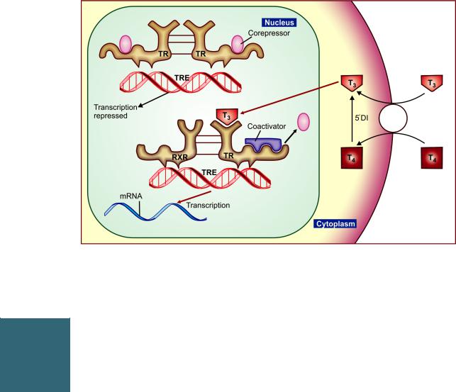
In contrast to the steroid receptor, the TR resides in the nucleus even in the unliganded inactive state. It is bound to the ‘thyroid hormone response element’ (TRE) in the enhancer region of the target genes along with corepressors (Fig. 18.4). This keeps gene transcription suppressed. When T3 binds to the ligand-binding domain of TR, it heterodimerizes with retinoid X receptor (RXR) and undergoes a conformation change releasing the corepressor and binding the coactivator. This induces gene transcription → production of specific mRNA and a specific pattern of protein synthesis → various metabolic and anatomic effects. The expression of certain genes is repressed by T3. In their case, the unliganded TR allows gene transcription, while binding of T3 to TR halts the process.
Many of the effects, e.g. tachycardia, arrhythmias, raised BP, tremor, hyperglycaemia are mediated, at least partly, by sensitization of adrenergic receptors to catecholamines. Induction of adenylyl cyclase, proliferation of β adrenoceptors and a better coupling between these two has been demonstrated.
Apart from the nuclear T3 receptor, other sites of thyroid hormone action have been described. It acts on cell membrane to enhance amino acid and glucose entry and on mitochondria to increase oxygen consumption. At these sites T4 appears to be equipotent to T3, while at the nuclear receptor T4 has much lower affinity, and even when bound to the TR, T4 does not promote gene transcription.
Relation between T4 and T3
• Thyroid secretes more T4 than T3, but in iodine deficient state this difference is reduced.
•T4 is the major circulating hormone because it is 15 times more tightly bound to plasma proteins.
•T3 is 5 times more potent than T4 and acts faster. Peak effect of T3 comes in 1–2 days while that of T4 takes 6–8 days.
18 CHAPTER
250 |
HORMONES AND RELATED DRUGS |
|
|
Fig. 18.4: Mechanism of action of thyroid hormone on nuclear thyroid hormone receptor (TR). T3—Triiodothyronine; T4—Thyroxine; TRE—Thyroid hormone response element; RXR—Retinoid X receptor; mRNA—Messenger ribonucleic acid; 5’DI—5’Deiodinase (See text for explanation)
SECTION 5
•T3 is more avidly bound to the nuclear receptor than T4 and the T4-receptor complex is unable to activate/derepress gene transcription.
•About 1/3 of T4 is converted to T3 in the thyroid cells, liver and kidney by type 1 deiodinase (D1) and released into circulation. In addition, T3 is generated within the target cells (skeletal
muscle, heart, brain, pituitary) by another type (D2) of deiodinase.
Thus, it may be cocluded that T3 is the active hormone, while T4 is mainly a transport form; functions as a prohormone of T3. However, it may directly produce some nongenomic actions.
Preparations
l-thyroxine sod.: ELTROXIN 25 μg, 50 μg, 100 μg tabs, ROXIN 100 µg tab, THYRONORM 12.5 μg, 25 μg, 50 μg, 62.5 μg, 75 μg, 88 μg, 100 μg, 112 μg, 125 μg, 137 μg, 150 μg tabs, THYROX 25 µg, 50 µg, 75 µg, 100 µg tabs.An injectable preparation for i.v. use is available elsewhere.
Triiodothyronine (Liothyronine) is not freely available in India. It is occasionally used i.v. along with l-thyroxine in myxoedema coma.
Clinically, l-thyroxine is preferred for all indications over liothyronine because of more sustained and uniform action as well as lower risk of cardiac arrhythmias.
Pharmacokinetics and interactions
Oral bioavailability of l-thyroxine is ~ 75%, but severe hypothyroidism can reduce oral absorption. It should be administered in empty stomach to avoid interference by food. Sucralfate, iron, calcium and proton pump inhibitors also reduce l-thyroxine absorption. CYP3A4 inducers like rifampin, phenytoin and carbamazepine accelerate metabolism of T4; dose of l-thyroxine may need enhancement.

THYROID HORMONES AND THYROID INHIBITORS |
251 |
|
|
USES
The most important use of thyroid hormone is for replacement therapy in deficiency states:
1.Cretinism It is due to failure of thyroid development or a defect in hormone synthesis (sporadic cretinism) or due to extreme iodine deficiency (endemic cretinism). It is usually detected during infancy or childhood; but screening of neonates is the best preventive strategy. Treatment with thyroxine (8–12 µg/kg) daily should be started as early as possible, because mental retardation that has already ensued is only partially reversible. Response is dramatic: physical growth and development are restored and further mental retardation is prevented.
2.Adult hypothyroidism (Myxoedema)
This is one of the commonest endocrine disorders which develops as a consequence of autoimmune thyroiditis or thyroidectomy; It may accompany simple goiter if iodine deficiency is severe. Antibodies against thyroid peroxidase or thyroglobulin are responsible for majority of cases of adult hypothyroidism. Important drugs that can cause hypothyroidism are 131I, iodides, lithium and
amiodarone. Treatment with T4 is most gratifying. It is often wise to start with a low dose—50 µg of l-thyroxine daily and increase every 2–3 weeks to an optimum of 100–200 µg/day (adjusted by clinical response and serum TSH levels). Further dose adjustments are made at 4–6 week intervals needed for reaching steady-state. Individualization of proper dose is critical, aiming at normalization of serum TSH levels. Increase in dose is mostly needed during pregnancy.
Subclinical hypothyroidism characterized by euthyroid status and normal free serum thyroxine (FT4) level (> 9 pmol/L) but raised TSH level (>10 mU/L) should be treated with T4. For TSH level between 6–10 mU/L, replacement therapy is optional. It is preferable if patient has other cardiovascular risk factors.
3. Myxoedema coma It is an emergency; characterized by progressive mental deterioration due to acute hypothyroidism: carries significant mortality. Rapid thyroid replacement is crucial.
Though liothyronine (T3) acts faster, its use is attended by higher risk of cardiac arrhythmias, angina, etc. Drug of choice is l-thyroxine (T4) 200–500 μg i.v. followed by 100 μg i.v. OD till oral therapy can be instituted. Some authorities recommend adding low dose i.v. T3 10 μg 8 hourly in younger patients with no arrhythmia or ischaemia. Alternatively oral T4 500 μg loading dose followed by 100–300 μg daily may be used, but in severe hypothyroidism, oral absorption is delayed and inconsistent.
Other essential measures needed are— warming the patient, i.v. corticosteroids to cover attendant adrenal insufficiency, ventilatory and cardiovascular support, correction of hyponatraemia and hypoglycaemia.
4. Nontoxic goiter It may be endemic or sporadic. Endemic is due to iodine deficiency which may be accentuated by factors present in water (excess calcium), food or milk (goitrin, thiocyanates). A defect in hormone synthesis may be responsible for sporadic cases. In both types, deficient production of thyroid hormone leads to excess TSH → thyroid enlarges, more efficient trapping of iodide occurs and probably greater proportion of T3 is synthesized → enough hormone to meet peripheral demands is produced so that the patient is clinically euthyroid. Thus, treatment with T4 is in fact replacement therapy in this condition as well, despite no overt hypothyroidism. Full maintenance doses must be given. Most cases of recent diffuse enlargement of thyroid regress. Long-standing goiter with degenerative and fibrotic changes and nodular goiter regress little or not at all. Thyroxine therapy may be withdrawn after a year or so in some cases if adequate iodine intake is ensured. Others need life-long therapy.
Endemic goiter and cretinism due to iodine deficiency in pregnant mother is preventable by ensuring daily ingestion of 150–200 µg of iodine. This is best achieved by iodizing edible salt by adding iodate (preferred over iodide). In India iodization of table salt (100 µg iodine/g salt) is required under the National Programme, but recently mandatory
iodization rule has been withdrawn.
5. Thyroidnodule Certainbenignfunctioning nodules regress when TSH is suppressed by
18 CHAPTER

252 |
HORMONES AND RELATED DRUGS |
|
|
SECTION 5
T4 therapy. Nonfunctional nodules and those nonresponsive to TSH (that are associated with low TSH levels) do not respond and should not be treated with levothyroxine. T4 therapy should be stopped if the nodule does not decrease in size within 6 months, and when it stops regressing after the initial response.
6.Papillary carcinoma of thyroid This type of cancer is often responsive to TSH. In
nonresectable cases, full doses of T4 suppress endogenous TSH production and may induce temporary regression.
7.Empirical uses T4 has been sometimes used in the
following conditions without any rationale; response is unpredictable.
Refractory anaemias. Mental depression
Menstrual disorders, infertility not corrected by usual treatment.
Chronic/non-healing ulcers. Obstinate constipation.
Thyroxine is not to be used for obesity and as a hypocholesterolemic agent.
THYROID INHIBITORS
These are drugs used to lower the functional capacity of the hyperactive thyroid gland.
Thyrotoxicosis is due to excessive secretion of thyroid hormones. The two main causes are
Graves’ disease and toxic nodular goiter. Graves’ disease is an autoimmune disorder: IgG class of antibodies to the TSH receptor are detected in blood. They bind to and stimulate thyroid cells, and produce other TSH like effects. Due to feedback inhibition, TSH levels are low. The accompanying exophthalmos is due to autoimmune inflammation of periorbital tissues.
Toxic nodular goiter, which produces thyroid hormone independent of TSH, mostly supervenes on old nontoxic goiters. It is more common in the elderly; ocular changes are generally absent.
CLASSIFICATION
1.Inhibit hormone synthesis (Antithyroid drugs)
Propylthiouracil, Methimazole, Carbimazole.
2.Inhibit iodide trapping (Ionic inhibitors)
Thiocyanates (–SCN), Perchlorates (–ClO4), Nitrates (–NO3).
3.Inhibit hormone release
Iodine, Iodides of Na and K, Organic iodide.
4.Destroy thyroid tissue
Radioactive iodine (131I, 125I, 123I).
Compounds in groups 1 and 2 may be collectively called goitrogens because, if given in excess, they cause enlargement of thyroid by feedback release of TSH.
In addition, certain drugs used in high doses for prolonged periods cause hypothyroidism/goiter as a side effect:
•Lithium: inhibits thyroid hormone release.
•Amiodarone: inhibits peripheral conversion of T4 to T3;
also interferes with thyroid hormone action.
•Sulfonamides, paraaminosalicylic acid: inhibit thyro-
globulin iodination and coupling reaction.
•Phenobarbitone, phenytoin, carbamazepine, rifampin: induce metabolic degradation of T4/T3.
Goitrin—found in plants (cabbage, turnip, mustard, etc.), is the cause of goiter in cattle who feed on these plants. May contribute to endemic goiter in certain iodine deficient regions.
ANTITHYROID DRUGS (Thioamides)
By convention, only the hormone synthesis inhibitors are called antithyroid drugs, though this term has also been applied to all thyroid inhibitors.
Thiourea derivatives were found to produce goiter and hypothyroidism in rats in the 1940s. Open chain compounds were found to be toxic. Subsequently, methyl and propyl thiouracil and thioimidazole derivatives methimazole and carbimazole were found to be safe and effective.
Antithyroid drugs bind to the thyroid peroxidase and prevent oxidation of iodide/ iodotyrosyl residues, thereby;
(i)Inhibit iodination of tyrosine residues in thyroglobulin
(ii)Inhibit coupling of iodotyrosine residues to
form T3 and T4.
Action (ii) has been observed at lower concentration of antithyroid drugs than action (i). Thyroid colloid is depleted over time and blood levels of T3/T4 are progressively lowered.

|
|
|
THYROID HORMONES AND THYROID INHIBITORS |
|
|
253 |
||||
|
|
|
|
|
|
|
|
|||
|
|
|
|
|
|
|
|
|||
|
TABLE 18.1 |
Differences between propylthiouracil and carbimazole |
|
|
|
|
|
|||
|
|
|
|
|
|
|
|
|
|
|
|
|
Propylthiouracil |
Carbimazole |
|
|
|
|
|
||
|
|
|
|
|
|
|
||||
|
1. |
Dose to dose less potent |
About 5 × more potent |
|
|
|
||||
|
2. |
Highly plasma protein bound |
Less bound |
|
|
|
|
|
||
|
3. |
Less transferred across placenta and in milk |
Larger amounts cross to foetus and in milk |
|
||||||
|
4. |
Plasma t½ 1–2 hours |
6–10 hours |
|
|
|
|
|
||
|
5. |
Single dose acts for 4–8 hours |
12–24 |
hours |
|
|
|
|
|
|
|
6. |
No active metabolite |
Produces active |
metabolite—methimazole |
|
|||||
|
7. |
Multiple (2–3) daily doses needed |
Mostly |
single daily |
dose |
|
|
|
||
|
8. |
Inhibits peripheral conversion of T4 to T3 |
Does |
not inhibit |
T4 |
to T3 |
conversion |
|
||
|
|
|
|
|
|
|
|
|
|
|
Thioamides do not interfere with trapping of iodide and do not modify the action of T3 and T4 on peripheral tissues or on pituitary. Goiter is not the result of potentiation of TSH action on thyroid, but is due to increased TSH release as a consequence of reduction in feedback inhibition. No goiter occurs if antithyroid drugs are given to hypophysectomised animals or if T4 is given along with them. Antithyroid drugs do not affect release of T3 and T4—their effects are not apparent till thyroid is depleted of its hormone content.
Propylthiouracil also inhibits peripheral conversion of T4 to T3 by D1 type of 5’DI, but not by D2 type. This may partly contribute to its antithyroid effects. Methimazole and carbimazole do not have this action (Table 18.1) and may even antagonize that of propylthiouracil.
Pharmacokinetics All antithyroid drugs are quickly absorbed orally, widely distributed in the body, enter milk and cross placenta; are metabolized in liver and excreted in urine primarily as metabolites. All are concentrated in thyroid: intrathyroid t½ is longer: effect of a single dose lasts longer than would be expected from the plasma t½. Carbimazole acts largely by getting converted to methimazole in the body and is longer acting than propythiouracil.
Adverse effects Hypothyroidism and goiter can occur due to overtreatment, but is reversible on stopping the drug. It is indicated by enlarge-
ment of thyroid, and is due to excess TSH production. Goiter does not develop with appropriate doses which restore T4 concentration to normal so that feedback TSH inhibition is maintained.
Important side effects are: g.i. intolerance, skin rashes and joint pain.
Loss or graying of hair, loss of taste, fever and liver damage are infrequent.
A rare but serious adverse effect is agranulocytosis (1 in 500 to 1000 cases); It is mostly reversible. There is partial cross reactivity between propylthiouracil and carbimazole.
Preparations and dose
Propylthiouracil: 50–150 mg TDS followed by 25–50 mg BD–TDS for maintenance. PTU 50 mg tab. Methimazole: 5–10 mg TDS initially, maintenance dose 5–15 mg daily in 1–2 divided doses.
Carbimazole: 5–15 mg TDS initially, maintenance dose 2.5–10 mg daily in 1–2 divided doses, NEO MERCAZOLE, THYROZOLE, ANTITHYROX 5 mg tab.
Carbimazole is more commonly used in India. Propylthiouracil (600–900 mg/day) may be preferred in thyroid storm for its inhibitory action on peripheral conversion of T4 to more active T3. It is also used in patients developing adverse effects with carbimazole.
Use Antithyroid drugs control thyrotoxicosis in both Graves’ disease and toxic nodular goiter. Clinical improvement starts after 1–2 weeks or more (depending on hormone content of thyroid gland). Iodide loaded patients (who have received
18 CHAPTER
254 |
HORMONES AND RELATED DRUGS |
|
|
SECTION 5
iodide containing contrast media/cough mixtures, amiodarone) are less responsive. Maintenance doses are titrated on the basis of clinical status of the patient. The following strategies are adopted.
(i) As definitive therapy (a) Remission may occur in upto half of the patients of Graves’ disease after 1–2 years of treatment: the drug can then be withdrawn. If symptoms recur—treatment is reinstituted.Thisis preferred in young patients with a short history of Graves’disease and a small goiter.
(b) Remissions are rare in toxic nodular goiter: surgery (or 131I) is preferred. However, in frail elderly patient with multinodular goiter who may be less responsive to 131I, permanent maintenance therapy with antithyroid drugs can be employed.
(ii)Preoperatively Surgeryinthyrotoxicpatients isrisky.Youngpatientswithfloridhyperthyroidism and substantial goiter are rendered euthyroid with carbimazole before performing subtotal thyroidectomy.
(iii)Along with 131I Initial control with antithy- roiddrug—1to2weeksgap—radioiodinedosing— resume antithyroid drug after 5–7 days and gradually withdraw over 3 months as the response to 131I develops. This approach is preferred in older patients who are to be treated with 131I, but require prompt control of severe hyperthyroidism.This will also prevent initial hyperthyroidism
following 131I due to release of stored T4. Advantages of antithyroid drugs over surgery/131I are:
(a)No surgical risk, scar or chances of injury to parathyroid glands or recurrent laryngeal nerve.
(b)Hypothyroidism, if induced, is reversible.
(c)Can be used even in children and young adults.
Disadvantages are:
(a)Prolonged (often life-long) treatment is needed because relapse rate is high.
(b)Not practicable in uncooperative/unintelligent patient.
(c)Drug toxicity.
Thyroidectomy and 131I are contraindicated during pregnancy. With antithyroid drugs risk of foetal hypothyroidism and goiter is there. However, low doses of propylthiouracil are preferred: its greater protein binding allows less transfer to the foetus. For the same reason it is to be preferred in the nursing mother. However, methimazole has also now been found to be safe during pregnancy.
Propylthiouracil is used in thyroid storm as well (see p. 256).
IONIC INHIBITORS
Certain monovalent anions inhibit iodide trapping by NIS into the thyroid probably because of similar hydrated ionic size—T4/T3 cannot be synthesized. Perchlorate is 10 times more potent than thiocyanate in blocking NIS, while nitrate is very weak.
They are toxic and not clinically used now. Thiocyanates: can cause liver, kidney, bone marrow and brain toxicity.
Perchlorates: produce rashes, fever, aplastic anaemia, agranulocytosis.
IODINE AND IODIDES
Though iodine is a constituent of thyroid hormones, it is the fastest acting thyroid inhibitor. In Grave’s disease the gland, if enlarged, shrinks, becomes firm and less vascular. The thyroid status starts returning to normal at a rate commensurate with complete stoppage of hormone release from the gland. The thyroid gland involutes and colloid is restored. The response to iodine and iodides is identical, because elemental iodine is reduced to iodide in the intestines. With daily administration, peak effects are seen in 10–15 days, after which ‘thyroid escape’ occurs and thyrotoxicosis may return with greater vengeance. Worsening of hyperthyroidism especially occurs in multinodular goiter.
All facets of thyroid function seem to be affected, but the most important action is inhibition of hormone release—‘thyroid constipation’. Endocytosis of colloid and proteolysis of thyroglobulin comes to a halt. The mechanism of action is not clear. Excess iodide inhibits its own transport into thyroid cells by interfering with expression of NIS on the cell membrane. In addition, it

THYROID HORMONES AND THYROID INHIBITORS |
255 |
|
|
attenuates TSH and cAMP induced thyroid stimulation. Excess iodide rapidly and briefly interferes with iodination of tyrosil and thyronil residues of thyroglobulin (probably by altering redox potential of thyroid cells) resulting in reduced T3/T4 synthesis (Wolff-Chaikoff effect). However, within a few days, the gland ‘escapes’ from this effect and hormone synthesis resumes.
Preparations and dose Lugol’s solution (5% iodine in 10% Pot. iodide solution): LUGOL’S SOLUTION, COLLOID IODINE 10%: 5–10 drops/day. COLLOSOL 8 mg iodine/5 ml liq.
Iodide (Sod./Pot.) 100–300 mg/day (therapeutic), 5–10 mg/ day (prophylactic) for endemic goiter.
Uses
1.Preoperative preparation for thyroidectomy in Graves’ disease: Iodine is generally given for
10days just preceding surgery. The aim is to make the gland firm, less vascular and easier to operate on. Though iodide itself will lower the thyroid status, it cannot be relied upon to attain euthyroidism which is done by use of carbimazole before starting iodide. Propranolol may be given additionally for rapid control of symptoms.
2.Thyroid storm Lugol’s iodine (6–10 drops) or iodine containing radiocontrast media (iopanoic acid/ipodate) orally are used to stop any further
release of T3/T4 from the thyroid and to decrease T4 to T3 conversion.
3.Prophylaxis of endemic goiter It is generally used as “iodized salt”(see p. 251).
4.Antiseptic As tincture iodine, povidone iodine, etc. see Ch. 65.
Adverse effects
1. Acute reaction It occurs only in individuals sensitive to iodine, and can be triggered even by a minute quantity. Manifestations are swelling of lips, eyelids, angioedema of larynx (may be dangerous), fever, joint pain, petechial haemorrhages, thrombocytopenia, lymphadenopathy. Further exposure to iodine should be stopped immediately.
2. Chronic overdose (iodism) Inflammation of mucous membranes, salivation, rhinorrhoea, sneezing, lacrimation, swelling of eyelids, burning sensation in mouth, headache, rashes, g.i. symptoms, etc. The symptoms regress on stopping iodide ingestion.
Long-term use of high doses can cause hypothyroidism and goiter.
Iodide may cause flaring of acne in adolescents. Given to pregnant or nursing mothers, it may be responsible for foetal/infantile goiter and hypothyroidism. Thyrotoxicosis may be aggravated in multinodular goiter.
RADIOACTIVE IODINE
The stable isotope of iodine is 127I. Its radioactive isotope of medicinal importance is:
131I: physical half-life 8 days.
The chemical behaviour of 131I is similar to the stable isotope.
131I emits X-rays as well as β particles. The former are useful in tracer studies, because they traverse the tissues and can be monitored by a counter, while the latter are utilized for their destructive effect on thyroid cells. 131I is concentrated by thyroid, incorporated in colloid—emits radiation from within the follicles. The β particles penetrate only 0.5–2 mm of tissue. The thyroid follicular cells are affected from within, undergo pyknosis and necrosis followed by fibrosis when a sufficiently large dose has been administered, without damage to neighbouring tissues. With carefully selected doses, it is possible to achieve partial ablation of thyroid.
Radioactive iodine is administered as sodium salt of 131I dissolved in water and taken orally. Diagnostic 25–100 µ curie is given; counting or scanning is done at intervals. No damage to thyroid cells occurs at this dose.
Therapeutic The most common indication is hyperthyroidism due to Graves’ disease or toxic nodular goiter. The average therapeutic dose is 3–6 m curie—calculated on the basis of previous tracer studies and thyroid size. Higher doses are generally required for toxic multinodular goiter
18 CHAPTER

256 |
HORMONES AND RELATED DRUGS |
|
|
SECTION 5
than for Graves’ disease. The response is slow— starts after 2 weeks and gradually increases, reaching peak at 3 months or so. Thyroid status is evaluated after 3 months, and a repeat dose, if needed, is given. About 20–40% patients require one or more repeat doses.
Advantages
1.Treatment with 131I is simple, conveniently given on outpatient basis and inexpensive.
2.No surgical risk, scar or injury to parathyroid glands/recurrent laryngeal nerves.
3.Once hyperthyroidism is controlled, cure is permanent.
Disadvantages
1.Hypothyroidism: About 5–10% patients of Graves’ disease treated with 131I become hypothyroid every year (upto 50% or more patients may ultimately require supplemental thyroxine treatment). This probably reflects the natural history of Graves’ disease, because only few patients of toxic nodular goiter treated with 131I develop hypothyroidism. Moreover,
eventual hypothyroidism is a complication of subtotal thyroidectomy/prolonged carbimazole therapy as well.
2.Long latent period of response.
3.Contraindicated during pregnancy—foetal thyroid will also be destroyed resulting in cretinism, other abnormalities if given during first trimester.
4.Not suitable for young patients: they are more likely to develop hypothyroidism later
and would then require life-long T4 treatment. Genetic damage/cancer is also feared, though there is no evidence for it.
131I is the treatment of choice after 25 years of age and if CHF, angina or any other contraindication to surgery is present.
Metastatic carcinoma of thyroid (especially papillary or those cases of follicular carcinoma which concentrate iodine), 131I may be used as palliative therapy after thyroidectomy. Much
higher doses are required and prior stimulation with TSH is recommended.
β ADRENERGIC BLOCKERS
Propranolol (and other nonselective β blockers) have emerged as an important form of therapy to rapidly alleviate manifestations of thyrotoxicosis that are due to sympathetic overactivity, viz. palpitation, tremor, nervousness, severe myopathy, sweating. They have little effect on thyroid function and the hypermetabolic state. They are used in hyperthyroidism in the following situations.
(i)While awaiting response to propylthiouracil/ carbimazole or 131I.
(ii)Along with iodide for preoperative preparation before subtotal thyroidectomy.
(iii)Thyroid storm (thyrotoxic crisis): This is
an emergency due to decompensated hyperthyroidism. Vigorous treatment with the following is indicated:
•Nonselective β blockers (e.g. propranolol) are the most valuable measure. They afford dramatic symptomatic relief. In addition, they
reduce peripheral conversion of T4 to T3. Propranolol 1–2 mg slow i.v. may be followed by 40–80 mg oral every 6 hours .
•Propylthiouracil 200–300 mg oral 6 hourly: reduces hormone synthesis as well as peripheral T4 to T3 conversion.
•Iopanoic acid (0.5–1 g OD oral) or ipodate are iodine containing radiocontrast media. They are potent inhibitors of thyroid hormone release
from thyroid, as well as of peripheral T4 to T3 conversion.
•Corticosteroids (hydrocortisone 100 mg i.v. 8 hourly followed by oral prednisolone): help to tide over crisis, cover any adrenal insuffi-
ciency and inhibit conversion of T4 to T3 in periphery.
•Diltiazem 60–120 mg BD oral may be added if tachycardia is not controlled by propranolol
alone, or when it is contraindicated.
•Rehydration, anxiolytics, external cooling and appropriate antibiotics are the other measures.

THYROID HORMONES AND THYROID INHIBITORS |
257 |
|
|
)PROBLEM DIRECTED STUDY
18.1A 20-year girl was diagnosed as a case of recent onset Graves’ disease with mild diffuse pulsatile thyroid enlargement. She was treated with tab. Carbimazole 5 mg 2 tab 3 times a day. Her symptoms started subsiding after 2 weeks and were fully controlled after 3 months. The thyroid swelling also subsided and she was maintained on a dose of carbimazole 5 mg twice daily. After one year she noticed that the neck swelling was reappearing and her body weight
increased by 2 kg in the last one month, but without recurrence of her earlier symptoms. She rather felt dull, sleepy and depressed. The serum TSH was 12 μU/ml and free thyroxine (FT4) was 9 pmol/L.
(a)Why was the initial response to carbimazole delayed? Could any additional medicine be given to her initially to afford more rapid symptomatic relief?
(b)What was the cause of reappearance of the neck swelling and her condition after 1 year? What measures need to be taken at this stage?
(see Appendix-1 for solution)
18 CHAPTER

Chapter 19 Insulin, Oral Hypoglycaemic
Drugs and Glucagon
Diabetes mellitus (DM) It is a metabolic disorder characterized by hyperglycaemia, glycosuria, hyperlipidaemia, negative nitrogen balance and sometimes ketonaemia. A widespread pathological change is thickening of capillary basement membrane, increase in vessel wall matrix and cellular proliferation resulting in vascular complications like lumen narrowing, early atherosclerosis, sclerosis of glomerular capillaries, retinopathy, neuropathy and peripheral vascular insufficiency.
Enhanced nonenzymatic glycosylation of tissue proteins due to persistent exposure to high glucose concentrations and the accumulation of larger quantities of sorbitol (a reduced product of glucose) in tissues are believed to be causative in the pathological changes of diabetes. The concentration of glycosylated haemoglobin (HbA1c) is taken as an index of protein glycosylation: it reflects the state of glycaemia over the preceding 2–3 months.
Two major types of diabetes mellitus are:
Type I Insulin-dependent diabetes mellitus (IDDM)/juvenile onset diabetes mellitus: There is β cell destruction in pancreatic islets; majority of cases are autoimmune (type 1A) antibodies that destroy β cells are detectable in blood, but some are idiopathic (type 1B)—no β cell antibody is found. In all type 1 cases
circulating insulin levels are low or very low, and patients are more prone to ketosis. This type is less common and has a low degree of genetic predisposition.
Type II Noninsulin-dependent diabetes mellitus (NIDDM)/maturity onset diabetes mellitus: There is no loss or moderate reduction in β cell mass; insulin in circulation is low, normal or even high, no anti-β-cell antibody is demonstrable; has a high degree of genetic predisposition; generally has a late onset (past middle age). Over 90% cases of diabetes are type 2 DM. Causes may be:
•Abnormality in gluco-receptor of β cells so
that they respond at higher glucose concentration or relative β cell deficiency. In either way, insulin secretion is impaired; may progress
to β cell failure.
•Reduced sensitivity of peripheral tissues to insulin: reduction in number of insulin receptors, ‘down regulation’ of insulin receptors. Many hypertensives are hyperinsulinaemic, but normoglycaemic; and have associated dyslipidaemia, hyperuricaemia, abdominal obesity (metabolic syndrome). Thus, there is relative insulin resistance, particularly at the level of
Approaches to drug therapy in type 2 DM
Improve |
insulin |
availability |
Overcome insulin resistance |
Exogenous insulin |
Biguanides |
||
Sulfonylureas |
|
Thiazolidinediones |
|
Meglitinide/phenylalanine analogues |
α glucosidase inhibitors |
||
Dipeptidyl peptidase-4 inhibitors (DPP-4Is) |
|
||
Major limitations (except for DPP-4Is) |
Major limitations |
||
Hypoglycaemic |
episodes |
Inability to achieve normoglycaemia by themselves |
|
Weight gain |
|
in many patients, especially moderate-to-severe cases |
|
Concern |
about |
premature atherosclerosis |
|
due to |
hyperinsulinaemia |
|
|
|
|
|
|

INSULIN, ORAL HYPOGLYCAEMIC DRUGS AND GLUCAGON |
259 |
|
|
Fig. 19.1: Human proinsulin; simplified structure depicting the main features. The connecting peptide, having 35 amino acids, is split off leaving insulin molecule with two chains joined by two disulfide bridges
liver, muscle and fat. Hyperinsulinaemia per se has been implicated in causing angiopathy.
•Excess of hyperglycaemic hormones (glucagon, etc.)/obesity: cause relative insulin defi-
ciency—the β cells lag behind.
Other rare forms of DM are those due to specific genetic defects (type-3) like ‘maturity onset diabetes of young’ (MODY), other endocrine disorders, pancreatectomy and ‘gestational diabetes mellitus’ (GDM, type-4).
INSULIN
Insulin was discovered in 1921 by Banting and Best who demonstrated the hypoglycaemic action of an extract of pancreas prepared after degeneration of the exocrine part due to ligation of pancreatic duct. It was first obtained in pure crystalline form in 1926 and the chemical structure was fully worked out in 1956 by Sanger.
Insulin is a two chain polypeptide having 51 amino acids and MW about 6000. The A-chain has 21 while B-chain has 30 amino acids. There are minor differences between human, pork and beef insulins:
|
Species |
|
A-chain |
|
B-chain |
|
|
|
|
8th AA |
10th AA |
|
30th AA |
|
Human |
|
THR |
ILEU |
|
THR |
|
Pork |
|
THR |
ILEU |
|
ALA |
|
Beef |
|
ALA |
VAL |
|
ALA |
Thus, pork insulin is more homologous to human insulin than is beef insulin. The A and B chains are held together by two disulfide bonds.
Insulin is synthesized in the β cells of pancreatic islets as a single chain peptide Preproinsulin (110 AA) from which 24 AAs are first removed to produce Proinsulin (Fig. 19.1). The connecting or ‘C’ peptide (35 AA) is split off by proteolysis in Golgi apparatus; both insulin and C peptide are stored in granules within the cell. The C peptide is secreted in the blood along with insulin.
Assay Insulin is bioassayed by measuring blood sugar depression in rabbits (1 U reduces blood glucose of a fasting rabbit to 45 mg/dl) or by its potency to induce hypoglycaemic convulsions in mice. 1 mg of the International Standard of insulin = 28 units. With the availability of pure preparations, it can now be assayed chemically and quantity expressed by weight. Plasma insulin can be measured by radioimmunoassay or enzyme immunoassay.
Regulation of insulin secretion
Under basal condition ~1U insulin is secreted per hour by human pancreas. Much larger quantity is secreted after every meal. Secretion of insulin from β cells is regulated by chemical, hormonal and neural mechanisms.
Chemical The β cells have a glucose sensing mechanism dependent on entry of glucose into β cells (through the aegis of a glucose transporter GLUT1) and its phosphorylation by glucokinase. Glucose entry and metabolism leads to activation of the glucosensor which indirectly inhibits the ATP-sensitive K+ channel (K+ATP) resulting in partial depolarization of the β cells (see Fig. 19.6). This increases intracellular Ca2+ availability (due to increased influx, decreased efflux and release from intracellular stores) → exocytotic release of insulin storing granules. Other nutrients that can evoke insulin release are—amino acids, fatty acids and ketone bodies, but glucose is the principal regulator and it stimulates synthesis of insulin as well. Glucose induces a brief pulse of insulin output within 2 min (first phase) followed by a delayed but more sustained second phase of insulin release.
19 CHAPTER

260 |
HORMONES AND RELATED DRUGS |
|
|
SECTION 5
Glucose and other nutrients are more effective in invoking insulin release when given orally than i.v. They generate chemical signals ‘incretins’ from the gut which act on β cells in the pancreas to cause anticipatory release of insulin. The incretins involved are glucagon-like peptide-1 (GLP- 1), glucose-dependent insulinotropic polypeptide (GIP), vasoactive intestinal peptide (VIP), pancreozymin-cholecys- tokinin, etc.; but different incretin may mediate signal from different nutrient. Glucagon and some of these peptides enhance insulin release by increasing cAMP formation in the β cells.
Hormonal A number of hormones, e.g. growth hormone, corticosteroids, thyroxine modify insulin release in response to glucose. PGE has been shown to inhibit insulin release. More important are the intra-islet paracrine interactions between the hormones produced by different types of islet cells. The β cells constitute the core of the islets and are the most abundant cell type. The α cells, comprising 25% of the islet cell mass, surround the core and secrete glucagon. The δ cells (5–10%) elaborating somatostatin are interspersed between the α cells. There are some PP (pancreatic polypeptide containing) cells as well.
•Somatostatin inhibits release of both insulin and glucagon.
•Glucagon evokes release of insulin as well as somatostatin.
•Insulin inhibits glucagon secretion. Amylin, another β cell polypeptide released with insulin, inhibits glucagon secretion through a central site of action in the brain.
The three hormones released from closely situated cells influence each other’s secretion and appear to provide fine tuning of their output in response to metabolic needs (Fig. 19.2).
Neural The islets are richly supplied by sympathetic and vagal nerves.
•Adrenergic α2 receptor activation decreases insulin release (predominant) by inhibiting β cell adenylyl cyclase.
•Adrenergic β2 stimulation increases insulin release (less prominent) by stimulating β cell adenylyl cyclase.
•Cholinergic—muscarinic activation by ACh or vagal stimulation causes insulin secretion
through IP3/DAG-increased intracellular Ca2+ in the β cells.
Fig. 19.2: Paracrine modulation of hormone secretion within the pancreatic islets of Langerhans
These neural influences appear to govern both basal as well as evoked insulin secretion, because the respective blocking agents have effects opposite to that mentioned above. The primary central site of regulation of insulin secretion is in the hypothalamus: stimulation of ventrolateral nuclei evokes insulin release, whereas stimulation of ventromedial nuclei has the opposite effect.
ACTIONS OF INSULIN
The overall effects of insulin are to dispose meal derived glucose, amino acids, fatty acids and favour storage of fuel. It is a major anabolic hormone: promotes synthesis of gylcogen, lipids and protein. The actions of insulin and the results of its deficiency can be summarized as:
1. Insulin facilitates glucose transport across cell membrane; skeletal muscle and fat are highly sensitive. The availability of glucose intracellularly is the limiting factor for its utilization in these and some other tissues. However, glucose entryinliver,brain,RBC,WBCandrenalmedullary cellsislargelyindependentof insulin.Ketoacidosis interferes with glucose utilization by brain and contributes to diabetic coma. Muscular activity induces glucose entry in muscle cells without the need for insulin. As such, exercise has insulin sparing effect.
The intracellular pool of vesicles containing glucose transporter glycoproteins GLUT4 (insulin activated) and GLUT1 is in dynamic equilibrium

INSULIN, ORAL HYPOGLYCAEMIC DRUGS AND GLUCAGON |
261 |
|
|
with the GLUT vesicles inserted into the membrane. This equilibrium is regulated by insulin to favour translocation to the membrane. Moreover, on a long-term basis, synthesis of GLUT4 is upregulated by insulin.
2.The first step in intracellular utilization of glucose is its phosphorylation to form glucose- 6-phosphate. This is enhanced by insulin through increased production of glucokinase. Insulin facilitates glycogen synthesis from glucose in liver, muscle and fat by stimulating the enzyme glycogen
synthase. It also inhibits glycogen degrading enzyme phosphorylase → decreased glycogenolysis in liver.
3.Insulin inhibits gluconeogenesis (from protein, FFA and glycerol) in liver by gene mediated decreased synthesis of phosphoenol pyruvate carboxykinase. In insulin deficiency, proteins and amino acids are funneled from peripheral tissues to liver where these substances are converted to carbohydrate and urea. Thus, in diabetes there
is underutilization and over production of glucose → hyperglycaemia → glycosuria.
4.Insulin inhibits lipolysis in adipose tissue and favours triglyceride synthesis. In diabetes increased amount of fat is broken down due to
unchecked action of lipolytic hormones (glucagon, Adr, thyroxine, etc.) → increased FFA and glycerol in blood → taken up by liver to produce acetyl-CoA. Normally acetyl-CoA is resynthesized to fatty acids and triglycerides, but this process is reduced in diabetics and acetyl CoA is diverted
to produce ketone bodies (acetone, acetoacetate, β-hydroxy-butyrate). The ketone bodies are relea-
sed in blood—partly used up by muscle and heart as energy source, but when their capacity is exceeded, ketonaemia and ketonuria result.
5.Insulin enhances transcription of vascular endothelial lipoprotein lipase and thus increases clearance of VLDL and chylomicrons.
6.Insulin facilitatesAA entry and their synthesis into proteins, as well as inhibits protein breakdown
in muscle and most other cells. Insulin deficiency leads to protein breakdown → AAs are released in blood→ takenupbyliverandconvertedtopyruvate, glucose and urea. The excess urea produced is excreted in urine resulting in negative nitrogen balance. Thus,catabolismtakestheupperhandover anabolism in the diabetic state.
Most of the above metabolic actions of insulin are exerted within seconds or minutes and are called the rapid actions. Others involving DNA mediated synthesis of glucose transporter and some enzymes of amino acid metabolism have a latency of few hours—the intermediate actions. In addition insulin exerts major long-term effects on multiplication and differentiation of many types of cells.
Mechanism of action Insulin acts on specific receptors located on the cell membrane of practically every cell, but their density depends on the cell type: liver and fat cells are very rich. The insulin receptor is a receptor tyrosine kinase (RTK) which is heterotetrameric glycoprotein consisting of 2 extracellular α and 2 transmembrane β subunits linked together by disulfide bonds. It is oriented across the cell membrane as a heterodimer (Fig. 19.3). The α subunits carry insulin binding sites, while the β subunits have tyrosine protein kinase activity.
Binding of insulin to α subunits induces aggregation and internalization of the receptor along with the bound insulin molecules. This activates tyrosine kinase activity of the β subunits → pairs of β subunits phosphorylate tyrosine residues
19 CHAPTER
Actions of insulin producing hypoglycaemia
Liver |
|
|
Muscle |
|
|
|
|
Adipose tissue |
|
▲ Increases glucose uptake |
▲ Increases |
glucose |
uptake |
▲ Increases glucose uptake and |
|||||
and |
glycogen |
synthesis |
and utilization |
|
|
storage as fat and glycogen |
|||
▲ Inhibits glycogenolysis |
▲ Inhibits |
proteolysis |
and |
▲ Inhibits lipolysis and |
|||||
and |
glucose |
output |
release |
of |
amino |
acids, |
release of FFA + glycerol |
||
▲ Inhibits gluconeogenesis |
pyruvate, |
lactate |
into blood |
which form substrate |
|||||
from |
protein, |
pyruvate, FFA |
which form the substrate for |
for |
gluconeogenesis |
||||
and |
glycerol |
|
gluconeogenesis |
in |
liver |
in |
liver |
||
|
|
|
|
|
|
|
|
|
|

262 |
HORMONES AND RELATED DRUGS |
|
|
SECTION 5
Fig. 19.3: A model of insulin receptor and mediation of its metabolic and cellular actions.
T—Tyrosine residue; GLUT4—Insulin dependent glucose transporter; IRS—Insulin receptor substrate proteins; PIP3—Phosphatidyl inositol trisphosphate; MAP kinase—Mitogen-activated protein kinase; T-PrK—Tyrosine protein kinase; Ras—Regulator of cell division and differentiation (protooncogene product).
on each other → expose the catalytic site to phosphorylate tyrosine residues of Insulin Receptor Substrate proteins (IRS1, IRS2, etc). In turn, a cascade of phosphorylation and dephosphorylation reactions is set into motion which amplifies the signal and results in stimulation or inhibition of enzymes involved in the rapid metabolic actions of insulin.
Certain second messengers like phosphatidyl inositol trisphosphate (PIP3) which are generated through activation of a specific PI3-kinase also mediate the action of insulin on metabolic enzymes.
Insulin stimulates glucose transport across cell membrane by ATP dependent translocation of glucose transporter GLUT4 to the plasma membrane. The second messenger PIP3and certain tyrosine phosphorylated guanine nucleotide exchange proteins playcrucialrolesintheinsulinsensitivetranslocationofGLUT4 from cytosol to the plasma membrane, especially in skeletal muscle and adipose tissue. Over a period of time insulin also promotes expression of the genesdirecting synthesis of GLUT4. Genes for a large number of enzymes and carriers are regulated by insulin through Ras/Raf and MAP-Kinase as well as through the phosphorylation cascade. Long-term effects of insulin are
exerted by generation of transcription factors promoting proliferation and differentiation of specific cells.
The internalized receptor-insulin complex is either degraded intracellularly or returned back to the surface from where the insulin is released extracellularly. The relative preponderance of these two processes differs among different tissues: maximum degradation occurs in liver, least in vascular endothelium.
Fate of insulin Insulin is distributed only extracellularly. It is a peptide; gets degraded in the g.i.t. if given orally. Injected insulin or that released from pancreas is metabolized primarily in liver and to a smaller extent in kidney and muscles. Nearly half of the insulin entering portal vein from pancreas is inactivated in the first passage through liver. Thus, normally liver is exposed to a much higher concentration (4–8 fold) of insulin than are other tissues.As noted above, degradation

|
|
|
INSULIN, ORAL HYPOGLYCAEMIC DRUGS AND GLUCAGON |
|
|
|
263 |
||||
|
|
|
|
|
|
|
|
||||
|
|
|
|
|
|
|
|
||||
|
TABLE 19.1 |
Types of insulin preparations and insulin analogues |
|
|
|
|
|
||||
|
|
|
|
|
|
|
|
|
|
|
|
|
Type |
|
|
Appearance |
Onset |
Peak |
Duration |
Can be mixed |
|
|
|
|
|
|
|
|
(hr) |
(hr) |
(hr) |
with |
|
|
|
|
|
|
|
|
|
|
|
|
|
|
|
|
Rapid |
acting |
|
|
|
|
|
|
|
|
|
|
Insulin |
lispro |
Clear |
0.2–0.3 |
1–1.5 |
3–5 |
Regular, |
NPH |
|
||
|
Insulin |
aspart |
Clear |
0.2–0.3 |
1–1.5 |
3–5 |
Regular, |
NPH |
|
||
|
Insulin |
glulisine |
Clear |
0.2–0.4 |
1–2 |
3–5 |
Regular, |
NPH |
|
||
|
Short acting |
|
|
|
|
|
|
|
|
||
|
Regular (soluble) insulin |
Clear |
0.5–1 |
2–3 |
6–8 |
All preparations |
|
||||
|
|
|
|
|
|
|
|
(except insulin |
|
||
|
|
|
|
|
|
|
|
glargine/detemir) |
|
||
|
Intermediate acting |
|
|
|
|
|
|
|
|
||
|
Insulin zinc suspension or Lente* |
Cloudy |
1–2 |
8–10 |
20–24 |
Regular |
|
|
|
||
|
Neutral protamine hagedorn |
Cloudy |
1–2 |
8–10 |
20–24 |
Regular |
|
|
|
||
|
(NPH) or isophane insulin |
|
|
|
|
|
|
|
|
||
|
Long acting |
|
|
|
|
|
|
|
|
||
|
Insulin |
glargine |
Clear |
2–4 |
— |
24 |
None |
|
|
|
|
|
Insulin detemir |
Clear |
1–4 |
— |
20–24 |
None |
|
|
|
||
* Lente insulin is a 7:3 mixture of ultralente (crystalline) and semilente (amorphous) insulin zinc suspension. Ultralente (long-acting) and semilente (short-acting) are not separately marketed. The older protamine zinc insulin is also not marketed.
of insulin after receptor mediated internalization occurs to variable extents in most target cells. During biotransformation the disulfide bonds are reduced—A and B chains are separated. These are further broken down to the constituent amino acids. The plasma t½ is 5–9 min.
Preparations of insulin
The older commercial preparations were produced from beef and pork pancreas. They contained ~1% (10,000 ppm) of other proteins (proinsulin, other polypeptides, pancreatic proteins, insulin derivatives, etc.) which were potentially antigenic. They are no longer produced and have been totally replaced by highly purified pork/beef insulins/ recombinant human insulins/insulin analogues.
Highly purified insulin preparations
In the 1970s improved purification techniques like gel filtration and ion-exchange chromatography were applied to produce ‘single peak’ and ‘monocomponent (MC)’ insulins which contain <10 ppm proinsulin. The MC insulins are more stable and cause less insulin resistance or injection site lipodystrophy. The immunogenicity of pork
MC insulin is similar to that of recombinant human insulin.
Types of insulin preparations
Regular (soluble) insulin It is a buffered neutral pH solution of unmodified insulin stabilized by a small amount of zinc. At the concentration of the injectable solution, the insulin molecules self aggregate to form hexamers around zinc ions. After s.c. injection, insulin monomers are released gradually by dilution, so that absorption occurs slowly. Peak action is produced only after 2–3 hours and action continues upto 6–8 hours. The absorption pattern is also affected by dose; higher doses act longer. When injected s.c. just before a meal, this pattern often creates a mismatch between need and availability of insulin to result in early postprandial hyperglycaemia and late postprandial hypoglycaemia. It is generally injected ½-1 hour before a meal. Regular insulin injected s.c. is also not suitable for providing a low constant basal level of action in the interdigestive period. The slow onset of action is not applicable to i.v. injection, because insulin hexamer dissociates rapidly to produce prompt action.
19 CHAPTER
264 |
HORMONES AND RELATED DRUGS |
|
|
SECTION 5
To overcome the above problems, some longacting ‘modified’ or ‘retard’ preparations of insulin were soon developed. Recently, both rapidly acting aswellaspeaklessandlong-actinginsulinanalogues have become available.
For obtaining retard preparations, insulin is rendered insoluble either by complexing it with protamine (a small molecular basic protein) or by precipitating it with excess zinc and increasing the particle size.
Lente insulin (Insulin-zinc suspension): Two types of insulin-zinc suspensions have been produced. The one with large particles is crystalline and practically insoluble in water (ultralente). It is long-acting. The other has smaller particles and is amorphous (semilente), is shortacting. Their 7:3 ratio mixture is called ‘Lente insulin’ and is intermediate-acting.
Isophane (Neutral Protamine Hagedorn or NPH) insulin: Protamine is added in a quantity just sufficient to complex all insulin molecules; neither of the two is present in free form and pH is neutral. On s.c. injection, the complex dissociates slowly to yield an intermediate duration of action. It is mostly combined with regular insulin (70:30 or 50:50) and injected s.c. twice daily before breakfast and before dinner (splitmixed regimen).
1.Highly purified (monocomponent) pork regular insulin: ACTRAPID MC, RAPIDICA 40 U/ml inj.
2.Highly purified (MC) pork lente insulin: LENTARD, MONOTARD MC, LENTINSULIN-HPI, ZINULIN 40 U/ml
3.Highly purified (MC) pork isophane (NPH) insulin: INSULATARD 40 U/ml inj.
4.Mixture of highly purified pork regular insulin (30%) and isophane insulin (70%): RAPIMIX, MIXTARD 40 U/ml inj.
Human insulins In the 1980s, the human insulins (having the same amino acid sequence as human insulin) were produced by recombinant DNA technology in Escherichia coli—‘proinsulin recombinant bacterial’ (prb) and in yeast— ‘precursor yeast recombinant’ (pyr), or by ‘enzymatic modification of porcine insulin’ (emp).
1.HUMAN ACTRAPID: Human regular insulin; 40 U/ ml, 100 U/ml, ACTRAPID HM PENFIL 100 U/ml pen
inj., WOSULIN-R 40 U/ml inj vial and 100 U/ml pen injector cartridge.
2.HUMAN MONOTRAD, HUMINSULIN-L: Human lente insulin; 40 U/ml, 100 U/ml.
3.HUMAN INSULATARD, HUMINSULIN-N: Human isophane insulin 40 U/ml. WOSULIN-N 40 U/ml inj. vial and 100 U/ml pen injector cartridge.
4.HUMAN ACTRAPHANE, HUMINSULIN 30/70, HUMAN MIXTARD: Human soluble insulin (30%) and isophane insulin (70%), 40 U/ml. and 100 U/ml vials. WOSULIN 30/70: 40 U/ml vial and 100 U/ml cartridge.
5.ACTRAPHANE HM PENFIL: Human soluble insulin 30% + isophane insulin 70% 100 U/ml pen injector.
6.INSUMAN 50/50: Human soluble insulin 50% + isophane insulin 50% 40 U/ml inj; HUMINSULIN 50:50, HUMAN MIXTARD 50; WOSULIN 50/50 40 U/ml vial, 100 U/ml cartridge.
In the USA pork and beef insulins are no longer manufactured, but they are still available in U.K., India and some European countries. In Britain now > 90% diabetics who use insulin are taking human insulins or insulin analogues. In India also human insulins and analogues are commonly used, except for considerations of cost. Human insulin is more water soluble as well as hydrophobic than porcine or bovine insulin. It has a slightly more rapid s.c. absorption, earlier and more defined peak and slightly shorter duration of action. Human insulin is also modified similarly to produce isophane (NPH) and lente preparations. Lente human insulin is no longer prepared in the USA.
The allegation that human insulin produces more hypoglycaemic unawareness has not been substantiated. However, after prolonged treatment, irrespective of the type of insulin, many diabetics develop relative hypoglycaemic unawareness/change in hypoglycaemic symptoms, because of autonomic neuropathy, changes in perception/attitude and other factors.
Clinical superiority of human insulin over pork MC insulin has not been demonstrated. Though new patients may be started on human insulins, the only indication for transfer from purified pork to human insulin is allergy to pork insulin. It is unwise to transfer stabilized patients from one to another species insulin without good reason.
Insulin analogues
Using recombinant DNA technology, analogues of insulin have been produced with modified pharmacokinetics on s.c. injection, but similar pharmacodynamic effects. Greater stability and consistency are the other advantages.

INSULIN, ORAL HYPOGLYCAEMIC DRUGS AND GLUCAGON |
265 |
|
|
Insulin lispro: Produced by reversing proline and lysine at the carboxy terminus B 28 and B 29 positions, it forms very weak hexamers that dissociate rapidly after s.c. injection resulting in a quick and more defined peak as well as shorter duration of action. Unlike regular insulin, it needs to be injected immediately before or even after the meal, so that dose can be altered according to the quantity of food consumed. A better control of meal-time glycaemia and a lower incidence of late post-prandial hypoglycaemia have been obtained. Using a regimen of 2–3 daily mealtime insulin lispro injections, a slightly greater reduction in HbA1c compared to regular insulin has been reported. Fewer hypoglycaemic episodes occurred.
HUMALOG 100 U/ml, 3 ml cartridge, 10 ml vial.
Insulin aspart: The proline at B 28 of human insulin is replaced by aspartic acid. This change reduces the tendency for self-aggregation, and a time-action profile similar to insulin lispro is obtained. It more closely mimics the physiological insulin release pattern after a meal, with the same advantages as above.
NOVOLOG, NOVORAPID 100 U/ml inj; Biphasic insulin aspart - NOVO MIX 30 FEXPEN injector.
Insulin glulisine: Another rapidly acting insulin analogue with lysine replacing asparagine at B 23 and glutamic acid replacing lysine at B 29. Properties and advantages are similar to insulin lispro. It has been particularly used for continuous subcutaneous insulin infusion (CSII) by a pump.
Insulin glargine: This long-acting biosynthetic insulin has 2 additional arginine residues at the carboxy terminus of B chain and glycine replaces asparagine at A 21. It remains soluble at pH4 of the formulation, but precipitates at neutral pH encountered on s.c. injection. A depot is created from which monomeric insulin dissociates slowly to enter the circulation. Onset of action is delayed, but relatively low blood levels of insulin are maintained for upto 24 hours. A smooth ‘peakless’ effect is obtained. Thus, it is suitable for once daily injection to provide background insulin action. Fasting and interdigestive blood glucose levels are effectively lowered irrespective of time of the day when injected or the site of s.c.
injection. It is mostly injected at bed time. Lower incidence of night-time hypoglycaemic episodes compared to isophane insulin has been reported. However, it does not control meal-time glycaemia, for which a rapid acting insulin or an oral hypoglycaemic is used concurrently. Because of acidic pH, it cannot be mixed with any other insulin preparation; must be injected separately.
LANTUS OPTISET 100 U/ml in 5 ml vial and 3 ml prefilled pen injector.
Insulin detemir Myristoyl (a fatty acid) radical is attached to the amino group of lysine at B29 of insulin chain. As a result, it binds to albumin after s.c. injection from which the free form becomes available slowly. A pattern of insulin action almost similar to that of insulin glargine is obtained, but twice daily dosing may be needed.
REACTIONS TO INSULIN
1. Hypoglycaemia This is the most frequent and potentially the most serious reaction. It is commonly seen in patients of ‘labile’ diabetes in whom insulin requirement fluctuates unpredictably. Hypoglycaemia can occur in any diabetic following inadvertent injection of large dose, by missing a meal after injection or by performing vigorous exercise. The symptoms can be divided into those due to counter-regulatory sympathetic stimulation—sweating, anxiety, palpitation, tremor; and those due to deprivation of the brain of its essential nutrient glucose (neuroglucopenic symptoms)—dizziness, headache, behavioural changes, visual disturbances, hunger, fatigue, weakness, muscular incoordination and sometimes fall in BP. Generally, the reflex sympathetic symptoms occur before the neuroglucopenic, but the warning symptoms of hypoglycaemia differ from patient to patient and also depend on the rate of fall in blood glucose level. After longterm treatment about 30% patients lose adrenergic symptoms. Diabetic neuropathy can abolish the autonomic symptoms. Hypoglycaemic unawareness tends to develop in patients who experience frequent episodes of hypoglycaemia.
Finally, when blood glucose falls further (to < 40 mg/dl) mental confusion, abnormal behaviour, seizures and coma occur. Irreversible
19 CHAPTER

266 |
HORMONES AND RELATED DRUGS |
|
|
SECTION 5
neurological deficits are the sequelae of prolonged hypoglycaemia.
Treatment Glucose must be given orally or i.v. (for severe cases)—reverses the symptoms rapidly. Glucagon 0.5–1 mg i.v. or Adr 0.2 mg s.c. (less desirable) may be given as an expedient measure in patients who are not able to take sugar orally and injectable glucose is not available.
2.Local reactions Swelling, erythema and stinging sometimes occur at the injected site, especially in the beginning. Lipodystrophy of the subcutaneous fat around the injection site may occur if the same site is injected repeatedly. This is rare with the newer preparations.
3.Allergy This is due to contaminating proteins, and is very rare with human/highly purified insulins. Urticaria, angioedema and anaphylaxis are the manifestations.
4.Edema Some patients develop short-lived dependent edema (due to Na+ retention) when insulin therapy is started.
Drug interactions
1.β adrenergic blockers prolong hypoglycaemia
by inhibiting compensatory mechanisms opera-
ting through β2 receptors (β1 selective blockers are less liable). Warning signs of hypoglycaemia
like palpitation, tremor and anxiety are masked. Rise in BP can occur due to unopposed α action of released Adr.
2.Thiazides, furosemide, corticosteroids, oral contraceptives, salbutamol, nifedipine tend to raise blood sugar and reduce effectiveness of insulin.
3.Acute ingestion of alcohol can precipitate hypoglycaemia by depleting hepatic glycogen.
4.Lithium, high dose aspirin and theophylline may also accentuate hypoglycaemia by enhancing insulin secretion and peripheral glucose utilization.
USES OF INSULIN
Diabetes mellitus The purpose of therapy in diabetes mellitus is to restore metabolism to normal, avoid symptoms due to hyperglycaemia and glucosuria, prevent short-term complications (infection, ketoacidosis, etc.) and long-term sequelae (cardiovascular, retinal, neurological, renal, etc.)
Insulin is effective in all forms of diabetes mellitus and is a must for type 1 cases, as well as for post pancreatectomy diabetes and gestational diabetes. Many type 2 cases can be controlled by diet, reduction in body weight and appropriate exercise supplemented, if required, by oral hypoglycaemics. Insulin is needed by such patients when:
•Not controlled by diet and exercise or when these are not practicable.
•Primary or secondary failure of oral hypoglycaemics or when these drugs are not tolerated.
•Under weight patients.
•Temporarily to tide over infections, trauma, surgery, pregnancy. In the perioperative period and during labour, monitored i.v. insulin infusion is preferable.
•Any complication of diabetes, e.g. ketoacidosis, nonketotic hyperosmolar coma, gangrene of extremities.
When instituted, insulin therapy has to be tailored according to the requirement and convenience of each patient. A tentative regimen is instituted and the insulin requirement is assessed by testing urine or blood glucose levels (glucose oxidase based spot tests and glucometers are available). Most type 1 patients require 0.4–0.8 U/kg/day. In type 2 patients, insulin dose varies (0.2–1.6 U/kg/day) with the severity of diabetes and body weight: obese patients require proportionately higher doses due to relative insulin resistance.
Any satisfactory insulin regimen should provide basal control by inhibiting hepatic glucose output, lipolysis and protein breakdown, as well as supply extra amount to meet postprandial needs for disposal of absorbed glucose and amino acids. A single daily injection of any long/intermediate/ short-acting insulin or a mixture of these cannot fulfil both these requirements. Either multiple (2-4) injections daily of long and short acting insulins or a single injection daily of long-acting insulin supplemented by oral hypoglycaemics for meal time glycaemia is used. A frequently selected regimen utilizes mixture of regular with lente/

INSULIN, ORAL HYPOGLYCAEMIC DRUGS AND GLUCAGON |
267 |
|
|
isophane insulin. The total daily dose of a 30:70 or 50:50 mixture of regular and NPH insulin is usually split into two (split-mixed regimen) and injected s.c. before breakfast and before dinner. Several variables, viz. site and depth of s.c. injection, posture, regional muscular activity, injected volume and type of insulin can alter the rate of absorption of s.c. injected insulin, so that the anticipated time-course of insulin action may not be obtained each time. The advantage is that only two daily injections are required, but the post-lunch glycaemia may not be adequately covered (see Fig. 19.4 A), and late postprandial hypoglycaemia may occur.
A more intensive regimen termed the ‘basalbolus regimen’ that is now advised needs 3–4 daily injections (see Fig. 19.4B). A long-acting insulin (glargine) is injected once daily either before breakfast or before bed-time for basal coverage along with 2–3 meal-time injections of a rapid acting preparation (insulin lispro or aspart). Such intensive regimens more completely meet the objective of achieving round-the-clock euglycaemia, but are more demanding and expensive. The large multicentric Diabetes Control and Complications Trial (DCCT) among type 1 patients has established that intensive insulin therapy markedly reduces the occurrence of primary diabetic retinopathy, neuropathy, nephropathy and slows progression of these complications in those who already have them, in comparison to conventional regimens which attain only intermittent euglycaemia. Thus, the risk of microvascular disease appears to be related to the glycaemia control. The ‘UK prospective diabetes study’ (UK PDS, 1998) has extended these observations to type 2 DM patients as well. Since the basis of pathological changes in both type 1 and type 2 DM is accumulation of glycosylated proteins and sorbitol in tissues as a result of exposure to high glucose concentrations, tight glycaemia control can delay end-organ damage in all diabetic subjects.
However, regimens attempting near normoglycaemia are associated with higher incidence
Fig. 19.4: Diagrammatic depiction of pattern of insulin action obtained with two commonly employed insulin regimens.
Arrow indicates subcutaneous insulin injection.
I. Ana—Insulin analogue (rapid acting); I. Glar—Insulin glargine; R.I—Regular insulin; NPH—Neutral protamine Hagedorn (isophane) insulin
of severe hypoglycaemic episodes. Moreover, |
|
|
injected insulin fails to reproduce the normal |
|
|
pattern of increased insulin secretion in response |
|
|
to each meal, and liver is exposed to the same |
CHAPTER |
|
concentration of insulin as other tissues, while |
||
|
||
normally it receives much higher concentration |
|
|
through portal circulation. As such, the overall |
|
|
desirability and practicability of intensive insulin |
19 |
|
therapy has to be determined in individual patients. |
Intensive insulin therapy is best avoided in young children (risk of hypoglycaemic brain damage) and in the elderly (more prone to hypoglycaemia and its serious consequences).
Diabetic ketoacidosis (Diabetic coma)
Ketoacidosis of different grades generally occurs in insulin dependent diabetics. It is infrequent in type 2 DM. The most common precipitating cause is infection; others are trauma, stroke, pancreatitis, stressful conditions and inadequate doses of insulin.

268 |
HORMONES AND RELATED DRUGS |
|
|
SECTION 5
Fig. 19.5: Schematic depiction of the development of diabetic ketoacidosis due to insulin lack. Symptoms produced are shown within boxes
Thedevelopmentofcardinalfeaturesofdiabetic ketoacidosis is outlined in Fig. 19.5. Patients may present with varying severity. Typically they are dehydrated, hyperventilating and have impaired consciousness. The principles of treatment remain the same, irrespective of severity, only the vigour with which therapy is instituted is varied.
1.Insulin Regular insulin is used to rapidly correct the metabolic abnormalities. A bolus dose of 0.1–0.2 U/kg i.v. is followed by 0.1 U/kg/hr infusion; the rate is doubled if no significant fall in blood glucose occurs in 2 hr. Fall in blood glucose level by 10% per hour can be considered adequate response.
Usually, within 4–6 hours blood glucose reaches 300 mg/dl. Then the rate of infusion is reduced to 2–3 U/hr. This is maintained till the patient becomes fully conscious and routine therapy with s.c. insulin is instituted.
2.Intravenous fluids It is vital to correct dehydration. Normal saline is infused i.v., initially at the rate of 1 L/hr, reducing progressively to 0.5
L/4 hours depending on the volume status. Once BP and heart rate have stabilized and adequate renal perfusion is assured change over to ½N saline. After the blood sugar has reached 300 mg/dl, 5% glucose in ½N saline is the most appropriate fluid because blood glucose falls before ketones are fully cleared from the circulation. Also glucose is needed to restore the depleted hepatic glycogen.
3.KCl Though upto 400 mEq of K+ may be lost in urine during ketoacidosis, serum K+ is usually normal due to exchange with intracellular stores. When insulin therapy is instituted ketosis subsides and K+ is driven back intracellularly— dangerous hypokalemia can occur. After 4 hours it is appropriate to add 10–20 mEq/hr KCl to the i.v. fluid. Further rate of infusion is guided by serum K+ measurements and ECG.
4.Sodium bicarbonate It is not routinely needed. Acidosis subsides as ketosis is controlled. However, if arterial blood pH is < 7.1, acidosis is not corrected spontaneously or hyperventilation

INSULIN, ORAL HYPOGLYCAEMIC DRUGS AND GLUCAGON |
269 |
|
|
is exhausting, 50 mEq of sod. bicarbonate is added to the i.v. fluid. Bicarbonate infusion is continued slowly till blood pH rises above 7.2.
5.Phosphate When serum PO4 is in the lownormal range, 5–10 m mol/hr of sod./pot. phosphate infusion is advocated. However, routine use of PO4 in all cases is still controversial.
6.Antibiotics and other supportive measures and treatment of precipitating cause must be instituted simultaneously.
Hyperosmolar (nonketotic hyperglycaemic) coma This usually occurs in elderly type 2 patients. Its cause is obscure, but appears to be precipitated by the same factors as ketoacidosis, especially those resulting in dehydration. Uncontrolled glycosuria of DM produces diuresis resulting in dehydration and haemoconcentration over several days → urine output is finally reduced and glucose accumulates in blood rapidly to > 800 mg/dl, plasma osmolarity is > 350 mOsm/ L → coma, and death can occur if not vigorously treated.
The general principles of treatment are the same as for ketoacidotic coma, except that faster fluid replacement is to be instituted and alkali is usually not required. These patients are prone to thrombosis (due to hyperviscosity and sluggish circulation), prophylactic heparin therapy is recommended.
Despite intensive therapy, mortality in hyperosmolar coma remains high. Treatment of precipitating factor and associated illness is vital.
Insulin resistance
Insulin resistance refers to suboptimal response of body tissues, especially liver, skeletal muscle and fat to physiological amounts of insulin. As already stated, relative insulin resistance is integral to type 2 DM. Advanced age, obesity and sedentary life-style promote insulin resistance.
Insulin sensitivity has been found to decline with age. Glucose entry into muscle and liver in response to insulin is deficient in individuals with large stores of body fat. Bigger adipocytes
have fewer insulin receptors. However, in most type 2 diabetics the transducer mechanism linking insulin receptor to the response appears to be faulty, rather than the receptor itself. Exercise increases insulin sensitivity and lack of it contributes to insulin resistance.
Pregnancyandoralcontraceptivesofteninduce relatively low grade and reversible insulin resistance. Other rare causes are—acromegaly, Cushing’s syndrome, pheochromocytoma, lipoatrophic diabetes mellitus. Hypertension is often accompanied with relative insulin resistance as part of metabolic syndrome.
Acute insulin resistance This form of insulin resistance develops rapidly and is usually a short term problem. Causes are—
(a)Infection, trauma, surgery, emotional stress induce release of corticosteroids and other hyperglycaemic hormones which oppose insulin action.
(b)Ketoacidosis—ketone bodies and FFA inhibit glucose uptake by brain and muscle. Also insulin binding may increase resulting in insulin resistance.
Treatment is to overcome the precipitating cause and to give high doses of regular insulin. The insulin requirement comes back to normal once the condition has been controlled.
Treatment is to overcome the precipitating cause and to give high doses of regular insulin. The insulin requirement comes back to normal once the condition has been controlled.
Newer insulin delivery devices A number of innovations have been made to improve ease and accuracy of insulin administration as well as to achieve tight glycaemia control. These are:
1.Insulin syringes Prefilled disposible syringes contain specific types or mixtures of regular and modified insulins.
2.Pen devices Fountain pen like: use insulin cartridges for s.c. injection through a needle. Preset amounts (in 2 U increments) are propelled by pushing a plunger; convenient in carrying and injecting.
3.Inhaled insulin An inhaled human insulin preparation was marketed in Europe and the USA, but withdrawn due to risk of pulmonary fibrosis and other complications. The
19 CHAPTER

270 |
HORMONES AND RELATED DRUGS |
|
|
SECTION 5
fine powder delivered through a nebulizer controlled mealtime glycaemia, but was not suitable for round-the-clock basal effect. Attempts are being made to overcome the shortcomings.
4. Insulin pumps Portable infusion devices connected to a subcutaneously placed cannula—provide ‘continuous subcutaneous insulin infusion’ (CSII). Only regular insulin or a fast acting insulin analogue is used. The pump can be programmed to deliver insulin at a low basal rate (approx. 1 U/hr) and premeal boluses (4–15 times the basal rate) to control post-prandial glycaemia. Though, theoretically more appealing, no definite advantage of CSII over multidose s.c. injection has been demonstrated. Moreover, cost, strict adherence to diet, exercise, care of the device and cannula, risk of pump failure, site infection, are too demanding on the patient. The CSII may be appropriate for selected type 2 DM cases only.
5. Implantable pumps Consist of an electromechanical mechanism which regulates insulin delivery from a percutaneously refillable reservoir. Mechanical pumps, propellant driven and osmotic pumps have been utilized.
6. Other routes of insulin delivery Intraperitoneal, oral (by complexing insulin into liposomes or coating it with impermeable polymer) and rectal routes are being tried. These have the advantage of providing higher concentrations in the portal circulation, which is more physiological.
ORAL HYPOGLYCAEMIC DRUGS
These drugs lower blood glucose levels and are effective orally. The chief draw back of insulin is—it must be given by injection. Orally active drugs have always been saught.
The early sulfonamides tested in 1940s produced hypoglycaemia as side effect. Taking this lead, the first clinically acceptable sulfonylurea tolbutamide was introduced in 1957. Others followed soon after. In the 1970s many so called ‘second generation’ sulfonylureas were developed which are 20–100 times more potent. Clinically useful biguanide phenformin was produced parallel to sulfonylureas in 1957. Newer approaches have constantly been explored and have lately yielded thiazolidinediones, meglitinide analogues, α-glucosidase inhibitors and the latest are dipeptidyl peptidase-4 (DPP-4) inhibitors.
CLASSIFICATION
A. Enhance Insulin secretion
1.Sulfonylureas (KATP Channel blockers)
First generation: Tolbutamide
Second generation: Glibenclamide(Glyburide), Glipizide, Gliclazide, Glimepiride
2.Meglitinide/phenylalanine analogues
Repaglinide, Nateglinide
3.Glucagon-like peptide-1 (GLP-1) receptor agonists (Injectable drugs)
Exenatide, Liraglutide
4.Dipeptidyl peptidase-4 (DPP-4) inhibitors
Sitagliptin, Vildagliptin, Saxagliptin, Alogliptin,
Linagliptin
B. Overcome Insulin resistance
1.Biguanide (AMPK activator)
Metformin
2.Thiazolidinediones (PPARγ activator)
Pioglitazone
C. Miscellaneous antidiabetic drugs
1.α-Glucosidase inhibitors
Acarbose, Miglitol, Voglibose
2.Amylin analogue
Pramlintide
3. Dopamine-D2 receptor agonist
Bromocriptin
4. Sodium-glucose cotransport-2 (SGLT-2) inhibitor
Dapagliflozin
Sulfonylureas (KATP Channel blockers)
The generic formula of sulfonylureas (SUs) is—
All SUs have similar pharmacological profile, their sole significant action being lowering of blood glucose level in normal subjects and in type 2 diabetics, but not in type 1 diabetics. Being more potent and clinically superior, only the second generation SUs are employed now. All first generation compounds have been discontinued except tolbutamide which is infrequently used.
Mechanism of action Sulfonylureas provoke a brisk release of insulin from pancreas, the mechanism of which is detailed in Fig. 19.6. The rate of insulin secretion at any glucose concentration is increased, i.e. insulin release is provoked even at low-glucose concentration risking production of severe and unpredictable hypoglycaemia. In type 2 DM the kinetics of insulin release in response to glucose or meals is delayed

INSULIN, ORAL HYPOGLYCAEMIC DRUGS AND GLUCAGON |
271 |
|
|
and subdued. The SUs primarily augment the 2nd phase insulin secretion with little effect on the 1st phase. That they do not cause hypoglycaemia in pancreatectomised animals and in type 1 diabetics (presence of at least 30% functional β cells is essential for their action), confirms their indirect action through pancreas.
A minor action reducing glucagon secretion, probably by increasing insulin and somatostatin release has been demonstrated. Hepatic degradation of insulin is also slowed.
Extrapancreatic action After few months of administration, the insulinaemic action of SUs declines, probably due to down regulation of sulfonylurea receptors (SUR1) on β cells, but improvement in glucose tolerance is maintained. In this phase, they sensitize the target tissues (especially liver) to the action of insulin. This is due to increase in number of insulin receptors and/or a postreceptor action—improving translation of receptor activation. It is hypothesized that long-term improvement in carbohydrate tolerance leads to overall lowering of circulating insulin concentration which reverses the down regulation of insulin receptors. An apparent increase in their number occurs. A direct extrapancreatic action of SUs to increase insulin receptors on target cells and to inhibit gluconeogenesis in liver has been proposed, but appears to have little clinical relevance.
Pharmacokinetics All SUs are well absorbed orally, and are 90% or more bound to plasma proteins: have low volumes of distribution (0.2–0.4 L/kg). They are primarily metabolized— may produce active metabolite. The metabolites (active/inactive) are excreted in urine. As such, they should be used cautiously in patients with liver or kidney dysfunction.
The distinctive features of different SUs are given in Table 19.2.
Interactions
Drugs that enhance SU action (may precipitate hypoglycaemia) are—
(a) Displace from protein binding: Phenylbutazone, sulfinpyrazone, salicylates, sulfonamides.
(b)Inhibit metabolism/excretion: Cimetidine ketoconazole, sulfonamides, warfarin, chloramphenicol, acute alcohol intake (also synergises by causing hypoglycaemia).
(c)Synergise with or prolong pharmacodynamic
action: Salicylates, propranolol (cardioselective
β1 blockers are less liable), sympatholytic antihypertensives, lithium, theophylline, alcohol (by inhibiting gluconeogenesis).
Drugs that decrease SU action (vitiate diabetes control) are—
(a)Induce metabolism: Phenobarbitone, phenytoin, rifampicin, chronic alcoholism.
(b)Opposite action/suppress insulin release:
Corticosteroids, thiazides, furosemide, oral contraceptives.
Adverse effects Incidence of adverse effects is quite low (3–7%).
1.Hypoglycaemia It is the commonest problem, may occasionally be severe and rarely fatal. It is more common in elderly, liver and kidney disease patients and when potentiating drugs are added. Tolbutamide carries lowest risk due to its low potency and short duration of action.
Treatment of hypoglycaemic episode is to give glucose, may be for a few days because hypoglycaemia may recur.
2.Nonspecific side effects Majority of diabetics started on SUs tend to gain 1–3 kg weight. This may be a consequence of their insulinaemic action. Nausea, vomiting, flatulence, diarrhoea or constipation, headache and paresthesias are generally mild and infrequent.
3.Hypersensitivity Rashes, photosensitivity, purpura, transient leukopenia, rarely agranulocytosis.
Flushing and a disulfiram-like reaction after alcohol is reported to occur in some individuals taking SUs.
Tolbutamide reduces iodide uptake by thyroid but hypothyroidism does not occur.
Safety of SUs during pregnancy is not established. Change over to insulin is advised.
19 CHAPTER

SECTION 5
TABLE 19.2  Important features of oral hypoglycaemics
|
Drug |
Preparations |
Plasma |
Duration Clearance Daily |
No. of |
Remarks |
|||||
|
|
|
|
t½ (hr) |
of action |
route* |
dose doses per |
|
|||
|
|
|
|
|
|
|
(hr) |
|
|
day |
|
|
|
|
|
|
|
|
|
|
|
|
|
SULFONYLUREAS |
|
|
|
|
|
|
|
|
|
|
|
1. |
Tolbutamide |
RASTINON, |
6 |
6–8 |
L |
0.5–3 g |
2–3 Weaker, shorter acting, flexible dosage, safer in |
||||
|
|
0.5 g tab. |
|
|
|
|
|
|
|
|
elderly and in those prone to hypoglycaemia. |
2. |
Glibenclamide |
DAONIL, |
|
2–4 |
24 |
L |
2.5–15 mg |
1–2 Potent but slow acting, higher incidence of |
|||
|
(Glyburide) |
EUGLUCON, BETANASE |
|
|
|
|
|
|
|
hypoglycaemia, single daily dose despite short t½ |
|
3. |
Glipizide |
2.5, 5 mg tab. |
|
|
|
|
|
|
|
due to active metabolite and sequestration in β cells. |
|
GLYNASE, GLIDE |
3–5 |
12 |
L |
5–20 mg |
1–2 |
Fast and shorter acting, higher daily dose to be |
|||||
|
|
MINIDIAB 5 mg tab |
|
|
|
|
|
|
|
devided, hypoglycaemia and weight gain less likely, |
|
|
|
|
|
|
|
|
|
|
|
|
preferable in elderly. |
4. |
Gliclazide |
DIAMICRON 80 mg tab. |
8–20 |
12–24 |
L |
40–240 mg |
1–2 Has antiplatelet action, generates only inactive |
||||
|
|
DIAZIDE 20, 80 mg tab |
|
|
|
|
|
|
|
metabolite, daily dose > 80mg to be divided. |
|
|
|
GLIZID 30, 40, 80 mg tab |
|
|
|
|
|
|
|
|
|
5. |
Glimepiride |
AMARYL, GLYPRIDE |
5–7 |
24 |
L |
1–6 mg |
1–2 |
Long acting, only inactive metabolite. Stronger extra- |
|||
|
|
GLIMER 1, 2 mg tab |
|
|
|
|
|
|
|
pancreatic action. Lower incidence of hypoglycaemia. |
|
MEGLITINIDE / PHENYLALANINE ANALOGUES |
|
|
|
|
|
|
|
|
|||
1. |
Repaglinide |
EUREPA, RAPLIN |
< 1 |
3–5 |
L |
1–8 mg |
3–4 |
Given ½ hr before each meal for limiting p.p. |
|||
|
|
REGAN 0.5, 1, 2 mg tab |
|
|
|
|
|
|
|
hyperglycaemia. |
|
2. |
Nateglinide |
GLINATE, NATELIDE |
1.5 |
2–4 |
L |
180–480 |
3–4 Stimulates 1st phase insulin secretion, less likely |
||||
|
|
60,120 mg tab |
|
|
|
|
|
mg |
|
to cause delayed hypoglycaemia. |
|
DPP-4 INHIBITORS |
|
|
|
|
|
|
|
|
|
|
|
1. |
Sitagliptin |
JANUVIA |
100 mg tab. |
~12 |
24 |
K |
100 mg |
1 |
Non-covalent binding to DPP-4; excreted unchanged in |
||
|
|
|
|
|
|
|
|
|
|
|
urine. Low risk of hypoglycaemia. Body weight neutral. |
2. |
Vildagliptin |
GALVUS, |
JALRA, |
2–4 |
12–24 |
L,K |
50–100 mg |
1–2 |
Covalent binding to DPP-4; Metabolized in liver. |
||
|
|
ZOMELIS |
50 mg cap |
|
|
|
|
|
|
|
Hepatotoxicity reported. |
BIGUANIDE |
|
|
|
|
|
|
|
|
|
|
|
1. |
Metformin |
GLYCIPHAGE, GLYCOMET |
1.5–3 |
6–8 |
K |
0.5–2.5 g |
1–2 No hypoglycaemia. Not metabolized. Lactic acidosis |
||||
|
|
0.5, 0.85 g tab, 0.5 g |
|
|
|
|
|
|
|
rare, only in kidney disease. |
|
|
|
and 1.0 g SR tabs |
|
|
|
|
|
|
|
|
|
THIAZOLIDINEDIONE |
|
|
|
|
|
|
|
|
|
||
1. |
Pioglitazone |
PIONORM, PIOREST, |
3–5 |
24 |
L |
15–45 mg |
1 |
May improve lipid profile. Reverses insulin resis- |
|||
|
|
PIOZONE 15, 30 mg tab |
|
|
|
|
|
|
|
tance. No hypoglycaemia, C/I in liver and heart disease. |
|
|
|
|
|
|
|
|
|
|
|
|
|
*L—Metabolized in liver; K—Excreted unchanged by kidney; p.p.—postprandial
272
DRUGS RELATED AND HORMONES

INSULIN, ORAL HYPOGLYCAEMIC DRUGS AND GLUCAGON |
273 |
|
|
Sulfonylureas are secreted in milk: should not be given to nursing mothers.
Chlorpropamide is one of the first SUs which has been discontinued because of long duration of action (> 2 days) and frequent hypoglycaemia. It was also prone to cause dilutional hyponatraemia (by sensitizing kidney to ADH action), cholestatic jaundice and alcohol flush.
Meglitinide / D-phenylalanine analogues
(KATP Channel blockers)
These are KATP channel blockers with a quick and short lasting insulinemic action.
Repaglinide This meglitinide analogue oral hypoglycaemic is designed to normalise mealtime glucose excursions. Though not a sulfonylurea, it acts in an analogous manner by binding to SUR → closure of ATP dependent K+ channels
→depolarisation → insulin release (see Fig. 19.6). Repaglinide is quickly absorbed and rapidly
metabolized. It induces fast onset short-lasting insulin release. Because of this characteristic its pattern of use is different from that of SUs. It is administered before each major meal to control postprandial hyperglycaemia; the dose should be omitted if a meal is missed. Because of short lasting action it may have a lower risk of serious hypoglycaemia. Side effects are mild headache, dyspepsia, arthralgia and weight gain.
Repaglinide is indicated only in selected type 2 diabetics who suffer pronounced post prandial hyperglycaemia, or to supplement metformin/long-acting insulin. It should be avoided in liver disease.
Nateglinide It is a D-phenylalanine derivative which principally stimulates the 1st phase insulin secretion by closing β cell KATP channels resulting in faster onset and shorter lasting hypoglycaemia than repaglinide. Ingested 10 min before meal, it limits postprandial hyperglycaemia in type 2 diabetics without producing late phase hypoglycaemia. There is little effect on fasting blood glucose level. Episodes of hypoglycaemia are less frequent than with SUs. Side effects are dizziness, nausea, flu like symptoms and joint pain. It is
used in type 2 DM along with other antidiabetics, to control postprandial rise in blood glucose.
Glucagon-like peptide-1 (GLP-1) receptor agonists
GLP-1 is an important incretin released from the gut in response to ingested glucose. It induces insulin release from pancreatic β cells, inhibits glucagon release from α cells, slows gastric emptying and suppresses appetite by activating specific GLP-1 receptors, which are cell surface GPCRs (see Fig. 19.6) expressed on β and α cells, central and peripheral neurones, gastrointestinal mucosa, etc. Characteristically GLP-1 induces insulin release only at high glucose concentration. The incretin system appears to promote β cell health as well. Failure of incretins has been implicated in the pathogenesis of β cell dysfunction of type 2 DM, particularly progression of the disease. GLP-1 based therapy appears to be the most effective measure for preserving β cell function in type 2 DM.
GLP-1 itself is not suitable for clinical use because of rapid degradation by the enzyme dipeptidyl peptidase-4 (DPP-4) which is expressed on the luminal membrane of capillary endothelial cells, kidney, liver gut mucosa and immune cells. Another incretin glucose-dependent insulinotropic peptide (GIP) also induces insulin release, but in human beings GLP-1 is the more important incretin and GIP has poor action in type 2 diabetics. The GIP receptor is distinct from GLP-1 receptor, but mediates mostly similar responses. Some more stable analogues of GLP-1 have been produced for clinical use in type-2 DM.
Exenatide It is a synthetic DPP-4 resistant analogue which activates GLP-1 receptors (Fig. 19.6) and produces the same responses. Being a peptide, it is inactive orally. After s.c. injection its plasma t½ is ~ 3 hours and duration of action 6–10 hours. It is marketed in USA, UK, Europe for use mainly as add-on drug to metformin/SU or a combination of these or pioglitazone in poorly controlled type 2 diabetics. Benefits noted are lowering of postprandial as well as fasting blood glucose, HbA1c and body weight. The most important side effect is nausea and vomiting occurring in ~ 50% recipients, but tolerance develops later.
19 CHAPTER
274 |
HORMONES AND RELATED DRUGS |
|
|
SECTION 5
Fig. 19.6: Mechanism of action of insulin secretagogues
The sulfonylureas (SU) and meglitinide analogues (Megli) block the sulfonylurea receptor (SUR1) which constitutes a subunit of the inwardly rectifying ATP-sensitive K+ channel (KATP) in the membrane of pancreatic β cells. The inward flow of K+ ions is thereby restricted, intracellular K+ concentration falls and the membrane is partially depolarized augmenting Ca2+ channel opening as well as release of Ca2+ from intracellular stores. The Ca2+ ions promote fusion of insulin containing intracellular granules with the plasma membrane and exocytotic release of insulin.
Incretins such as glucagon-like peptide 1 (GLP1) and glucose-dependent insulinotropic polypeptide (GIP) act upon their own G-protein coupled receptors on the β cell membrane to activate adenylyl cyclase and generate cAMP, which also promotes exocytosis of insulin. Exenatide (Exe) and liraglutide (Lira) are GLP1 receptor agonists—produce the same response as GLP1. The incretins GLP1 and GIP are rapidly inactivated by the capillary endothelial enzyme dipeptidyl peptidase-4 (DPP-4). Their action is enhanced by DPP-4 inhibitors sitagliptin (sita) and vildagliptin (vilda). The DPP-4 inhibitors thus markedly accentuate the insulin response to ingested glucose/meal and attenuate postprandial glycaemia.
Liraglutide This recently developed long-acting GLP-1 agonist is closely related to the native peptide but its tight binding to plasma proteins extends t½ to > 12 hours and duration of action to > 24 hours. Injected s.c. once daily, alone or added to oral metformin ± SU or pioglitazone, it has achieved improved glycaemic control in type 2 diabetics. Nausea and diarrhoea are the frequent side effects, but decrease in incidence over time. Use of liraglutide is attended by weight loss, and it is being evaluated as an antiobesity drug even for nondiabetics.
Hypoglycaemia is rare with exenatide/liraglutide monotherapy, but can occur when combined with SUs/metformin.
Dipeptidyl peptidase-4 (DPP-4) inhibitors
Realizing the key role of the enzyme DPP-4 in rapid degradation of endogenous GLP-1, orally active inhibitors of this enzyme have been developed as indirectly acting insulin secretagogues. In the past few years, DPP-4 inhibitors
have emerged as important adjunctive drugs in type 2 DM.
Sitagliptin This is the first DPP-4 inhibitor introduced in USA in 2006 and now available world wide. It is a competitive and selective DPP-4 inhibitor which potentiates the action of GLP-1 (Fig. 19.6) and GIP, boosts post prandial insulin release, decreases glucagon secretion and lowers meal-time as well as fasting blood glucose in type 2 diabetics. No effect on gastric emptying and appetite have been noted. It is body weight neutral and carries low risk of hypoglycaemia unless combined with SUs or insulin. The HbA1c lowering caused by sitagliptin is equivalent to that with metformin. Further lowering of HbA1c occurs when it is added to pioglitazone/SUs/insulin

INSULIN, ORAL HYPOGLYCAEMIC DRUGS AND GLUCAGON |
275 |
|
|
with or without metformin. However, sitagliptin monotherapy is recommended only when metformin cannot be used. Most professional guidelines recommend DPP-4 inhibitors primarily as adjuvant drugs in type 2 diabetics not well controlled by metformin/SUs/pioglitazone or insulin. Though clinical efficacy of all DPP-4 inhibitors is comparable, one metaanalysis has found sitagliptin to cause greater reduction of fasting blood glucose than vildagliptin.
Sitagliptin is well absorbed orally, is little metabolized and is largely excreted unchanged in urine with a t½ averaging 12 hours. Dose reduction is needed in renal impairment, but not in liver disease. Sitagliptin is well tolerated, side effects are nausea, loose stools, headache, rashes, allergic reactions and edema. Nasopharyngitis and cough occurs in some patients, which has been ascribed to prevention of substance P degradation. Pancreatitis is rare.
Vildagliptin This is the second DPP-4 inhibitor available in Europe and India which binds to the enzyme covalently. The complex dissociates very slowly resulting in persistent DPP-4 inhibition even after the free drug has been cleared from circulation. This explains the longer duration of action (12–24 hours) despite short plasma t½ (2–4 hours). The major route of elemination is by hepatic metabolism; only 20–25% is excreted unchanged in urine. Dose reduction is needed in moderately severe liver and kidney disease. No significant drug interactions have been reported. Vildagliptin is less selective than sitagliptin for DPP-4; causes some inhibition of DPP-8, DPP-9 as well, but the clinical significance of this feature is not known. The tolerability of vildagliptin is similar to that of sitagliptin, but hepatotoxicity has been reported. Vildagliptin may require twice daily dosing; though single daily dose suffices in most cases when combined with another hypoglycaemic.
Saxagliptin It has been available in USA since 2009, and is recently marketed in India. Like vildagliptin, it binds covalently with DPP-4 and acts for 24 hours despite a plasma t½ of 2–4
hours. It is metabolized by CYP3A4 and generates an active metabolite that has a t½ of 3–7 hours. Drug interactions with CYP3A4 inhibitors are possible.
Dose: 5 mg OD; reduce by half in moderately severe renal failure, but not in liver disease.
ONGLYZA 2.5, 5 mg tabs
Alogliptin is marketed in Japan and Linagliptin has been recently approved in USA.
Biguanide (AMPK activator)
Two biguanide antidiabetics, phenformin and metformin were introduced in the 1950s. Because of higher risk of lactic acidosis, phenformin was withdrawn and has been banned in India since 2003.
H — N — C — N — C — N — CH3
| || | || |
H NH H NH CH3
METFORMIN
Metformin It differs markedly from SUs: causes little or no hypoglycaemia in nondiabetic subjects, and even in diabetics, episodes of hypoglycaemia are rare. It does not stimulate pancreatic β cells. Metformin is reported to improve lipid profile as well in type 2 diabetics.
Mechanism of action Biguanides do not cause insulin release, but presence of insulin is essential for their action. Metformin is not effective in pancreatectomized animals and in type 1 diabetics. Though the details are not clear, recent studies have recognized activation of AMPdependent protein kinase (AMPK) to play a crucial role in mediating the actions of metformin, the key features of which are:
1.Suppresses hepatic gluconeogenesis and glucose output from liver. This is the major action responsible for lowering of blood glucose in diabetics.
2.Enhances insulin-mediated glucose uptake and disposal in skeletal muscle and fat. Insulin resistance exhibited by type-2 diabetics is thus overcome. This translates into—
• glycogen storage in skeletal muscle
19 CHAPTER

276 |
HORMONES AND RELATED DRUGS |
|
|
SECTION 5
•reduced lipogenesis in adipose tissue and enhanced fatty acid oxidation.
3.Interferes with mitochondrial respiratory chain and promotes peripheral glucose utilization through anaerobic glycolysis.
AMPK activation by metformin appears to be an indirect consequence of interference with cellular respiration and lowering of intracellular ATP and other energy sources.
Metformin also retards intestinal absorption of glucose, other hexoses, amino acids and Vit B12.
Pharmacokinetics The important features of metformin pharmacokinetics are given in Table 19.2. Clearance of metformin approximates g.f.r. It accumulates in renal failure and increases the risk of lactic acidosis.
Adverse effects Side effects with metformin are frequent, but generally not serious. Abdominal pain, anorexia, bloating, nausea, metallic taste, mild diarrhoea and tiredness are the usual complaints, which tend to subside with time. Metformin does not cause hypoglycaemia except in overdose.
Lactic acidosis Small increase in blood lactate occurs with metformin, but lactic acidosis is rare (<1 per 10,000 patient years) because it is poorly concentrated in hepatic cells. Alcohol ingestion can precipitate lactic acidosis.
Vit B12 deficiency due to interference with its absorption can occur with high dose of metformin.
In addition to general restrictions for use of oral hypoglycaemics (see below), metformin is contraindicated in hypotensive states, heart failure, severe respiratory, hepatic and renal disease, as well as in alcoholics because of increased risk of lactic acidosis.
Drugs like cimetidine, furosemide may compete with metformin excretion and enhance its toxicity.
Uses Metformin is now established as a first choice drug for all type 2 DM patients, except when not tolerated or contraindicated.
Advantages of metformin are:
• nonhypoglycaemic
•weight loss promoting
•has potential to prevent macrovascular as well as microvascular complications of diabetes
•no acceleration of β cell exhaustion/ failure in type 2 DM.
•antihyperglycaemic efficacy (HbA1c reduction by 0.8–1.2%) equivalent to other oral drugs.
•can be combined with any other oral or injectable antidiabetic, if one drug is not adequate.
The limiting feature is g.i. intolerance,
especially at higher doses, but lack of serious toxicity is well established by decades of use.
Infertility: Metformin has been found to improve ovulation and fertility in some infertile women with polycystic ovary. This benefit is observed irrespective of the glycaemic status of the woman. It may be due to mitigation of insulin resistance and lowering of circulating insulin levels.
Thiazolidinedione (PPARγ agonist)
Pioglitazone Only one thiazolidinedione
Pioglitazone is currently available. Rosiglitazone, the other member, is banned in India since 2010 and has been withdrawn in Europe due to unacceptable increase in risk of myocardial infarction, CHF, stroke and death. This class of oral antidiabetic drugs are selective agonists for the nuclear peroxisome proliferator-activated receptor γ (PPARγ) which is expressed mainly in fat cells, but also in muscle and some other cells. It enhances the transcription of several insulin responsive genes. Glitazones tend to reverse insulin resistance by enhancing GLUT4 expression and translocation. Entry of glucose into muscle and fat is improved. Hepatic gluconeogenesis is also suppressed. Activation of genes regulating fatty acid metabolism and lipogenesis in adipose tissue contributes to the insulin sensitizing action. Lipolysis and plasma fatty acid levels are reduced. Adipocyte turnover and differentiation is accelerated by glitazones. Thus, fatty tissue is a major site of their action. The magnitude of blood glucose reduction is somewhat less than SUs and metformin. Improved glycaemic control results in lowering of circulating HbA1C and insulin levels in type 2 DM patients.

INSULIN, ORAL HYPOGLYCAEMIC DRUGS AND GLUCAGON |
277 |
|
|
Pioglitazone, in addition, lowers serum triglyceride level and raises HDL level without much change in LDL level, probably because it acts on PPARα as well to induce expression of reverse cholesterol transporter and some apoproteins.
Pioglitazone is well tolerated; adverse effects are plasma volume expansion, edema, weight gain, headache, myalgia and mild anaemia. Monotherapy with glitazones is not associated with hypoglycaemic episodes. Few cases of hepatic dysfunction have been reported; CHF may be precipitated or worsened. Monitoring of liver function is advised. It is contraindicated in liver disease and in CHF. Glitazones increase the risk of fractures, especially in elderly women.
Pioglitazone is metabolized by both CYP2C8 and CYP3A4. Failure of oral contraception may occur during pioglitazone therapy. Ketoconazole inhibits and rifampin induces metabolism of pioglitazone.
Pioglitazone is indicated in type 2 DM, but not in type 1 DM. It reduces blood glucose and HbA1c (by 0.5–1.2%) without increasing circulating insulin. About 25% patients may not respond (nonresponders), probably due to low baseline insulin levels. It should be stopped if HbA1c reduction is < 0.5% at 6 months. Pioglitazone is primarily used to supplement SUs/metformin and in case of insulin resistance. However, it is not likely to be effective when β cell failure has set in, which may be the cause of loss of efficacy to a combination of SUs + metformin. It may also be used as monotherapy (along with diet and exercise) in mild cases.
Several reports describe greater fluid retention, weight gain and precipitation of CHF after combined use of glitazones with insulin. Experts advise avoiding such combination. Pioglitazone should not be used during pregnancy. The Diabetes Prevention Programme (2005) has shown that glitazones have the potential to delay progression of prediabetics to overt type 2 DM. They may help to conserve β cell function in diabetics.
α Glucosidase inhibitors
Acarbose It is a complex oligosaccharide which reversibly inhibits α-glucosidases, the final
enzymes for the digestion of carbohydrates in the brush border of small intestine mucosa. It slows down and decreases digestion and absorption of polysaccharides (starch, etc.) and sucrose. In addition, GLP-1 release is promoted which may contribute to the effect. Postprandial glycaemia is reduced without significant increase in insulin levels. Regular use lowers HbA1c modestly (by 0.4–0.8%), but change in body weight and lipid levels is minimal. The stop-NIDDM trial (2002) has shown that long-term acarbose treatment in prediabetics reduces occurrence of type 2 DM as well as hypertension and cardiac disease. In diabetics, it reduces cardiovascular events.
Acarbose is a mild antihyperglycaemic and not a hypoglycaemic; may be used as an adjuvant to diet (with or without metformin/SU) in obese diabetics. Dose 50–100 mg TDS is taken at the beginning of each major meal. Only a small fraction of the dose is absorbed. Flatulence, abdominal discomfort and loose stool are produced in about 50% patients due to fermentation of unabsorbed carbohydrates. Patient acceptability of α-glucosidase inhibitors is poor due to uncomfortable g.i. symptoms. Hepatic transaminases may rise, but liver damage is rare.
GLUCOBAY 50, 100 mg tabs, ASUCROSE, GLUCAR 50 mg tabs.
Miglitol It has a smaller molecule than acarbose, and it is a stronger inhibitor of sucrase. Potency for other α-glucosidases is equivalent to acarbose. Absorption of miglitol is substantial, but variable. The absorbed drug is excreted by the kidney. No systemic toxicity is known.
Dose: 25–100 mg TDS at beginning of each meal. MIGTOR, DIAMIG, ELITOX 25, 50 mg tabs.
Voglibose Has properties, use and side effects similar to that of acarbose.
Dose: 200–300 mg TDS just before meals. VOGLITOR, VOLIX, VOLIBO 0.2, 0.3 g tabs.
Amylin analogue
Amylin, also called ‘islet amyloid polypeptide’ (IAP), is produced by pancreatic β cells and acts in the brain to reduce glucagon secretion from α cells, delay gastric emptying, retard glucose absorption and promote satiety.
19 CHAPTER

278 |
HORMONES AND RELATED DRUGS |
|
|
SECTION 5
Pramlintide It is a synthetic amylin analogue which on s.c. injection before meal attenuates postprandial glycaemia and exerts a centrally mediated anorectic action. The duration of action is 2–3 hours. It has been used as an adjuvant to meal time insulin injection to suppress the glycaemic peak in both type 1 and type 2 diabetics. Reduction in body weight is an additional benefit.
Dopamine D2 agonist
Bromocriptine Recently (2009) a quick release oral formulation of bromocriptine has been approved by US-FDA for adjunctive treatment of type 2 DM. Taken early in the morning it is thought to act on the hypothalamic dopaminergic control of the circadian rhythm of hormone (GH, prolactin, ACTH, etc.) release and reset it to reduce insulin resistance. Bromocriptin can be taken alone to supplement diet+exercise or added to metformin or SU or both. Started at 0.8 mg OD and increased upto 4.8 mg OD (as needed) it has been shown to marginally improve glycaemic control and lower HbA1c by upto 0.5%.
Sodium-glucose co-transport-2 (SGLT-2) inhibitor
Practically all the glucose filtered at the glomerulus is reabsorbed in the proximal tubules. The major transporter which accomplishes this is SGLT-2, whose inhibition induces glucosuria and lowers blood glucose in type 2 DM, as well as causes weight loss.
Dapagliflozin This SGLT-2 inhibitor has been recently tested in type 2 DM patients. After single daily dose it produces round-the-clock glucosuria and lowers blood glucose levels. The concerns which appear inbuilt due to its mechanism of action are—glycosuria which can predispose to urinary and genital infections, electrolyte imbalance and increased urinary frequency. Tolerability and safety of this
drug is yet to be established.
Status of oral hypoglycaemics in diabetes mellitus
After 8 years of prospective study involving large number of patients, the University Group Diabetes Programme (UGDP) of USA (1970) presented findings that cardiovascular mortality was higher in patients treated with biguanides and SUs than in those treated with diet and exercise alone or with insulin. A decline in their use followed. Subsequent studies both refuted and supported these conclusions.
The controversy has been settled by the UK PDS trial which found that both SUs and metformin did not increase cardiovascular mortality over > 10 years observation period.
Related to degree of glycaemia control, both insulin and SUs reduced microvascular complications (retinopathy, neuropathy, nephropathy) in type 2 DM, but did not have significant effect on macrovascular complications (coronary artery disease, stroke, etc). Metformin, however, could reduce macrovascular complications as well; it decreased risk of death and other diabetes related endpoints in overweight patients. This may be related to the fact that both SUs and exogenous insulin improve glycaemic control by increasing insulin supply rather than by reducing insulin resistance, while metformin can lower insulin resistance which is a pathogenic factor in type 2 DM. All oral hypoglycaemics do however control symptoms that are due to hyperglycaemia and glycosuria, and are much more convenient than insulin.
Oral hypoglycaemics are indicated only in type 2 diabetes, in addition to diet and exercise. They are most effective in patients with—
1.Age above 40 years at onset of disease.
2.Obesity at the time of presentation.
3.Duration of disease < 5 years when starting treatment.
4.Fasting blood sugar < 200 mg/dl.
5.Insulin requirement < 40 U/day.
6.No ketoacidosis or a history of it, or any other complication.
The Diabetes Prevention Programme (2002) has established that in middle aged, obese prediabetics metformin prevented progression to type 2 DM, but not in older nonobese prediabetics. Glitazones also appear to have prophylactic potential. Long-term acarbose therapy as well can delay type 2 DM.
Oral hypoglycaemics should be used to supplement dietary management and not to replace it. In view of the prophylactic and outcome benefits, current recommendation is to institute metformin therapy right at the diagnosis of type 2 DM, along with dietary and other lifestyle measures, without waiting to see if the latter alone are sufficient. Metformin may delay progression of diabetic severity by favourably affecting β cell

INSULIN, ORAL HYPOGLYCAEMIC DRUGS AND GLUCAGON |
279 |
|
|
health and retarding β cell failure. It is especially valuable for obese patients; may also aid weight reduction. Further, it has the potential to reduce the risk of myocardial infarction and stroke. Thus, unless contraindicated/not tolerated, metformin is prescribed to all type 2 diabetics, despite its inferior patient acceptability due to g.i. side effects.
Many type 2 DM patients do not attain desired level of glycaemia control and HbA1c reduction (to < 7%) with metformin alone, and a second drug is needed. SUs are the most commonly selected 2nd drug. They have good patient acceptability, convenient dosing and high efficacy, but can cause weight gain and hypoglycaemia. There is some evidence that SUs given over longterm (2–10 years) expedite β cell apoptosis and failure. Receptor desensitization may also be a cause, and SUs tend to lose efficacy in few years (5–10% per year failure rate). There is no difference in the clinical efficacy of different 2nd generation SUs. However, this does not indicate that choice among them is irrelevant. Differences among them are mainly in dose, onset and duration of action which govern flexibility of regimens. Some specific features of various SUs are given in Table 19.2. If a particular SU proves inadequate in a given patient, another one may still work.
Patients with near normal fasting blood glucose but prominent post-prandial hyperglycaemia, or those experiencing late postmeal hypoglycaemia may do better with a premeal meglitinide/phenyl alanine analogue.
Pioglitazone is usually the 3rd choice drug; may be added to metformin or a combination of metformin + SU. Though it reduces insulin resistance, tends to preserve β cell function and does not cause hypoglycaemia, it is infrequently selected for monotherapy. Its major limitations are—tendency to fluid retention, weight gain, increased risk of heart failure and fractures, need to monitor liver function and inefficacy in a significant number of patients.
Acarbose-like drugs are mild antihyperglycaemics, mostly used as supplementary drugs to a combination hypoglycaemic regimen. They are disliked by many patients because of bloating, indigestion and other abdominal symptoms.
The latest hypoglycaemics gaining popularity are the DPP-4 inhibitors. Their favourable features are:
•Insulin release is glucose dependent; therefore not likely to induce hypoglycaemia.
•Suppress glucagon release, thus lowering fasting blood glucose as well.
•Improve β cell health and retard progression
of β cell failure.
•Body weight neutral.
•Mostly single daily dose, well tolerated with few side effects, no serious toxicity, no drug
interactions, except with saxagliptin. However, they are new drugs and have not withstood the test of time yet. Their impact on cardiovascular mortality and other outcomes is yet to be measured. As such, most professional guidelines place them as second line/add on antidiabetic drugs. They are especially valuable for patients having body weight problem and those experiencing frequent episodes of hypoglycaemia.
Upto 50% patients of type 2 DM initially treated with oral hypoglycaemics ultimately need insulin. Moreover, when a diabetic on oral hypoglycaemics presents with infection, severe trauma or stress, pregnancy, ketoacidosis or any other complication, or has to be operated upon— switchover to insulin (see Flow chart in Fig. 19.7). Metformin and/or SUs or DPP-4 inhibitors can also be combined with insulin, particularly when a single daily injection of long-acting (e.g. glargine) insulin is used to provide basal control. The oral drug given before meals serves to check postprandial glycaemia.
Epalrestat Sorbitol is a minor metabolite of glucose generated by the enzyme aldose reductase. In diabetics, excess sorbitol is produced and gets deposited in nerves and other tissues. This is involved in the pathogenesis of diabetic neuropathy and other complications. Epalrestat is an aldose reductase inhibitor developed in Japan which has been found to delay sorbitol accumulation in sciatic nerve/other tissues of diabetics imparting potential to delay progression of diabetic neuropathy. In trials it has caused modest improvement in nerve conduction, neuropathic pain and other symptoms. However, magnitude of benefit and safety are yet to be defined. Nausea, vomiting and elevation of liver enzymes are the adverse effects.
Dose: 50 mg TDS before meals; ALRISTA 50 mg tab.
19 CHAPTER
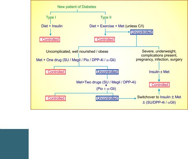
280 |
HORMONES AND RELATED DRUGS |
|
|
SECTION 5
Fig. 19.7: Simplified flow chart of management approaches in diabetes mellitus. Met—Metformin; SU—Sulfonylurea; Megli—Meglitinide/d-phenylalanine analogue; DPP-4i—Dipeptidyl peptidase-4 inhibitor; α Gli—α Glucosidase inhibitor; Pio—Pioglitazone
Note: A meglitinide drug is indicated only in patients with predominant postprandial hyperglycaemia. An α glucosidase inhibitor can be additional add on drug.
GLUCAGON
A hyperglycaemic principle was demonstrated to be present in the pancreatic islets just two years after the discovery of insulin in 1921. It was named ‘glucagon’. Glucagon is a single chain polypeptide containing 29 amino acids, MW 3500. It is secreted by the α cells of the islets of Langerhans and commercially produced now by recombinant DNA technology.
Regulation of Secretion Like insulin, glucagon is also derived by cleavage of a larger peptide prohormone. Its secretion is regulated by glucose levels, other nutrients, paracrine hormones and nervous system. Glucose has opposite effects on insulin and glucagon release, i.e. high glucose level inhibits glucagon secretion. The incretin GLP-1, FFA and ketone bodies also inhibit glucagon release. Amino acids, however, induce both insulin and glucagon secretion. Insulin, amylin and somatostatin, elaborated by the neighbouring β and δ cells, inhibit glucagon secretion. Sympathetic stimulation consistently and parasympathetic stimulation under certain conditions evokes glucagon release.
Actions Glucagon is hyperglycaemic; most of its actions are opposite to that of insulin. Glucagon causes hyperglycaemia primarily by enhancing glycogenolysis and gluconeogenesis in liver; suppression of glucose utilization in muscle and fat contributes modestly. Glucagon is considered to be the hormone of fuel mobilization. Its secretion is increased during fasting, and is largely responsible for the high fasting blood glucose levels in type 2 diabetics. It plays an essential role in the development of diabetic ketoacidosis. Increased secretion of glucagon has been shown to attend all forms of severe tissue injury.
Glucagonincreasestheforceandrateofcardiaccontraction and this is not antagonized by β blockers. It has a relaxant action on the gut and inhibits gastric acid production.
Mechanism of action Glucagon, through its own receptor and coupling Gs protein activates adenylyl cyclase and increases cAMP in liver, fat cells, heart and other tissues; most of its actions are mediated through this cyclic nucleotide.
Glucagon is inactive orally; that released from pancreas is broken down in liver, kidney, plasma and other tissues. Its t½ is 3–6 min.

|
INSULIN, ORAL HYPOGLYCAEMIC DRUGS AND GLUCAGON |
281 |
|
|
|
Uses |
Other hyperglycaemics |
|
1.Hypoglycaemia due to insulin or oral hypoglycaemics; use of glucagon is; only an expedient measure for the emergency, and must be followed by oral glucose/sugar given repeatedly till the blood glucose level stabilizes. It may not work if hepatic glycogen is already depleted.
Dose: 0.5–1.0 mg i.v. or i.m. GLUCAGON 1 mg inj.
2.Cardiogenic shock to stimulate the heart in β adrenergic blocker treated patients. However, action is not very marked.
3.To facilitate radiographic examination of upper/lower g.i. tract by relaxing stomach and intestines.
Diazoxide Chemically related to thiazides, it inhibits insulin release from β cells and causes hyperglycaemia lasting 4–8 hours. Its action on ATP sensitive K+ channels of β cells is opposite to that of SUs. Other actions which may contribute to hyperglycaemia are decreased peripheral utilization of glucose and release of catecholamines. It has been used to prevent hypoglycaemia in insulinomas. Other actions are vasodilatation, fall in BP and antidiuresis.
Somatostatin It causes hyperglycaemia primarily by inhibiting insulin release.
Streptozocin It is obtained from Streptomyces achromogenes. Causes selective damage to insulin secreting β cells. It has been used to produce experimental diabetes in animals and to treat insulin secreting tumours of pancreas.
)PROBLEM DIRECTED STUDY
19.1During routine medical checkup a 50-year male office executive with sedentary lifestyle was diagnosed to have developed type 2 diabetes mellitus. His fasting and post-meal blood glucose
was 130 mg/dl and 190 mg/dl respectively, HbA1C was 7.8%, BP was 130/82 mm Hg and body mass index was 27 kg/m2. He was asymptomatic and investigations revealed no end organ damage. He was advised suitable diet, exercise and other lifestyle modifications.
(a) Should he be prescribed an antidiabetic medication as well? If so, which drug/combination of drugs should be selected, and why?
(see Appendix-1 for solution)
19 CHAPTER