

- •Preface
- •Contents
- •List of Abbreviations
- •6. Adverse Drug Effects
- •7b. Cholinergic System and Drugs
- •9. Adrenergic System and Drugs
- •11. Histamine and Antihistaminics
- •12. 5-Hydroxytryptamine, its Antagonists and Drug Therapy of Migraine
- •16. Drugs for Cough and Bronchial Asthma
- •17a. Introduction
- •17b. Anterior Pituitary Hormones
- •20. Corticosteroids
- •21. Androgens and Drugs for Erectile Dysfunction
- •24. Drugs Affecting Calcium Balance
- •25. Skeletal Muscle Relaxants
- •26. Local Anaesthetics
- •27. General Anaesthetics
- •28. Ethyl and Methyl Alcohols
- •29 Sedative-Hypnotics
- •30. Antiepileptic Drugs
- •31. Antiparkinsonian Drugs
- •32. Drugs Used in Mental Illness: Antipsychotic and Antimanic Drugs
- •38. Antiarrhythmic Drugs
- •40. Antihypertensive Drugs
- •41b. Diuretics
- •42. Antidiuretics
- •46. Drugs for Peptic Ulcer and Gastroesophageal Reflux Disease
- •48. Drugs for Constipation and Diarrhoea
- •51. Beta-Lactam Antibiotics
- •53. Aminoglycoside Antibiotics
- •55. Antitubercular Drugs
- •56. Antileprotic Drugs
- •57. Antifungal Drugs
- •58. Antiviral Drugs
- •59. Antimalarial Drugs
- •61. Anthelmintic Drugs
- •62. Anticancer Drugs
- •63. Immunosuppressant Drugs
- •64. Drugs Acting on Skin and Mucous Membranes
- •66. Chelating Agents
- •67. Vitamins
- •68. Vaccines and Sera
- •69. Drug Interactions
- •Appendices
- •Selected References for Further Reading
- •Index

Chapter 38 Antiarrhythmic Drugs
These are drugs used to prevent or treat irregularities of cardiac rhythm.
Nearly 3 out of 4 patients of acute myocardial infarction (MI) and about half of those given a general anaesthetic exhibit at least some irregularity of cardiac rhythm. Arrhythmias are the most important cause of sudden cardiac death. However, only few arrhythmias need to be treated with antiarrhythmic drugs.
Abnormal automaticity or impaired conduction or both underlie cardiac arrhythmias. The generation and propagation of cardiac impulse and properties of excitability and refractoriness are described on p. 492 to 494. Ischaemia, electrolyte and pH imbalance, mechanical injury, stretching (due to heart failure), neurogenic and drug influences, including antiarrhythmic drugs themselves, can cause arrhythmias by altering electrophysiological properties of cardiac fibres.
Important mechanisms of cardiac arrhythmias are:
A. Enhanced/ectopic pacemaker activity The slope of phase-4 depolarization may be increased pathologically in the automatic fibres or such activity may appear in ordinary
fibres. Ectopic impulse may also result from current of injury. Myocardial cells damaged by ischaemia become partially depolarized: a current may flow between these and normally polarized fibres (injury current) and initiate an impulse.
B. After-depolarizations These are secondary depolarizations accompanying a normal or premature action potential (AP), Fig. 38.1.
Early after-depolarization (EAD) Repolarization during phase-3 is interrupted and membrane potential oscillates. If the amplitude of oscillations is sufficiently large, neighbouring tissue is activated and a series of impulses are propagated. EADs are frequently associated with long Q-T interval due to slow repolarization and markedly prolonged APs. They result from depression of delayed rectifier K+ current.
Delayed after-depolarization (DAD) After attaining resting membrane potential (RMP) a secondary deflection occurs which may reach threshold potential and initiate a single premature AP. This generally results from Ca2+ overload (digitalis toxicity, ischaemia-reperfusion).
Because an AP is needed to trigger after-depolarizations, arrhythmias based on these have been called triggered arrhythmias.
C. Reentry Due primarily to abnormality of conduction, an impulse may recirculate in the heart and cause repetitive activation without the need for any new impulse to be generated. These are called reentrant arrhythmias.
Fig. 38.1: Action potential in a nonautomatic ventricular fibre (in red) followed by early or delayed after-depolarizations.

ANTIARRHYTHMIC DRUGS |
527 |
|
|
(i) Circus movement reentry It occurs in an anatomically defined circuit. A premature impulse, temporarily blocked in one direction by refractory tissue, makes a one-way transit around an obstacle (natural orifices in the heart, A-V nodal region) or through an abnormal tract, finds the original spot in an advanced state of recovery and reexcites it, setting up recurrent activation of adjacent myocardium (Fig. 38.2). This type of reentry is often responsible for PSVT, atrial flutter and atrioventricular reciprocal rhythm in WPW.
Reentry occurring in an anatomically fixed circuit can be permanently cured by radiofrequency catheter ablation
of |
the defined pathway. |
(ii) |
Functional reentry In this type of reentry there is no |
fixed ‘obstacle’ or ‘pathway’. Rather, a functional obstacle (core of the circuit) and unidirectional conduction pathway is created by a premature impulse which travels through electrophysiologically inhomogeneous myocardium. On encountering refractory tissue in one direction, the wavefront travels through partially recovered fibres—gets markedly slowed and can set up small reentry circuits which may constantly shift location. Functional reentry may be responsible for ventricular extrasystoles, polymorphic ventricular tachycardia, atrial/ventricular fibrillation.
Fig. 38.2: Diagrammatic depiction of circus movement reentry in atrium
For reentry to occur, the path length of the circuit should be greater than the wave length (ERP × conduction velocity) of the impulse. Slow conduction in the reentrant circuit may be caused by:
(a)Partial depolarization of the membrane—decreased slope of phase 0 depolarization, i.e. depressed fast channel response.
(b)Cells changing over from fast channel to slow channel depolarization which conducts extremely slowly. When a fibre is depolarized to a RMP of about –60 mv, the Na+ (fast) channels are inactivated, but the fibre can still develop Ca2+ (slow) channel response.
Reentry can be abolished both by marked slowing of conduction (converting unidirectional block to bidirectional block) as well as by acceleration of impulse (retrograde impulse reaches so early as to meet refractory tissue).
(iii) Fractionation of impulse When atrial ERP is brief and inhomogeneous(undervagaloveractivity),animpulsegenerated early in diastole gets conducted irregularly over the atrium, i.e. it moves rapidly through fibres with short ERP (which have completely recovered) slowly through fibres with longer ERP (partially recovered) and not at all through those still refractory.Thus, asynchronous activation of atrial fibres occurs → atrial fibrillation (AF). This arrhythmia must be initiated by a premature depolarization, but is self sustaining, because passageofanirregularimpulseleavesamoreirregularrefractory trace and perpetuates the inhomogeneity of ERPs.
The important cardiac arrhythmias are:
1.Extrasystoles (ES) are premature ectopic beats due to abnormal automaticity or afterdepolarization arising from an ectopic focus in the atrium (AES), A-V node (nodal ES) or ventricle (VES). The QRS complex in VES is broader and abnormal in shape.
2.Paroxysmal supraventricular tachycardia (PSVT) is sudden onset episodes of atrial tachycardia (rate 150–200/min) with 1:1 atrioventricular conduction: mostly due to circus movement type of reentry occurring within or around the A-V node or using an accessory pathway between atria and ventricle (Wolff-Parkinson-White syndrome or WPW).
3.Atrial flutter (AFI) Atria beat at a rate of 200– 350/min and there is a physiological 2:1 to 4:1 or higher A-V block (because A-V node cannot transmit impulses faster than 200/min). This is mostly due to a stable re-entrant circuit in the right atrium, but some cases may be due to rapid discharge of an atrial focus.
4.Atrial fibrillation (AF) Atrial fibres are activated asynchronously at a rate of 350–550/min (due to electrophysiological inhomogeneity of atrial fibres), associated with grossly irregular and often fast (100–160/min) ventricular response. Atria remain dilated and quiver like a bag of worms.
5.Ventricular tachycardia (VT) is a run of 4 or more consecutive ventricular extrasystoles. It may be a sustained or nonsustained arrhythmia, and is due either to discharges from an ectopic focus, after-depolarizations or single site (monomorphic) or multiple site (polymorphic) reentry circuits.
38 CHAPTER

528
SECTION 8
CARDIOVASCULAR DRUGS
6.Torsades de pointes (French: twisting of points) is a life-threatening form of polymorphic ventricular tachycardia with rapid asynchronous complexes and an undulating baseline on ECG. It is generally associated with long Q-T interval.
7.Ventricular fibrillation (VF) is grossly irregular, rapid and fractionated activation of
ventricles resulting in incoordinated contraction of its fibres with loss of pumping function. It is fatal unless reverted within 2–5 min; is the most common cause of sudden cardiac death.
8.Atrio-ventricular (A-V) block is due to depression of impulse conduction through the A-V node and bundle of His, mostly due to vagal influence or ischaemia.
First degree A-V block: Slowed conduction resulting in prolonged P-R interval.
Second degree A-V block: Some supraventricular complexes are not conducted: drop beats.
Third degree A-V block: No supraventricular complexes are conducted; ventricle generates its own impulse; complete heart block.
Proarrhythmic potential of antiarrhythmic drugs
Most antiarrhythmics can themselves be the cause of serious arrhythmias, especially during long-term prophylactic use. Two multicentric trials ‘Cardiac Arrhythmia Suppression Trial I and II’ (CAST I, II, 1991, 1992) showed that post-MI patients randomized to receive on a long-term basis encainide, flecainide, moricizine had higher incidence of sudden death, though initially the same drugs had suppressed VES in these patients. It is possible that during transient episodes of ischaemia, the intraventricular conduction slowing action of these drugs gets markedly accentuated resulting in VT and VF. Similar increased mortality has been reported by the “Mortality in the survival with D-sotalol (SWORD) trial. It is therefore not prudent to try and suppress all extrasystoles/ arrhythmias, especially those not causing symptoms, with chronic prophylactic antiarrhythmic therapy. Only the β blockers and amiodarone have been found to decrease cardiac mortality in the long term.
CLASSIFICATION
Antiarrhythmic drugs act by blocking myocardial Na+, K+ or Ca2+ channels. Some have additional or even primary autonomic effects. Classification
Drugs that prolong Q-T interval
(have potential to precipitate Torsades de pointes)
1. Antiarrhythmics : Quinidine, procainamide, disopyramide, propafenone, amiodarone
2. Antimalarials : Quinine, mefloquine, artemisinin, halofantrine
3.Antibacterials : Sparfloxacin, moxifloxacin
4.Antihistaminics : Terfenadine, astemizole, ebastine
5.Antidepressants: Amitryptyline and other tricyclics
6.Antipsychotics : Thioridazine, pimozide,
aripiprazole, ziprasidone
7. Prokinetic |
: Cisapride |
of antiarrhythmic drugs has been difficult, because many drugs have more than one action. Vaughan Williams and Singh (1969) proposed a 4 class system which takes into account the primary electrophysiological action of a drug that may serve to indicate the type of clinical effects and therapeutic utility. However, different drugs within a class have their own specific set of properties.
A simplified clinical classification of antiarrhythmic drugs is given at the end of the chapter.
CLASS I
The primary action of drugs in this class is to limit the conductance of Na+ (and K+) across cell membrane—a local anaesthetic action. They also reduce rate of phase-4 depolarization in automatic cells.
SUBCLASS IA
The subclass IA containing the oldest antiarrhythmic drugs quinidine and procainamide are open state Na+ channel blockers which also moderately delay channel recovery (channel
recovery time τrecovery 1–10s), suppress A-V conduction and prolong refractoriness. The Na+
channel blockade is greater at higher frequency (premature depolarization is affected more). These actions serve to extinguish ectopic pacemakers that are often responsible for triggered arrhythmias and abolish reentry by converting unidirectional block into bidirectional block.

ANTIARRHYTHMIC DRUGS |
529 |
|
|
Antiarrhythmic drugs
Class |
|
Actions |
|
|
|
|
Drugs |
|
|
|
|
|
|
|
|
|
|
I. |
Membrane stabilizing |
agents |
|
|
|
|
|
|
|
(Na+ channel blockers) |
|
|
|
|
|
||
|
A. |
Moderately decrease dv/dt |
of |
0 |
phase |
Quinidine, |
Procainamide, Disopyramide |
|
|
B. |
Little decrease in |
dv/dt of |
0 |
phase |
Lidocaine, |
Mexiletine |
|
|
C. |
Marked decrease |
in dv/dt |
of |
0 |
phase |
Propafenone, Flecainide |
|
II. |
Antiadrenergic agents (β blockers) |
|
Propranolol, Esmolol, Sotalol (also class III) |
|||||
III. |
Agents widening AP |
|
|
|
|
Amiodarone, Dronedarone |
||
|
(prolong repolarization and ERP) |
|
|
Dofetilide, |
Ibutilide |
|||
IV. |
Calcium channel blockers |
|
|
|
Verapamil, |
Diltiazem |
||
Note: Class IA agents also have Class III property; Propranolol has Class I action as well; sotalol has both Class II and Class III actions.
In addition |
|
|
|
|
1. |
For PSVT |
: |
Adenosine, Digoxin |
|
2. |
For A-V |
block |
: |
Sympathomimetics—Isoprenaline, etc. |
|
|
|
|
Anticholinergics—Atropine. |
3. |
Digitalis |
is used in |
AF, |
AFl and PSVT to control ventricular rate. |
Quinidine
It is the dextro isomer of the antimalarial alkaloid quinine found in cinchona bark. In addition to Na+ channel blockade, quinidine has cardiac antivagal action which augments prolongation of atrial ERP and minimizes RP disparity of atrial fibres. A-V node ERP is increased by direct action of quinidine, but decreased by its antivagal action; overall effect is inconsistent. Quinidine depresses myocardial contractility; failure may be precipitated in damaged hearts.
ECG: Quinidine increases P-R and Q-T intervals and tends to broaden QRS complex. Changes in the shape of T wave may be seen reflecting effect on repolarization.
Mechanism of action: Quinidine blocks myocardial Na+ channels in the open state—reduces automaticity and maximal rate of 0 phase depolarization in a frequency dependent manner. Prolongation of APD is due to K+ channel block, while lengthening of ERP is caused by its moderate effect on recovery of Na+ and K+ channels. At high concentrations it also inhibits L type Ca2+ channels. Quinidine decreases the availability of Na+ channels as well as delays their reactivation.
The other actions of quinidine are fall in BP (due to weak α adrenergic blockade and direct cardiac depression), decreased skeletal muscle contractility, augmented uterine contractions, vomiting, diarrhoea and neurological effects like ringing in ears, vertigo, deafness, visual disturbances and mental changes (Cinchonism). Like its levo isomer, it has antimalarial action, and has been used as a parenteral alternative to quinine for falciparum malaria. The important drug interactions of quinidine are:
•Rise in blood levels and toxicity of digoxin due to displacement from tissue binding and inhibition of P-
glycoprotein mediated renal and biliary clearance of digoxin.
• Marked fall in BP in patients receiving vasodilators.
•Risk of torsades de pointes is increased by hypokalaemia caused by diuretics.
•Synergistic cardiac depression with β-blockers, verapamil, K+ salts.
•Quinidine inhibits CYP2D6: prolongs t½ of propafenone and inhibits conversion of codeine to morphine.
Use: Though quinidine is effective in many atrial and ventricular arrhythmias, it is seldom used now, because of risk of adverse effects, including that of torsades de pointes, sudden cardiac arrest or VF; idiosyncratic angioedema, vascular collapse, thrombocytopenia, etc. In a dose of 100–200 mg TDS quinidine may rarely be used to maintain sinus rhythm after termination of AF or AFI.
QUINIDINE SULPHATE 200 mg tab; QUININGA 300 mg tab, 600 mg/2 ml inj; NATCARDINE 100 mg tab.
Procainamide
It is orally active amide derivative of the local anaesthetic procaine, with cardiac electrophysiological actions almost identical to those of quinidine, viz. slowing of 0 phase depolarization and impulse conduction, prolongation of APD, ERP, QRS complex and Q-T interval. Significant differences between the two are:
38 CHAPTER

530 |
CARDIOVASCULAR DRUGS |
|
|
SECTION 8
•It is less effective in suppressing ectopic automaticity.
•It causes less marked depression of contractility and A-V conduction.
•Antivagal action is absent.
•It is not an α blocker: causes less fall in BP; at high doses, fall in BP is due to ganglionic blockade.
Pharmacokinetics Oral bioavailability of procainamide is about 75%, peak plasma concentration occurs in 1 hour. It is metabolized in liver, primarily by acetylation to N-acetyl- procainamide (NAPA) which has no Na+ channel blocking property but blocks K+ channels and prolongs repolarization: APD is lengthened. There are fast and slow acetylators of procainamide (as there are for isoniazid). Plasma t½ is relatively short (3–4 hours).
Dose: For abolition of arrhythmia—0.5–1 g oral or i.m. followed by 0.25–0.5 g every 2 hours; or 500 mg i.v. loading dose (25 mg/min injection) followed by 2 mg/kg/hour. Maintenance dose—0.5 g every 4–6 hours.
PRONESTYL 250 mg tab., 1 g/10 ml inj.
Adverse effects Gastrointestinal tolerance of procainamide is better than quinidine, but nausea and vomiting do occur. CNS: weakness, mental confusion and hallucinations are noted at higher doses.
Flushing and hypotension are seen on rapid i.v. injection. Cardiac toxicity, ability to cause torsades de pointes are similar to quinidine.
Hypersensitivity reactions are rashes, fever, angioedema. Agranulocytosis and aplastic anaemia is rare.
Long-term high dose procainamide therapy can cause systemic lupus erythematosus (SLE), especially in slow acetylators.
Use Procainamide (i.v.) is occasionally used to terminate monomorphic VT and some supraventricular arrhythmias. Many WPW reciprocal VTs respond and it has been used to prevent recurrences of VF. However, procainamide is not suitable for prolonged oral therapy because of poor efficacy and high risk of lupus.
Disopyramide
It is a quinidine like Class IA drug that has prominent cardiac depressant and anticholinergic actions, but no α adrenergic blocking property. Disopyramide usually has no effect on sinus rate because of opposing direct depressant and antivagal actions. Prolongation of P-R interval and QRS broadening are less marked.
Pharmacokinetics Bioavailability of oral disopyramide is about 80%. It is partly metabolized in liver by dealkylation, nearly half is excreted unchanged in urine; plasma t½ is
6–8 hrs. The t½ is increased in patients of MI and in renal insufficiency.
Dose: 100–150 mg 6–8 hourly oral.
NORPACE 100, 150 mg cap, REGUBEAT 100 mg tab.
Adverse effects Disopyramide causes less g.i. effects. Anticholinergic side effects are the most prominent: dry mouth, constipation, urinary retention (especially in elderly males) and blurred vision.
Depression of cardiac contractility is more prominent. Cardiac decompensation and hypotension may occur in patients with damaged hearts because it also increases peripheral resistance, so that cardiac output may be markedly decreased.
Contraindications are—sick sinus, cardiac failure and prostate hypertrophy.
Use The primary indication of disopyramide is as a second line drug for prevention of recurrences of ventricular arrhythmia. It may also be used for maintenance therapy after cardioversion of AF or AFl.
SUBCLASS IB
These drugs block Na+ channels more in the inactivated than in the open state, but do not delay channel recovery (channel recovery time < 1S). They do not depress A-V conduction or prolong (may even shorten) APD, ERP and Q-T.
Lidocaine (Lignocaine)
It is the most commonly used local anaesthetic (see Ch. 26). In addition, it is a popular antiarrhythmic in intensive care units.
The most prominent cardiac action of lidocaine is suppression of automaticity in ectopic foci. Enhanced phase-4 depolarization in partially depolarized or stretched PFs, and after-depolari- zations are antagonized, but SA node automaticity is not depressed.
The rate of 0 phase depolarization and conduction velocity in A-V bundle or ventricles is not decreased. Lidocaine decreases APD in PF and ventricular muscle, but has practically no effect on APD and ERP of atrial fibres. Atrial reentry is not affected. However, it can suppress reentrant ventricular arrhythmias either by abolishing oneway block or by producing two way block.
Lidocaine is a blocker of inactivated Na+ channels more than that of open state. As such,

ANTIARRHYTHMIC DRUGS |
531 |
|
|
it is relatively selective for partially depolarized cells and those with longer APD (whose Na+ channels remain inactivated for longer period). While normal ventricular and conducting fibres are minimally affected, depolarized/damaged fibres are significantly depressed. Brevity of atrial AP and lack of lidocaine effect on channel recovery might explain its inefficacy in atrial arrhythmias.
Lidocaine has minimal effect on normal ECG; QT interval may decrease. It causes little depression of cardiac contractility or arterial BP. There are no significant autonomic actions: all cardiac effects are direct actions.
Pharmacokinetics Lidocaine is inactive orally due to high first pass metabolism in liver. Action of an i.v. bolus lasts only 10–20 min because of rapid redistribution. It is hydrolysed, deethylated and conjugated; metabolites are excreted in urine. Metabolism of lidocaine is hepatic blood flow dependent.
The t½ of early distribution phase is 8 min while that of later elimination phase is nearly 2 hours. Its t½ is prolonged in CHF, because of decrease in volume of distribution and hepatic blood flow.
Dose and preparations Lidocaine is given only by i.v. route: 50–100 mg bolus followed by 20–40 mg every 10– 20 min or 1–3 mg/min infusion.
XYLOCARD, GESICARD 20 mg/ml inj. (5, 50 ml vials). These preparations for cardiac use contain no preservative. The local anaesthetic preparations should not be used for this purpose.
Propranolol prolongs t½ of lidocaine by reducing hepatic blood flow. Cimetidine also increases plasma levels of lidocaine.
Adverse effects The main toxicity is dose related neurological effects:
Drowsiness, nausea, paresthesias, blurred vision, disorientation, nystagmus, twitchings and fits. Lidocaine has practically no proarrhythmic potential and is the least cardiotoxic antiarrhythmic. Only excessive doses cause cardiac depression and hypotension.
Use Lidocaine is safe if given by slow i.v. injection; is used to suppress VT and prevent VF. It is ineffective in atrial arrhythmias. Because of rapidly developing and titratable action it is a good drug in the emergency setting, e.g. arrhythmias following acute MI or during cardiac surgery. In acute MI, i.v. infusion of lidocaine can prevent VF, but a metaanalysis has shown that it fails to improve survival; may even increase short term mortality. Therefore, lidocaine is no longer administered prophylactically to all MI patients, but may be used in selected cases, and to treat ventricular arrhythmias when they occur.
Efficacy of lidocaine in chronic ventricular arrhythmia is poor, but it suppresses VT due to digitalis toxicity, for which it is used because it does not worsen A-V block.
Mexiletine
It is a local anaesthetic and an orally active antiarrhythmic; chemically and pharmacologically similar to lidocaine. Automaticity in PF is reduced both by decreasing phase-4 slope and by increasing threshold voltage. By reducing the rate of 0 phase depolarization in ischaemic PF it may convert one-way block to two-way block.
Mexiletine is almost completely absorbed orally, 90% metabolized in liver and excreted in urine; plasma t½ 9–12 hours.
Bradycardia, hypotension and accentuation of A-V block may attend i.v. injection of mexiletine.
Neurological side effects—tremor, nausea and vomiting are common; dizziness, confusion, blurred vision, ataxia can occur.
Dose: 100–250 mg i.v. over 10 min., 1 mg/min infusion. Oral: 150–200 mg TDS with meals.
MEXITIL 50, 150 mg caps, 250 mg/10 ml. inj.
Use Parenteral mexiletine may be used in post-infarction sinister ventricular arrhythmias as alternative to lidocaine. Oral use to chronically suppress VES/VT is of questionable merit.
SUBCLASS IC
These are the most potent Na+ channel blockers with more prominent action on open state and the longest recovery times (> 10S). They markedly delay conduction, prolong P-R interval, broaden QRS complex, but have variable effect on APD. Drugs of this subclass have high
38 CHAPTER

532
SECTION 8
CARDIOVASCULAR DRUGS
proarrhythmic potential when administered chronically; sudden deaths have occurred.
Propafenone By blocking Na+ channels propafenone considerably depresses impulse transmission and has profound effect on HisPurkinje as well as accessory pathway conduction. Anterograde as well as retrograde conduction in the bypass tract of WPW syndrome is retarded. Propafenone prolongs APD and has β adrenergic blocking property—can precipitate CHF and bronchospasm. Sino-atrial block has occurred occasionally.
Propafenone is absorbed orally and undergoes variable first pass metabolism; there being extensive or poor metabolizers because the major metabolic isoenzyme CYP2D6 is deficient in poor metabolizers. CYP2D6 inhibitors like fluoxetine increase its bioavailability and plasma concentration. Bioavailability and t½ differs considerably among individuals. Some metabolites are active. Side effects are nausea, vomiting, bitter taste, constipation and blurred vision. As mentioned above, it can worsen CHF, asthma and increase the risk of sudden death.
Propafenone is used for prophylaxis and treatment of ventricular arrhythmias, reentrant tachycardias involving AV node/accessory pathway and to maintain sinus rhythm in AF. However, it can occasionally increase ventricular rate in AFl by slowing atrial rate and allowing 1:1 A-V transmission. Some reentrant VTs may also be worsened.
Dose: 150 mg BD–300 mg TDS; RHYTHMONORM 150 mg tab.
Flecainide It is the prototype class IC antiarrhythmic which markedly delays Na+ channel recovery. In contrast to propafenone, flecainide has no consistent effect on APD and no β adrenergic blocking property. It suppresses VES, VT, WPW tachycardia and prevents recurrences of AF and PSVT. But in the CAST study it was found to increase mortality in patients recovering from MI; can itself provoke arrhythmias during chronic therapy. It is reserved for resistant cases of paroxysmal AF and for life-threatening sustained VT in patients not having associated CHF.
CLASS II
The primary action of class II drugs is to suppress adrenergically mediated ectopic activity.
Propranolol (see Ch. 10) It is the most commonly selected β blocker for treatment and prevention of cardiac arrhythmias; has some quinidine like direct membrane stabilizing action at high doses. However, in the clinically used dose range—antiarrhythmic action is exerted primarily because of cardiac adrenergic blockade. In a normal resting individual propranolol has only mild depressant action on SA node automaticity, but marked decrease in the slope of phase-4 depolarization and automaticity occurs in SA node, PF and other ectopic foci when the same has been increased under adrenergic influence. The other most important action is to prolong the ERP of A-V node (an antiadrenergic action). This impedes A-V conduction so that no paradoxical tachycardia can occur when atrial rate is reduced in AF or AFl.
Slow channel responses and after-depolari- zations that have been induced by catecholamines (CAs) are suppressed. Reentrant arrhythmias that involve A-V node (many PSVTs) or that are dependent on slow channel/depressed fast channel responsemaybeabolishedbyitsmarkeddepressant action on these modalities.
The most prominent ECG change is prolongation of PR interval. Depression of cardiac contractility and BP are mild.
Administration For rapid action, propranolol may be injected i.v. 1 mg/min (max. 5 mg) under close monitoring. The usual oral antiarrhythmic dose is 40–80 mg 2–4 times a day.
Use Propranolol is very useful in treating inappropriate sinus tachycardia. Atrial and nodal ESs, especially those provoked by emotion or exercise are suppressed by propranolol, but need to be treated only when symptomatic and disturbing. Propranolol is less effective than adenosine and verapamil for termination of PSVT (success rate ~ 60%), but can be used to prevent recurrences.
Propranolol rarely abolishes AF or AFl, but is used to control ventricular rate. It is highly effective in sympathetically mediated arrhythmias seen in pheochromocytoma and during anaesthesia

ANTIARRHYTHMIC DRUGS |
533 |
|
|
with halothane. Digitalis induced tachyarrhythmias may be suppressed.
Non-sustained VT may be treated with a β blocker (propranolol, esmolol), but its efficacy in terminating sustained VT is low. However, propranolol may prevent recurrences of VT and its antiischaemic action may be protective. Prophylactic treatment with β blockers reduces mortality in post-MI patients. Propranolol or esmolol injected i.v. may terminate torsades de pointes. Along with a class IA or IC drug, it may be used for WPW reciprocal rhythms.
Sotalol (see p. 148) It is a nonselective β blocker having prominent Class III action of prolonging repolarization by blocking cardiac inward rectifier K+ channels. It is not a Na+ channel blocker— does not depress conduction in fast response tissue, but delays A-V conduction and prolongs its ERP. Sotalol is effective in polymorphic VT, some WPW arrhythmias and for maintaining sinus rhythm in AF/AFl. Due to prolongation of APD and Q-T, risk of dose-dependent torsades de pointes is the major limitation. It is contraindicated in patients with long Q-T interval.
Esmolol (see p. 149) This quick and short acting β1 blocker administered i.v. is very useful for emergency control of ventricular rate in AF/AFl. It can terminate supraventricular tachycardia, and is mainly used for arrhythmias associated with anaesthesia where rapidly developing β adrenergic blockade is desired.
MINIBLOCK 100 mg/10 ml, 250 mg/10 ml inj.; 0.5 mg/kg in 1 min followed by 0.05–0.2 mg/kg/min i.v. infusion.
CLASS III
The characteristic action of this class is prolongation of repolarization (phase-3); AP is widened and ERP is increased. The tissue remains refractory even after full repolarization: reentrant arrhythmias are terminated.
Amiodarone
This unusual iodine containing highly lipophilic long-acting antiarrhythmic drug exerts multiple actions:
•Prolongs APD and Q-T interval attributable to block of myocardial delayed rectifier K+ channels. This also appears to reduce nonuniformity of refractoriness among different fibres.
•Preferentially blocks inactivated Na+ channels (like lidocaine) with relatively rapid rate of channel recovery: more effective in depressing conduction in cells that are partially depolarized or have longer APD.
•Partially inhibits myocardial Ca2+ channels, has noncompetitive β adrenergic blocking property and alters thyroid function. Thus amiodarone
is a multichannel blocker with some additional activities.
Conduction is slowed and ectopic automaticity is markedly depressed, but that of SA node is only slightly affected. Effect of oral doses on cardiac contractility and BP are minimal, but i.v. injection frequently causes myocardial depression and hypotension.
Despite prolongation of APD, the arrhythmia (torsades de pointes) provoking potential of amiodarone is low, probably because it does not exhibit ‘reverse use-dependence’ of APD prolongation or because of its multiple antiarrhythmic mechanisms. The prolongation of APD by other class III drugs is more marked at slower rates of activation (encouraging EAD) than at higher rates (reverse usedependence), while with amiodarone it is independent of rate of activation.
Pharmacokinetics Amiodarone is incompletely and slowly absorbed from the g.i.t. On daily oral ingestion the action develops over several days, even weeks. However, on i.v. injection, action develops rapidly. It accumulates in muscle and fat from which it is slowly released and then metabolized in liver mainly by CYP3A4. One metabolite is active. The duration of action is exceptionally long; t½ 3–8 weeks.
Dose: Amiodarone is mainly used orally 400–600 mg/day for few weeks, followed by 100–200 mg OD for maintenance therapy. 100–300 mg (5 mg/kg) slow i.v. injection over 30–60 min.
CORDARONE, ALDARONE, EURYTHMIC 100, 200 mg tabs, 150 mg/3 ml inj.
Use Amiodarone is effective in a wide range of ventricular and supraventricular arrhythmias including PSVT, nodal and ventricular tachycardia,
38 CHAPTER
534
SECTION 8
CARDIOVASCULAR DRUGS
AF, AFl, etc. Resistant VT and recurrent VF are the most important indications. It is also used to maintain sinus rhythm in AF when other drugs have failed. Rapid termination of ventricular (VT and VF) and supraventricular arrhythmias can be obtained by i.v. injection. WPW tachyarrhythmia is terminated by suppression of both normal and aberrant pathways.
Long duration of action makes amiodarone suitable for chronic prophylactic therapy. Apart from propranolol, it is the only antiarrhythmic drug which in the long term has been found to reduce sudden cardiac death. Because of high and broad spectrum efficacy and relatively low proarrhythmic potential, amiodarone is a commonly used antiarrhythmic, despite its organ toxicity.
Adverse effects These are dose-related and increase with duration of therapy. Fall in BP, bradycardia and myocardial depression occurs on i.v. injection and after drug cumulation.
Nausea, gastrointestinal upset may attend oral medication, especially during the loading phase. Photosensitization and sun burn like skin pigmentation occurs in about 10% patients. Corneal microdeposits are common with longterm use, may cause headlight dazzle, but are reversible on discontinuation.
Pulmonary alveolitis and fibrosis is the most serious toxicity of prolonged use, but is rare if daily dose is kept below 200 mg.
Peripheral neuropathy generally manifests as weakness of shoulder and pelvic muscles. Liver damage is rare. Amiodarone interferes with thyroid function in many ways including inhibition of peripheral conversion of T4 to T3 and interaction with thyroid hormone receptor. Goiter, hypothyroidism and rarely hyperthyroidism may develop on chronic use.
Interactions Amiodarone can increase digoxin and warfarin levels by reducing their renal clearance. Additive A-V block can occur in patients receiving β blockers or calcium channel blockers. Inducers and inhibitors of CYP3A4 respectively decrease and increase amiodarone levels.
Dronedarone This is a recently introduced noniodinated congener of amiodarone, less toxic, but also less effective class III antiarrhythmic. Clinical utility of dronedarone is limited to supraventricular arrhythmias; primary indication being maintenance of sinus rhythm in haemodynamically stable patients of paroxysmal/nonpermanent AF, and to control ventricular rate during AF/AFl.
In clinical trials, recurrence time for AF was increased by 2–3 times in dronedarone recipients. Like amiodarone, dronedarone is a multichannel blocker, inhibits delayed rectifier and other types of cardiac K+ channels, inward Na+ channel and L-type Ca2+ channel. The noncompetitive β adrenergic blocking activity is more marked compared to amiodarone, but it does not interfere with thyroid function. It increases myocardial APD, ERP and slows A-V conduction.
Dronedarone is less lipophilic and is metabolized by CYP3A4 and CYP2D6; inhibitors of these isoenzymes (Ketoconazole, erythromycin, metoprolol, etc.) markedly increase its blood levels. The elimination t½ is 24 hours. Side effects are mainly gastrointestinal disturbances, bradycardia, weakness, cough and dermatological reactions. Though it prolongs Q-T interval, risk of torsades de pointes is very low. Hypothyroidism, pulmonary fibrosis and peripheral neuropathy does not occur. Dronedarone is contraindicated in moderate-to-severe CHF, 2nd/3rd degree A-V block and in permanent AF.
Dose: 400 mg BD oral; MULTAQ 400 mg tab.
Note: On the basis of two clinical trials PALLAS and ATHENA, the US-FDA in Dec 2011 issued a safety alert that dronedarone should not be used in AF patients who cannot and will not be converted to sinus rhythm (permanent AF), because it doubles the rate of stroke, heart failure and cardiovascular death in such patients. If during dronedarone therapy the patient is found to have AF, he should either be cardioverted or dronedarone should be stopped. A recent meta-analysis has also noted unfavourable cardiovascular and mortality outcomes with dronedarone, especially in patients with cardiovascular risk factors.
Dofetilide This newer antiarrhythmic prolongs APD and ERP by selectively blocking rapid component of delayed rectifier K+ current without affecting other channels or receptors; has no autonomic or peripheral actions. It is therefore labelled as pure class III antiarrhythmic.

ANTIARRHYTHMIC DRUGS |
535 |
|
|
Oral dofetilide can convert AF or AFl to sinus rhythm in ~30% cases, but is more effective in maintaining sinus rhythm in converted patients—its primary indication. Significantly, chronic therapy with dofetilide in patients with high risk of sudden cardiac death/post MI cases has not increased mortality, despite provoking torsades de pointes in some recipients. It is mainly excreted unchanged in urine and produces few side effects.
Ibutilide is another new class III antiarrhythmic used i.v. for pharmacological conversion of AFl and AF to sinus rhythm. Efficacy is higher in recent onset AF/AFl and in AFl compared to AF. Induction of Torsades de pointes is a risk.
CLASS IV
The primary action of this class of drugs is to inhibit Ca2+ mediated slow channel inward current.
Verapamil
Of the many Ca2+ channel blockers, verapamil has the most prominent cardiac electrophysiological action (Table 38.1). It blocks L type Ca2+ channels and delays their recovery. Its antiarrhythmic aspects are described here, while other aspects are covered in Ch. 39 and 40.
The basic action of verapamil is to depress Ca2+ mediated depolarization. This suppresses automaticity and reentry dependent on slow channel response. Phase-4 depolarization in SA node is reduced resulting in bradycardia. Reflex sympathetic stimulation due to vasodilatation partly counteracts the direct bradycardia producing action. Delayed after-depolarizations in PFs are dampened.
The most consistent action of verapamil is prolongation of A-V nodal ERP. As a result
A-V conduction is markedly slowed (P-R interval increases) and reentry involving A-V node is terminated. Intraventricular conduction, however, is not affected. Verapamil has negative inotropic action due to interference with Ca2+ mediated excitation-contraction coupling in myocardium.
Uses and precautions
1. PSVT—Verapamil can terminate attacks of PSVT; 5 mg i.v. over 2–3 min is effective in ~ 80% cases. However, i.v. verapamil carries the risk of marked bradycardia, A-V block, cardiac arrest and hypotension. It should not be used if PSVT is accompanied with hypotension or CHF. For preventing recurrences of PSVT, verapamil 60 to 120 mg TDS may be given orally.
2. To control ventricular rate in AF or AFl; Verapamil causes a dose dependent (40–120 mg TDS oral) reduction in ventricular rate in AF and AFl, and is a first line drug for this purpose. In case of inadequate response, digoxin may be added to it. Verapamil can also be injected i.v. for emergency control of ventricular rate in AF and AFl.
Reentrant supraventricular and nodal arrhythmias are susceptible to verapamil, but it is contraindicated in broad QRS complex WPW tachycardia in which it may abbreviate the ERP of bypass tract. A class IA (procainamide) or IC (propafenone) drug which prolongs ERP of bypass tract and depresses conduction is to be combined with verapamil so as to concurrently depress A-V conduction.
Verapamil has poor efficacy in ventricular arrhythmias. In contrast to β blockers, verapamil
TABLE 38.1  Electrophysiological actions of calcium channel blockers
|
|
|
Verapamil |
Diltiazem |
Nifedipine |
|
|
|
|
|
|
1. |
SA node automaticity |
↓ |
↓,– |
— |
|
2. |
Ventricular automaticity |
↓,– |
— |
— |
|
3. |
ERP |
: atrial |
— |
— |
— |
|
|
: A-V nodal |
↑↑ |
↑ |
↑↓ |
|
|
: ventricular |
— |
— |
— |
|
|
: bypass tract |
↑↓ |
↑↓ |
— |
4. |
ECG |
: R-R interval |
↑ |
↑↓ |
↓ |
|
|
P-R interval |
↑ |
↑ |
— |
38 CHAPTER
↑ : increase; ↓ : decrease; —: no change

536 |
CARDIOVASCULAR DRUGS |
|
|
SECTION 8
prophylaxis does not reduce mortality in postMI patients. In some patients of VT, i.v. injection of verapamil has precipitated VF: therefore contraindicated. It is also not recommended for digitalis toxicity, because additive A-V block may occur. It is contraindicated in partial heart block and sick sinus.
CALAPTIN 40, 80 mg tab; 120, 240 mg SR tab, 5 mg/2 ml inj.
Diltiazem The direct cardiac actions of diltiazem are similar to those of verapamil. However, bradycardia and depression of cardiac contractility are less marked. It is an alternative to verapamil for termination as well as prophylaxis of PSVT.
For rapid control of ventricular rate in AF or AFl, i.v. diltiazem is preferred over verapamil, because it can be more easily titrated to the target heart rate, causes less hypotension or myocardial depression and can be used even in the presence of mild-to-moderate CHF.
DILZEM 30, 60, 90 mg tabs, 25 mg/5 ml inj.
Drugs for PSVT
An attack of PSVT can be terminated by reflex vagal stimulation through Valsalva maneuver, splashing ice cold water on face, hyperflexion (head between knees), etc. Alternatively, or if it does not work, the drug of choice is adenosine (i.v.). Other alternatives are i.v. injection of verapamil/diltiazem/esmolol. To prevent recurrences, oral therapy with verapamil, diltiazem or propranolol alone or combined with digoxin may be prescribed.
Adenosine
Administered by rapid i.v. injection (over 1–3 sec) either as the free base (6–12 mg) or as ATP (10–20 mg), adenosine terminates within 30 sec. more than 90% episodes of PSVT involving the A-V node. It activates ACh sensitive K+ channels and causes membrane hyperpolarization through interaction with A1 type of adenosine GPCRs on SA node (pacemaker depression → bradycardia), A-V node (prolongation of ERP → slowing of conduction) and atrium (shortening of AP, reduced excita-bility). It indirectly reduces
Ca2+ current in A-V node. Depression of the reentrant circuit through A-V node is responsible for termination of majority of PSVTs. Adrenergically induced DADs in ventricle are also suppressed. Coronary dilatation occurs transiently.
ADENOJECT, ADENOCOR 3 mg adenosine (base) per ml in 2 ml and 10 ml amp.
Adenosine has a very short t½ in blood (~10 sec) due to uptake into RBCs and endothelial cells where it is converted to 5-AMP and inosine. Almost complete elimination occurs in a single passage through coronary circulation. Injected ATP is rapidly converted to adenosine. Dipyridamole potentiates its action by inhibiting uptake, while theophylline/ caffeine antagonize its action by blocking adenosine receptors. Higher doses may be required in heavy tea/coffee drinkers. Patients on carbamazepine are at greater risk of developing heart block. Advantages of adenosine for termination of PSVT are:
•Efficacy equivalent to or better than verapamil.
•Action lasts < 1 min; adverse effects (even
cardiac arrest, if it occurs) are transient.
•No haemodynamic deterioration; can be given to patients with hypotension, CHF or those receiving β blockers. Verapamil is contraindicated in these situations.
•Safe in wide QRS tachycardia (verapamil is unsafe).
•Effective in patients not responding to
verapamil.
However, adenosine produces transient dyspnoea, chest pain, fall in BP and flushing in 30–60% patients; ventricular standstill for few sec or VF occurs in some patients. Bronchospasm may be precipitated in asthmatics; verapamil is the drug of choice for such patients. Adenosine has to be rapidly injected in a large vein and has brief action. Therefore, it is not suitable for prophylaxis in recurrent cases.
Other uses of adenosine
(a)Diagnosis of tachycardias dependent on A-V node.
(b)To induce brief coronary vasodilatation during certain diagnostic/interventional procedures.
(c)To produce controlled hypotension during surgery.

ANTIARRHYTHMIC DRUGS |
537 |
|
|
Drugs for A-V Block
The definitive treatment of chronic heart block is pacing with an implanted cardiac pacemaker. Drugs are of value only for acute/transient A-V block and as an interim measure.
Atropine: When A-V block is due to vagal overactivity, e.g. digitalis toxicity, some cases of MI; it can be improved by atropine 0.6–1.2 mg i.m. Atropine abbreviates A-V node ERP and increases conduction velocity in bundle of His.
Sympathomimetics (Adr, isoprenaline): These drugs may overcome partial heart block by facilitating A-V conduction and shortening ERP of conducting tissues.
They may also be used in complete (3rd degree) heart block to maintain a sufficient idioventricular rate (by increasing automaticity of ventricular pacemakers) till external pacemaker can be implanted.
Choice and use of antiarrhythmic drugs
Mere detection of an arrhythmia does not necessitate treatment.
Asymptomatic arrhythmias and those which do not jeopardize haemodynamics, e.g. most AES and occasional VES, first degree A-V block, bundle branch block, etc. in an otherwise normal heart, do not require antiarrhythmic treatment; reassurance is enough. If a patient is particularly disturbed by AES, propranolol is the best option. Chronic prophylactic therapy with class I and class IV antiarrhythmics does not appear to afford survival benefit, except in few selected cases. Only
propranolol and to some extent amiodarone have been shown to reduce cardiovascular mortality in the long-term. On the other hand, vigorous therapy is indicated when:
•Arrhythmia is life-threatening, e.g. sustained VT, torsades de pointes, VF.
•Arrhythmia is causing hypotension, breathlessness, activity limitation or cardiac failure.
•Palpitation is marked, e.g. in PSVT, sustained VT, AF, torsades de pointes.
•When simple arrhythmia may lead to more serious ones, e.g. after MI (warning arrhythmias).
In the above situations antiarrhythmics are mostly needed for short periods. Majority of antiarrhythmic drugs have narrow margin of safety. A simple clinical classification of antiarrhythmic drugs is presented in the box below.
The selection of an antiarrhythmic in a patient depends on:
(a)ECG diagnosis
(b)Possible mechanism underlying the arrhythmia
(c)Mechanism of action and range of antiarrhythmic activity of the drug
(d)Pharmacokinetic profile of the drug.
(e)Haemodynamic effects of the drug.
The aim is to improve cardiovascular function either by restoring sinus rhythm, or by controlling ventricular rate, or by conversion to a more desirable pattern of electrical and mechanical activity.
Despite extensive investigation, choice of an antiarrhythmic is still largely empirical. A practical guide to the choice and use of antiarrhythmic drugs is summarized in the box on next page.
38 CHAPTER
Clinical classification of antiarrhythmic drugs
Supraventricular |
Supraventricular |
and |
Ventricular arrhythmias |
arrhythmias only |
ventricular arrhythmias |
only |
|
|
|
|
|
Adenosine |
Amiodarone |
Procainamide |
Lidocaine |
Verapamil |
β blockers |
Disopyramide |
Mexiletine |
Diltiazem |
Propranolol |
Quinidine |
|
Dronedarone |
Sotalol |
Flecainide |
|
Digoxin |
Esmolol |
Propafenone |
|
|
|
|
|

538 |
CARDIOVASCULAR DRUGS |
|
|
SECTION 8
Choice of antiarrhythmics for cardiac arrhythmias
|
Arrhythmia |
|
Clinical objective |
|
Drug(s) |
|||||
|
|
|
|
|
|
|
|
|||
1. |
Atrial |
extrasystoles |
|
|
|
|
No drug if asymptomatic or non-disturbing |
|||
|
Symptomatic |
: Suppression, symptom relief |
Propranolol |
(only if disturbing) |
||||||
|
|
|
|
|
|
|
||||
2. |
Paroxysmal |
supraventricular |
: Termination |
of |
PSVT |
i.v. adenosine/verapamil/diltiazem/esmolol |
||||
|
tachycardia |
(PSVT) |
: Prevention |
of recurrence |
Oral verapamil/diltiazem/propranolol/sotalol |
|||||
|
|
|
|
|
|
|||||
3. |
Atrial |
fibrillation (AF) |
: Reversal to |
SR |
Cardioversion |
|||||
|
|
|
|
(for paroxysmal/persistent AF) |
i.v. amiodarone |
|||||
|
|
|
|
: Maintenance of |
SR |
Sotalol/propafenone/amiodarone/ |
||||
|
|
|
|
|
|
|
|
dronedarone/disopyramide |
||
|
|
|
|
: Ventricular |
rate |
control |
oral verapamil/diltiazem/propranolol + |
|||
|
|
|
|
(for permanent |
AF/during |
digoxin |
|
|||
|
|
|
|
recurrence of AF) |
|
|
|
|||
|
|
|
|
: Urgent vent. rate control |
i.v. esmolol/verapamil/amiodarone |
|||||
|
|
|
|
|
|
|||||
4. |
Atrial |
flutter (AFl) |
: Reversal to |
SR |
Cardioversion, radiofrequency ablation, |
|||||
|
|
|
|
|
|
|
|
Propafenone (after rate control with |
||
|
|
|
|
|
|
|
|
verapamil/propranolol) |
||
|
|
|
|
: Ventricular |
rate |
control |
Propranolol/verapamil/diltiazem + digoxin |
|||
|
|
|
|
|
|
|
|
or Amiodarone |
||
|
|
|
|
|
|
|||||
5. |
Wolff-Parkinson-White |
: Termination |
|
|
Radiofrequency ablation, cardioversion |
|||||
|
syndrome (WPW) |
: Maintenance |
|
|
|
|
||||
|
tachycardia |
|
- |
narrow QRS |
|
Propafenone/procainamide |
||||
|
|
|
|
- |
wide QRS |
|
Propafenone + verapamil/propranolol |
|||
|
|
|
|
|
|
|
|
or |
Amiodarone/sotalol |
|
|
|
|
|
|
|
|
|
|
||
6. |
Acute-MI arrhythmia |
|
|
|
|
|
|
|
||
|
Bradycardia |
|
: Reversal to normal rate |
Atropine (i.v.)—no effect—Pacing |
||||||
|
Vent. extrasystoles/ |
: Abolition to |
prevent |
i.v. Lidocaine/procainamide/amiodarone |
||||||
|
tachycardia |
|
serious arrhythmia |
Cardioversion (if haemodynamically unstable) |
||||||
|
|
|
|
|
|
|
|
|
||
7. |
Chronic vent. tachycardia |
|
|
|
|
|
|
|
||
|
Nonsustained VT |
: Suppression |
|
Propranolol/amiodarone (oral) |
||||||
|
Sustained VT |
: Abolition |
|
|
i.v. Amiodarone + propranolol |
|||||
|
|
|
|
|
|
|
|
or |
cardioversion |
|
|
|
|
|
|
|
|
|
or |
propafenone/lidocaine (i.v.) |
|
|
|
|
|
: Maintenance therapy |
Amiodarone/sotalol |
|||||
|
|
|
|
(prevention |
of VF/arrest) |
Implantable |
defibrillator |
|||
|
|
|
|
|
|
|
|
|||
8. |
Ventricular |
fibrillation (VF) |
: Termination |
|
|
Defibrillation |
+ amiodarone (i.v.) |
|||
|
|
|
|
: Recurrence |
prevention |
Amiodarone |
(oral)/propranolol |
|||
SR—Sinus rhythm
)PROBLEM DIRECTED STUDY
38.1A sales executive aged 55 years presented with palpitation felt off-and-on, both during activity as well as at rest for the last one month or so. He also complained of tiredness and anxiety. The pulse was irregular in volume and frequency with average rate 104/min, respiration 20/min, BP 130/84 mm Hg, apex beat was irregular, with an average rate 120/min. Heart sounds were irregular, but there was no murmur. The ECG showed atrial fibrillation (AF) with no sign of ischaemia. A diagnosis of persistent AF was made, and it was decided to electrically cardiovert him. He was put on warfarin sod. 5 mg twice daily for 2 days followed by 5 mg once daily and dose to be adjusted to an INR between 2–2.5. This was to be maintained for 1 month before attempting cardioversion.
(a)Why the patient has been put on warfarin therapy before attempting cardioversion?
(b)Can some drug be given to control and regularize his heart rate in the mean time? If so, which drug(s)?
(c)If electrical cardioversion does not succeed, can some drug be given to revert him to sinus rhythm (SR)?
(d)After cardioversion, can some drug(s) be given to maintain SR and prevent recurrence of AF?
(see Appendix-1 for solution)

Chapter 39 Antianginal and Other
Anti-ischaemic Drugs
ANTIANGINAL DRUGS
Antianginal drugs are those that prevent, abort or terminate attacks of angina pectoris.
Angina pectoris Is a pain syndrome due to induction of an adverse oxygen supply/demand situation in a portion of the myocardium. Two principal forms are recognized:
(a) Classical angina (common form) Attacks are predictably provoked (stable angina) by exercise, emotion, eating or coitus and subside when the increased energy demand is withdrawn. The underlying pathology is—severe arteriosclerotic affliction of larger coronary arteries (conducting vessels) which run epicardially and send perforating branches to supply the deeper tissue (Fig. 39.1). The coronary obstruction is ‘fixed’; blood flow fails to increase during increased demand despite local factors mediated dilatation of resistance vessels (Fig. 39.2) and ischaemic pain is felt. Due to inadequacy of ischaemic left ventricle, the end diastolic left ventricular pressure rises from 5 to about 25 mm Hg—produces subendocardial ‘crunch’ during diastole (blood flow to the subendocardial region
occurs only during diastole) and aggravates the ischaemia in this region. Thus, a form of acutely developing and rapidly reversible left ventricular failure results which is relieved by taking rest and reducing the myocardial workload.
Drugs that are useful, primarily reduce cardiac work (directly by acting on heart or indirectly by reducing preload hence end diastolic pressure, and afterload). They may also cause favourable redistribution of blood flow to the ischaemic areas.
(b) Variant/Prinzmetal/Vasospastic angina
(uncommon form) Attacks occur at rest or during sleep and are unpredictable. They are due to
Fig. 39.1: Diagrammatic representation of subendocardial ‘crunch’ during an attack of angina
Fig. 39.2: Diagrammatic representation of coronary artery calibre changes in classical and variant angina

540
SECTION 8
CARDIOVASCULAR DRUGS
recurrent localized (occasionally diffuse) coronary vasospasm (Fig. 39.2) which may be superimposed on arteriosclerotic coronary artery disease. Abnormally reactive and hypertrophied segments in the coronary arteries have been demonstrated. Drugs are aimed at preventing and relieving the coronary vasospasm.
Unstable angina (UA) with rapid increase in duration and severity of attacks is mostly due to rupture of an atheromatous plaque attracting platelet deposition and progressive occlusion of the coronary artery; occasionally with associated coronary vasospasm.
Chronically reduced blood supply causes atrophy of cardiac muscle with fibrous replacement (reduced myocardial work capacity → CHF) and may damage conducting tissue to produce unstable cardiac rhythms. Antianginal drugs relieve cardiac ischaemia but do not alter the course of coronary artery pathology: no permanent benefit is afforded. On the other hand, aspirin, ACE inhibitors and statins (hypocholesterolaemic) can modify coronary artery disease and improve prognosis.
Glyceryl trinitrate, the drug unsurpassed in its ability to abort and terminate anginal attack, was introduced by Murrell in 1879. Other organic nitrates were added later, but a breakthrough was achieved in 1963 when propranolol was used for chronic prophylaxis. The calcium channel blockers have been a major contribution of the 1970s. A number of vasodilator and other drugs have been promoted from time to time, but none is as uniformly effective. Some potassium channel openers (nicorandil), metabolic modulator (trimetazidine) and late Na+ current inhibitor (ranolazine) have been introduced lately.
CLASSIFICATION
1.Nitrates
(a)Short acting: Glyceryl trinitrate (GTN, Nitroglycerine)
(b)Long acting: Isosorbide dinitrate (short acting by sublingual route), Isosorbide mononitrate, Erythrityl tetranitrate, Pentaerythritol tetranitrate
2.β Blockers Propranolol, Metoprolol, Atenolol and others.
3.Calcium channel blockers
(a)Phenyl alkylamine: Verapamil
(b)Benzothiazepine: Diltiazem
(c)Dihydropyridines: Nifedipine, Felodipine, Amlodipine, Nitrendipine, Nimodipine, Lacidipine, Lercanidipine, Benidipine
4.Potassium channel opener Nicorandil
5.Others Dipyridamole, Trimetazidine,
Ranolazine, Ivabradine, Oxyphedrine
Clinical classification
A.Used to abort or terminate attack GTN, Isosorbide dinitrate (sublingually).
B. Used for chronic prophylaxis All other drugs.
NITRATES (GTN as prototype)
All organic nitrates share the same action; differ only in time course. The only major action is direct nonspecific smooth muscle relaxation.
Preload reduction The most prominent action is exerted on vascular smooth muscle. Nitrates dilate veins more than arteries → peripheral pooling of blood → decreased venous return, i.e. preload on heart is reduced → end diastolic size and pressure are reduced → decreased cardiac work according to Laplace relationship—which describes the effectiveness of ventricular wall tension in elevating intraventricular pressure and the extent to which fibre shortening results in systolic ejection.
Wall tension = intraventricular pressure × ventricular radius
Thus, reduction in ventricular radius decreases the tension that must be generated in the ventricular wall—hence decreased O2 consumption. Reduction in cardiac output (c.o.) occurs at rest but is less marked during angina due to better ventricular emptying. The decrease in end diastolic pressure abolishes the subendocardial crunch by restoring the pressure gradient across ventricular wall due to which subendocardial perfusion occurs during diastole. It is through their action on peripheral veins that nitrates exert major beneficial effects in classical angina.

ANTIANGINAL AND OTHER ANTI-ISCHAEMIC DRUGS |
541 |
|
|
Afterload reduction Nitrates also produce some arteriolar dilatation → slightly decrease total peripheral resistance (t.p.r.) or afterload on heart; BP falls somewhat; systolic more than diastolic (reflex sympathetic activity tends to maintain diastolic BP). This action contributes to the reduction in cardiac work which is directly proportional to aortic impedance.
With usual doses, and if the patient does not stand still (which favours pooling of blood in the legs), tachycardia is not prominent. With large doses and if the mean BP falls significantly, reflex sympathetic stimulation occurs → tachycardia, increased cardiac contractility → increased cardiac work → angina may be precipitated. Fainting and cold sweat occur due to cerebral ischaemia. All these can be prevented by lying down and raising the foot end.
Redistribution of coronary flow In the arterial tree, nitrates preferentially relax bigger conducting (angiographically visible) coronary arteries than arterioles or resistance vessels. This pattern of action may cause favourable redistribution of blood flow to ischaemic areas in angina patients. Dilatation of conducting vessels all over by nitrate along with ischaemia-induced dilatation of autoregulatory resistance vessels only in the ischaemic zone increases blood flow to this area (see Fig. 39.4B), while in the non-ischaemic zones, resistance vessels maintain their tone → flow does not increase, or may decrease to compensate for increased flow to ischaemic zone. In fact, nitrates do not appreciably increase total coronary flow in angina patients.
Mechanism of relief of angina The relaxant effect on larger coronary vessels is the principal action of nitrates benefiting variant angina by counteracting coronary spasm. In classical angina undoubtedly the primary effect is to reduce cardiac work by action on peripheral vasculature, though increased blood supply to ischaemic area may contribute. Exercise tolerance of angina patients is improved because the same amount of exercise causes lesser augmentation of cardiac work.
Heart and peripheral blood flow |
Nitrates have |
|
||
no direct stimulant or depressant action on the |
|
|||
heart. They dilate cutaneous (especially over face |
|
|||
and neck → flushing) and meningeal vessels |
|
|||
causing headache. Splanchnic and renal blood |
|
|||
flow decreases to compensate for vasodilatation |
|
|||
in other areas. Nitrates tend to decongest lungs |
|
|||
by shifting blood to systemic circulation. |
|
|||
Other smooth muscles |
Bronchi, biliary tract |
|
||
and esophagus are mildly relaxed. Effect on |
|
|||
intestine, ureter, uterus is variable and insignificant. |
|
|||
Mechanism of action |
Organic nitrates are |
|
||
rapidly denitrated enzymatically in the smooth |
|
|||
muscle cell to release the reactive free radical |
|
|||
nitric oxide (NO) which activates cytosolic |
CHAPTER |
|||
guanylyl cyclase → increased cGMP → causes |
||||
|
||||
dephosphorylation of myosin light chain kinase |
|
|||
(MLCK) through a cGMP dependent protein |
|
|||
kinase (Fig. 39.3). Reduced availability of |
39 |
|||
phosphorylated (active) MLCK |
interferes with |
|||
activation of myosin → it fails to interact with |
||||
|
||||
|
||||
actin to cause contraction. Consequently relaxation |
|
|||
occurs. Raised intracellular cGMP may also reduce |
|
|||
Ca2+ entry—contributing to relaxation. |
|
|||
Fig. 39.3: Mechanism of vascular smooth muscle relaxant action of nitrodilators like glyceryl trinitrate and calcium channel blockers; (- - - →) Inhibition CAM—Calmodulin; NO—Nitric oxide; MLCK—Myosin light chain kinase; MLCK-P—Phosphorylated MLCK; GTP—Guanosine triphosphate; cGMP—Cyclic guanosine monophosphate

542 |
CARDIOVASCULAR DRUGS |
|
|
SECTION 8
Veins express greater amount of mitochondrial aldehyde dehydrogenase, the enzyme that generates NO from GTN, than arteries. This may account for the predominant venodilator action. It has been suggested that preferential dilatation of epicardial conducting arteries over autoregulatory arterioles is also due to differential distribution of nitrate metabolizing enzymes in these vessels.
Platelets Though platelets are poor in mitochondrial aldehyde dehydrogenase, the NO generated from nitrates activates cGMP production in platelets as well, leading to a mild antiaggregatory effect. This action may be valuable in unstable angina.
Pharmacokinetics Organic nitrates are lipidsoluble: well absorbed from buccal mucosa, intestines and skin. Ingested orally, all except isosorbide mononitrate undergo extensive and variable first pass metabolism in liver. They are rapidly denitrated by a glutathione reductase and a mitochondrial aldehyde dehydrogenase. The partly denitrated metabolites are less active, but have longer t½. Though nitrates have been traditionally classified into short-acting and long-acting, it is the rate of absorption from the site of administration and the rate of metabolism that govern the duration of action of a particular nitrate. For example, GTN and isosorbide dinitrate are both short-acting from sublingual but longer-acting from oral route.
Adverse effects These are mostly due to vasodilatation.
1.Fullness in head, throbbing headache; some degree of tolerance develops on continued use.
2.Flushing, weakness, sweating, palpitation, dizziness and fainting; these are mitigated by lying down. Erect posture and alcohol accentuate these symptoms.
3.Methemoglobinemia: is not significant with clinically used doses. However, in severe
anaemia, this can further reduce O2 carrying capacity of blood.
4.Rashes are rare, though relatively more common with pentaerythritol tetranitrate.
Tolerance Attenuation of haemodynamic and antiischaemic effect of nitrates occurs in a dose and duration of exposure dependent manner if they are continuously present in the body. This
tolerance weans off rapidly (within hours) when the body is free of the drug. Clinically, no significant tolerance develops on intermittent use of sublingual GTN for attacks of angina. However, it may become important when GTN is used orally, transdermally or by continuous i.v. infusion round the clock, as well as with the use of long acting agents, especially sustained release formulations. Cross tolerance occurs among all nitrates. Tolerance occurs more readily with higher doses.
The mechanism of nitrate tolerance is not well understood. Reduced ability to generate NO due to depletion of cellular SH radicals has been demonstrated experimentally. However, thiol replinishing agents only partially overcome nitrate tolerance. This form of therapy has not met clinical success. Other changes which interfere with NO production could be involved. Products formed during generation of NO inhibit mitochondrial aldehyde dehydrogenase.Activation of compensatory mechanisms including volume expansion, sympathetic and renin-angiotensin system stimulation or other humoral pathways as well as oxidative stress due to free radicals generated during denitration may contribute to nitrate tolerance.
The most practical way to prevent nitrate tolerance is to provide nitrate free intervals everyday.
Dependence On organic nitrates is now well recognized. Sudden withdrawal after prolonged exposure has resulted in spasm of coronary and peripheral blood vessels. MI and sudden deaths have been recorded. Angina threshold is lowered during the nitrate free interval in some patients: episodes of angina may increase. In such patients an antianginal drug of another class should be added. Withdrawal of nitrates should be gradual.
Interactions Sildenafil causes dangerous potentiation of nitrate action: severe hypotension, MI and deaths are on record (see p. 304). Additive hypotension is also possible when nitrate is given to a patient receiving other vasodilators.
INDIVIDUAL DRUGS
1. Glyceryl trinitrate (GTN, Nitroglycerine)
It is a volatile liquid which is adsorbed on the inert matrix of the tablet and rendered nonexplosive. The tablets must be stored in a tightly closed glass (not plastic) container lest

ANTIANGINAL AND OTHER ANTI-ISCHAEMIC DRUGS |
543 |
|
|
the drug should evaporate away. The sublingual route is used when terminating an attack or aborting an imminent one is the aim. The tablet may be crushed under the teeth and spread over buccal mucosa. It acts within 1–2 min (peak blood level in 3–6 min) because of direct absorption into systemic circulation (bypassing liver where almost 90% is metabolized).
Plasma t½ is 2 min, duration of action depends on the period it remains available for absorption from buccal mucosa. When anginal pain is relieved, the remaining part of tablet may be spit or swallowed. A sublingual spray formulation has been recently marketed—acts more rapidly than sublingual tablet. Hepatic metabolizing capacity can be overwhelmed by administering a large dose (5–15 mg) orally. Sustained release oral capsules containing much larger amounts of GTN can be used for chronic prophylaxis.
Nitroglycerine is readily absorbed from the skin. In the early 1970s, cutaneous application as ointment was found to produce haemodynamic effects for 4–6 hours. A transdermal patch in which
the drug is incorporated into a polymer bonded to adhesive plaster (see p. 6) has been developed which provides steady delivery for 24 hours. It starts working within 60 min and has a bioavailability of 70–90%. However, development of tolerance and dependence may jeopardise its value. It is advised that the patch be taken off for 8 hours daily. A transmucosal dosage form which has to be stuck to the gums under the upper lip has also been produced—acts in 5 min and releases the drug for 4–6 hours.
Intravenous infusion of GTN provides rapid, steady, titratable plasma concentration for as long as desired. It has been successfully used for unstable angina, coronary vasospasm, LVF accompanying MI, hypertension during cardiac surgery, etc. Begin with 5 µg/min, adjust according to need. Early institution of infusion may limit the size of infarct in MI.
2. Isosorbide dinitrate It is a solid but similar in properties to GTN; can be used sublingually at the time of attack (slightly slower
TABLE 39.1  Organic nitrates available for angina pectoris
|
Drug |
Preparations |
Dose & route |
Duration of action |
|
|
|
|
|
1. |
GTN (Nitroglycerine) |
ANGISED 0.5 mg tab |
0.5 mg sublingual |
10–30 min |
|
|
NITROLINGUAL, GTN SPRAY |
0.4–0.8 mg s.l. spray |
10–30 min |
|
|
0.4 mg per spray |
|
|
|
|
ANGISPAN-TR 2.5, 6.5 mg SR cap. |
5–15 mg oral |
4–8 hr |
|
|
NITROCONTIN, CORODIL 2.6, |
|
|
|
|
6.4 mg tabs. |
|
|
|
|
NITRODERM-TTS 5 or 10 mg patch |
One patch for 14–16 |
Till applied, |
|
|
|
hr per day |
max 24 hr. |
|
|
MYOVIN, MILLISROL, NITROJECT |
5–20 µg/min i.v. |
Till infused |
|
|
5 mg/ml inj |
|
|
2. |
Isosorbide dinitrate |
SORBITRATE 5, 10 mg tab, |
5–10 mg sublingual |
20–40 min |
|
|
ISORDIL 5 mg sublingual |
10–20 mg oral |
2–3 hr |
|
|
& 10 mg oral tab. |
|
|
|
|
DITRATE 5, 10 mg tab; 20, 40 mg |
20–40 mg oral |
6–10 hr |
|
|
SR tab |
|
|
3. |
Isosorbide-5- |
MONOTRATE 10, 20, 40 mg tab, |
20–40 mg oral |
6–10 hr |
|
mononitrate |
25, 50 mg SR tabs |
|
|
|
|
5-MONO, MONOSORBITRATE |
|
|
|
|
10, 20, 40 mg tab. |
|
|
4. |
Erythrityl-tetranitrate |
CARDILATE 5, 15 mg tab |
15–60 mg oral |
4–6 hr |
5. |
Pentaerythritol- |
PERITRATE 10 mg tab |
10–40 mg oral |
3–5 hr |
|
tetranitrate |
PERITRATE-SA 80 mg SR tab |
80 mg oral |
8–12 hr |
39 CHAPTER
544
SECTION 8
CARDIOVASCULAR DRUGS
in action than GTN, peak in 5–8 min) as well as orally for chronic prophylaxis. Presystemic metabolism on oral administration is pronounced and variable. The t½ is 40 min, but sustained release formulation may afford protection for 6–10 hours. Last dose should not be taken later than 6 PM to allow nitrate level to fall during sleep at night.
3. Isosorbide mononitrate This is an active metabolite of isosorbide dinitrate. When administered orally it undergoes little first pass metabolism: bioavailability is high, interindividual differences are minimal and it is longer acting (t½ 4–6 hr). Last dose is to be taken in the afternoon; SR tablet once a day in the morning.
4. Erythrityl tetranitrate and pentaerythritol tetranitrate These are longer-acting nitrates used only for chronic prophylaxis. Sustained release oral preparations are now available for 2–3 times a day dosing.
There has been considerable scepticism in the past about the efficacy of orally administered longacting nitrates. Studies with high doses have shown that firstpass metabolism in liver can be saturated and haemodynamic effects lasting 4–6 hours do occur.
USES
1. Angina pectoris Nitrates are effective in classical as well as variant angina. For aborting or terminating an attack, sublingual GTN tablet or spray, or isosorbide dinitrate is taken on ‘as and when required’ basis. GTN produces relief within 3 min in 75% patients, the rest may require another dose or take longer (upto 9 min). Nitrates increase exercise tolerance and postpone ECG changes of ischaemia. Longer-acting formulations (oral, transdermal) of GTN or other nitrates are used on regular schedule for chronic prophylaxis. However, development of tolerance and dependence may limit the usefulness of this approach: 6–8 drug free hours daily are advisable. Moreover, chronic nitrate therapy in angina does not decrease
cardiac mortality. In terms of prognostic benefit chronic prophylactic therapy with CCBs is superior to long-acting nitrates in variant angina.
2. Acute coronary syndromes (ACS) These are characterized by rapid worsening of anginal status of the patient : include unstable angina (UA) and non-ST segment elevation myocardial infarction (NSTEMI). It needs aggressive therapy with a combination of drugs intended to prevent furthercoronaryocclusion,increasecoronaryblood flow and decrease myocardial stress (oxygen demand). Nitrates are useful by decreasing preload (myocardial work) as well as by increasing coronary flow (dilatation and antagonism of coronary spasm, if present). Initially GTN is given sublingually, but if pain persists after 3 tablets 5 min apart, i.v. infusion of GTN is started. The role of nitrates appears to be limited to relief of pain, because no mortality benefit has been demonstrated in large randomized clinical trials such as GISSI-3 (1994) and ISIS-4 (1995).
Antiplatelet drugs like aspirin, clopidogrel, GPIIb/IIIa antagonists, with or without heparin are the primary measures in UA/NSTEMI. The β blockers are indicated in all patients (if there are no contraindications) to reduce myocardial oxygen demand. A CCB is indicated only when coronary spasm is not effectively counteracted by the nitrate. Revascularization by thrombolytics/coronary angioplasty with stents/coronary bypass surgery is considered in high risk patients.
3. Myocardial infarction (MI) Administered by carefully titrated i.v. infusion to avoid hypotension and tachycardia, GTN is frequently used during evolving MI with the aim of relieving chest pain, pulmonary congestion and limiting the area of necrosis by favourably altering O2 balance in the marginal partially ischaemic zone (a consequence of preload reduction). However, evidence that it decreases mortality is not robust; prognostic benefits appear marginal. Proper patient selection is important. GTN should not
be |
administered if: |
|
|
• |
Systolic BP is < 90 mm Hg |
|
|
• |
Heart |
rate is < 50 or > 100 |
beats/min |
• |
Right |
ventricular infarction is |
suspected |

ANTIANGINAL AND OTHER ANTI-ISCHAEMIC DRUGS |
545 |
|
|
•Hypotension caused by nitrate limits the administration of β blockers which have more powerful salutary effects.*
•Patient has taken sildenafil in the past 24 hours.
4.CHF and acute LVF The role of vasodilators in CHF is described in Ch. 37. Nitrates afford relief by venous pooling of blood (which can
be aided by sitting posture while managing acute LVF or severe chronic CHF) → reduced venous return (preload) → decreased end diastolic volume → improvement in left ventricular function by Laplace law and regression of pulmonary congestion. Intravenous GTN is the preparation of choice for emergency use. Rate of infusion must be guided by continuous haemodynamic monitoring.
5.Biliary colic due to disease or morphine— responds to sublingual GTN or isosorbide dinitrate.
6.Esophageal spasm Sublingual GTN promptly relieves pain. Nitrates taken before a meal facilitate feeding in esophageal achalasia by reducing esophageal tone.
7. Cyanide poisoning Nitrates generate methaemoglobin which has high affinity for cyanide radical and forms cyanomethaemoglobin. However, this may again dissociate to release cyanide. Therefore, sodium thiosulfate is given to form Sod. thiocyanate which is poorly dissociable and is excreted in urine.
Cytochrome and other oxidative enzymes are thus protected from cyanide; even that which has complexed CN is reactivated. However, early treatment is critical. The antidotes should be repeated as required.
* American Heart Association/American College of Cardiology guidelines for the management of patients with acute myocardial infarction. Circulation 2004, 110, 588-636.
Haemoglobin

 Sod. nitrite (10 ml of 3% solution i.v.)
Methaemoglobin

 Cyanide
Cyanomethaemoglobin

 Sod. thiosulfate (50 ml of 25% solution i.v.)
Methaemoglobin + Sod. thiocyanate
↓
Excreted in urine Sodium nitrite is used for this purpose because it is a very weak vasodilator; large doses (>300 mg) sufficient to generate enough methaemoglobin can be injected i.v. without producing hypotension.
β BLOCKERS (see Ch. 10)
These drugs do not dilate coronaries or other blood vessels; total coronary flow is rather reduced due to blockade of dilator β2 receptors. However, flow to the ischaemic subendocardial region is not reduced because of favourable redistribution and decrease in ventricular wall tension. β blockers act by reducing cardiac work and O2 consumption as a consequence of decreased heart rate, inotropic state and mean BP. This is marginal at rest. More importantly, β blockers limit increase in cardiac work that occurs during exercise or anxiety by antiadrenergic action on heart.
All β blockers are nearly equally effective in decreasing frequency and severity of attacks and in increasing exercise tolerance in classical angina, but cardioselective agents (atenolol, metoprolol) are preferred over nonselective β1 + β2 blockers (e.g. propranolol). The latter are particularly prone to worsen variant angina due to unopposed α receptor mediated coronary constriction that may accentuate the coronary spasm. Long term β blocker therapy clearly lowers risk of sudden cardiac death among ischaemic heart disease patients.
In angina pectoris, β-blockers are to be taken on a regular schedule; not on ‘as and when required’ basis. The dose has to be individualized. Abrupt discontinuation after chronic use may precipitate severe attacks, even MI.
39 CHAPTER

546 |
CARDIOVASCULAR DRUGS |
|
|
SECTION 8
Voltage sensitive calcium channels
|
|
|
|
L-type |
|
T-type |
N-type |
|
||
|
|
|
(Long lasting current) |
(Transient current) |
(Neuronal) |
|
||||
|
|
|
|
|
|
|
||||
1. Conductance |
25 pS |
|
|
8 pS |
12–20 pS |
|
||||
2. |
Activation |
threshold |
High |
|
|
Low |
Medium |
|
||
3. |
Inactivation rate |
Slow |
|
|
Fast |
Medium |
|
|||
4. |
Location |
and |
• |
Excitation-contraction |
• |
SA node—pace- |
• Only on |
neurones |
||
|
function |
|
|
coupling |
in cardiac and |
|
maker activity |
in CNS, |
sympathetic |
|
|
|
|
|
smooth |
muscle |
• |
‘T’ current and |
and myenteric plexuses |
||
|
|
|
• SA, A-V |
node—con- |
|
repetitive spikes |
—transmitter release |
|||
|
|
|
|
ductivity |
|
|
|
in thalamic and |
|
|
|
|
|
• |
Endocrine |
cells— |
|
other neurones |
|
|
|
|
|
|
|
hormone |
release |
• |
Endocrine cells— |
|
|
|
|
|
|
• |
Neurones—transmitter |
|
hormone release |
|
|
||
|
|
|
|
release |
|
|
• |
Certain arteries— |
|
|
|
|
|
|
|
|
|
|
constriction |
ω-Conotoxin |
|
5. |
Blocker |
|
Nifedipine, |
diltiazem, |
Mibefradil, flunari- |
|||||
|
verapamil |
|
verapamil |
|
|
zine, ethosuximide |
|
|
||
Unstable angina (UA)/Non-ST-elevation MI (NSTEMI) Unless contraindicated, β blockers are routinely used in UA/NSTEMI. However, they should be given only after starting nitrate ± calcium channel blocker to counteract coronary vasospasm, if present (β blockers carry the risk of worsening coronary vasospasm). β blockers reduce myocardial O2 demand and afford additional benefit by reducing risk of impending MI/sudden cardiac death.
CALCIUM CHANNEL BLOCKERS
Verapamil was developed in Germany in 1962 as a coronary dilator. It had additional cardiodepressant property, but its mechanism of action was not known. Fleckenstein (1967) showed that it interfered with Ca2+ movement into the cell. In the subsequent years, a large number of chemically diverse Ca2+ channel blockers (CCBs) with different pharmacological profiles have been produced.
Three important classes of calcium channel blockers are examplified by:
Verapamil—a phenyl alkylamine, hydrophilic papaverine congener.
Nifedipine—a dihydropyridine (lipophilic). Diltiazem—a hydrophilic benzothiazepine.
The dihydropyridines (DHPs) are the most potent Ca2+ channel blockers, and this subclass has proliferated exceptionally.
Calcium channels
Three types of Ca2+ channels have been described in smooth muscles (other excitable cells as well):
(a) Voltage sensitive channel |
Activated when membrane |
potential drops to around –40 |
mV or lower. |
(b)Receptor operated channel Activated by Adr and other agonists—independent of membrane depolarization (NA contracts even depolarized aortic smooth muscle by promoting influx of Ca2+ through this channel and releasing Ca2+ from sarcoplasmic reticulum).
(c)Leak channel Small amounts of Ca2+ leak into the resting cell and are pumped out by Ca2+ATPase. Mechanical stretch promotes inward movement of Ca2+, through the leak channel or through separate stretch sensitive channel.
The voltage sensitive Ca2+ channels are heterogeneous: three major types have been identified (see box):
AllvoltagesensitiveCa2+ channelsaremembranespanning funnel shaped glycoproteins that function as ion selective valves. They are composed of a major α1 subunit which encloses the ion channel and other modulatory subunits like α2,β, γ and δ. In L-type Ca2+ channels each subunit exists in multiple isoforms which may be site specific, e.g.
Skeletal muscle L-channels are: α1s . α2/δa . β1 . γ Cardiac muscle L-channels are: α1ca . α2/δc . β2 Smooth muscle L-channels are: α1cb . α2/δ . β3
Even smooth muscle L-channels differ between vascular and nonvascular. Moreover, distribution may be heterogeneous in different parts of the vascular bed.
Only the voltage sensitive L-type channels are blocked by the CCBs. The 3 groups of CCBs viz. phenylalkylamines (verapamil), benzothiazepine (diltiazem) and dihydropyridines

ANTIANGINAL AND OTHER ANTI-ISCHAEMIC DRUGS |
547 |
|
|
(nifedipine) bind to their own specific binding sites on the α1 subunit; all restricting Ca2+ entry, though characteristics of channel blockade differ. Further, different drugs may have differing affinities for various site specific isoforms of the L-channels. This may account for the differences in action exhibited by various CCBs. The vascular smooth muscle has a more depolarized membrane (RMP about –40 mV) than heart. This may contribute to vascular selectivity of certain CCBs.
PHARMACOLOGICAL ACTIONS AND ADVERSE EFFECTS
The common property of all three subclasses of CCBs is to inhibit Ca2+ mediated slow channel component of action potential (AP) in smooth/ cardiac muscle cell. The two most important actions of CCBs are:
(i)Smooth muscle (especially vascular) relaxation.
(ii)Negative chronotropic, inotropic and dromotropic action on heart.
Smooth muscle Smooth muscles depolarize primarily by inward Ca2+ movement through voltage sensitive channel. These Ca2+ ions trigger release of more Ca2+ from intracellular stores and together bring about excitation-contraction coupling through phosphorylation of myosin light chain as depicted in Fig. 39.3. The CCBs cause relaxation by decreasing intracellular availability of Ca2+. They markedly relax arterioles but have mild effect on veins. Extravascular smooth muscle (bronchial, biliary, intestinal, vesical, uterine) is also relaxed.
The dihydropyridines (DHPs) have the most marked smooth muscle relaxant and vasodilator action; verapamil is somewhat weaker followed by diltiazem.
Nitrendipine and few other DHPs have been shown to release NO from endothelium and inhibit cAMP-phosphodiesterase resulting in raised smooth muscle cAMP. These additional mechanisms may account for their predominant smooth muscle relaxant action. Released endothelial NO may exert
antiatherosclerotic action.
Heart In the working atrial and ventricular fibres, Ca2+ moves in during the plateau phase of AP and releases more Ca2+ from sarcoplasmic reticulum. This Ca2+ surge causes contraction
through binding to troponin—allowing interaction |
|
|
of myosin with actin (see Fig. 37.3). The CCBs |
|
|
would thus have negative inotropic action. |
|
|
The 0 phase depolarization in SA and A-V |
|
|
nodes is largely Ca2+ mediated. Automaticity and |
|
|
conductivity of these cells appear to be dependent |
|
|
on the rate of recovery of the Ca2+ channel. |
|
|
The L-type Ca2+ channels activate as well as |
|
|
inactivate at a slow rate. Consequently, Ca2+ depo- |
|
|
larized cells (SA and A-V nodal) have a |
|
|
considerably less steep 0 phase and longer |
|
|
refractory period. The recovery process which |
|
|
restores the channel to the state from which it |
|
|
can again be activated (Fig. 39.4) by membrane |
|
|
depolarization is delayed by verapamil and |
|
|
diltiazem (resulting in depression of pacemaker |
|
|
CHAPTER |
||
is enhanced at higher rates of stimulation, that |
||
activity and conduction), but not by DHPs (they |
|
|
have no negative chronotropic/dromotropic |
|
|
action). Moreover, channel blockade by verapamil |
|
|
by nifedipine is independent of frequency, while |
39 |
|
diltiazem is intermediate. Thus, verapamil slows |
||
|
||
|
||
sinus rate and A-V conduction, but nifedipine does |
|
|
not. Effect of diltiazem on sinus node automaticity |
|
|
and A-V conduction is similar to that of verapamil. |
|
Fig. 39.4: Activation–inactivation–recovery cycle of cardiac Ca2+ channels

548
SECTION 8
CARDIOVASCULAR DRUGS
TABLE 39.2  Comparative properties of representative calcium channel blockers
|
|
Verapamil |
Nifedipine |
Diltiazem |
|
|
|
|
|
1. |
Channel blocking potency |
++ |
+++ |
+ |
2. |
Frequency dependence of |
++ |
– |
+ |
|
channel blockade |
|
|
|
3. |
Channel recovery rate |
Much delayed |
No effect |
Delayed |
4. |
Cardiac effects (In vivo |
|
|
|
|
at usual clinical doses) |
↓ |
↑ |
↓, – |
|
Heart rate |
|||
|
A-V conduction velocity |
↓↓ |
– |
↓↓ |
|
Contractility |
–, ↓ |
↑ |
↓,↑ |
|
Output |
–, ↓ |
↑ |
–, ↑ |
5. |
Vascular smooth muscle |
++ |
+++ |
+ |
|
relaxation |
|
|
|
6. |
Clinical use in |
Arrhythmia |
Angina |
Angina |
|
|
Angina |
Hypertension |
Hypertension |
|
|
(Hypertension) |
|
Arrhythmia |
|
|
|
|
|
The relative potencies to block slow channels in smooth muscle do not parallel those in the heart. The DHPs are more selective for smooth muscle L channels. At concentrations which cause vasodilatation they have negligible negative inotropic action which is most prominent in verapamil. Diltiazem causes less depression of contractility than verapamil. Important differences between the three representative CCBs are summarized in Table 39.2. Their cardiac electrophysiological effects are compared in Table 38.1.
Verapamil It dilates arterioles and has some α adrenergic blocking activity—decreases t.p.r. but BP is only modestly lowered. The pronounced direct cardiodepressant effect is partially offset in vivo by reflex effects of peripheral vasodilatation. The HR generally decreases, A-V conduction is slowed, but c.o. is maintained by reflex sympathetic stimulation and reduction in aortic impedance. However, ventricular contractility may be markedly impaired in CHF patients. Coronary flow is increased.
Dose: 40–160 mg TDS oral, 5 mg by slow i.v. injection. CALAPTIN 40, 80 mg tabs, 120, 240 mg SR tabs, 5 mg/2 ml inj. VASOPTEN 40, 80, 120 mg tab.
Adverse effects Nausea, constipation and bradycardia are more common than with other
CCBs, while flushing, headache and ankle edema are less common. Hypotension is occasional and tachycardia (common with DHPs) is absent. It can accentuate conduction defects (contraindicated in 2nd and 3rd degree A-V block) and precipitate CHF in patients with preexisting disease. Cardiac arrest has occurred on i.v. injection and when it is given to patients with sick sinus.
Interactions Verapamil should not be given with β blockers—additive sinus depression, conduction defects or asystole may occur.
It increases plasma digoxin level by decreasing its excretion: toxicity can develop.
It should not be used along with other cardiac depressants like quinidine and disopyramide.
Diltiazem It is somewhat less potent vasodilator than nifedipine and verapamil, and has modest direct negative inotropic action, but direct depression of SA node and A-V conduction are equivalent to verapamil. Usual clinical doses produce consistent fall in BP with little change or decrease in HR. Large dose or i.v. injection decreases t.p.r. markedly which may elicit reflex cardiac effects. Diltiazem dilates coronaries.
Dose: 30–60 mg TDS–QID oral; DILZEM, 30, 60 mg tabs, 90 mg SR tab; 25 mg/5 ml inj; ANGIZEM 30, 60, 90, 120, 180 mg tab, DILTIME 30, 60 mg tab; 90, 120 mg SR tab.

ANTIANGINAL AND OTHER ANTI-ISCHAEMIC DRUGS |
549 |
|
|
Adverse effects Side effects are milder, but the profile is similar to verapamil. Like verapamil, it also increases plasma digoxin level.
Diltiazem should not be given to patients with preexisting sinus, A-V nodal or myocardial disease. Only low doses should be given to patients on β blockers.
Nifedipine It is the prototype DHP with a rapid onset and short duration of action. The overriding action of nifedipine is arteriolar dilatation → t.p.r. decreases, BP falls. The direct depressant action on heart requires much higher dose, but a weak negative inotropic action can be unmasked after β blockade. As described above, it does not depress SA node or A-V conduction. Reflex sympathetic stimulation of heart predominates producing tachycardia, increased contractility and c.o. No decrease in venous return along with lowering of afterload aids increase in c.o. Coronary flow is increased.
Dose: 5–20 mg BD–TDS oral.
CALCIGARD, DEPIN, NIFELAT 5, 10 mg cap, also 10 mg, 20 mg S.R. (RETARD) tab; ADALAT RETARD 10, 20 mg SR tab.
Adverse effects Frequent side effects are palpitation, flushing, ankle edema, hypotension, headache, drowsiness and nausea. These are related to peaks of drug level in blood: can be minimized by low starting dose or fractionation of dose or use of retard formulation. Ankle edema is not due to fluid retention, but because of greater dilatation of precapillary than postcapillary vessels. Nifedipine has paradoxically increased the frequency of angina in some patients. Higher mortality among post MI patients has been confirmed. However, it has been safely administered with β blockers and digoxin.
By its relaxant effect on bladder nifedipine can increase urine voiding difficulty in elderly males. Gastroesophageal reflux may be worsened by all DHPs due to relaxation of lower esophageal sphincter. It has also been reported to hamper diabetes control by decreasing insulin release.
Other dihydropyridines (DHPs)
All DHPs have pharmacodynamic profile similar to nifedipine. However, minor differences in organ
selectivity and major differences in pharmacokinetic characteristics exhist (Table 39.3). The slower and longer acting ones induce less reflex sympathetic stimulation. Tachycardia, propensity to increase cardiac work, flushing, headache, dizziness are subdued. They are the favoured DHPs because of milder side effects and because increased mortality among post-MI patients is reported with the regular short-acting nifedipine formulation.
Felodipine It differs from nifedipine in having greater vascular selectivity, larger tissue distribution and longer t½. The extended release preparation is suitable for once daily administration.
Dose: 5–10 mg OD, max. 10 mg BD.
FELOGARD, PLENDIL, RENDIL 2.5, 5, 10 mg ER tab.
Amlodipine Pharmacokinetically it is the most distinct DHP and the most popular. Oral absorption is slow, but complete; peak blood level occurs. after 6 to 9 hr—the early vasodilator side effects (palpitation, flushing, headache, postural dizziness) are largely avoided. Because of less extensive and less variable first pass metabolism, its oral bioavailability is higher and more consistent. Volume of distribution and t½ are exceptionally long: diurnal fluctuation in blood level is small and action extends over the next morning.
Dose: 5–10 mg OD; AMLOPRES, AMCARD, AMLOPIN, MYODURA 2.5, 5, 10 mg tabs.
S(–)Amlodipine The single enantiomer preparation is effective at half the dose and is claimed to cause less ankle edema.
Dose: 2.5–5 mg OD;
S-NUMLO, S-AMCARD, ASOMEX, ESAM 2.5, 5 mg tabs.
Nitrendipine A DHP with oral bioavailability of 10–30% and elimination t½ of 4–12 hours. It has been shown to release NO from the endothelium and inhibit cAMP phosphodiesterase. These may be the additional mechanisms of vasodilator action. The endothelial NO may retard atherosclerosis. Ventricular contractility and A-V conduction are not depressed. Nitrendipine is indicated in hypertension and angina pectoris.
Dose: 5–20 mg OD; NITREPIN, CARDIF 10, 20 mg tabs.
39 CHAPTER

550
SECTION 8
CARDIOVASCULAR DRUGS
Lacidipine A highly vasoselective newer DHP suitable for once daily administration. It is claimed to attain higher concentration in vascular smooth muscle membrane, and is approved only for use as antihypertensive.
Dose: 4 mg OD, increase to 6 mg OD if required. LACIVAS, SINOPIL 2, 4 mg tabs.
Nimodipine It is a short-acting DHP which penetrates blood-brain barrier very efficiently due to high lipid solubility. As such, it is believed to selectively relax cerebral vasculature and is approved for prevention and treatment of neurological deficit due to cerebral vasospasm following subarachnoid haemorrhage or ruptured congenital intracranial aneurysms. Side effects are headache, flushing, dizziness, palpitation and nausea.
Dose: 30–60 mg 4–6 hourly for 3 weeks following subarachnoid haemorrhage; VASOTOP, NIMODIP, NIMOTIDE 30 mg tab; 10 mg/50 ml inj.
Lercanidipine Another DHP similar to nifedipine, but with longer duration of action. Peak plasma concentrations occur at 1.5–3 hrs; t½ is 5–10 hours. It is indicated in hypertension at a dose of 10–20 mg OD.
LEREZ, LERKA 10, 20 mg tabs.
Benidipine A long-acting DHP that owes its long duration of action to slow dissociation from the DHP receptor on the smooth muscle cell. Marketed only in India and Japan, it is indicated in hypertension and angina pectoris.
Dose: 4–8 mg OD; CARITEC 4, 8 mg tab.
PHARMACOKINETICS
The pharmacokinetic parameters of representative Ca2+ channel blockers are tabulated in Table 39.3.
All are 90–100% absorbed orally, peak occurring at 1–3 hr (except amlodipine 6–9 hr). The oral bioavailability of Ca2+ channel blockers is incomplete with marked interand intra-individual variations. This is due to high first pass metabolism (modest and less variable for amlodipine). All are highly plasma protein bound (min.: diltiazem 80%, max.: felodipine 99%).
The Ca2+ channel blockers are high clearance drugs with extensive tissue distribution. All are > 90% metabolized in liver and excreted in urine. Some metabolites are active. The elimination t½ are in the range of 2–6 hr, but that of amlodipine is exceptionally long; followed by lacidipine, nitrendipine and felodipine.
On chronic use verapamil decreases its own metabolism—bioavailability is nearly doubled and t½ is prolonged.
USES
Calcium channel blockers can be safely given to patients with obstructive lung disease and peripheral vascular disease in whom β blockers are contraindicated. The problem of rebound worsening of angina on withdrawal after chronic use is less marked with CCBs than with β blockers.
1. Angina pectoris All CCBs are effective in reducing frequency and severity of classical as well as variant angina. Benefit in classical angina appears to be primarily due to reduction in cardiac work: mainly as a result of reduced afterload and the BP × HR product. Though, they can increase coronary flow in normal individuals, this is unlikely to be significant in patients with fixed arterial obstruction. Exercise tolerance is increased.
TABLE 39.3 |
Pharmacokinetic characteristics of calcium channel blockers |
|
|
|
Drug |
Bioavailability |
Vd |
CL |
Active |
Elimin. |
|
|
|
(L/Kg) |
(L/hr/Kg) |
metabolite |
t½ (hr) |
|
|
|
|
|
|
|
1. |
Verapamil |
15–30% |
5.0 |
0.9 |
Yes |
4–6 |
2. |
Diltiazem |
40–60% |
3.0 |
0.7 |
Yes |
5–6 |
3. |
Nifedipine |
30–60% |
0.8 |
0.42 |
Minor |
2–5 |
4. |
Felodipine |
15–25% |
10.0 |
1.0 |
None |
12–18 |
5. |
Amlodipine |
60–65% |
21.0 |
0.42 |
None |
35–45 |
|
|
|
|
|
|
|

ANTIANGINAL AND OTHER ANTI-ISCHAEMIC DRUGS |
551 |
|
|
Many controlled studies and metaanalysis have concluded that myocardial ischaemia may be aggravated by short-acting DHPs. This may be due to decreased coronary flow secondary to fall in mean arterial pressure, reflex tachycardia and coronary steal. The direct cardiac effect of verapamil and diltiazem to reduce O2 requirement. In addition, less marked reflex sympathetic stimulation makes them unlikely to aggravate ischaemia.
Trials using high dose regular short-acting nifedipine formulation have reported increased mortality among MI patients. The sudden rush of sympathetic activity evoked by each dose of these preparations has been held responsible for the deleterious effect. The slow and long-acting DHPs do not share this disadvantage. There is some evidence that verapamil and diltiazem reduce reinfarction and mortality in MI patients (similar to that achieved by β blockers) with uncompromised ventricular function.
Myocardial infarction: The concensus opinion is against use of CCBs in evolving MI as well as to prevent further attacks, but verapamil/diltiazem may be employed for secondary prophylaxis when β blockers are contraindicated.
The capacity of CCBs to prevent arterial spasm is undoubtedly responsible for the beneficial effect in variant angina. Reduction of cardiac O2 demand would also work in the same direction. No significant difference in efficacy among different CCBs has been noted in angina pectoris.
CCBs are not a first line treatment of unstable angina, but may be used as add on therapy to nitrates when coronary vasospasm is prominent andisnotcounteractedbynitratealone.Antiplatelet drugs and β blockers + a nitrate are the primary drugs which reduce infarction and mortality in UA. Use of nifedipine/DHPs in non β blocked patients is to be avoided.
2. Hypertension All DHPs, diltiazem and verapamil are among the first line drugs for hypertension (see Ch. 40).
3. Cardiac arrhythmias Verapamil and diltiazem are highly effective in PSVT and for control
of ventricular rate in supraventricular arrhythmias (see Ch. 38).
4.Hypertrophic cardiomyopathy The negative inotropic action of verapamil can be salutary in this condition.
5.Other uses Nifedipine is an alternative drug for premature labour (see p. 333). Verapamil has been used to suppress nocturnal leg cramps. The DHPs reduce severity of Raynaud’s episodes.
DRUG COMBINATIONS IN ANGINA
Along with any of the drugs used for chronic prophylaxis of angina, sublingual short-acting nitrate is allowed on ‘as and when’ required basis to abort and terminate anginal attacks when they occur. In addition to the symptomatic treatment with antianginal drugs, therapy aimed at modifying course of coronary artery disease (CAD), and at cardioprotection with antiplatelet drugs, statins and ACE inhibitors is advised by professional guidelines. The β blockers ward-off attacks of angina as well as afford cardioprotection.
Of the three major classes of antianginal drugs described above, generally one agent is used initially; choice depends on the stage and severity of disease, associated cardiac/other medical conditions and individual acceptability of side effects. The antianginal efficacy and tolerability of long-acting nitrates (including transdermal GTN), β blockers and long-acting CCBs is similar. However, direct comparative studies have found β blockers to achieve greater reduction in the number of anginal attacks than CCBs, but objective measurements and outcome were not different. When monotherapy is unable to provide adequate relief in tolerated doses, concurrent use of 2 or 3 drugs may be required.
I. β blocker + long-acting nitrate combination is rational in classical angina because:
(a)Tachycardia due to nitrate is blocked by β blocker.
(b)The tendency of β blocker to cause ventricular dilatation is counteracted by nitrate.
(c)The tendency of β blocker to reduce total coronary flow is opposed by nitrate.
39 CHAPTER

552 |
CARDIOVASCULAR DRUGS |
|
|
SECTION 8
II. The above advantages may also be obtained by combining a slow acting DHP (in place of nitrate) with β blocker. The DHPs are particularly suitable if there is an element of coronary vasospasm in classical angina. However, verapamil or diltiazem should not be used with β blocker since their depressant effects on SA and A-V node may add up.
III. Nitrates primarily decrease preload, while CCBs have a greater effect on afterload and on coronary flow. Their concurrent use may decrease cardiac work and improve coronary perfusion to an extent not possible with either drug alone. This combination may be especially valuable in severe vasospastic angina, and when β blockers are contraindicated.
IV. In the more severe and resistant cases of classical angina, combined use of all the three classes is indicated. Since their primary mechanism of benefit is different, supraadditive results may be obtained.
•Nitrates primarily decrease preload.
•CCBs mainly reduce afterload + increase coronary flow.
•β blockers decrease cardiac work primarily by direct action on heart.
Verapamil/diltiazem should be avoided in such combinations.
In randomized comparative studies, combinations have been found superior to monotherapy only in more severe cases, but not in mild angina. Recent evidence suggests a greater role of reflex vasospasm of arteriosclerotic segments of coronary arteries in precipitating attacks of angina. As such, coronary dilator action of DHPs/nitrates may be more relevant.
POTASSIUM CHANNEL OPENERS
Minoxidil and diazoxide are K+ channel openers which were used earlier in severe hypertension and hypertensive emergencies. Novel K+ channel openers like nicorandil, pinacidil and cromakalim have been developed in the 1990s.
The chemical (intracellular 150 mM vs extracellular 4–5 mM) and electrical (inside –90 mV) gradients for K+ across the plasma membrane are in opposite directions. As such, depending on the channel, this ion can move in either direction. Such movement is regulated by multiple types of K+ channels, viz:
• Voltage dependent K+ channel
•ATP activated K+ channel
•Ca2+ activated K+ channel
•Receptor operated K+ channel
•Na+ activated K+ channel
•Cell volume sensitive K+ channel
These channels regulate K+ movement outward as well as inward, serve diverse functions and exhibit different sensitivities to drugs. As such, K+ channel openers exhibit considerable diversity in action.
The above mentioned drugs open ATP activated K+ channels in smooth muscles. Their most prominent action is hyperpolarization and relaxation of vascular as well as visceral smooth muscle. The hypotensive K+ channel opener diazoxide reduces insulin secretion, while sulfonylureas promote insulin release by blocking K+ channels in pancreatic β cells. Nicorandil has been introduced as an antianginal drug in the 1990s.
Nicorandil This dual mechanism antianginal drug activates ATP sensitive K+ channels (KATP) thereby hyperpolarizing vascular smooth muscle. The vasodilator action is partly antagonized by K+ channel blocker glibenclamide. Like nitrates it also acts as a NO donor—relaxes blood vessels by increasing cGMP. Thus, arterial dilatation is coupled with venodilatation. Coronary flow is increased; dilatation of both epicardial conducting vessels and deeper resistance vessels has been demonstrated. No significant cardiac effects on contractility and conduction have been noted.
Beneficial effects on angina frequency and exercise tolerance comparable to nitrates and CCBs have been obtained in stable as well as vasospastic angina. Nicorandil is believed to exert cardioprotective action by simulating ‘ischaemic preconditioning’ as a result of activation of mitochondrial KATP channels. Ischaemic preconditioning in a phenomenon in which brief periods of ischaemia and reperfusion exert a cardioprotective effect on subsequent total vascular occlusion, and involves opening of mito.KATP channels.
A large ‘Impact of nicorandil in angina’ (IONA, 2002) randomized trial found nicorandil to reduce acute coronary events in high risk stable angina patients.
Nicorandil is well absorbed orally, nearly completely metabolized in liver and is excreted in urine. It exhibits biphasic elimination; the initial rapid phase t½ is 1 hour and later slow phase t½ is 12 hours.
Side effects of nicorandil are flushing, palpitation, weakness, headache, dizziness, nausea
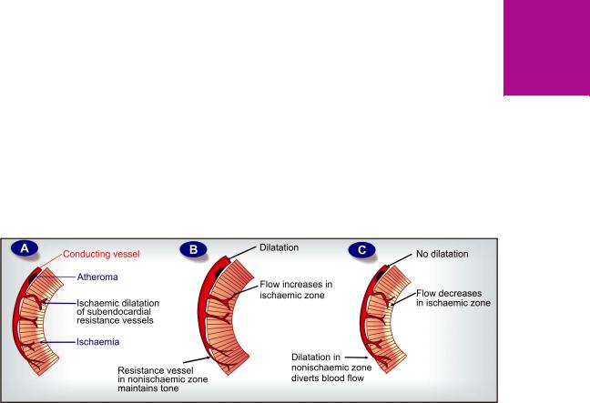
ANTIANGINAL AND OTHER ANTI-ISCHAEMIC DRUGS |
553 |
|
|
and vomiting. Large painful aphthous ulcers in the mouth, which heal on stopping nicorandil have been reported. Nitrate like tolerance does not occur with nicorandil, but it has the potential to interact with sildenafil.
Dose: 5–20 mg BD; NIKORAN, 5, 10 mg tabs, 2 mg/vial, 48 mg/vial inj; KORANDIL 5, 10 mg tabs.
Though nicorandil is an alternative antianginal drug, its efficacy and long term effects are less well established. It has failed to acquire wide acceptance, but may be useful in resistant angina when combined with other drugs. Administered i.v. during angioplasty for acute MI, it is believed to improve outcome.
OTHER ANTIANGINAL DRUGS
1. Dipyridamole It is a powerful coronary dilator; increases total coronary flow by preventing uptake and degradation of adenosine which is a local mediator involved in autoregulation of coronary flow in response to ischaemia. It dilates resistance vessels and abolishes autoregulation, but has no effect on larger conducting coronary vessels. Cardiac work is not decreased because venous return is not reduced. BP is minimally altered. Accordingly, it fails to relieve anginal symptoms or avert ECG changes.
The pharmacological success but therapeutic failure of dipyridamole has been explained on the basis of ‘coronary steal’ phenomenon (Fig. 39.5C). By dilating resistance vessels in nonischaemic zone as well, it diverts the already reduced blood flow away from the ischaemic zone.
Dipyridamole inhibits platelet aggregation. By potentiating PGI2 and increasing cAMP in platelets, it enhances antiaggregatory influences. Though not useful as an antianginal drug, it is being employed for prophylaxis
of coronary and cerebral thrombosis in post-MI and poststroke patients, as well as to prevent thrombosis in patients with prosthetic heart valves (see Ch. 44).
Dose: 25–100 mg TDS; PERSANTIN, CARDIWELL 25, 75, 100 mg tab.
2. Trimetazidine This antianginal drug acts by nonhaemodynamic mechanisms. There is no effect on determinants of myocardial O2 consumption, such as HR and BP, both at rest as well as during exercise, but angina frequency is reduced and exercise capacity is increased. In patients not adequately controlled by long-acting nitrate/β blocker/CCB, addition of trimetazidine further reduced anginal attacks and increased exercise duration. The mechanism of action of trimetazidine is uncertain, but it may improve cellular tolerance to ischaemia by:
•Inhibiting mitochondrial long chain 3-ketoacyl-CoA- thiolase (LC3-KAT) a key enzyme in fatty acid oxidation— thereby reducing fatty acid metabolism and increasing glucose metabolism in myocardium. Ischaemic myocardium shifts to utilizing fatty acid as substrate, thereby
increasing requirement of O2 for the same amount of ATP generated. Since oxidation of fatty acid requires
more O2, shift back of substrate to glucose would reduce O2 demand.Trimetazidine has been labelled as pFOX (fatty acid oxidation pathway) inhibitor.
•Limiting intracellular acidosis and Na+, Ca2+ accumulation during ischaemia.
•Protecting against O• free radical induced membrane damage.
Trimetazidine is absorbed orally, partly metabolized and largely excreted unchanged in urine; t½ is 6 hr. It is generally well tolerated; side effects
39 CHAPTER
Fig. 39.5: Diagrammatic representation of coronary haemodynamics. A—in classical angina, B — Selective nitrate action on conducting vessels, which along with ischaemic dilatation of resistance vessels, increases flow to the subendocardial region → relief of angina. C—Dipyridamole action on all resistance vessels increases blood flow to nonischaemic zone to the detriment of ischaemic zone → coronary steal
554
SECTION 8
CARDIOVASCULAR DRUGS
are—gastric burning, dizziness, fatigue and muscle cramps. Reversible parkinsonism has been reported in the elderly.
Trimetazidine has also been advocated for visual disturbances, tinnitus, Méniére’s disease, dizziness, etc., but conclusive evidence of efficacy in these conditions is lacking. For ischaemic heart disease, it has been widely used in France, Spain, some other European countries and India, but not in the UK or USA. It is mostly an add on medication to conventional therapy in angina and post-MI patients.
Dose: 20 mg TDS.
FLAVEDON, CARVIDON, TRIVEDON 20 mg tabs, 35 mg modified release tab.
3. Ranolazine This novel antianginal drug primarily acts by inhibiting a late Na+ current (late INa) in the myocardium which indirectly facilitates Ca2+ entry through Na+/Ca2+ exchanger. Reduction in Ca2+ overload in the myocardium during ischaemia decreases contractility and has a cardioprotective effect. Sparing of fatty acid oxidation during ischaemia in favour of more O2 efficient carbohydrate oxidation by inhibiting LC3KAT has also been demonstrated. This was earlier believed to be the main mechanism of antianginal action of ranolazine, but is now considered secondary. Ranolazine has no effect on HR and BP, but prolongs exercise duration in angina patient.
The efficacy of ranolazine in decreasing frequency of anginal attacks and in prolonging exercise duration has been demonstrated both as monotherapy as well as when added to conventional drugs in multicentric randomized trials: MARISA (monotherapy assessment of ranolazine in stable angina, 2004), CARISA (Combination assessment of ranolazine in stable angina, 2004), ERICA (Efficacy of ranolazine in chronic angina, 2005). In the MERLINTIMI36 (2007) trial on non ST-elevation acute coronary syndrome patients, ranolazine use was associated with a lower rate of ventricular arrhythmias. Incidence of AF was also decreased. However, ranolazine prolongs Q-T interval, and should not be used along with other Q-T prolonging drugs (class I and III antiarrhythmics, and other drugs listed on p. 528).
Oral absorption of ranolazine is slow taking 4–6 hours with a bioavailability of 30–50%. It is metabolized in liver mainly by CYP3A4 and excreted in urine, with an average t½ of 7 hours.
Side effects reported are dizziness, weakness, constipation, postural hypotension, headache and dyspepsia. It should not be given to patients taking CYP3A4 inhibitors.
Dose: 0.5–1.0 g BD as SR tab; RANOZEX, RANK, CARTINEX, REVULANT, RANOLAZ 0.5 g SR tab.
4. Ivabradine This ‘pure’ heart rate lowering antianginal drug has been introduced recently as an alternative to β blockers. The only significant action of ivabradine is blockade of cardiac pacemaker (sino-atrial) cell ‘f’ channels, which are ‘funny’ cation channels that open during early part of slow diastolic (phase 4) depolarization. The resulting inward current (If) determines the slope of phase 4 depolarization. Selective blockade of If current by ivabradine results in heart rate reduction without any other electrophysiological or negative inotropic or negative lucitropic (slowing of myocardial relaxation) effect. Heart rate reduction decreases cardiac O2 demand and prolongation of diastole tends to improve myocardial perfusion (O2 supply). Accordingly, in clinical trials, ivabradine has been found to improve exercise tolerance in stable angina and reduce angina frequency.
Ivabradine is well absorbed orally, 40% bioavailable due to first pass metabolism; degraded by CYP3A4 and excreted in urine with a t½ of 2 hours. Apart from excess bradycardia, the most important adverse effect is visual disturbance. Extrasystoles, prolongation of P-R interval, headache, dizziness and nausea are the other problems. It should not be used if heart rate is <60/min, in sick sinus and in AF. Concurrent use of drugs which prolong Q-T or which inhibit CYP3A4 is contraindicated.
Ivabradine is indicated in chronic stable angina in patients with sinus rhythm who are intolerant to β blockers or when the latter are contraindicated. It can also be used in inappropriate sinus tachycardia.
Dose: Initially 5 mg BD, increase if needed to 7.5 mg BD, Elderly 2.5 mg BD.
IVABRAD, BRADIA 5, 7.5 mg tab.
5. Oxyphedrine This drug is claimed to improve myocardial metabolism so that heart can sustain hypoxia better. Though used in angina and MI for over 3 decades, its efficacy and status in coronary artery disease is not defined. It can diminish or alter taste sensation.
Dose: 8–24 mg TDS oral, 4–8 mg i.v. OD-BD; ILDAMEN 8, 24 mg tab., 4 mg/2 ml inj.

ANTIANGINAL AND OTHER ANTI-ISCHAEMIC DRUGS |
555 |
|
|
DRUGS FOR PERIPHERAL VASCULAR
DISEASES
Peripheral vascular diseases (PVDs) are either primarily occlusive (Buerger’s disease with intermittent claudication of legs), or mainly vasospastic (Raynaud’s phenomenon with episodic blanching ± cyanosis of fingers followed by hyperaemia), or both as in arteriosclerotic/diabetic vascular insufficiency, ischaemic leg ulcers, frost bite, gangrene, cerebrovascular inadequacy, etc. Increased cardiovascular risk is associated with all PVDs. Measures that reduce cardiovascular risk (smoking cessation, BP normalization, antiplatelet drugs, diabetes control, statins, weight management, exercise training) have solutary effect on PVDs as well. In addition, vasodilators and some other drugs have been used.
1. Cyclandelate It is a papaverine like general smooth muscle relaxant which increases cutaneous, skeletal muscle and cranial blood flow in normal individuals. However, efficacy in PVDs is not different from placebo. Side effects are flushing, palpitation and headache.
Dose: 200–400 mg TDS; CYCLOSPASMOL, CYCLASYN 200, 400 mg tab/cap.
2. Xanthinol nicotinate (Nicotinyl xanthinate) It is a compound of xanthine and nicotinic acid, both of which are vasodilators. It increases blood flow in many vascular beds and has been promoted for cerebrovascular disorders and PVDs, but therapeutic benefits are insignificant.
Dose: 300–600 mg TDS oral; 300 mg by i.m. or slow i.v. injection; COMPLAMINA 150 mg tab, 500 mg retard tab, 300 mg/2 ml inj.
3. Pentoxiphylline (Oxpentifylline) An analogue of theophylline and a weak phosphodiesterase (PDE) inhibitor, it has been shown to increase blood flow in ischaemic areas by reducing whole blood viscosity and by improving flexibility of RBCs. The rheological (dealing with property of flow) action rather than vasodilatation is said to be responsible for improving passage of blood through microcirculation. Thus, the ‘steal’ phenomenon is not likely. Oral doses do not affect heart rate, t.p.r. or BP.
Pentoxiphylline is usually well tolerated: side effects are nausea, vomiting, dyspepsia and bloating which can be minimized by taking the drug after meals.
Dose: 400 mg BD–TDS; TRENTAL-400, FLEXITAL 400 mg SR tab, 300 mg/15 ml for slow i.v. injection.
Pentoxiphylline is mainly used in intermittent claudication (calf pain on walking) due to occlusive vascular disease (Buerger’s disease); walking distance is increased. Other
conditions claimed to be improved are: trophic leg ulcers, transient ischaemic attacks (TIAs), nonhaemorrhagic stroke, and chronic cerebrovascular insufficiency. However, overall benefits are modest and restricted to a fraction of patients.
4. Cilostazole This is a PDE-3 inhibitor (like the inodilator inamrinone, p.524) which increases intracellular cAMP in platelets and vascular smooth muscle resulting in antiaggregatory and vasodilator effects. In clinical trials it has increased walking distance in patients with intermittent claudication and appears to be more effective than pentoxiphylline. Since long-term oral milrinone therapy for heart failure has increased cardiac mortality and has been discontinued, concern is expressed about long-term safety of cilostazole. However, no increase in cardiovascular mortality has so far been observed with cilostazole, but a warning has been issued not to use cilostazole in patients with heart failure. It is also not to be used in patients who have pain even at rest, or in those with tissue necrosis.
The most common side effect is headache. Others are palpitation, dizziness, nausea, vomiting, weakness and increase in ventricular ectopics or nonsustained VT. Cilostazole is extensively metabolized by CYP3A4 and CYP2C19 into active and inactive metabolites, together having elimination t½ of ~ 12 hours. It should not be administered along with inhibitors of CYP3A4 and CYP2C19, which increase its toxicity.
Cilostazole is indicated for intermittent claudication in patients with no rest pain or heart failure.
Dose: 100 mg BD, 30 min before or 2 hour after food. PLETOZ, CILODOC, STILOZ 50, 100 mg tabs.
Comment Apart from the above drugs, β adrenergic agonists like isoxsuprine, CCBs like nifedipine and α blockers like prazosin, phenoxybenzamine have been used in PVDs. However, no vasodilator can overcome organic obstruction. Because ischaemia itself is the most potent vasodilator stimulus in skeletal muscle and cerebral beds, vasodilators can even divert the blood to nonischaemic areas (steal phenomenon). They obviously are more useful when vasospasm is wholely or partly involved, e.g. in Raynaud’s phenomenon. PGI2 has been employed in severe cases with rest pain (p. 190).
39 CHAPTER

556 |
CARDIOVASCULAR DRUGS |
|
|
SECTION 8
DRUG THERAPY IN MYOCARDIAL
INFARCTION
According to severity, the acute coronary syndromes may be graded into:
•Unstable angina (UA): Vascular obstruction is incomplete, myocardial necrosis is absent—biochemical markers of ischaemia (see p. 632) do not appear in blood, and ST segment is not elevated in ECG.
•Non ST segment elevation myocardial infarction (NSTEMI): Vascular obstruction is incomplete, but is attended by relatively smaller area of myocardial necrosis; biochemical markers appear in blood, but ST segment is not elevated.
•ST segment elevation myocardial infarction (STEMI):
Vascular obstruction is complete, larger area of myocardium is necrosed, biochemical markers are prominent and ST segment in ECG is elevated.
However, UA and NSTEMI may progress to STEMI.
Myocardial infarction (MI) is ischaemic necrosis of a portion of the myocardium due to sudden occlusion of a branch of coronary artery. An acute thrombus at the site of atherosclerotic obstruction is the usual cause. About ¼ patients die before therapy can be instituted. The remaining are best treated in specialized coronary care units with continuous monitoring of the haemodynamic parameters, biochemical markers and ECG to guide the selection of drugs and dosage. Those who receive such facility can be greatly benefitted by drug therapy, which according to individual needs is directed to:
1.Pain, anxiety and apprehension After pain is not relieved by 3 doses of GTN given 5 min apart, an opioid analgesic (morphine/pethidine) or diazepam is administered parenterally.
2.Oxygenation By O2 inhalation and assisted respiration, if needed.
3.Maintenance of blood volume, tissue perfusion and microcirculation Slow i.v.
infusion of saline/low molecular weight dextran (avoid volume overload).
4. Correction of acidosis Acidosis occurs due to lactic acid production; can be corrected by i.v. sod. bicarbonate infusion.
5. Prevention and treatment of arrhythmias
Prophylactic i.v. infusion of a β blocker (unless
contraindicated) as soon as the MI patient is seen and its continuation orally for a few days has been shown to reduce the incidence of arrhythmias and mortality. β blockers used early in evolving MI can reduce the infarct size (myocardial salvage) and subsequent complications.
Tachyarrhythmias may be treated with i.v. lidocaine, procainamide or amiodarone. Routine prophylactic lidocaine infusion is not recommended now. Bradycardia and heart block may be managed with atropine or electrical pacing.
6.Pump failure The objective is to increase c.o. and/or decrease filling pressure without unduly increasing cardiac work or lowering BP. Drugs used for this purpose are:
(a) Furosemide: indicated if pulmonary wedge pressure is > 20 mm Hg. It decreases cardiac preload.
(b) Vasodilators: venous or combined dilator is selected according to the monitored haemodynamic parameters. Drugs like GTN (i.v.), or nitroprusside have been mainly used.
(c) Inotropic agents: dopamine or dobutamine i.v. infusion (rarely digoxin if AF present) may be needed to augment the pumping action of heart and tide over the crisis.
7.Prevention of thrombus extension, embolism, venous thrombosis Aspirin
(162–325 mg) should be given for chewing and swallowing as soon as MI is suspected (if not already being taken on a regular basis). This is continued at 80–160 mg/day. Anticoagulants (heparin followed by oral anticoagulants) are used primarily to prevent deep vein thrombosis (increased risk due to bed rest) and pulmonary/ systemic arterial embolism. Its value in checking coronary artery thrombus extension is uncertain. Any benefit is short-term; anticoagulants are not prescribed on long-term basis now (see Ch. 44).
8. Thrombolysis and reperfusion Fibrinolytic agents, i.e. plasminogen activators—streptokinase/ urokinase/alteplase to achieve reperfusion of
ANTIANGINAL AND OTHER ANTI-ISCHAEMIC DRUGS |
557 |
|
|
the infarcted area (see Ch. 44). Unless thrombolysis can be started within 1–2 hours of MI symptom onset, primary percutaneous coronary intervention (PCI) with stenting is now the preferred revascularization procedure, wherever available.
9. Prevention of remodeling and subsequent CHF ACE inhibitors/ARBs are of proven efficacy and afford long-term survival benefit (see Ch. 36).
10. Prevention of future attacks
(a)Platelet inhibitors—aspirin or clopidogrel given on long-term basis are routinely prescribed (see Ch. 44).
(b)β blockers—reduce risk of reinfarction, CHF
and mortality. All patients not having any contraindication are put on a β blocker for at least 2 years.
(c)Control of hyperlipidaemia—dietary substitution with unsaturated fats, hypolipidemic drugs especially statins (see Ch. 45).
) PROBLEM DIRECTED STUDY |
|
|
|
|
|
39.1 A 55-year-old man presented with complaints of tightness and discomfort over middle |
|
|
part of chest felt episodically, particularly after walking briskly or climbing stairs or during sex. |
|
CHAPTER |
This is relieved within 5–10 minutes of rest. One or two episodes occur practically every day. |
|
|
|
|
|
He is a past smoker who quit smoking 5 years back when he was diagnosed to have chronic |
|
|
obstructive pulmonary disease (COPD), for which he regularly takes 2 inhalations of Ipratropium Br. |
|
|
3 times a day and 2 puffs of salbutamol inhalation whenever he feels out of breath. The pulse |
|
39 |
was 90/min and BP 124/82 mm Hg. The resting ECG was normal, but stress test was positive. |
|
|
A diagnosis of exertional angina was made and he was prescribed—Tab glyceryl trinitrate |
|
|
|
|
|
|
||
0.5 mg to be put under the tongue as soon as he begins to feel the chest discomfort, as well |
|
|
as before undertaking any physical exertion. |
|
|
(a) Should he be prescribed another drug to be taken on a regular basis to prevent episodes |
|
|
of angina? If so, which drugs can be given to him and which cannot be given? |
|
|
(b) Should additional medication be given to prevent long-term complications and improve survival? |
|
|
(see Appendix-1 for solution) |
|
|
|
|
|