

- •Preface
- •Contents
- •List of Abbreviations
- •6. Adverse Drug Effects
- •7b. Cholinergic System and Drugs
- •9. Adrenergic System and Drugs
- •11. Histamine and Antihistaminics
- •12. 5-Hydroxytryptamine, its Antagonists and Drug Therapy of Migraine
- •16. Drugs for Cough and Bronchial Asthma
- •17a. Introduction
- •17b. Anterior Pituitary Hormones
- •20. Corticosteroids
- •21. Androgens and Drugs for Erectile Dysfunction
- •24. Drugs Affecting Calcium Balance
- •25. Skeletal Muscle Relaxants
- •26. Local Anaesthetics
- •27. General Anaesthetics
- •28. Ethyl and Methyl Alcohols
- •29 Sedative-Hypnotics
- •30. Antiepileptic Drugs
- •31. Antiparkinsonian Drugs
- •32. Drugs Used in Mental Illness: Antipsychotic and Antimanic Drugs
- •38. Antiarrhythmic Drugs
- •40. Antihypertensive Drugs
- •41b. Diuretics
- •42. Antidiuretics
- •46. Drugs for Peptic Ulcer and Gastroesophageal Reflux Disease
- •48. Drugs for Constipation and Diarrhoea
- •51. Beta-Lactam Antibiotics
- •53. Aminoglycoside Antibiotics
- •55. Antitubercular Drugs
- •56. Antileprotic Drugs
- •57. Antifungal Drugs
- •58. Antiviral Drugs
- •59. Antimalarial Drugs
- •61. Anthelmintic Drugs
- •62. Anticancer Drugs
- •63. Immunosuppressant Drugs
- •64. Drugs Acting on Skin and Mucous Membranes
- •66. Chelating Agents
- •67. Vitamins
- •68. Vaccines and Sera
- •69. Drug Interactions
- •Appendices
- •Selected References for Further Reading
- •Index

Chapter 42 Antidiuretics
Antidiuretics (more precisely ‘anti-aquaretics’, because they inhibit water excretion without affecting salt excretion) are drugs that reduce urine volume, particularly in diabetes insipidus (DI) which is their primary indication. Drugs are:
1.Antidiuretic hormone (ADH, Vasopressin), Desmopressin, Lypressin, Terlipressin
2.Thiazide diuretics, Amiloride.
3.Miscellaneous: Indomethacin, Chlorpropamide, Carbamazepine.
ANTIDIURETIC HORMONE
(Argenine Vasopressin-AVP)
It is a nonapeptide secreted by posterior pituitary (neurohypophysis) along with oxytocin (see Ch. 23). It is synthesized in the hypothalamic (supraoptic and paraventricular) nerve cell bodies as a large precursor peptide along with its binding protein ‘neurophysin’. Both are transported down the axons to the nerve endings in the median eminence and pars nervosa. Osmoreceptors present in hypothalamus and volume receptors present in left atrium, ventricles and pulmonary veins primarily regulate the rate of ADH release governed by body hydration. Osmoreceptors are also present in the hepatic portal system which sense ingested salt and release ADH even before plasma osmolarity is increased by the ingested salt. Impulses from baroreceptors and higher centres also impinge on the nuclei synthesizing ADH and affect its release. The two main physiological stimuli for ADH release are rise in plasma osmolarity and contraction of e.c.f. volume.
Several neurotransmitters, hormones and drugs modify ADH secretion. It is enhanced by angiotensin II, prostaglandins (PGs), histamine, neuropeptide Y and ACh. GABA and atrial natriuretic peptide (ANP) decrease its release. Opioids have agent-specific and dose dependent action. Low-dose morphine
inhibits ADH secretion, but high doses enhance it. Opioid peptides are mostly inhibitory. Nicotine and imipramine stimulate, while alcohol, haloperidol, phenytoin and glucocorticoids decrease ADH release.
The human ADH is 8-arginine-vasopressin (AVP); 8-lysine-vasopressin (lypressin) is found in swine and has been synthetically prepared. Other more potent and longer acting peptide analogues of AVP having agonistic as well as antagonistic action have been prepared.
ADH (Vasopressin) receptors
These are G protein coupled cell membrane receptors; two subtypes V1 and V2 have been identified, cloned and structurally characterized.
V1 Receptors All vasopressin receptors except those on renal CD cells, AscLH cells and vascular endothelium are of the V1 type. These are further divided into V1a and V1b subtypes:
V1a receptors are present on vascular smooth muscle (including that of vasa recta in renal medulla), uterine and other visceral smooth muscles, interstitial cells in renal medulla, cortical CD cells, adipose tissue, brain, platelets, liver, etc. The V1b receptors are localized to the anterior pituitary, certain areas in brain and in pancreas.
The V1 receptors function mainly through the phospholipase C–IP3/DAG pathway—release Ca2+ from intracellular stores—causing vasoconstriction, visceral smooth muscle contraction, glycogenolysis, platelet aggregation,ACTH release, etc. These actions are augmented by enhanced influx of Ca2+ through Ca2+ channels as well as by DAG mediated protein kinase C activation which phosphorylates relevant proteins. V1 receptors, in addition, activate phospholipase A2—release arachidonic acid resulting in generation of PGs and other eicosanoids which contribute to many of the V1 mediated effects. Persistent V1 receptor
594 |
DRUGS ACTING ON KIDNEY |
|
|
SECTION 9
stimulation activates protooncogenes (possibly through IP3/DAG pathway) resulting in growth (hypertrophy) of vascular smooth muscle and other responsive cells.
V2 Receptors These are located primarily on the collecting duct (CD) principal cells in the kidney—regulate their water permeability through cAMP production. Some V2 receptors are also present on AscLH cells which activate Na+K+2Cl¯ cotransporter. Vasodilatory V2 receptors are present on endothelium of blood vessels.
The V2 receptors are more sensitive (respond at lower concentrations) to AVP than are V1 receptors.
Selective peptide agonists and antagonists of the subtypes of vasopressin receptors are:
|
|
|
Selective agonist |
V1a |
|
Receptor |
[Phe2, Ile2, Orn8] AVP |
V1b |
Receptor |
Deamino [D-3 {pyridyl)-Ala2] AVP |
|
V2 |
Receptor |
Desmopressin (dDAVP) |
|
|
|
|
Selective antagonist |
V1a |
|
Receptor |
d(CH2)5 [Tyr (Me2)] AVP |
V1b |
Receptor |
dp [Tyr (me2)] AVP |
|
V2 |
Receptor |
d(CH2)5 [D-Ile2, Ile4, Ala-NH29] AVP |
|
Some orally active nonpeptide V1a, V1b and V2 receptor antagonists have been produced. Tolvaptan and Mozavaptan are nonpeptide V2 selective antagonists that are now in clinical use.
Actions
Kidney AVP acts on the collecting duct (CD) principalcellstoincreasetheirwaterpermeability— waterfromtheductlumendiffusestotheinterstitium by equilibrating with the hyperosmolar renal medulla(see Fig.IX.1).Inman,maximalosmolarity of urine that can be attained is 4 times higher than plasma. WhenAVP is absent, CD cells remain impermeable to water → dilute urine (produced by the diluting segment) is passed as such. Graded effect occurs at lower concentrations ofAVP: urine volume closely balances fluid intake.
Mechanism of action Vasopressin is instrumental in rapid adjustments of water excretion according to the state of body hydration, as well as in dealing with conditions prevailing over long-
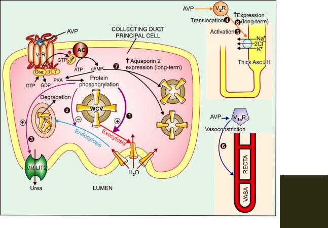
term. The V2 subtype of ADH receptors are present on the basolateral membrane of principal cells in CDs (see Fig. 42.1). Activation of these receptors increases cAMP formation intracellularly → activation of cAMP dependent protein kinase A → phosphorylation of relevant proteins which promote exocytosis of ‘aquaporin-2’ water channel containing vesicles (WCVs) through the apical membrane → more aqueous channels get inserted into the apical membrane. The rate of endocytosis and degradation of WCVs is concurrently reduced. The water permeability of CD cells is increased in proportion to the population of aquaporin-2 channels in the apical membrane at any given time. Continued V2 receptor stimulation (during chronic water deprivation) in addition upregulates aquaporin-2 synthesis through cAMP response element of the gene encoding aquaporin-2.
Other aquaporins like aquaporin-1 (in PT) and aquaporin-3,4 (on basolateral membrane of CD cells) also participate in water transport at these sites.
To achieve maximum concentration of urine, activation of V2 receptors increases urea permeability of terminal part of CDs in inner medulla by stimulating a vasopressin regulated urea transporter (VRUT or UT-1)—which in turn augments medullary hypertonicity. Recently, V2 receptor mediated actions of AVP on AscLH have also been demonstrated which further reinforce medullary hypertonicity by translocating to luminal membrane and activating the Na+K+2Cl¯ cotransporter in the short-term and increasing its synthesis in the long-term.
The V1 receptors also participate in the renal response to AVP. Activation of V1 receptors constricts vasa recta to diminish blood flow to inner medulla which reduces washing off effect and helps in maintaining high osmolarity in this region. Thus, it contributes to antidiuresis. On the other hand, activation of medullary interstitial cell V1 receptors enhance PG synthesis which attenuate cAMP generation in CD cells and oppose V2 mediated antidiuresis. V1 receptors are also present on CD cells. Their stimulation activates
ANTIDIURETICS |
595 |
|
|
Fig. 42.1: Mechanisms of rapid and long-term anti-aquaretic action of vasopressin
All V2 receptor (V2R) mediated actions are exerted through the adenylyl cyclase (AC)-cyclic AMP (cAMP) pathway, while the V1a receptor (V1aR) mediated action is exerted via the phospholipase C—IP3: DAG pathway.
Rapid actions
(1)Translocation of water channel containing vesicles (WCVs) and exocytotic insertion of aquaporin 2 water channels into the apical membrane of principal cells of collecting ducts; the primary action responsible for antidiuresis.
(2)Inhibition of endocytotic removal of aquaporin 2 channels from the apical membrane.
(3)Activation of vasopressin regulated urea transporter (VRUT) at apical membrane of collecting ducts in the inner medulla.
(4)Translocation of Na+K+2Cl¯ cotransporter to the luminal membrane of cells in thick ascending limb of loop of Henle (AscLH).
(5)Activation of Na+K+2Cl¯ cotransporter in AscLH cells.
(6)V1a receptor (V1aR) mediated vasoconstriction of vasa recta
Long-term actions
(7)Gene mediated increased expression of aquaporin 2 channels in collecting duct cells.
(8)Gene mediated increased expression of Na+K+2Cl¯ cotransporter in AscLH cells.
PKA—cAMP dependent protein kinase.
42 CHAPTER

596 |
DRUGS ACTING ON KIDNEY |
|
|
SECTION 9
PKc which directly diminishes responsiveness of CD cells to V2 receptors and restrains V2 mediated water permeability. The logic of this apparent paradox may lie in the fact that these V1 actions are produced at much higher concentrations of AVP, so that physiologically they may serve to restrict V2 effects only when blood levels of AVP are very high.
Lithium and demeclocycline partially antagonize AVP action (probably by limiting cAMP formation), reduce the urine concentrating ability of the kidney, produce polyuria and polydipsia. They have been used in patients with inappropriate ADH secretion. On the other hand NSAIDs (especially indomethacin) augment AVP induced antidiuresis by inhibiting renal PG synthesis. Carbamazepine and chlorpropamide also potentiate AVP action on kidney.
Blood vessels AVP constricts blood vessels through V1 receptors and can raise BP (hence the name vasopressin), but much higher concentration is needed than for maximal antidiuresis. The cutaneous, mesenteric, skeletal muscle, fat depot, thyroid, and coronary beds are particularly constricted. Though vasoconstrictor action of AVP does not appear to be physiologically important, some recent studies indicate that it may play a role in CHF, haemorrhage, hypotensive states, etc. Prolonged exposure to AVP causes vascular smooth muscle hypertrophy.
The V2 receptor mediated vasodilatation can be unmasked when AVP is administered in the presence of a V1 antagonist. It can also be demonstrated by the use of selective V2 agonist desmopressin, and is due to endothelium dependent NO production.
Other actions Most visceral smooth muscles contract. Increased peristalsis in gut (especially large bowel), evacuation and expulsion of gases may occur.
Uterus is contracted by AVP acting on oxytocin receptors. In the nonpregnant and early pregnancy uterus, AVP is equipotent to oxytocin. Only at term sensitivity to oxytocin increases selectively.
CNS Exogenously administered AVP does not penetrate blood-brain barrier. However, it is now recognized as a peptide neurotransmitter in many
areas of brain and spinal cord. AVP may be involved in regulation of temperature, systemic circulation, ACTH release, and in learning of tasks.
AVP induces platelet aggregation and hepatic glycogenolysis. It releases coagulation factor VIII and von Willebrand’s factor from vascular endothelium through V2 receptors.
Pharmacokinetics AVP is inactive orally because it is destroyed by trypsin. It can be administered by any parenteral route or by intranasal application. The peptide chain of AVP is rapidly cleaved enzymatically in many organs, especially in liver and kidney; plasma t½ is short ~25 min. However, the action of aqueous vasopressin lasts 3–4 hours.
Aqueous vasopressin (AVP) inj: POSTACTON 10 U inj; for i.v., i.m. or s.c. administration.
VASOPRESSIN ANALOGUES
Lypressin It is 8-lysine vasopressin. Though somewhat less potent than AVP, it acts on both V1 and V2 receptors and has longer duration of action (4–6 hours). It is being used in place of AVP—mostly for V1 receptor mediated actions.
PETRESIN, VASOPIN 20 IU/ml inj; 10 IU i.m. or s.c. or 20 IU diluted in 100–200 ml of dextrose solution and infused i.v. over 10–20 min.
Terlipressin This synthetic prodrug of vasopressin is specifically used for bleeding esophageal varices; may produce less severe adverse effects than lypressin.
Dose: 2 mg i.v., repeat 1–2 mg every 4–6 hours as needed. GLYPRESSIN 1 mg freeze dried powder with 5 ml diluent for inj, T-PRESSIN, TERLINIS 1 mg/10 ml inj.
Desmopressin (dDAVP) This synthetic peptide is a selective V2 agonist; 12 times more potent antidiuretic than AVP, but has negligible vasoconstrictor activity. It is also longer acting because enzymatic degradation is slow; t½ 1–2 hours; duration of action 8–12 hours. Desmopressin is the preparation of choice for all V2 receptor related indications. The intranasal route is preferred, though bioavailability is only 10–20%. An oral formulation has been recently marketed with a bioavailability of 1–2%; oral dose

ANTIDIURETICS |
597 |
|
|
is 10–15 times higher than intranasal dose, but systemic effects are produced and nasal side effects are avoided. Many patients find oral tablet more convenient.
Dose: Intranasal: Adults 10–40 µg/day in 2–3 divided doses, children 5–10 µg at bed time.
Oral: 0.1–0.2 mg TDS.
Parenteral (s.c. or i.v.) 2–4 µg/day in 2–3 divided doses. MINIRIN 100 µg/ml nasal spray (10 µg per actuation); 100 µg/ml intranasal solution in 2.5 ml bottle with applicator; 0.1 mg tablets; 4 µg/ml inj.
Uses
A. Based on V2 actions (Desmopressin is the drug of choice)
1.Diabetes insipidus DI of pituitary origin (neurogenic) is the most important indication for vasopressin. It is ineffective in renal (nephrogenic) DI, since kidney is unresponsive to ADH. Lifelong therapy is required, except in some cases of head injury or neurosurgery, where DI occurs transiently.
The dose of desmopressin is individualized by measuring 24 hour urine volume. Aqueous vasopressin or lypressin injection is impracticable for long-term treatment. It can be used in transient DI and to differentiate neurogenic from nephrogenic DI—urine volume is reduced and its osmolarity increased if DI is due to deficiency of ADH, but not when it is due to unresponsiveness of kidney to ADH. Desmopressin 2 µg i.m. is the preparation of choice now for the same purpose.
2.Bedwetting in children and nocturia in adults Intranasal or oral desmopressin at bed-
time controls primary nocturia by reducing urine volume. Nocturnal voids are reduced to nearly half and first sleep period in adults is increased by ~2 hr. Fluid intake must be restricted 1 hr before and till 8 hr after the dose to avoid fluid retention. Monitor BP and body weight periodically to check fluid overload. Withdraw for one week every 3 months for reassessment.
3. Renal concentration test 5–10 U i.m. of aqueous vasopressin or 2 µg of desmopressin causes maximum urinary concentration.
4. Haemophilia, von Willebrand’s disease
AVP may check bleeding by releasing coagulation factor VIII and von Willebrand’s factor. Desmopressin is the preferred preparation in a dose of 0.3 µg/kg diluted in 50 ml saline and infused i.v. over 30 min.
B. Based on V1 actions
1. Bleeding esophageal varices Vasopressin/ terlipressin often stop bleeding by constricting mesenteric blood vessels and reducing blood flow through the liver to the varices, allowing clot formation. Terlipressin stops bleeding in ~80% and has been shown to improve survival. It has replaced AVP because of fewer adverse effects and greater convenience in use. Octreotide (a somatostatin analogue) injected i.v. is an alternative. However, definitive therapy of varices remains endoscopic obliteration by sclerotherapy.
2. |
Before abdominal radiography |
AVP/lypressin has |
been |
occasionally used to drive out |
gases from bowel. |
Adverse effects Because of V2 selectivity, desmopressin produces fewer adverse effects than vasopressin, lypressin or terlipressin. However, transient headache and flushing are frequent.
Nasal irritation, congestion, rhinitis, ulceration and epistaxis can occur on local application. Systemic side effects are: belching, nausea, abdominal cramps, pallor, urge to defecate, backache in females (due to uterine contraction). Fluid retention and hyponatraemia may develop.
Symptoms of hyponatremia are due to shift of water intracellularly resulting in cerebral edema producing headache, mental confusion, lassitude, nausea, vomiting and even
seizures. Children are more susceptible.
AVP can cause bradycardia, increase cardiac afterload and precipitate angina by constricting coronary vessels. It is contraindicated in patients with ischaemic heart disease, hypertension, chronic nephritis and psychogenic polydipsia. Urticaria and other allergies are possible with any preparation.
THIAZIDES
Diuretic thiazides paradoxically exert an antidiuretic effect in DI. High ceiling diuretics
42 CHAPTER

598
SECTION 9
DRUGS ACTING ON KIDNEY
are also effective but are less desirable because of their short and brisk action. Thiazides reduce urine volume in both pituitary origin as well as renal DI. They are especially valuable for the latter in which AVP is ineffective. However, their efficacy is low; urine can never become hypertonic as can occur with AVP in neurogenic DI. The mechanism of action is not well understood, possible explanation is:
Thiazides induce a state of sustained electrolyte depletion so that glomerular filtrate is more completely reabsorbed iso-osmotically in PT. Further, because of reduced salt reabsorption in the cortical diluting segment, a smaller volume of less dilute urine is presented to the CDs and the same is passed out. That salt restriction has a similar effect, substantiates this mechanism of action. Secondly, thiazides reduce g.f.r. and thus the fluid load on tubules.
Hydrochlorothiazide 25–50 mg TDS or equivalent dose of a longer acting agent is commonly used. Though less effective than AVP, it is more convenient and cheap even for pituitary origin DI; may reduce polyuria to some extent.
K+ supplements are needed.
Amiloride is the drug of choice for lithium induced nephrogenic DI (see p. 590).
Indomethacin has also been found to reduce
polyuria in renal DI to some extent by reducing renal PG synthesis. It can be combined with a thiazide ± amiloride in nephrogenic DI. Other NSAIDs are less active.
Chlorpropamide It is a long-acting sulfonylurea oral hypoglycaemic, found to reduce urine volume in DI of pituitary origin but not in renal DI. It sensitizes the kidney to ADH action; thus its efficacy depends on small amounts of the circulating hormone; it is not active when ADH is totally absent.
Carbamazepine It is an antiepileptic (see Ch. 30) which reduces urine volume in DI of pituitary origin, but mechanism of action is not clear. Higher doses are needed; adverse effects are marked; it is of little value in treatment of DI.
VASOPRESSIN ANTAGONISTS
Tolvaptan It is an orally active nonpeptide selective V2 receptor antagonist that has been recently introduced for the treatment of hyponatraemia due to CHF, cirrhosis of liver or syndrome of inappropriate ADH secretion (SIADH). It increases free water clearance by the kidney (aquaretic) and helps to correct the low plasma Na+ levels. In clinical trials symptoms of worsening heart failure were improved. However, too rapid correction of hyponatraemia should not be attempted, because thrombotic complications can occur due to haemoconcentration. The most frequent side effect is thirst and dry mouth. Others are fever, g.i. upset and hyperglycaemia. Tolvaptan is metabolized by CYP3A4; should not be given to patients receiving inhibitors of this isoenzyme. The t½ is 6–8 hours, and it is given once daily.
Mozavaptan (V2 selective antagonist) and Conivaptan (V1a+V2 antagonist) are the other vasopressin antagonists that are in clinical use.

SECTION 10
DRUGS AFFECTING BLOOD AND
BLOOD FORMATION
Chapter 43 Haematinics and
Erythropoietin
Haematinics These are substances required in the formation of blood, and are used for treatment of anaemias.
Anaemia occurs when the balance between production and destruction of RBCs is disturbed by:
(a)Blood loss (acute or chronic)
(b)Impaired red cell formation due to:
•Deficiency of essential factors, i.e. iron, vitamin B12, folic acid.
•Bone marrow depression (hypoplastic anaemia), erythropoietin deficiency.
(c)Increased destruction of RBCs (haemolytic anaemia)
In this chapter essential factors required for normal formation or pigmentation of RBCs will be covered.
IRON
Distribution of iron in body Iron is an essential body constituent. Total body iron in an adult is 2.5–5 g (average 3.5 g). It is more in men (50 mg/kg) than in women (38 mg/kg). It
is distributed into: |
|
|
Haemoglobin (Hb) |
: 66% |
|
Iron stores as ferritin and |
: |
25% |
haemosiderin |
|
|
Myoglobin (in muscles) |
: |
3% |
Parenchymal iron (in enzymes, etc.) |
: |
6% |
Haemoglobin is a protoporphyrin; each molecule having 4 iron containing haeme residues. It has 0.33% iron; thus loss of 100 ml of blood (containing 15 g Hb) means loss of 50 mg elemental iron. To raise the Hb level of blood by 1 g/dl— about 200 mg of iron is needed. Iron is stored only in ferric form, in combination with a large protein apoferritin.
Iron has for long been considered important for the body. Lauha bhasma (calcined iron) has been used in ancient Indian medicine. According to Greek thought Mars is the God of strength, and iron is dedicated to Mars: as such, iron was used to treat weakness, which is common in anaemia. In 1713 iron was shown to be present in blood. In the early 19th century Blaud developed his famous ‘Blaud’s pill’ consisting of ferrous sulfate and potassium carbonate for anaemia. All important aspects of iron metabolism have been learned in the past 60 years.
aggregates
Apoferritin + Fe3+  Ferritin
  Haemosiderin (not reutilized)
Ferritin can get saturated to different extents; at full saturation it can hold 30% iron by weight. The most important storage sites are reticuloendothelial (RE) cells. Parenchymal iron occurs as prosthetic group in many cellular enzymes—
600 |
DRUGS AFFECTING BLOOD AND BLOOD FORMATION |
|
|
cytochromes, peroxidases, catalases, xanthine oxidase and some mitochondrial enzymes. Though, the primary reflection of iron deficiency occurs in blood, severe deficiency affects practically every cell.
Daily requirement To make good average daily
loss, iron requirements |
are: |
|
|
|
Adult male |
: 0.5–1 mg (13 µg/kg) |
|||
Adult female |
: 1–2 mg (21 |
µg/kg) |
||
(menstruating) |
|
|
|
|
Infants |
: |
60 |
µg/kg |
|
Children |
: |
25 |
µg/kg |
|
Pregnancy |
: |
3–5 mg (80 |
µg/kg) |
|
(last 2 trimesters)
Dietary sources of iron
Rich : Liver, egg yolk, oyster, dry beans, dry fruits, wheat germ, yeast.
Medium : Meat, chicken, fish, spinach, banana, apple.
Poor : Milk and its products, root vegetables.
Iron absorption
The average daily diet contains 10–20 mg of iron. Its absorption occurs all over the intestine, but
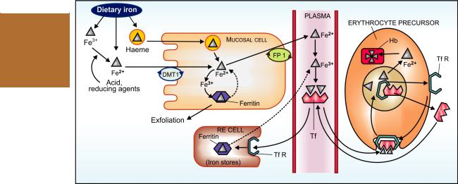
majority in the upper part. Dietary iron is present either as haeme or as inorganic iron. Absorption of haeme iron is better (upto 35% compared to inorganic iron which averages 5%) and occurs directly without the aid of a carrier (Fig. 43.1). However, it is a smaller fraction of dietary iron. The major part of dietary iron is inorganic and in the ferric form. It needs to be reduced to the ferrous form before absorption. Two separate iron transporters in the intestinal mucosal cells function to effect iron absorption. At the luminal membrane the divalent metal transporter 1 (DMT1) carrys ferrous iron into the mucosal cell. This along with the iron released from haeme is transported across the basolateral membrane by another iron transporter ferroportin (FP). These iron transporters are regulated according to the body needs. Absorption of haeme iron is largely independent of other foods simultaneously ingested, but that of inorganic iron is affected by several factors.
Factors facilitating iron absorption
1.Acid: by favouring dissolution and reduction of ferric iron.
SECTION 10
Fig. 43.1: Schematic depiction of intestinal absorption, transport, utilization and storage of iron (see text for description) Fe2+—Ferrous iron; Fe3+—Ferric iron; DMT1—Divalent metal transporter 1; Hb—Haemoglobin; RE cell— Reticuloendothelial cell; FP1—Ferroportin; Tf—Transferrin; TfR—Transferrin receptor

HAEMATINICS AND ERYTHROPOIETIN |
601 |
|
|
2.Reducing substances: ascorbic acid, amino acids containing SH radical. These agents reduce ferric iron and form absorbable complexes.
3.Meat: by increasing HCl secretion and providing haeme iron.
Factors impeding iron absorption
1. |
Alkalies (antacids) render iron insoluble, |
||
|
oppose its |
reduction. |
|
2. |
Phosphates |
(rich in egg yolk) By |
|
3. |
Phytates (in maize, wheat) |
complexing |
|
4. |
Tetracyclines |
iron |
|
5.Presence of other foods in the stomach. In general, bioavailability of iron from cereal
based diets is low.
Mucosal block The gut has a mechanism to prevent entry of excess iron in the body. Iron reaching inside mucosal cell is either transported to plasma or oxidised to ferric form and complexed with apoferritin to form ferritin (Fig. 43.1). This ferritin generally remains stored in the mucosal cells and is lost when they are shed (lifespan 2–4 days). This is called the ‘Ferritin curtain’.
The iron status of the body and erythropoietic activity govern the balance between these two processes, probably through a ‘haematopoietic transcription factor’, and thus the amount of iron that will enter the body. A larger percentage is absorbed during iron deficiency. When body iron is low or erythropoiesis is occurring briskly, ferritin is either not formed or dissociates soon— the released iron is transported to the blood.
Mucosal block however, can be overwhelmed by gross excess of iron.
Transport, utilization, storage and excretion
Free iron is highly toxic. As such, on entering plasma it is immediately converted enzymatically to the ferric form and complexed with a glycoprotein transferrin (Tf). Iron circulates in plasma bound to Tf (two Fe3+ residues per molecule). The total plasma iron content (~3 mg) is recycled 10 times everyday (turnover of iron is 30 mg/day).
Iron is transported inside erythropoietic and other cells through attachment of transferrin to specific membrane bound transferrin receptors (TfRs). The complex is engulfed by receptor mediated endocytosis. Iron dissociates from the complex at the acidic pH of the intracellular vesicles; the released iron is utilized for haemoglobin synthesis or other purposes, while Tf and TfR are returned to the cell surface to carry fresh loads. In iron deficiency and haemolytic states when brisk erythropoiesis is occurring, erythropoietic cells express more TfRs, but other cells do not. Thus, the erythron becomes selectively more efficient in trapping iron.
After entering the storage cells through TfRs, iron is stored in RE cells (in liver, spleen, bone marrow), as well as in hepatocytes and myocytes as ferritin and haemosiderin. Apoferritin synthesis is regulated by iron status of the body. When it is low—the ‘iron regulating element’ (IRE) on mRNA is blocked—transcription of apoferritin does not occur, while more Tf is produced. On the other hand, more apoferritin is synthesized to trap iron when iron stores are rich. Plasma iron derived from destruction of old RBCs (lifespan ~120 days), from stores and from intestinal absorption forms a common pool that is available for erythropoiesis, to all other cells and for restorage.
Iron is tenaciously conserved by the body; daily excretion in adult male is 0.5–1 mg, mainly as exfoliated g.i. mucosal cells, some RBCs and in bile (all lost in faeces). Other routes are desquamated skin, very little in urine and sweat. In menstruating women, monthly menstrual loss may be averaged to 0.5–1 mg/day. Excess iron is required during pregnancy for expansion of RBC mass, transfer to foetus and loss during delivery; totalling to about 700 mg. This is to be met in the later 2 trimesters.
Preparations and dose Oral iron
The preferred route of iron administration is oral. Dissociable ferrous salts are inexpensive, have high iron content and are better absorbed than
43 CHAPTER

602
SECTION 10
DRUGS AFFECTING BLOOD AND BLOOD FORMATION
TABLE 43.1  Some combination preparations of iron
Trade name |
Iron compound |
Other ingredients |
|
|
|
|
|
CONVIRON Cap |
Fe. sulfate (dried) 60 mg |
B12 15 µg, folic acid 1.5 mg, B6 1.5 mg, vit. C 75 mg |
|
FESOVIT-SPANSULE Cap |
Fe. sulfate (dried) 150 mg |
B12 15 µg, folic acid 1 mg, nicotinamide 50 mg, |
|
|
|
|
B6 2 mg |
FERSOLATE-CM tab |
Fe. sulfate (dried) 195 mg |
Cu sulfate 2.6 mg, Mn. sulfate 2 mg |
|
FEFOL SPANSULE Cap |
Fe. sulfate 150 mg |
Folic acid 0.5 mg |
|
HEMGLOB syr (15 ml) |
Fe. gluconate 300 mg |
B12 15 µg, B1 5 mg, B2 5 mg, B6 1.5 mg, |
|
|
|
|
niacinamide 45 mg |
AUTRIN Cap |
Fe. fumarate 300 mg |
B12 15 µg, folic acid 1.5 mg |
|
DUMASULES Cap |
Fe. fumarate 300 mg |
B12 7.5 µg, folic acid 0.75 mg, B1 5 mg, |
|
|
|
|
niacinamide 50 mg, vit. C 75 mg, B6 1.5 mg |
HEMSYNERAL Cap |
Fe. fumarate 200 mg |
B12 15 µg, folic acid 1.5 mg |
|
ANEMIDOX Cap |
Fe fumarate 360 mg |
Folic acid 1.5 mg, vit B12 15 μg, Cal. carb. |
|
|
|
|
200 mg, vit C 75 mg, vit D 400 i.u. |
HEMSI syr. (5 ml) |
Fe. fumarate |
100 mg |
Vit B12 5 μg, folic acid 0.5 mg, Zn 3.3 mg, |
|
|
|
Cu 0.035 mg, Mn 0.2 mg |
FERRICARB Cap |
Carbonyl iron |
(100 mg iron) |
Folic acid 1.5 mg, vit B12 15 μg, zinc sulfate |
|
|
|
88 mg, pyridoxine 3 mg, sod. selenite 60 μg |
HBFAST tab |
Carbonyl iron (100 mg iron) |
Folic acid 0.35 mg |
|
HEMATRINE Cap |
Fe. succinate 100 mg |
B12 2.5 µg, folic acid 0.5 mg, vit. C 25 mg, |
|
|
|
|
niacinamide 15 mg |
POLYRON tab, BIOFER tab |
Ferric hydroxy polymaltose |
Folic acid 0.35 mg |
|
POLYFER chewable tab |
(Iron 100 mg) |
|
|
MUMFER syr (5 ml) |
Ferric hydroxy polymaltose |
|
|
|
(50 mg iron) |
|
|
drops (1 ml) |
—do—(50 mg iron) |
|
|
FERROCHELATE syr (5 ml) |
Ferric ammon. cit. (Iron 60 mg) |
B12 5 µg, folic acid 1 mg |
|
drops (1 ml) |
—do—(Iron 20 mg) |
B12 4 µg, folic acid 0.2 mg |
|
RARICAP tab |
Iron cal. complex (Iron 25 mg) |
Folic acid 0.3 mg |
|
PROBOFEX Cap |
Fe. aminoate (60 mg iron) |
B12 15 µg, folic acid 1.5 mg, B6 3 mg |
|
DEXORANGE Cap, |
Ferric ammon. cit. 160 mg |
B12 7.5 µg, folic acid 0.5 mg, Zn 7.5 mg |
|
syrup (15 ml) |
|
|
(as sulfate) |
|
|
|
|
Combination of iron with strychnine, arsenic and yohimbine and all fixed dose combination of haemoglobin in any form are banned in India.

HAEMATINICS AND ERYTHROPOIETIN |
603 |
|
|
ferric salts, especially at higher doses. Gastric irritation and constipation (the most important side effects of oral iron) are related to the total quantity of elemental iron administered. If viewed in terms of iron content, nearly all preparations have the same degree of gastric tolerance, the limits of which are fairly well defined in individual patients. Some simple oral preparations are:
1.Ferrous sulfate: (hydrated salt 20% iron, dried salt 32% iron) is the cheapest; may be preferred on this account. It often leaves a metallic taste in mouth; FERSOLATE 200 mg tab.
2.Ferrous gluconate (12% iron): FERRONICUM
300 mg tab, 400 mg/15 ml elixer.
3. Ferrous fumarate (33% iron): is less water soluble than ferrous sulfate and tasteless;
NORI-A 200 mg tab.
4. Colloidal ferric hydroxide (50% iron):
FERRI DROPS 50 mg/ml drops.
5. Carbonyl iron: It is high purity metallic iron in very fine powder form (particle size < 5 μM), prepared by decomposition of iron pentacarbonyl, a highly toxic compound. It is claimed to be absorbed from intestines over a long time, and gastric tolerance may be better. However, bioavailability is about 3/4th that of ferrous sulfate.
Other forms of iron present in oral formulations are:
Ferrous succinate (35% iron) Iron choline citrate
Iron calcium complex (5% iron) Ferric ammonium citrate (20% iron) Ferrous aminoate (10% iron)
Ferric glycerophosphate Ferric hydroxy polymaltose
These are claimed to be better absorbed and/or produce less bowel upset, but this is primarily due to lower iron content. They are generally more expensive.
A number of oral formulations containing one of the iron compounds along with one to many vitamins, yeast, amino acids and other minerals are widely marketed and promoted. Some of these are listed in Table 43.1, but should be considered irrational.
A Technical Advisory Board (India) has recommended that B complex vitamins and zinc should not be included in iron and folic acid containing haematinic preparations.
Ferric hydroxy polymaltose has been marketed by many pharmaceuticals and vigorously promoted for its high iron content, no metallic taste, good g.i. tolerability and direct absorption from the intestines. Because the complex releases little free iron in the gut lumen—g.i. irritation is minimal. However, the high bioavailability observed in rats has not been found in humans and reports of its poor efficacy in treating iron deficiency anaemia have appeared. Its therapeutic efficacy is questionable.
The elemental iron content and not the quantity of iron compound per dose unit should be taken into consideration. Sustained release preparations are more expensive and not rational because most of the iron is absorbed in the upper intestine, while these preparations release part of their iron content lower down. Bioavailability of iron from such preparations, though claimed to be good, is actually variable. Liquid formulations may stain teeth: should be put on the back of tongue and swallowed. In general, they are less satisfactory.
A total of 200 mg elemental iron (infants and children 3–5 mg/kg) given daily in 3 divided doses produces the maximal haemopoietic response. Prophylactic dose is 30 mg iron daily. Absorption is much better when iron preparations are taken in empty stomach. However, side effects are also more; some prefer giving larger amounts after meals, while others like to give smaller doses in between meals.
Adverse effects of oral iron These are common at therapeutic doses and are related to elemental iron content. Individuals differ in susceptibility. Side effects are:
Epigastric pain, heartburn, nausea, vomiting, bloating, staining of teeth, metallic taste, colic, etc. Tolerance to oral iron can be improved by initiating therapy at low dose and gradually escalating to the optimum dose.
Constipation is more common (believed to be due to astringent action of iron) than diarrhoea (thought to reflect irritant action). However, these may be caused by alteration of intestinal flora as well.
43 CHAPTER
604 |
DRUGS AFFECTING BLOOD AND BLOOD FORMATION |
|
|
SECTION 10
Parenteral iron
Iron therapy by injection is indicated only when:
1.Oral iron is not tolerated: bowel upset is too much.
2.Failure to absorb oral iron: malabsorption; inflammatory bowel disease. Chronic inflammation (rheumatoid arthritis) decreases iron absorption, as well as the rate at which iron can be utilized.
3.Non-compliance to oral iron.
4.In presence of severe deficiency with chronic bleeding.
5.Along with erythropoietin: oral ion may not
be absorbed at sufficient rate to meet the demands of induced rapid erythropoiesis.
Parenteral iron therapy needs calculation of the total iron requirement of the patient for which several formulae have been devised. A simple one is:
Iron requirement (mg) =
4.4 × body weight (kg) × Hb deficit (g/dl)
This formula includes iron needed for replenishment of stores. The rate of response with parenteral iron is not faster than with optimal doses given orally, except probably in the first 2–3 weeks when dose of oral iron is being built up. However, iron stores can be replenished in a shorter time by parenteral therapy, because after correction of anaemia, a smaller fraction of ingested iron is absorbed.
The ionized salts of iron used orally cannot be injected because they have strong protein precipitating action and free iron in plasma is highly toxic. Four organically complexed formulations of iron are currently available in India; two of these Iron-dextran and Iron-sorbitol- citric acid have been in use for over 50 years, while two relatively new ones Ferrous sucrose and Ferric carboxymaltose have been added in the past few years. The newer formulations are less risky and have improved ease of administration. Few other formulations are marketed elsewhere.
Iron-dextran It is a high molecular weight colloidal solution containing 50 mg elemental iron/ ml; is the only preparation that can be injected i.m. as well as i.v. By i.m. route it is absorbed through lymphatics, circulates without binding to transferrin and is engulfed by RE cells where iron dissociates and is made available to the erythron for haeme synthesis. In the injected muscle 10–30% of the dose remains locally bound and becomes unavailable for utilization for several weeks. Thus, 25% extra needs to be added to the calculated dose. Iron-dextran is not excreted in urine or in bile. Because dextran is antigenic, anaphylactic reactions are more common than with the newer preparations.
IMFERON, FERRI INJ: iron dextran 100 mg in 2 ml for i.m./ i.v. injection.
Intramuscular: Injection is given deeply in the gluteal region using Z track technique (to avoid staining of the skin). Iron dextran can be injected 2 ml daily, or on alternate days, or 5 ml each side on the same day (local pain lasting weeks may occur with the higher dose).
Intravenous: After a test dose of 0.5 ml irondextran injected i.v. over 5–10 min, 2 ml can be injected per day taking 10 min for the injection. Alternatively, the total calculated dose is diluted in 500 ml of glucose/saline solution and infused i.v. over 6–8 hours under constant observation. Injection should be terminated if the patient complains of giddiness, paresthesias or tightness in the chest.
Adverse effects
Local Pain at site of i.m. injection, pigmentation of skin, sterile abscess—especially in old and debilitated patient.
Systemic Fever, headache, joint pains, flushing, palpitation, chest pain, dyspnoea, lymph node enlargement.
An anaphylactoid reaction resulting in vascular collapse and death occurs rarely.
Iron-sorbitol-citric acid It is a low molecular weight complex which can be injected only i.m., from where absorption occurs directly into circulation and not through lymphatics. No local

HAEMATINICS AND ERYTHROPOIETIN |
605 |
|
|
binding in muscle occurs, but about 30% of the dose is excreted in urine; the calculated total dose has to be increased accordingly. Patient may be alarmed because the urine turns brown after some time. Iron-sorbitol-citric acid binds to transferrin in plasma and may saturate it if present in large quantity. That is why it is not suitable for i.v. injection or infusion, as the remaining free iron is highly toxic. Even with the recommended i.m. dose, incidence of immediate reaction, including ventricular tachycardia, A-V block, other irregularities, hypotension, flushing is higher. It is contraindicated in patients with kidney disease. This formulation is not favoured now.
Dose: 75 mg i.m. (max 100 mg) daily or on alternate days. FERIMAX: iron-sorbitol-citric acid 75 mg, folic acid 0.75 mg, hydroxocobalamin 75 μg in 1.5 ml amp.
Ferrous-sucrose This newer formulation is a high molecular weight complex of iron hydroxide with sucrose, that on i.v. injection is taken up by RE cells, where iron dissociates and is utilized. It is safer than the older formulations and a dose of 100 mg (max 200 mg) can be injected i.v. taking 5 min, once daily to once weekly till the total calculated dose (including that to replenish stores) is administered. However, total dose i.v. infusion is not possible. The solution is highly alkaline ruling out i.m./s.c. injection.
The incidence of hypersensitivity reaction is very low. Though, some consider a test dose to be unnecessary, the British guidelines recommend it before the first dose. Other side effects are also milder. This preparation is particularly indicated for anaemia in kidney disease patients, but reports of kidney damage are on record. Oral iron should not be given concurrently and till 5 days after the last injection.
UNIFERON, ICOR, MICROFER: ferrous sucrose 50 mg in 2.5 ml and 100 mg in 5 ml amp. for i.v. inj.
Ferric carboxymaltose It is the latest formulation of iron in which a ferric hydroxide core is stabilized by a carbohydrate shell. The macromolecule is rapidly taken up by the RE cells, primarily in bone marrow (upto 80%), as well as in liver and spleen. Iron is released and delivered subsequently to the target cells. It is administered either as daily 100 mg i.v. injection, or upto 1000 mg is diluted with 100 ml saline (not glucose solution) and infused i.v. taking 15 min or more. Infusion may be repeated after a week. It should
not be injected i.m. In clinical trials, it has caused a rapid increase in haemoglobin level in anaemia patients and replenished stores. The incidence of acute reaction is very low. Pain at injection site, and rashes have occurred, but anaphylaxis is rare. Headache, nausea, abdominal pain are generally mild. Hypotension, flushing and chest pain are infrequent. Due to lack of safety data, it is not recommended for children <14 years.
ENCICARB INJ: Ferric carboxymaltose 50 mg/ml in 2 ml and 10 ml vials.
Use
1. Iron deficiency anaemia It is the most important indication for medicinal iron. Iron deficiency is the commonest cause of anaemia, especially in developing countries where a sizable percentage of population is anaemic. The RBC are microcytic and hypochromic due to deficient Hb synthesis. Other metabolic manifestations are seen when iron deficiency is severe. Apart from nutritional deficiency, chronic bleeding from g.i. tract (ulcers, inflammatory bowel disease, hookworm infestation) is a common cause. Iron deficiency also accompanies repeated attacks of malaria and chronic inflammatory diseases. The cause of iron deficiency should be identified and treated. Iron should be normally administered orally; parenteral therapy is to be reserved for special circumstances. A rise in Hb level by 0.5–1 g/dl per week is an optimum response to iron therapy. It is faster in the beginning and when anaemia is severe. Later, the rate of increase in Hb% declines. However, therapy should be continued till normal Hb level is attained (generally takes 1–3 months depending on the severity) and 2–3 months or more thereafter to replenish the stores.
Prophylaxis: The amount of iron available from average diet and the absorptive processes in the intestine place a ceiling on iron absorption of ~3 mg/day. Thus, iron balance is precarious in most menstruating women. Later half of pregnancy and infancy are periods when iron deficiency will develop unless medicinal iron is supplemented. In these situations as well as others (chronic
43 CHAPTER

606 |
DRUGS AFFECTING BLOOD AND BLOOD FORMATION |
|
|
SECTION 10
illness, menorrhagia, after acute blood loss, etc.) prophylactic use of iron is indicated.
2. Megaloblastic anaemia When brisk haemopoiesis is induced by vit B12 or folate therapy, iron deficiency may be unmasked. The iron status of these patients should be evaluated and iron given accordingly.
ACUTE IRON POISONING
It occurs mostly in infants and children: 10–20 iron tablets or equivalent of the liquid preparation (> 60 mg/kg iron) may cause serious toxicity in them. It is very rare in adults.
Manifestations are vomiting, abdominal pain, haematemesis, diarrhoea, lethargy, cyanosis, dehydration, acidosis, convulsions; finally shock, cardiovascular collapse and death. In few cases death occurs early (within 6 hours), but is typically delayed to 12– 36 hours, with apparent improvement in the intervening period. The pathological lesion is haemorrhage and inflammation in the gut, hepatic necrosis and brain damage.
Treatment It should be prompt.
To prevent further absorption of iron from gut
(a)Induce vomiting or perform gastric lavage with sodium bicarbonate solution—to render iron insoluble.
(b)Give egg yolk and milk orally: to complex iron. Activated charcoal does not adsorb iron.
To bind and remove iron already absorbed
Desferrioxamine (an iron chelating agent—see Ch. 66) is the drug of choice. It should be injected i.m. (preferably) 0.5–1 g (50 mg/kg) repeated 4–12 hourly as required, or i.v. (if shock is present) 10–15 mg/kg/hour; max 75 mg/kg in a day till serum iron falls below 300 µg/dl. Early therapy with desferrioxamine has drastically reduced mortality of iron poisoning.
Alternatively DTPA or calcium edetate (see Ch. 66) may be used if desferrioxamine is not available. BAL is contraindicated because its iron chelate is also toxic.
Supportive measures Fluid and electrolyte balance should be maintained and acidosis corrected by appropriate i.v. infusion. Respiration and BP may need support. Diazepam i.v. should be cautiously used to control convulsions, if they occur.
Miscellaneous/Adjuvant haematinics
1.Copper Haeme synthesis is interfered in copper deficiency. However, copper is a trace metal for man and clinical deficiency is very rare. Its routine use is, therefore, not justified. However, when copper deficiency is demonstrated, 0.5–5 mg of copper sulphate/day may be given therapeutically; prophylactic dose is 0.1 mg/day. It is present in some haematinic combinations (see Table 43.1).
2.Pyridoxine (see Ch. 67) Pyridoxine responsive anaemia is a rare entity. It is due to inherent abnormality in haeme synthesis. Sideroblastic anaemia associated with isoniazid and pyrazinamide (which interfere with pyridoxine metabolism and action) therapy needs to be treated with pyridoxine. Some other sideroblastic anaemias show partial improvement with large doses of pyridoxine. However, routine use of pyridoxine in anaemia is wasteful.
3.Riboflavin (see Ch. 67) Hypoplastic anaemia occurs in riboflavin deficiency which is generally a part of multiple deficiencies in protein-calorie malnutrition. In the absence of specific deficiency, use of riboflavin in anaemia is of
no value.
MATURATION FACTORS
Deficiency of vit B12 and folic acid, which are B group vitamins, results in megaloblastic anaemia characterized by the presence of large red cell precursors in bone marrow and their large and shortlived progeny in peripheral blood. Vit B12 and folic acid are therefore called maturation factors. The basic defect is in DNA synthesis. Apart from haemopoietic, other rapidly proliferating tissues also suffer.
VITAMIN-B12
Cyanocobalamin and hydroxocobalamin are complex cobalt containing compounds present in the diet and referred to as vit B12.
Thomas Addison (1849) described cases of anaemia not responding to iron. This was later called ‘pernicious’ (incurable, deadly) anaemia and its relation with atrophy of gastric mucosa was realized. Minot and Murphy (1926)

HAEMATINICS AND ERYTHROPOIETIN |
607 |
|
|
treated such patients by including liver in diet and received Nobel prize. Castle (1927–32) propounded the hypothesis that there was an extrinsic factor present in diet which combined with an intrinsic factor produced by stomach to give rise to the haemopoietic principle. Vit B12 was isolated in 1948 and was shown to be the extrinsic factor as well as the haemopoietic principle, the intrinsic factor only helped in its absorption.
Vit B12 occurs as water soluble, thermostable red crystals. It is synthesized in nature only by microorganisms; plants and animals acquire it from them.
Dietary sources Liver, kidney, sea fish, egg yolk, meat, cheese are the main vit B12 containing constituents of diet. The only vegetable source is legumes (pulses) which get it from microorganisms harboured in their root nodules.
Vit B12 is synthesized by the colonic microflora, but this is not available for absorption in man. The commercial source is Streptomyces griseus; as a byproduct of streptomycin industry.
Daily requirement 1–3 µg, pregnancy and lactation 3–5 µg.
Metabolic functions Vit B12 is intricately linked with folate metabolism in many ways; megaloblastic anaemia occurring due to deficiency of either is indistinguishable. In addition, vit B12 has some independent metabolic functions as well. The active coenzyme forms of B12 generated in the body are deoxyadenosyl-cobalamin (DAB12) and methyl-cobalamin (methyl B12).
(i) Vit B12 is essential for the conversion of homocysteine to methionine
methyl-THFA |
B12 |
methionine |
THFA |
methyl-B12 |
homocysteine |
Methionine is needed as a methyl group donor in many metabolic reactions and for protein synthesis. This reaction is also critical in making tetrahydrofolic acid (THFA) available for reutilization. In B12 deficiency THFA gets trapped in the methyl form and a number of one carbon transfer reactions suffer (see under folic acid).
(ii) Purine and pyrimidine synthesis is affected primarily due to defective ‘one carbon’ transfer because of ‘folate trap’. The most important of
these is inavailability of thymidylate for DNA production.
(iii) Malonic acid Succinic acid
is an important step in propionic acid metabolism. It links the carbohydrate and lipid metabolisms. This reaction does not require folate and has been considered to be responsible for demyelination seen in B12 deficiency, but not in pure folate deficiency. That myelin is lipoidal, supports this contention.
(iv)Now it appears that interference with the reaction:
Methionine |
|
S-adenosyl methionine |
|
may be more important in the neurological damage of B12 deficiency, because it is needed in the synthesis of phospholipids and myelin.
(v) Vit B12 is essential for cell growth and multiplication.
Utilization of vit B12 Vit B12 is present in food as protein conjugates and is released by cooking or by proteolysis in stomach facilitated by gastric acid. Intrinsic factor (a glycoprotein, MW60,000) secreted by stomach forms a complex with B12—attaches to specific receptors present on intestinal mucosal cells and is absorbed by active carrier mediated transport. This mechanism is essential for absorption of vit B12 ingested in physiological amounts. However, when gross excess is taken, a small fraction is absorbed without the help of intrinsic factor.
Vit B12 is transported in blood in combination with a specific β globulin transcobalamin II (TCII). Congenital absence of TCII or presence of abnormal protein (TCI or TCIII, in liver and bone marrow disease) may interfere with delivery of vit B12 to tissues. Vit B12 is especially taken up by liver cells and stored: about 2/3 to 4/5 of body’s content (2–8 mg) is present in liver.
Vit B12 is not degraded in the body. It is excreted mainly in bile (3–7 µg/day); all but 0.5–1 µg of this is reabsorbed—considerable enterohepatic circulation occurs. Thus, in the absence of intrinsic factor or when there is
43 CHAPTER

608 |
DRUGS AFFECTING BLOOD AND BLOOD FORMATION |
|
|
SECTION 10
malabsorption, B12 deficiency develops much more rapidly than when it is due to nutritional deficiency. It takes 3–5 years of total absence of B12 in diet to deplete normal body stores.
Vit B12 is directly and completely absorbed after i.m. or deep s.c. injection. Normally, only traces of B12 are excreted in urine, but when pharmacological doses (> 100 µg) are given orally or parenterally—a large part is excreted in urine, because the plasma protein binding sites get saturated and free vit B12 is filtered at the glomerulus. Hydroxocobalamin is more protein bound and better retained than cyanocobalamin.
Deficiency Vit B12 deficiency occurs due to:
1.Addisonian pernicious anaemia: is an autoimmune
disorder which results in destruction of gastric parietal cells → absence of intrinsic factor in gastric juice (along with achlorhydria) → inability to absorb vit B12.It is rare in India.
2.Other causes of gastric mucosal damage, e.g. chronic gastritis, gastric carcinoma, gastrectomy, etc.
3.Malabsorption (damaged intestinal mucosa), bowel resection, inflammatory bowel disease.
4.Consumption of vit B12 by abnormal flora in intestine (blind loop syndrome) or fish tape worm.
5.Nutritional deficiency: is a less common cause; may occur in strict vegetarians.
6.Increased demand: pregnancy, infancy.
Manifestations of deficiency are:
(a)Megaloblastic anaemia (generally the first manifestation), neutrophils with hypersegmented nuclei, giant platelets.
(b)Glossitis, g.i. disturbances: damage to epithelial structures.
(c)Neurological: subacute combined degeneration of spinal cord; peripheral neuritis—dimi- nished vibration and position sense, paresthesias, depressed stretch reflexes; mental changes—poor memory, mood changes, hallucinations, etc. are late effects.
Preparations, dose, administration
Cyanocobalamin: 35 μg/5 ml liq. Hydroxocobalamin: 500 μg, 1000 μg inj.
In India both oral and injectable vit B12 is available mostly as combination preparation along with other vitamins, with or without iron. The leading ones are listed in Tables 43.1 and 67.2. Some selected brands with their vit B12 content are:
NEUROBION FORTE (1000 μg/3 ml inj; 15 μg/tab.), OPTINEURON (1000 μg/3 ml inj.), NEUROXIN-12 (500 μg/10 ml inj.), POLYBION (15 μg/cap), BECOSULES (5 μg/cap), FESOVIT (15 μg/cap), AUTRIN (15 μg/cap) FERRICARB (15 μg/cap).
Because of higher protein binding and better retention in blood, hydroxocobalamin is preferred for parenteral administration to treat vit B12 deficiency. In Britain it has totally replaced cyanocobalamin, which is restricted to oral use. However, professionals in USA consider that hydroxocobalamin may induce antibody formation in some patients and its blood level may decline as rapidly (within 1 month) as that of cyanocobalamin. Therefore, they use cyanocobalamin orally as well as parenterally.
Prophylactic dose: 3–10 μg/day orally in those at risk of developing deficiency.
Therapeutic dose: Oral vit B12 is not dependable for treatment of confirmed vit B12 deficiency because its absorption from the intestine is unreliable. Injected vit B12 is a must when deficiency is due to lack of intrinsic factor (pernicious anaemia, other gastric causes), since the absorptive mechanism is totally non-functional. Various regimens are in use. The one followed in Britain is—hydroxocobalamin 1 mg i.m./s.c. daily for 2 weeks or till neurological symptoms (when present) abate, followed by 1 mg injected every 2 months for maintenance. The regimen popular in USA is—cyanocobalamin 100 μg i.m./ s.c. daily for 1 week, then weekly for 1 month, and then monthly for maintenance indefinitely.
Methylcobalamin (methyl B12) is the active coenzyme form of vit B12 for synthesis of methionine and S-adenosylmethionine that is needed for integrity of myelin. This preparation of vit B12 in a dose of 1.5 mg/day has been especially promoted for correcting the neurological defects in diabetic, alcoholic and other forms of peripheral neuropathy. However, in USA and many other countries, it is used only as a nutritional supplement, and not as a drug.
Methylcobalamin BIOCOBAL, DIACOBAL, METHYLCOBAL 0.5 mg tab.

HAEMATINICS AND ERYTHROPOIETIN |
609 |
|
|
Uses
1.Treatment of vit B12 deficiency: vit B12 is used as outlined above. It is wise to add 1–5 mg of oral folic acid and an iron preparation, because reinstitution of brisk haemopoiesis may unmask
deficiency of these factors. Response to vit B12 is dramatic—symptomatic improvement starts in
2days: appetite improves, patient feels better; mucosal lesions heal in 1–2 weeks; reticulocyte count increases; Hb% and haematocrit start rising after 2 weeks; platelet count normalises in 10 days and WBC count in 2–3 weeks. Time taken for complete recovery of anaemia depends on the severity of disease to start with. Neurological parameters improve more slowly—may take several months; full recovery may not occur if
vit B12 deficiency has been severe or had persisted for 6 months or more.
2.Prophylaxis: needs to be given only when there are definite predisposing factors for development of deficiency (see above).
3.Mega doses of vit B12 have been used in neuropathies, psychiatric disorders, cutaneous sarcoid and as a general tonic to allay fatigue, improve growth—value is questionable.
4.Tobacco amblyopia: hydroxocobalamin is of some benefit—it probably traps cyanide derived from tobacco to form cyanocobalamin.
Adverse effects Even large doses of vit B12 are quite safe. Allergic reactions have occurred on injection, probably due to contaminants. Anaphylactoid reactions (probably to sulfite contained in the formulation) have occurred on i.v. injection: this route should never be employed.
FOLIC ACID
It occurs as yellow crystals which are insoluble in water, but its sodium salt is freely water soluble. Chemically it is Pteroyl glutamic acid (PGA) consisting of pteridine + paraaminobenzoic acid (PABA) + glutamic acid.
Wills (1932–37) had found that liver extract contained a factor, other than vit B12, which could cure megaloblastic anaemia. Mitchell in 1941 isolated an antianaemia principle from
spinach and called it ‘folic acid’ (from leaf). Later the Will’s factor was shown to be identical to folic acid.
Dietary sources Liver, green leafy vegetables (spinach), egg, meat, milk. It is synthesized by gut flora, but this is largely unavailable for absorption.
Daily requirement of an adult is < 0.1 mg but dietary allowance of 0.2 mg/day is recommended. During pregnancy, lactation or any condition of high metabolic activity, 0.8 mg/day is considered appropriate.
Utilization Folic acid is present in food as polyglutamates; the additional glutamate residues are split off primarily in the upper intestine before being absorbed. Reduction to DHFA and methylation also occurs at this site. It is transported in blood mostly as methyl-THFA which is partly bound to plasma proteins. Small, physiological amounts of folate are absorbed by specific carriermediated active transport in the intestinal mucosa. Large pharmacological doses may gain entry by passive diffusion, but only a fraction is absorbed.
Folic acid is rapidly extracted by tissues and stored in cells as polyglutamates. Liver takes up a large part and secretes methyl-THFA in bile which is mostly reabsorbed from intestine: enterohepatic circulation occurs. Alcohol interferes with release of methyl-THFA from hepatocytes. The total body store of folates is 5–10 mg. Normally, only traces are excreted, but when pharmacological doses are given, 50–90% of the absorbed dose may be excreted in urine.
Metabolic functions Folic acid is inactive as such and is reduced to the coenzyme form in two steps: FA → DHFA → THFA by folate reductase (FRase) and dihydrofolate reductase (DHFRase). THFA mediates a number of one carbon transfer reactions by carrying a methyl group as an adduct (see under vit. B12 also).
1.Conversion of homocysteine to methionine:
vit B12 acts as an intermediary carrier of methyl group (see p. 607). This is the most important reaction which releases THFAfrom the methylated form.
2.Generation of thymidylate, an essential constituent of DNA:
43 CHAPTER
610 |
DRUGS AFFECTING BLOOD AND BLOOD FORMATION |
|
|
SECTION 10
Deoxyuridylate
 methylene-THFA

 Glycine
Thymidylate |
DHFA |
THFA |
Serine |
DHFRase
3.Conversion of serine to glycine: needs THFA and results in the formation of methylene-THFA which is utilized in thymidylate synthesis.
4.Purine synthesis: de novo building of purine ring requires formyl-THFA and methenyl-THFA (generated from methylene-THFA) to introduce carbon atoms at position 2 and 8.
5.Generation and utilization of ‘formate pool’.
6.Histidine metabolism: for mediating formimino group transfer.
Ascorbic acid protects folates in the reduced form. Other cofactors, e.g. pyridoxal, etc. are required for some of the above reactions.
Deficiency Folate deficiency occurs due to:
(a)Inadequate dietary intake
(b)Malabsorption: especially involving upper intestine— coeliac disease, tropical sprue, regional ileitis, etc. Deficiency develops more rapidly as both dietary and biliary folate is not absorbed.
(c)Biliary fistula; bile containing folate for recirculation is drained.
(d)Chronic alcoholism: intake of folate is generally poor. Moreover, its release from liver cells and recirculation are interfered.
(e)Increased demand: pregnancy, lactation, rapid growth periods, haemolytic anaemia and other diseases with high cell turnover rates.
(f)Drug induced: prolonged therapy with anticonvulsants (phenytoin, phenobarbitone, primidone) and oral contraceptives—interfere with absorption and storage of folate.
Manifestations of deficiency are:
(i)Megaloblastic anaemia, indistinguishable from
that due to vit B12 deficiency. However, folate deficiency develops more rapidly if external supply is cut off: body stores last 3–4 months only. In malabsorptive conditions megaloblastosis may appear in weeks.
(ii)Epithelial damage: glossitis, enteritis, diarrhoea, steatorrhoea.
(iii)Neural tube defects, including spina bifida in the offspring, due to maternal folate deficiency.
(iv) General debility, weight loss, sterility. However, neurological symptoms do not appear in pure folate deficiency.
Preparations and dose
Folic acid: FOLVITE, FOLITAB 5 mg tab;
Liquid oral preparations and injectables are available only in combination formulation (see Tables 43-1 and 67-2). Oral therapy is adequate except when malabsorption is present or in severely ill patient—given i.m.
Dose: therapeutic 2 to 5 mg/day, prophylactic 0.5 mg/day. Folinic acid; CALCIUM LEUCOVORIN 3 mg/ml inj.
FASTOVORIN 3 mg, 15 mg amps, 50 mg vial; RECOVORIN 15 mg tab, 15 mg, 50 mg vial for inj.
Uses
1. Megaloblastic anaemias due to:
(a)Nutritional folate deficiency; manifests ear-
lier than vit B12 deficiency. Oral folic acid 2–5 mg/day is adequate, but in acutely ill patients, therapy may be initiated with injection of folic acid 5 mg/day. Response occurs as quickly as with vit B12.
(b)Increased demand: pregnancy, lactation, infancy, during treatment of severe iron deficiency anaemia, haemolytic anaemias.
(c)Pernicious anaemia: folate stores may be low
and deficiency may be unmasked when vit B12 induces brisk haemopoiesis. Folic acid has only secondary and adjuvant role in this condition.
Folic acid should never be given alone to
patients with vit B12 deficiency, because haematological response may occur, but neurological defect may worsen due to diversion of meagre amount of vit B12 present in body to haemopoiesis.
(d)Malabsorption syndromes: Tropical sprue, coeliac disease, idiopathic steatorrhoea, etc.
(e)Antiepileptic therapy: Megaloblastic anaemia can occur due to prolonged phenytoin/phenobarbitone therapy (see Ch. 30). This is treated by folic acid, but large doses should be avoided as they may antagonize anticonvulsant effect.
2. Prophylaxis of folate deficiency: only when definite predisposing factors are present. Routine folate supplementation (1 mg/day) is recommended during pregnancy to reduce the risk of neural tube defects in the newborn.

HAEMATINICS AND ERYTHROPOIETIN |
611 |
|
|
3. Methotrexate toxicity Folinic acid (Leucovorin, citrovorum factor, 5-formyl-THFA) is an active coenzyme form which does not need to be reduced by DHFRase before it can act. Methotrexate is a DHFRase inhibitor; its toxicity is not counteracted by folic acid, but antagonized by folinic acid (3.0 mg i.v. repeated as required).
Folinic acid is expensive and not needed for the correction of simple folate deficiency for which folic acid is good enough.
4. Citrovorum factor rescue In certain malignancies, high dose of methotrexate is injected i.v. and is followed within ½ –1 hour with 1–3 mg i.v. of folinic acid to rescue the normal cells. It is ineffective if given > 3 hours after methotrexate.
5. To enhance anticancer efficacy of 5-fluorouracil (5-FU) Folinic acid is now routinely infused i.v. along with 5-FU (see p. 864), because THFA is required for inhibition of thymidylate synthase by 5-FU.
Adverse effects Oral folic acid is entirely nontoxic. Injections rarely cause sensitivity reactions.
ERYTHROPOIETIN
Erythropoietin (EPO) is a sialoglycoprotein hormone (MW 34000) produced by peritubular cells of the kidney that is essential for normal erythropoiesis. Anaemia and hypoxia are sensed by kidney cells and induce rapid secretion of EPO → acts on erythroid marrow and:
(a)Stimulates proliferation of colony forming cells of the erythroid series.
(b)Induces haemoglobin formation and erythroblast maturation.
(c)Releases reticulocytes in the circulation. EPO binds to specific receptors on the surface of its target cells. The EPO receptor is a JAK-STAT-binding receptor that alters phosphorylation of intracellular proteins and activates transcription factors to regulate gene expression. It induces erythropoiesis in a dose dependent manner, but has no effect on RBC lifespan.
The recombinant human erythropoietin (Epoetin α, β) is administered by i.v. or s.c. injection and has a plasma t½ of 6–10 hr, but action lasts several days.
Use The primary indication for epoetin is anaemia of chronic renal failure which is due to low levels of EPO. Only smptomatic patients with Hb ≤ 8 g/dl should be considered for EPO therapy. Epoetin 25–100 U/kg s.c. or i.v. 3 times a week (max. 600 U/kg/week) raises haematocrit and haemoglobin, reduces need for transfusions and improves quality of life. It is prudent to start with a low dose and titrate upwards to keep haematocrit between 30–36%, and Hb 10–11 g (max 12 g) per dl. Trials have found higher mortality if Hb level was raised to normal (13.5 g/dl). Some recent studies have indicated that dose reduction by about 30% is possible when epoetin is given s.c. compared to i.v. Exercise capacity and overall wellbeing of the patients is improved. Most patients have low iron stores; require concurrent parenteral/oral iron therapy for an optimum response. Other uses are:
1.Anaemia in AIDS patients treated with zidovudine.
2.Cancer chemotherapy induced anaemia.
3.Preoperative increased blood production for autologous transfusion during surgery.
Adverse effects Epoetin is nonimmunogenic. Adverse effects are related to sudden increase in haematocrit, blood viscosity and peripheral vascular resistance (due to correction of anaemia). These are—increased clot formation in the A-V shunts (most patients are on dialysis), hypertensive episodes, serious thromboembolic events, occasionally seizures. Flu like symptoms lasting 2–4 hr occur in some patients.
HEMAX 2000 IU/ml and 4000 IU/ml vials; EPREX 2000 IU, 4000 IU and 10000 IU in 1 ml prefilled syringes; ZYROP (epoetin β) 2000 IU and 4000 IU vials.
Recently, a hyperglycosylated modified EPO Darbepoetin has been introduced that has a t½ >24 hours, is longer acting and can be administered once every 2–4 weeks.
43 CHAPTER

612 |
DRUGS AFFECTING BLOOD AND BLOOD FORMATION |
|
|
)PROBLEM DIRECTED STUDY
43.1A lady aged 40 years consults you for treatment of her anaemia that is not improving with medicine prescribed by a local doctor. She told that she is suffering from weakness, fatigue and occasional giddiness for the last 4–5 months. She went to a local doctor 2 months ago who got her blood tested, which showed Hb was 7.5 g/dl. A liquid medicine was prescribed, that she has been taking 1 tablespoonful daily without any benefit. The medicine was found to be syrup Ferric ammonium citrate 160 mg/15 ml along with folic acid 0.5 mg and vit B12
7.5μg. She also revealed that she suffers from heart burn, and has been taking a tablet (Rabeprazole 20 mg) once daily for the last 2–3 years. Repeat blood testing showed Hb to be 7.6 g/dl, haematocrit was 27%, RBCs were microcytic-hypochromic, and other values were consistent with iron deficiency anaemia. Her periods were normal and detailed examination showed no evidence of bleeding from any site.
(a)What could be the reason for her failure to improve with oral iron therapy that she has been taking?
(b)Can she still be treated with oral iron, or does she require parenteral iron therapy? What treatment would be appropriate for her?
(see Appendix-1 for solution)
SECTION 10

Chapter 44 Drugs Affecting Coagulation,
Bleeding and Thrombosis
Haemostasis (arrest of blood loss) and blood coagulation involve complex interactions between the injured vessel wall, platelets and coagulation factors. Acascading series of proteolytic reactions (Fig. 44.1) is started by:
(i)Contact activation of Hageman factor: intrinsic system, in which all factors needed for coagulation are present in plasma. This pathway is responsible for clotting when blood is kept in a glass tube, and for amplification of the common pathway. This is slow and takes several minutes to activate factor X.
(ii)Tissue thromboplastin: extrinsic system, needs a tissue factor, but activates factor X in seconds. In the normal course, coagulation after injury to vessel wall occurs by this pathway.
The subsequent events are common in the two systems and result in polymerization of fibrinogen to form fibrin strands. Blood cells are trapped in the meshwork of fibrin strands producing clot.
Two in vitro tests ‘activated partial thromboplastin time’ (aPTT) and ‘prothrombin time’ (PT) are employed for testing integrity of the intrinsic, extrinsic and common pathways of the coagulation cascade. The results are interpreted as:
|
PT |
aPTT |
Intrinsic pathway |
Normal |
Prolonged |
interfered |
(12–14S) |
|
Extrinsic pathway |
Prolonged |
Normal |
interfered |
|
(26–32S) |
Common pathway |
Prolonged |
Prolonged |
interfered |
|
|
Most clotting factors are proteins present in plasma in the inactive (zymogen) form. By partial proteolysis they themselves become an active protease and activate the next factor. In addition to its critical role in cleaving and polymerizing fibrinogen, thrombin activates many upstream factors (especially f. XI,VIII andV) of the intrinsic
and common pathways—amplifying its own generation and continuation of clot formation. It is also a potent activator of platelets.
On the other hand, factors like antithrombin, protein C, protein S, antithromboplastin and the fibrinolysin system tend to oppose coagulation and lyse formed clot.Thus, a check and balance system operates to maintain blood in a fluid state while in circulation and allows rapid haemostasis following injury.
COAGULANTS
These are substances which promote coagulation, and are indicated in haemorrhagic states.
Fresh whole blood or plasma provide all the factors needed for coagulation and are the best therapy for deficiency of any clotting factor; also they act immediately. Other drugs used to restore
haemostasis are: |
|
|
|
1. |
Vitamin K |
|
|
K1 |
(from plants, |
: |
Phytonadione |
|
fat-soluble) |
|
(Phylloquinone) |
K3 |
(synthetic) |
|
|
—Fat-soluble |
: |
Menadione, |
|
|
|
|
Acetomenaphthone |
—Water-soluble |
: |
Menadione sod. bisulfite |
|
|
|
: |
Menadione |
|
|
|
sod. diphosphate |
2.Miscellaneous
Fibrinogen (human) Antihaemophilic factor Desmopressin
Adrenochrome monosemicarbazone Rutin, Ethamsylate
VITAMIN K
It is a fat-soluble dietary principle required for the synthesis of clotting factors.
614 |
DRUGS AFFECTING BLOOD AND BLOOD FORMATION |
|
|
Fig. 44.1: The coagulation cascade. The vit. K dependent factors have been encircled,
Factors inactivated by heparin (H) in red; the more important inhibited steps are highlighted by thick arrow. a—activated form; Pl.Ph.—Platelet phospholipid; HMW—High molecular weight; TF—Tissue factor (factor III)
SECTION 10
Dam (1929) produced bleeding disorder in chicken by feeding deficient diet. This was later found to be due to decreased concentration of prothrombin in blood and that it could be cured by a fat soluble fraction of hog liver. This factor was called Koagulations vitamin (vit K) and soon its structure was worked out. A similar vitamin was isolated in 1939 from alfalfa grass and labelled vit K1, while that from sardine (sea fish) meal was labelled K2. Synthetic compounds have been produced and labelled K3.
Chemistry and source Vit K has a basic naphthoquinone structure, with or without a side chain (R) at position 3. The side chain in K1 is phytyl, in K2 prenyl, while in K3 there is no side chain.
Dietary sources are—green leafy vegetables, such as cabbage, spinach; and liver, cheese, etc.
Daily requirement It is uncertain, because a variable amount of menaquinone (vit K2) produced by colonic bacteria becomes available. Even 3–10 µg/day external source may be sufficient. However, the total requirement of an adult has been estimated to be 50–100 µg/day.
Action Vit K acts as a cofactor at a late stage in the synthesis by liver of coagulation proteins— prothrombin, factors VII, IX and X. The vit K dependent change (γ carboxylation of glutamate residues of these zymogen proteins; see Fig. 44.2) confers on them the capacity to bind Ca2+ and to get bound to phospholipid surfaces—properties essential for participation in the coagulation cascade.
Utilization Fat-soluble forms of vit K are absorbed from the intestine via lymph and require

DRUGS AFFECTING COAGULATION, BLEEDING AND THROMBOSIS |
615 |
|
|
bile salts for absorption, while water-soluble forms are absorbed directly into portal blood. An active transport process in the jejunum has been demonstrated for K1, while K2 and K3 are absorbed by simple diffusion. Vit K is only temporarily concentrated in liver, but there are no significant stores in the body. It is metabolized in liver by side chain cleavage and glucuronide conjugation; metabolites are excreted in bile and urine.
Deficiency Deficiency of vit K occurs due to liver disease, obstructive jaundice, malabsorption, long-term antimicrobial therapy which alters intestinal flora. However, deficient diet is rarely responsible. The most important manifestation is bleeding tendency due to lowering of the levels of prothrombin and other clotting factors in blood. Haematuria is usually first to occur; other common sites of bleeding are g.i.t., nose and under the skin—ecchymoses.
Preparations
Phytonadione: VITAMIN-K, KVI, K-WIN 10 mg/ml for i.m. injection.
Menadione: 0.66 mg in GYNAE CVP with vit C 75 mg, ferrous gluconate 67 mg, Cal. lactate 300 mg and citras bioflavonoid 150 mg per cap:
Acetomenaphthone: ACETOMENADIONE 5, 10 mg tab; KAPILIN 10 mg tab.
Menadione sod. bisulfite: 20 mg, in CADISPER-C with vit C 100 mg, adrenochrome monosemicarbazone, 1 mg, rutin 60 mg, methylhesperidin 40 mg, Cal. phosphate 100 mg per tab.
STYPTOCID 10 mg with adrenochrome monosemicarbazone 0.5 mg, rutin 50 mg, vit C 37.5 mg, vit D 200 i.u., Cal. phosphate 260 mg per tab.
Use The only use of vit K is in prophylaxis and treatment of bleeding due to deficiency of clotting factors in the following situations:
(a)Dietary deficiency: of vit K is very rare in adults. However, when it occurs 5–10 mg/day oral or parenteral vit K rapidly corrects the defects.
(b)Prolonged antimicrobial therapy: treat in the same way as dietary deficiency of vit K.
(c)Obstructive jaundice or malabsorption syndromes (sprue, regional ileitis, steatorrhoea, etc.): vit K 10 mg i.m./day, or orally along with bile salts.
(d)Liver disease (cirrhosis, viral hepatitis): associated bleeding responds poorly to vit K. Because of hepatocellular damage, synthesis of clotting factors is inadequate despite the presence of vit K. However, vit K may be of some use if its absorption had been affected due to lack of bile salts.
(e)Newborns: All newborns have low levels of prothrombin and other clotting factors. Further decrease occurs in the next few days. The cause is both lower capacity to synthesize clotting factors as well as deficiency of vit K. The defect is exaggerated in the premature infant. Vit K 1 mg i.m. soon after birth has been recommended routinely. Some prefer administering 5–10 mg i.m. to the mother 4–12 hours before delivery. Haemorrhagic disease of the newborn can be effectively prevented/treated by such medication.
Menadione (K3) should not be used for this purpose (see below).
(f)Overdose of oral anticoagulants: This is the most important indication of vit K. Phytonadione
(K1) is the preparation of choice, because it acts most rapidly; dose depends on the severity of hypoprothrombinaemia (measured INR) and bleeding. Unnecessary high dose is to be avoided because it will render the patient unresponsive to oral anticoagulants for several days.
Severe: 10 mg i.m. followed by 5 mg 4 hourly; bleeding generally stops in 6–12 hours, but normal levels of coagulation factors are restored only after 24 hr. This dose of vit K will block anticoagulant action for 7–10 days.
Moderate: 10 mg i.m. followed by 5 mg once or twice according to response.
Mild: Just omit a few doses of the anticoagulant.
(g) Prolonged high dose salicylate therapy causes hypoprothrombinemia; vit K should be given prophylactically. If bleeding occurs—treat as for oral anticoagulants.
Adverse effects Phytonadione injected i.m. or given orally hardly produces any adverse effect; allergic reactions are rare. Severe anaphylactoid
44 CHAPTER

616 |
DRUGS AFFECTING BLOOD AND BLOOD FORMATION |
|
|
SECTION 10
reactions can occur on i.v. injection of emulsified formulation; this route should not be used.
Menadione and its water-soluble derivatives can cause haemolysis in a dose-dependent manner. Patients with G-6-PD deficiency and neonates are especially susceptible. Inthenewbornmenadioneoritssaltscanprecipitatekernicterus:
(a)by inducing haemolysis and increasing bilirubin load.
(b)by competitively inhibiting glucuronidation of bilirubin. Glucuronide conjugation is, as such, inadequate in neonates.
Because of poor efficacy and higher toxicity, there is little justification to use menadione and its water soluble salts for any indication.
Fibrinogen The fibrinogen fraction of human plasma is employed to control bleeding in haemophilia, antihaemophilic globulin (AHG) deficiency and acute afibrinogenemic states; 0.5 g is infused i.v.
FIBRINAL 0.5 g/bottle for i.v. infusion.
Antihaemophilic factor It is concentrated human AHG prepared from pooled human plasma. It is indicated (along with human fibrinogen) in haemophilia and AHG deficiency. It is highly effective in controlling bleeding episodes, but action is short-lasting (1 to 2 days).
Dose: 5–10 U/kg by i.v. infusion, repeated 6–12 hourly. FIBRINAL-H,ANTIHAEMOPHILIC FACTOR: 150 U or 200 U + fibrinogen 0.5 g/bottle for i.v. infusion.
Desmopressin It releases factor VIII and von Willebrand’s factor from vascular endothelium and checks bleeding in haemophilia and von Willebrand’s disease (see p. 597).
Adrenochrome monosemicarbazone It is believed to reduce capillary fragility, control oozing from raw surfaces and prevent microvessel bleeding, e.g. epistaxis, haematuria, secondary haemorrhage from wounds, etc. Its efficacy is uncertain.
Dose: 1–5 mg oral, i.m.
STYPTOCHROME 3 mg/2 ml inj., STYPTOCID: 2 mg/ 2 ml inj.
Rutin It is a plant glycoside claimed to reduce capillary bleeding. It has been used in a dose of 60 mg oral BD– TDS along with vit C which is believed to facilitate its action. Its efficacy is uncertain.
In CADISPER-C 60 mg tab.
Ethamsylate It reduces capillary bleeding when platelets are adequate; probably exerts antihyaluronidase action or corrects abnormalities of platelet adhesion, but does not stabilize fibrin (not an antifibrinolytic). Ethamsylate has been used in the prevention and treatment of capillary bleeding in menorrhagia, after abortion, PPH, epistaxis, malena, hematuria and after tooth extraction, but efficacy is unsubstantiated. Side effects are nausea, rash, headache, and fall in BP (only after i.v. injection).
Dose: 250–500 mg TDS oral/i.v.; ETHAMSYL, DICYNENE, HEMSYL, K. STAT 250, 500 mg tabs; 250 mg/2 ml inj.
LOCAL HAEMOSTATICS (STYPTICS)
After injury to arterioles and other smaller blood vessels, normal haemostasis occurs successively by contraction of injured vessel wall (lasting few minutes), adhesion and aggregation of platelets to form a plug, formation of a blood clot, and finally in due course dissolution of the clot by fibrinolysis. External bleeding is usually stopped by manual pressure, cotton-gauze pressure pack or by suturing. Control of bleeding may be aided by local haemostatics (styptics) that are substances used to stop bleeding from a local and approachable site. They are particularly effective on oozing surfaces, e.g. tooth socket, abrasions, etc. Absorbable materials like fibrin (prepared from human plasma and dryed as sheet or foam), gelatin foam, oxidized cellulose (as strips which can be cut and placed in the wound) provide a meshwork which activates the clotting mechanism and checks bleeding. Left in situ these materials are absorbed in 1–4 weeks and generally cause no foreign body reaction. Thrombin obtained from bovine plasma may be applied as dry powder or freshly prepared solution to the bleeding surface in haemophiliacs. Vasoconstrictors like 0.1% Adr solution may be soaked in sterile cottongauze and packed in the bleeding tooth socket or nose in case of epistaxis to check bleeding when spontaneous vasoconstriction is inadequate. Astringents such as tannic acid or metallic salts are occasionally applied for bleeding gums, bleeding piles, etc.
SCLEROSING AGENTS
These are irritants, cause inflammation, coagulation and ultimately fibrosis, when injected into haemorrhoids (piles) or varicose vein mass. They are used only for local injection.
1.Sod. tetradecyl sulfate (3% with benzyl alcohol 2%): 0.5–2 ml at each site. SETROL 2 ml inj.
2.Polidocanol (3% inj): 2 ml; ASKLEROL 2 ml inj.
ANTICOAGULANTS
These are drugs used to reduce the coagulability of blood. They may be classified into:

DRUGS AFFECTING COAGULATION, BLEEDING AND THROMBOSIS |
617 |
|
|
I.Used in vivo
A. Parenteral anticoagulants
(i)Indirect thrombin inhibitors: Heparin, Low molecular weight heparins, Fondaparinux, Danaparoid
(ii)Direct thrombin inhibitors: Lepirudin, Bivalirudin, Argatroban
B. Oral anticoagulants
(i)Coumarin derivatives: Bishydroxycoumarin (dicumarol), Warfarin sod, Acenocoumarol (Nicoumalone), Ethylbiscoumacetate
(ii)Indandione derivative: Phenindione.
(iii)Direct factor Xa inhibitors: Rivaroxaban
(iv)Oral direct thrombin inhibitor: Dabigatran etexilate
II.Used in vitro
A. Heparin:
150 U to prevent clotting of 100 ml blood.
B.Calcium complexing agents:
Sodium citrate: 1.65 g for 350 ml of blood; used to
keep blood in the fluid state for transfusion; ANTICOAGULANT ACID CITRATE DEXTROSE SOLUTION 2.2 g/100 ml (75 ml is used for 1 unit of blood).
Sodium oxalate:
10 mg for 1 ml blood
Sodium edetate:
2 mg for 1 ml blood
HEPARIN
McLean, a medical student, discovered in 1916 that liver contains a powerful anticoagulant. Howell and Holt (1918) named it ‘heparin’ because it was obtained from liver. However, it could be used clinically only in 1937 when sufficient degree of purification was achieved.
Chemistry and occurrence Heparin is a non-uniform mixture of straight chain mucopolysaccharides with MW 10,000 to 20,000. It contains polymers of two sulfated disaccharide units:
D-glucosamine-L- |
chain length and proportion |
iduronic acid |
of the two disaccharide units |
D-glucosamine-D- varies. Some glucosamine |
|
glucuronic acid |
residues are N-acetylated. |
Heparin carries strong electronegative charges and is the strongest organic acid present in the body. It occurs in mast cells as a much bigger
molecule (MW ~75,000) loosely bound to the granular protein. Thus, heparin is present in all tissues containing mast cells; richest sources are lung, liver and intestinal mucosa. Commercially it is produced from ox lung and pig intestinal mucosa.
ACTIONS
1. Anticoagulant Heparin is a powerful and instantaneously acting anticoagulant, effective both in vivo and in vitro. It acts indirectly by activating plasma antithrombin III (AT III, a serine proteinase inhibitor). The heparin-AT III complex then binds to clotting factors of the intrinsic and common pathways (Xa, IIa, IXa, XIa, XIIa and XIIIa) and inactivates them but not factor VIIa operative in the extrinsic pathway. At low concentrations of heparin, factor Xa mediated conversion of prothrombin to thrombin is selectively affected. The anticoagulant action is exerted mainly by inhibition of factor Xa as well as thrombin (IIa) mediated conversion of fibrinogen to fibrin.
Low concentrations of heparin prolong aPTT without significantly prolonging PT. High concentrations prolong both. Thus, low concentrations interfere selectively with the intrinsic pathway, affecting amplification and continuation of clotting, while high concentrations affect the common pathway as well.
Antithrombin III is itself a substrate for the protease clotting factors; binds with the protease to form a stable complex (suicide inhibitor). However, in the absence of heparin, the two interact very slowly. Heparin enhances the action of AT III in two ways:
(a)Long heparin molecule provides a scaffolding for the clotting factors (mainly Xa and IIa) as well as AT III to get bound and interact with each other.
(b)Heparin induces conformational change inATIII to expose its interactive sites.Aspecific pentasaccharide sequence, which is present in only some of the heparin molecules, binds to AT III with high affinity to induce the conformational change needed for rapid interaction with clotting factors. This has been synthesized and named fondaparinux.
Inhibition of IIa requires both the mechanisms, but Xa inhibition can occur by mechanism ‘b’ alone. This probably explains why low molecular weight heparin, which is insufficient to provide a long scaffolding, selectively inhibits factor Xa.
Higher doses of heparin given for some time cause reduction in AT-III levels, probably a
44 CHAPTER

618 |
DRUGS AFFECTING BLOOD AND BLOOD FORMATION |
|
|
SECTION 10
compensatory phenomenon. Sudden stoppage of conventional-dose therapy may result in rebound increase in coagulability for few days.
2.Antiplatelet Heparin in higher doses inhibits platelet aggregation and prolongs bleeding time.
3.Lipaemia clearing Injection of heparin clears turbid post-prandial lipaemic plasma by releasing a lipoprotein lipase from the vessel wall and tissues, which hydrolyses triglycerides of chylomicra and very low density lipoproteins to free fatty acids. These then pass into tissues and the plasma looks clear. This action requires lower concentration of heparin than that needed for anticoagulation.
Facilitation of fatty acid transport may be the physiological function of heparin; but since, it is not found in circulating blood and its storage form in tissues is much less active, this seems only conjectural.
PHARMACOKINETICS
Heparin is a large, highly ionized molecule; therefore not absorbed orally. Injected i.v. it acts instantaneously, but after s.c. injection anticoagulant effect develops after ~60 min. Bioavailability of s.c. heparin is inconsistent. Heparin does not cross blood-brain barrier or placenta (it is the anticoagulant of choice during pregnancy). It is metabolized in liver by heparinase and fragments are excreted in urine.
Heparin released from mast cells is degraded by tissue macrophages—it is not a physiologically circulating anticoagulant.
After i.v. injection of doses < 100 U/kg, the t½ averages 1 hr. Beyond this, dose-dependent inactivation is seen and t½ is prolonged to 1–4 hrs. The t½ is longer in cirrhotics and kidney failure patients, and shorter in patients with pulmonary embolism.
Unitage and administration Because of variable molecular size of unfractionated heparin (UFH), it is standardized only by bioassay: 1 U is the amount of heparin that will prevent 1 ml of citrated sheep plasma from clotting
for 1 hour after the addition of 0.2 ml of 1% CaCl2 solution. Heparin sod. 1 mg has 120–140 U of activity. HEPARIN SOD., BEPARINE, NUPARIN 1000 and 5000 U/ ml in 5 ml vials for injection.
Heparin should not be mixed with penicillin, tetracyclines, hydrocortisone or NA in the same syringe or infusion bottle. Heparinized blood is not suitable for blood counts (alters the shape of RBCs and WBCs), fragility testing and complement fixation tests.
Dosage Heparin is conventionally given i.v. in a bolus dose of 5,000–10,000 U (children 50–100 U/kg), followed by continuous infusion of 750–1000 U/hr. Intermittent i.v. bolus doses of UFH are no longer recommended. The rate of infusion is controlled by aPTT measurement which is kept at 50–80 sec. or 1.5–2.5 times the patient’s pretreatment value. If this test is not available, whole blood clotting time should be measured and kept at ~2 times the normal value.
Deep s.c. injection of 10,000–20,000 U every 8–12 hrs can be given if i.v. infusion is not possible. Needle used should be fine and trauma should be minimum to avoid haematoma formation. Haematomas are more common with i.m. injection—this route should not be used.
Low dose (s.c.) regimen 5000 U is injected s.c. every 8–12 hours, started before surgery and continued for 7–10 days or till the patient starts moving about. This regimen has been found to prevent postoperative deep vein thrombosis without increasing surgical bleeding. It also does not prolong aPTT or clotting time. However, it should not be used in case of neurosurgery or when spinal anaesthesia is to be given. The patients should not be receiving aspirin or oral anticoagulants. It is ineffective in high-risk situations, e.g. hip joint or pelvic surgery.
ADVERSE EFFECTS
1.Bleeding due to overdose is the most serious complication of heparin therapy. Haematuria is generally the first sign. With proper monitoring, serious bleeding occurs only in 1–3% patients.
2.Thrombocytopenia is another common problem. Generally it is mild and transient; occurs due to aggregation of platelets. Occasionally serious thromboembolic events result. In some patients antibodies are formed to the heparinplatelet complex and marked depletion of platelets occurs—heparin should be discontinued in such cases. Even low molecular weight (LMW) heparins are not safe in such patients.
3.Transient and reversible alopecia is infrequent. Serum transaminase levels may rise.

DRUGS AFFECTING COAGULATION, BLEEDING AND THROMBOSIS |
619 |
|
|
4.Osteoporosis may develop on long-term use of relatively high doses.
5.Hypersensitivity reactions are rare; manifestations are urticaria, rigor, fever and anaphylaxis. Patients with allergic diathesis are more liable.
Contraindications
1.Bleeding disorders, history of heparin induced thrombocytopenia.
2.Severe hypertension (risk of cerebral haemorrhage), threatened abortion, piles, g.i. ulcers (risk of aggravated bleeding).
3.Subacute bacterial endocarditis (risk of embolism), large malignancies (risk of bleeding in the central necrosed area of the tumour), tuberculosis (risk of hemoptysis).
4.Ocular and neurosurgery, lumbar puncture.
5.Chronic alcoholics, cirrhosis, renal failure.
6.Aspirin and other antiplatelet drugs should be used very cautiously during heparin therapy.
Low molecular weight (LMW) heparins
Heparin has been fractionated into LMW forms (MW 3000–7000) by different techniques. LMW heparins have a different anticoagulant profile; i.e. selectively inhibit factor Xa with little effect on IIa. They act only by inducing conformational change in AT III and not by providing a scaffolding for interaction of AT III with thrombin. As a result, LMW heparins have smaller effect on aPTT and whole blood clotting time than unfractionated heparin (UFH) relative to antifactor Xa activity. Also, they have lesser antiplatelet action—less interference with haemostasis. Thrombocytopenia is less frequent. A lower incidence of haemorrhagic complications compared to UFH has been reported in some studies, but not in others. However, major bleeding may be less frequent. They are eliminated primarily by renal excretion; are not to be used in patients with renal failure. The more important advantages of LMW heparins are pharmacokinetic:
•Better subcutaneous bioavailability (70–90%) compared to UFH (20–30%): Variability in response is minimized.
•Longer and more consistent monoexponential t½: (4–6 hours); making possible once daily s.c. administration.
•Since aPTT/clotting times are not prolonged, laboratory monitoring is not needed; dose is calculated on body weight basis.
•Risk of osteoporosis after long term use is much less with LMW heparin compared with UFH.
Most studies have found LMW heparins to be equally efficacious to UFH except during cardiopulmonary bypass surgery, in which high dose UFH is still the preferred anticoagulant, because LMW heparin and fondaparinux are less effective in preventing catheter thrombosis and their effects are not fully reversed by protamine. Indications of LMW heparins are:
1.Prophylaxis of deep vein thrombosis and pulmonary embolism in high-risk patients undergoing surgery; stroke or other immobilized patients.
2.Treatment of established deep vein thrombosis.
3.Unstable angina and MI: they have largely replaced continuous infusion of UFH.
4.To maintain patency of cannulae and shunts in dialysis patients.
A number of LMW heparins have been marketed. They differ in composition, pharmacokinetics and dosage.
Enoxaparin: CLEXANE 20 mg (0.2 ml) and 40 mg (0.4 ml) prefilled syringes; 20–40 mg OD, s.c. (start 2 hour before surgery).
Reviparin: CLIVARINE 13.8 mg (eq. to 1432 anti Xa IU) in 0.25 ml prefilled syringe; 0.25 ml s.c. once daily for 5-10 days.
Nadroparin: FRAXIPARINE 3075 IU (0.3 ml) and 4100 IU (0.4 ml) inj., CARDIOPARIN 4000 anti Xa IU/0.4 ml, 6000 anti Xa IU/0.6 ml, 100, 000 anti Xa IU/10 ml inj.
Dalteparin: 2500 IU OD for prophylaxis; 100 U/Kg 12 hourly or 200 U/Kg 24 hourly for treatment of deep vein thrombosis. FRAGMIN 2500, 5000 IU prefilled syringes.
Parnaparin: 0.6 ml s.c. OD for unstable angina and prophylaxis of DVT; FLUXUM 3200 IU (0.3 ml), 6400 IU
(0.6 ml) inj.
Ardeparin: 2500-5000 IU OD; INDEPARIN 2500 IU, 5000 IU prefilled syringes.
Fondaparinux: The pentasaccharide with specific sequence that binds to AT III with high
44 CHAPTER

620 |
DRUGS AFFECTING BLOOD AND BLOOD FORMATION |
|
|
SECTION 10
affinity to selectively inactivate factor Xa without binding thrombin (factor IIa), has been recently produced synthetically and given the name fondaparinux. It is being increasingly used and has been marketed in India as well. The bioavailability of fondaparinux injected s.c. is 100% and it is longer acting (t½ 17 hours). Metabolism is minimal, and it is largely excreted unchanged by the kidney. As such, it is not to be used in renal failure patients. Fondaparinux is less likely to cause thrombocytopenia compared to even LMW heparins. Risk of osteoporosis after prolonged use is also minimal. Fondaparinux does not require laboratory monitoring of aPTT, and is a longer acting alternative to LMW heparins with the above advantages.
Dose: 5–10 mg s.c. once daily. FONDAPARINUX, ARIXTRA 5 mg/0.4 ml, 7.5 mg/0.6 ml and 10 mg/0.8 ml prefilled single dose syringe.
Danaparoid is a preparation containing mainly heparan sulfate which is a heparin-like substance found in many tissues, having less potent anticoagulant action than heparin. Danaparoid is obtained from pig gut mucosa, and is used in cases with heparin induced thrombocytopenia.
DIRECT THROMBIN INHIBITORS
Unlike heparin, these recently developed anticoagulants bind directly to thrombin and inactivate it without the need to combine with and activate AT III.
Lepirudin This recombinant preparation of hirudin (a polypeptide anticoagulant secreted by salivary glands of leech) binds firmly to the catalytic as well as the substrate recognition sites of thrombin and inhibits it directly. Injected i.v., it is indicated only in patients who are at risk of heparin induced thrombocytopenia. On repeated/prolonged administration, antibodies against the lepirudin-thrombin complex may develop resulting in prolonged anticoagulant effect and possibility of anaphylaxis. Its action cannot be reversed by protamine or any other antidote.
Bivalirudin It is a smaller peptide prepared synthetically which has actions and uses similar to lepirudin. However, its action is slowly reversible due to cleavage of its peptide bonds by thrombin itself.
Argatroban This is a synthetic nonpeptide compound which binds reversibly to the catalytic site of thrombin, but not to the substrate recognition site. As such, it produces a rapid and short-lasting antithrombin action. Administered by i.v. infusion, it can be used in place of lepirudin for short-term indications in patients with heparin induced thrombocytopenia.
HEPARIN ANTAGONIST
Protamine sulfate It is a strongly basic, low molecular weight protein obtained from the sperm of certain fish. Given i.v. it neutralises heparin weight for weight, i.e. 1 mg is needed for every 100 U of heparin. For the treatment of heparin induced bleeding, due consideration must be given to the amount of heparin that may have been degraded by the patient’s body in the mean time. However, it is needed infrequently because the action of heparin disappears by itself in a few hours, and whole blood transfusion is needed to replenish the loss when bleeding occurs. Protamine is more commonly used when heparin action needs to be terminated rapidly, e.g. after cardiac or vascular surgery. Protamine does not neutralize fondaparinux, and it only partially reverses the anticoagulant effect of LMW heparins.
In the absence of heparin, protamine itself acts as a weak anticoagulant by interacting with platelets and fibrinogen. Being basic in nature it can release histamine in the body. Hypersensitivity reactions have occurred. Rapid i.v. injection causes flushing and breathing difficulty.
PROTA, PROTAMINE SULFATE 50 mg in 5 ml inj.
ORAL ANTICOAGULANTS
A haemorrhagic disease was described in cattle in 1924 which was due to feeding them on spoiled sweet clover hay. The disorder was found to be due to prothrombin deficiency and the toxic principle was identified as bishydroxycoumarin in 1939. It was cured by feeding alfalfa grass. First clinical use of bishydroxycoumarin was made in 1941 and many congeners were added later. Warfarin was initially used as rat poison; demonstration of its safety led to clinical trial; it is now a commonly employed oral anticoagulant.
Action and mechanism
Warfarin and its congeners act as anticoagulants only in vivo, not in vitro. This is so because they act indirectly by interfering with the synthesis of vit K dependent clotting factors in liver. They apparently behave as competitive antagonists of vit K and lower the plasma levels of functional clotting factors in a dose-dependent manner. In fact, they inhibit the enzyme vit K epoxide
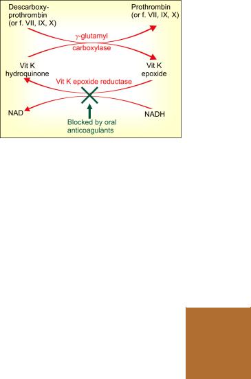
DRUGS AFFECTING COAGULATION, BLEEDING AND THROMBOSIS |
621 |
|
|
reductase (VKOR) and interfere with regeneration of the active hydroquinone form of vit K (Fig. 44.2) which acts as a cofactor for the enzyme γ-glutamyl carboxylase that carries out the final step of γ carboxylating glutamate residues of prothrombin and factors VII, IX and X. This carboxylation is essential for the ability of the clotting factors to bind Ca2+ and to get bound to phospholipid surfaces, necessary for the coagulation sequence to proceed.
Factor VII has the shortest plasma t½ (6 hr), its level falls first when warfarin is given, followed by factor IX (t½ 24 hr), factor X (t½ 40 hr) and prothrombin (t½ 60 hr). Though the synthesis of clotting factors diminishes within 2–4 hours of warfarin administration, anticoagulant effect develops gradually over the next 1–3 days as the levels of the clotting factors already present in plasma decline progressively. Thus, there is always a delay between administration of these drugs and the anticoagulant effect. Larger initial doses hasten the effect only slightly.
Therapeutic effect occurs when synthesis of clotting factors is reduced by 40–50%.
Protein C, protein S (both having anticoagulant property) and osteocalcin contain glutamate residues that require vit. K dependent γ carboxylation. These are also inhibited by oral anticoagulants, but density of adult bone is not affected, though new bone formation may be depressed.
Fig. 44.2: Mechanism of action of oral anticoagulants NAD—Nicotinamide adenine dinucleotide; NADH—its reduced form
The differences between different oral anticoagulants are primarily pharmacokinetic and in the adverse side effects produced by them. These are summarized in Table 44.1.
Racemic Warfarin sod. It is the most popular oral anticoagulant. The commercial preparation of warfarin is a mixture of R (dextrorotatory) and S (levorotatory) enantiomers. The S form is more potent and is metabolized relatively faster by ring oxidation carried out by CYP2C9, while R form is less potent and degraded by side chain reduction carried out by CYP1A and CYP3A4.
TABLE 44.1 |
|
Pharmacokinetic and adverse effect profile of oral anticoagulants |
|||||||
|
|
|
|
|
|
|
|
|
|
|
Drug |
|
t½ |
Duration |
|
|
Dose (mg) |
Adverse side effects |
|
|
|
|
|
(hour) |
of action |
|
|
|
(other than bleeding) |
|
|
|
|
|
|
|
|||
|
|
|
|
|
(days) |
|
Loading |
Maintenance* |
|
|
|
|
|
|
|
|
|
||
1. |
Bishydroxycoumarin |
25–100 |
4–7 |
|
200 for |
50–100 |
Frequent g.i.t. disturbances |
||
|
|
|
|
(dose dependent) |
|
2 days |
|
|
|
2. |
Warfarin sod. |
|
36–48 |
3–6 |
5–10 |
2–10£ |
Alopecia, dermatitis, diarrhoea |
||
3. |
Acenocoumarol |
|
18–24 |
2–3 |
8–12 |
2–8 |
Oral ulceration, g.i.t. distur- |
||
|
(Nicoumalone) |
|
|
|
|
|
|
bances, dermatitis, urticaria, |
|
|
|
|
|
|
|
|
|
|
alopecia |
4. |
Ethylbiscoumacetate |
2 |
1–3 |
900 |
300–600 |
Alopecia, bad taste |
|||
5. |
Phenindione |
|
5 |
1–3 |
200 |
50–100 |
Orange urine, rashes, fever, |
||
|
|
|
|
|
|
|
|
|
leukopenia, hepatitis, nephro- |
|
|
|
|
|
|
|
|
|
pathy, agranulocytosis |
|
|
|
|
|
|
|
|
|
|
44 CHAPTER
* Daily maintenance dose: to be adjusted by measurement of prothrombin time (INR). £ To be taken in a single dose at the same hour (usually bed time) each day.

622 |
DRUGS AFFECTING BLOOD AND BLOOD FORMATION |
|
|
SECTION 10
Both are partially conjugated with glucuronic acid and undergo some enterohepatic circulation; finally excreted in urine.
Warfarin is rapidly and completely absorbed from intestines and is 99% plasma protein bound. It crosses placenta and is secreted in milk; however, quantity of active form is generally insufficient to affect the suckling infant.
UNIWARFIN 1, 2, 5 mg tabs; WARF-5: 5 mg tab.
Bishydroxycoumarin (Dicumarol) It is slowly and unpredictably absorbed orally. Its metabolism is dose dependent—t½ is prolonged at higher doses. Has poor g.i. tolerance; not preferred now.
DICOUMAROL 50 mg tab.
Acenocoumarol (Nicoumalone) The t½ of acenocoumarol as such is 8 hours, but an active metabolite is produced so that overall t½ is about 24 hours. Acts more rapidly.
ACITROM, 1, 2, 4 mg tabs.
Ethyl biscoumacetate It has a rapid and brief action; occasionally used to initiate therapy, but difficult to maintain.
Phenindione Apart from risk of bleeding, it produces more serious organ toxicity; should not be used.
DINDEVAN 50 mg tab.
Adverse effects Bleeding as a result of extension of the desired pharmacological action is the most important problem causing ecchymosis, epistaxis, hematuria, bleeding in the g.i.t. Intracranial or other internal haemorrhages may even be fatal. Bleeding is more likely if therapy is not properly monitored, or when INR exceeds 4, or interacting drugs/contraindications are present.
Treatment: of bleeding due to oral anticoagulants consists of:
•Withhold the anticoagulant.
•Give fresh blood transfusion; this supplies clotting factors and replenishes lost blood. Alternatively fresh frozen plasma may be used
as a source of clotting factors.
•Give vit K1 which is the specific antidote (see p. 615), but it takes 6–24 hours for the clotting factors to be resynthesized and released in blood after vit K administration.
Adverse effects unrelated to anticoagulation are given in Table 44.1. Cutaneous necrosis is a rare
complication that can occur with any oral anticoagulant.
Phenindione produces serious toxicity; should not be used.
Warfarin and acenocoumarol are considered to be the most suitable and better tolerated drugs.
Dose regulation The dose of oral anticoagulant must be individualised by repeated measurement of prothrombin time; the aim is to achieve a therapeutic effect without unduly increasing the chances of bleeding.
The optimum ratio of PT during treatment with the oral anticoagulant to the normal value (of the testing laboratory) has been defined for various indications. But this value differs depending on whether rabbit brain or human brain thromboplastin (Tp) has been used for the test. A standardized system called the International Normalized Ratio (INR) based on the use of human brain Tp has been developed by WHO and adopted in all countries.
Recommended INR for various indications of oral anticoagulants
1. |
Prophylaxis of deep vein thrombosis |
INR |
|||
|
|||||
|
and similar |
indications |
|
|
2–2.5 |
2. |
Treatment |
of deep vein |
thrombosis, |
|
|
|
pulmonary |
embolism, TIAs, |
hip surgery |
2–3 |
|
3. |
Recurrent thromboembolism, |
arterial |
|
||
|
disease (MI), prosthetic |
heart valves |
3–3.5 |
||
Factors enhancing effect of oral anticoagulants are:
•Debility, malnutrition, malabsorption and prolonged antibiotic therapy: the supply of vit
K to liver is reduced in these conditions.
• Liver disease, chronic alcoholism: synthesis of clotting factors may be deficient.
•Hyperthyroidism: the clotting factors are degraded faster.
•Newborns: have low levels of vit K and clotting factors (there should be no need of these drugs in neonates anyway).
Factors decreasing effect of oral anticoagulants are:
•Pregnancy: plasma level of clotting factors is higher.
•Nephrotic syndrome: drug bound to plasma protein is lost in urine.

DRUGS AFFECTING COAGULATION, BLEEDING AND THROMBOSIS |
623 |
|
|
•Genetic warfarin resistance: the affinity of warfarin (as well as of vit K epoxide) to bind to the reductase (VKOR) enzyme, which generates the active vit K hydroquinone, is low. Dose of oral anticoagulant is 4–5 times higher.
Contraindications All contraindications to heparin (see p. 619) apply to these drugs as well. Factors which enhance the effect of oral anticoagulants (see above) should also be taken into consideration.
Oral anticoagulants should not be used during pregnancy. Warfarin given in early pregnancy increases birth defects, especially skeletal abnormalities. It can produce foetal warfarin syndrome—hypoplasia of nose, eye socket, hand bones, and growth retardation. Given later in pregnancy, it can cause CNS defects, foetal haemorrhage, foetal death and accentuates neonatal hypoprothrombinemia.
Drug interactions A large number of drugs interact with oral anticoagulants at pharmacokinetic or pharmacodynamic level, and either enhance or decrease their effect. These interactions are clinically important (may be fatal if bleeding occurs) and may involve more than one mechanism; the exact mechanism of an interaction is not always definable.
A. Enhanced anticoagulant action
1.Broad-spectrum antibiotics: inhibit gut flora and reduce vit K production.
2.Newer cephalosporins (ceftriaxone, cefoperazone) cause hypoprothrombinaemia by the same mechanism as warfarin —additive action.
3.Aspirin: inhibits platelet aggregation and causes g.i. bleeding—this may be hazardous in anticoagulated patients. High doses of salicylates have synergistic hypoprothrombinemic action and also displace warfarin from protein binding site.
4.Long acting sulfonamides, indomethacin, phenytoin and probenecid: displace warfarin from plasma protein binding.
5.Chloramphenicol, erythromycin, celecoxib, cimetidine, allopurinol, amiodarone and metronidazole: inhibit warfarin metabolism.
6.Tolbutamide and phenytoin: inhibit warfarin metabolism and vice versa.
7.Liquid paraffin (habitual use): reduces vit K absorption.
B. Reduced anticoagulant action
1.Barbiturates (but not benzodiazepines), carbamazepine, rifampin and griseofulvin induce the metabolism of oral anticoagulants. The dose of anticoagulant determined during therapy with these drugs would be higher: if the same is continued after withdrawing the inducer—marked hypoprothrombinemia can occur—fatal bleeding is on record.
2.Oral contraceptives: increase blood levels of clotting factors.
DIRECT FACTOR Xa INHIBITORS
Recently some orally active drugs have been produced which directly bind to and inactivate factor Xa, instead of inhibiting its synthesis. They, therefore, act rapidly without a lag time (as in case of warfarin, etc.), and have short-lasting action.
Rivaroxaban
It is an orally active direct inhibitor of activated factor Xa which has become available for prophylaxis and treatment of DVT. Its anticoagulant action develops rapidly within 3–4 hours of ingestion and lasts for ~24 hours. It is largely metabolized, but also excreted unchanged in urine; plasma t½ is 7–11 hours. Another advantage is that it requires no laboratory monitoring of PT or aPTT, and is recommended in a fixed dose of 10 mg once daily starting 6–10 hours after surgery for prophylaxis of venous thromboembolism following total knee/hip replacement. In comparative trials, its efficacy has been found similar to a regimen of LMW heparin followed by warfarin. Rivaroxaban has also been found equally effective as warfarin for preventing stroke in patients with atrial fibrillation. Side effects reported are bleeding, nausea, hypotension, tachycardia and edema.
ORAL DIRECT THROMBIN INHIBITOR
Dabigatran etexilate It is a prodrug which after oral administration is rapidly hydrolysed to dabigatran, a direct thrombin inhibitor which reversibly blocks the catalytic site of thrombin and produces a rapid (within 2 hours) anticoagulant action. Though oral bioavailability is low, the anticoagulant effect is consistent, and no laboratory monitoring is required. The plasma t½ is 12–14 hours and duration of action 24 hours. In the UK, Canada and Europe it is approved for prevention of venous thromboembolism following hip/knee joint replacement surgery. Administered in a dose of 110 mg (75 mg for elderly > 75 years) once
44 CHAPTER
624
SECTION 10
DRUGS AFFECTING BLOOD AND BLOOD FORMATION
TABLE 44.2  Some comparative aspects of heparin and oral anticoagulants
|
|
Heparin |
Warfarin |
|
|
|
|
1. |
Chemistry |
Mucopolysaccharide |
Coumarin derivative |
2. |
Source |
Hog lung, pig intestine |
Synthetic |
3. |
Route of admin. |
Parenteral (i.v., s.c.) |
Oral |
4. |
Onset of action |
Immediate |
Delayed (1–3 days) |
5. |
Duration of action |
4–6 hrs |
3–6 days |
6. |
Activity |
In vitro and in vivo |
In vivo only |
7. |
Mechanism |
Blocks action of factor X and |
Inhibits synthesis of clotting factors |
|
|
thrombin |
|
8. |
Antagonist |
Protamine sulphate |
Vit K |
9. |
Variability in response |
Little |
Marked |
10. |
Lab. control |
aPTT/clotting time |
Prothrombin time/INR |
|
|
(desirable) |
(essential) |
11. |
Drug interactions |
Few and not significant |
Many and significant |
12. |
Use |
To initiate therapy |
For maintenance |
|
|
|
|
daily, it has been found comparable to warfarin. In another large trial dabigatran etexilate 150 mg twice daily has yielded superior results to warfarin for prevention of embolism and stroke in patients of atrial fibrillation. In the USA it is approved for this indication. Adverse effects are bleeding and less commonly hepatobiliary disorders.
USES OF ANTICOAGULANTS
The aim of using anticoagulants is to prevent thrombus extension and embolic complications by reducing the rate of fibrin formation. They do not dissolve already formed clot, but prevent recurrences. Heparin is utilized for rapid and shortlived action, while oral anticoagulants are suitable for maintenance therapy. Generally, the two are started together; heparin is discontinued after 4–7 days when warfarin has taken effect.
The important features of heparin and oral anticoagulants are compared in Table 44.2.
1. Deep vein thrombosis (DVT) and pulmonary embolism (PE) Because venous thrombi are mainly fibrin thrombi, anticoagulants are expected to be highly effective. The best evidence of efficacy of anticoagulants comes from treatment and prevention of venous thrombosis and pulmonary embolism. Prophylaxis is recommended for all high risk patients including bedridden, elderly, postoperative, postpartum, poststroke and leg fracture patients. When deep vein thrombosis/
pulmonary embolism has occurred, immediate heparin/LMW heparin followed by warfarin therapy should be instituted. Three months anticoagulant therapy (continued further if risk factor persists) has been recommended by American College of Chest Physicians (2001).
Introduction of low dose s.c. heparin prophylaxis for patients undergoing elective surgery has considerably reduced the incidence of leg vein thrombosis and pulmonary embolism in the postoperative period. It has been extended to other situations needing prolonged immobilization. It is based on the premise that inhibition of small amount of activated factor X prevents further amplification of active products—particularly thrombin. This is the regimen of choice which does not need laboratory monitoring; spontaneous bleeding does not occur. LMW heparin/fondaparinux have now practically replaced UFH, except in case of major surgery and in high risk cases, because action of UFH can be terminated rapidly.
Anticoagulants are of little value in chronic peripheral vascular diseases.
2. Myocardial infarction (MI) Arterial thrombi are mainly platelet thrombi; anticoagulants are of questionable value. Their use in acute MI has declined. They do not alter immediate mortality of MI. It was hoped that anticoagulants will

DRUGS AFFECTING COAGULATION, BLEEDING AND THROMBOSIS |
625 |
|
|
prevent extension of the thrombus and ward off a recurrent attack. This has not been supported by the collected statistics. They may benefit by preventing mural thrombi at the site of infarction and venous thrombi in leg veins. Thus, anticoagulants may be given for a short period till patient becomes ambulatory. For secondary prophylaxis against a subsequent attack— anticoagulants are inferior to antiplatelet drugs.
Heparin (i.v.) or preferably LMW heparin/ fondaparinux s.c. once or twice daily for 2–8 days followed by oral anticoagulants for 3 months or continuation of LMW heparin for 2–3 months are generally given after recanalization of coronary artery by fibrinolytic therapy. Heparin is also used during coronary angioplasty and stent placement.
3.Unstable angina Short-term use of heparin has reduced the occurrence of MI in unstable angina patients; aspirin is equally effective. Current recommendation is to use aspirin + heparin/LMW heparin followed by warfarin.
4.Rheumatic heart disease; Atrial fibrillation (AF) All atrial fibrillation patients should be protected against thromboembolism from fibrillating atria and the resulting stroke. For this purpose, the effective options are warfarin/low dose heparin/low dose aspirin. The ‘Stroke prevention in Atrial Fibrillation’ trial and a metaanalysis have shown warfarin to be more effective than aspirin. Current guideline is to give warfarin to a target INR of 2–3 in AF patients with high risk for stroke (elderly, heart failure, etc.), and to reserve aspirin for low risk patients or for those unable to take warfarin. Anticoagulants are given for 3–4 weeks before and after attempting conversion of AF to sinus rhythm.
5.Cerebrovascular disease Anticoagulants are of little value in cerebral thrombosis. They have been used with the aim of preventing clot propagation, but all the trials conducted, including International Stroke Trial (IST), have failed to demonstrate significant benefit. Neurological sequelae are similar whether anticoagulants are used or not. Moreover, in the initial stages it is
difficult to rule out cerebral haemorrhage (unless CAT scan is done) in which they can be devastating. They may be used in cerebral embolism, because showers of emboli are often recurrent and can be prevented by anticoagulants. A late start (after one week) anticoagulant therapy is advocated by many in case of large embolic stroke. Oral anticoagulants may be beneficial in transient ischaemic attacks (TIAs), but antiplatelet drugs are simpler to use and probably better.
6. Vascular surgery, prosthetic heart valves, retinal vessel thrombosis, extracorporeal circulation, haemodialysis Anticoagulants are indicated along with antiplatelet drugs for prevention of thromboembolism.
Heparin flushes (200 U in 2 ml) every 4–8 hr are used to keep patent long-term intravascular cannulae/catheters.
7. Defibrination syndrome or ‘disseminated intravascular coagulation’ occurs in abruptio placentae and other obstetric conditions, certain malignancies and infections. The coagulation factors get consumed for the formation of intravascular microclots and blood is incoagulable. Heparin paradoxically checks bleeding in such patients by preserving the clotting factors. However, in some cases heparin may aggravate bleeding.
FIBRINOLYTICS (Thrombolytics)
These are drugs used to lyse thrombi/clot to recanalize occluded blood vessels (mainly coronary artery). They are therapeutic rather than prophylactic and work by activating the natural fibrinolytic system (Fig. 44.3).
Haemostatic plug of platelets formed at the site of injury to blood vessels is reinforced by fibrin deposition to form a thrombus. Once repair is over, the fibrinolytic system is activated to remove the fibrin. The enzyme responsible for digesting fibrin is a serine protease Plasmin generated from plasminogen by tissue plasminogen activator (t-PA), which is produced primarily by vascular endothelium. Plasminogen circulates in plasma as well as remains bound to fibrin. The t-PA selectively activates fibrin-bound plasminogen within the thrombus, and any plasmin that leaks is inactivated by circulating antiplasmins. Fibrin bound plasmin
44 CHAPTER
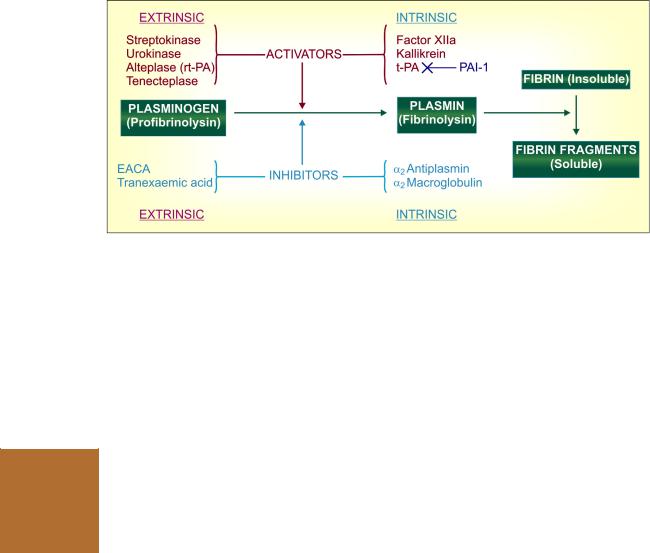
626 |
DRUGS AFFECTING BLOOD AND BLOOD FORMATION |
|
|
Fig. 44.3: The plasminogen-plasmin system
t-PA—Tissue plasminogen activator; rt-PA—Recombinant t-PA; PAI-1—Plasminogen activator inhibitor-1
SECTION 10
is not inactivated by antiplasmins because of common binding site for both fibrin and antiplasmin.The t-PA itself is inactivated by plasminogen activator inhibitor-1 and -2 (PAI-1, PAI-2).
When excessive amounts of plasminogen are activated (by administered fibrinolytics), the α2 antiplasmin is exhausted and active plasmin persists in plasma. Plasmin is a rather nonspecific protease: degrades coagulation factors (including fibrinogen) and some other plasma proteins as well. Thus, activation of circulating plasminogen induces a lytic state whose major complication is haemorrhage. Even selective activation of thrombus bound plasmin can cause bleeding by dissolving physiological thrombi.
In general, venous thrombi are lysed more easily by fibrinolytics than arterial, and recent thrombi respond better. They have little effect on thrombi > 3 days old. The clinically important fibrinolytics are:
Streptokinase |
Alteplase (rt-PA) |
Urokinase |
Reteplase |
|
Tenecteplase |
Streptokinase (Stk) Obtained from β haemolytic Streptococci group C, it is the first fibrinolytic drug to be used clinically, but is not employed now except for considerations of cost. Streptokinase is inactive as such; combines with circulating plasminogen molecules to form an activator complex which then causes limited proteolysis of other plasminogen molecules to generate the active enzyme plasmin. Stk. is non-fibrin specific, i.e. activates both circulating as well as fibrin bound plasminogen. Therefore, it depletes circulating fibrinogen and predisposes to bleeding. Compared to newer more fibrin-specific tissue plasminogen activators
(alteplase, etc.) it is less effective in opening occluded coronary arteries, and causes less reduction in MI related mortality.
There are several other disadvantages as well with Stk. Antistreptococcal antibodies due to past infections inactivate considerable fraction of the initial dose of Stk. A loading dose therefore is necessary. Plasma t½ is estimated to be 30–80 min. Stk is antigenic—can cause hypersensitivity reactions; anaphylaxis occurs in 1–2% patients. It cannot be used second time due to neutralization by antibodies generated in response to the earlier dose. Fever, hypotension and arrhythmias are reported. However, being less expensive, it is still used in resource poor areas, but not in Europe or USA.
STREPTASE, (freeze dried powder in vials) 2.5 lac, 7.5 lac and 15 lac IU/vial, ESKINASE, CARDIOSTREP 7.5 lac, 15 lac IU/vial.
For MI: 7.5–15 lac IU infused i.v. over 1 hr.
For deep vein thrombosis and pulmonary embolism: 2.5 lac IU loading dose over ½–1 hr, followed by 1 lac IU/hr for 24 hr.
Urokinase It is an enzyme isolated from human urine; but commercially prepared from cultured human kidney cells. It activates plasminogen directly and has a plasma t½ of 10–15 min. It is nonantigenic. Fever occurs during treatment, but hypotension and allergic phenomena are rare. Urokinase is Indicated in patients in whom streptokinase has been given for an earlier episode, but is seldom used now.
UROKINASE, UROPASE, 2.5 lac, 5 lac, 7.5 lac, 10 lac IU per vial inj.
For MI: 2.5 lac IU i.v. over 10 min followed by 5 lac IU over next 60 min (stop in between if full recanalization occurs) or 6000 IU/min for upto 2 hr.
For venous thrombosis and pulmonary embolism: 4400 IU/kg over 10 min i.v. followed by 4400 IU/kg/hr for 12 hr.

DRUGS AFFECTING COAGULATION, BLEEDING AND THROMBOSIS |
627 |
|
|
Alteplase (recombinant tissue plasminogen activator (rt-PA) Produced by recombinant DNA technology from human tissue culture, it is moderately specific for fibrin-bound plasminogen, so that circulating fibrinogen is lowered only by ~ 50%. It is rapidly cleared by liver and inactivated by plasminogen activator inhibitor-1 (PAI-1). The plasma t½ is 4–8 min. Because of the short t½, it needs to be given by slow i.v. infusion and often requires heparin coadministration. It is nonantigenic, but nausea, mild hypotension and fever may occur. It is expensive.
ACTILYSE 50 mg vial with 50 ml solvent water.
For MI: (accelerated regimen) 15 mg i.v. bolus injection followed by 50 mg over 30 min, then 35 mg over the next 1 hr. (total 90 min).
For pulmonary embolism: 100 mg i.v. infused over 2 hr. For ischaemic stroke: 0.9 mg/kg by i.v. infusion over 60 min, with 10% of the dose injected in the first minute.
Reteplase It is a modified form of rt-PA that is longer acting, but somewhat less specific for fibrin-bound plasminogen. The longer duration of action enables bolus dose administration (10 mg over 10 min repeated after 30 min).
Tenecteplase This genetically engineered substitution mutant of native t-PA has higher fibrin selectivity, slower plasma clearance (longer duration of action) and resistance to inhibition by PAI-1. It is the only fibrinolytic agent that can be injected i.v. as a single bolus dose over 10 sec, while alteplase requires 90 min infusion. This feature makes it possible to institute fibrinolytic therapy immediately on diagnosis of ST segment elevation myocardial infarction (STEMI), even during transport of the patient to the hospital. Several randomized multicentric trials have assessed its efficacy in STEMI and found it to be at least equally efficacious to alteplase. Risk of noncerebral bleeding may be lower with tenecteplase, but cranial bleeding incidence is similar.
Dose: 0.5 mg/kg single i.v. bolus injection. ELAXIM 30 mg, 50 mg per vial inj.
Uses of fibrinolytics
1. Acute myocardial infarction is the chief indication. Fibrinolytics are an alternative first line approach to emergency percutaneous coronary intervention (PCI) with stent placement.
Recanalization of thrombosed coronary artery has been achieved in 50–90% cases. Time lag in starting the infusion is critical for reducing area of necrosis, preserving ventricular function and reducing mortality. The benefits of i.v. thrombolytic therapy have been established by large randomised studies. Aspirin with or without heparin is generally started concurrently or soon after thrombolysis to prevent reocclusion.
Alteplase has advantages over streptokinase, including higher thrombolytic efficacy. However, incidence of haemorrhage is not lower. Its stronger lytic effect on physiological haemostatic plugs may compensate for the lesser systemic fibrinolytic state.
2. Deep vein thrombosis in leg, pelvis, shoulder etc.; up to 60% patients can be successfully treated. Thrombolytics can decrease subsequent pain and swelling, but the main advantage is preservation of venous valves and may be a reduced risk of pulmonary embolism, though at the risk of haemorrhage. Comparable results have been obtained with Stk, urokinase and alteplase.
3. Pulmonary embolism Fibrinolytic therapy is indicated in large, life-threatening PE. The lung function may be better preserved, but reduction in mortality is not established.
4. Peripheral arterial occlusion Fibrinolytics recanalise ~40% limb artery occlusions, especially those treated within 72 hr. However, it is indicated only when surgical thrombectomy is not possible. Regional intraarterial fibrinolytics have been used for limb arteries with greater success. Peripheral arterial thrombolysis is followed by shortterm heparin and long-term aspirin therapy.
Fibrinolytics have no role in chronic peripheral vascular diseases.
5. Stroke: Thrombolytic therapy of ischaemic stroke is controversial. Possibility of improved neurological outcome is to be balanced with risk of intracranial haemorrhage. No net benefit was concluded by the ATLANTIS trial in patients treated at 3–5 hours of stroke onset. However, alteplase is approved for use in ischaemic stroke, and current opinion strongly recommends use of i.v. alteplase in carefully selected patients who
44 CHAPTER

628 |
DRUGS AFFECTING BLOOD AND BLOOD FORMATION |
|
|
SECTION 10
Contraindications to thrombolytic therapy
1.H/o Intracranial haemorrhage
2.H/o Ischaemic stroke in past 3 months
3.H/o Head injury in past 3 months
4.Intracranial tumour/vascular abnormality/ aneurysms
5.Active bleeding/bleeding disorders
6.Peptic ulcer, esophageal varices
7.Any wound or recent fracture or tooth extraction
8.H/o major surgery within 3 weeks
9.Uncontrolled hypertension
10.Pregnancy
can be treated within 3 hours of onset, and in whom intracranial haemorrhage is ruled out along with all risk factors for bleeding (see contraindications in box).
Evaluation All patients with STEMI are candidates for reperfusion therapy. No consistent benefit of fibrinolytics has been demonstrated in non-STEMI cases, while possibility of haemorrhage is increased. Only selected cases of NSTEMI may be treated with fibrinolytics. Both short-term and long-term outcome is determined by early restoration of flow in the occluded artery, regardless of whether it is achieved by thrombolysis or by PCI. Best results are obtained if perfusion can be restored within the first hour (the golden hour). While the efficacy of fibrinolytics in dissolving the thrombus diminishes with passage of time (little benefit after 6 hours of MI onset), reperfusion by PCI is affected to a lesser extent by the time lapse. Thrombolysis may be favoured if it can be started within 1–2 hours of onset. After 3 hours, PCI is favoured. Moreover, PCI has the advantage of lower bleeding risk, higher grade of flow in the reperfused artery and reduction in the rate of nonfatal recurrent MI compared to thrombolysis. As such, PCI has yielded superior results compared to fibrinolytics and is being preferred at centres where it can be performed swiftly with requisite expertise. Primary PCI is the procedure of choice for patients with contraindications to thrombolytics (see box). Fibrinolytic therapy requires careful patient selection, but often can be instituted with less delay, and even at centres not well equipped for PCI.
Another approach is ‘facilitated PCI’ wherein full or reduced dose fibrinoytic therapy is followed at the earliest by PCI. The results of this approach are comparable to those of primary PCI. The European as well as American (ACC, AHA) guidelines provide that STEMI patients should be treated with primary PCI or with fibrinolytic drugs followed by immediate rescue PCI, if reperfusion fails with the fibrinolytic. Aspirin and heparin are continued after thrombolysis.
ANTIFIBRINOLYTIC DRUGS
These are drugs which inhibit plasminogen activation and dissolution of clot, and are used to check fibrinolysis associated bleeding.
Epsilon amino-caproic acid (EACA) It is a lysine analogue which combines with the lysinebinding sites of plasminogen and plasmin so that the latter is not able to bind to fibrin and lyse it. It is a specific antidote for fibrinolytic agents and has been used in many hyperplasminaemic states associated with excessive intravascular fibrinolysis resulting in bleeding. The primary indication is to counteract the effect of fibrinolytic drugs and bleeding due to their use. In haemophiliacs, it has adjunctive value for controlling bleeding due to tooth extraction, prostatectomy, trauma, etc.
In haematuria it can cause ureteric obstruction by the unlysed clots. Therefore, fibrinolysis must be established firmly before using it. It can cause intravascular thrombosis. Rapid i.v. injection results in hypotension, bradycardia and may be arrhythmias. It should be used cautiously when renal function is impaired. Myopathy occurs rarely. The large dose needed is a limitation, and tranexamic acid is mostly preferred.
Initial priming dose is 5 g oral/i.v., followed by 1 g hourly till bleeding stops (max. 30 g in 24 hrs).
AMICAR, HEMOCID, HAMOSTAT 0.5 g tab., 1.25 g/5 ml syr., 5 g/20 ml inj.
Tranexamic acid Like EACA, it binds to the lysine binding site on plasminogen and prevents its combination with fibrin leading to fibrinolysis. It is 7 times more potent than EACA, and is

DRUGS AFFECTING COAGULATION, BLEEDING AND THROMBOSIS |
629 |
|
|
preferred for prevention/control of excessive bleeding due to:
•Fibrinolytic drugs.
•Cardio-pulmonary bypass surgery.
•Tonsillectomy, prostatic surgery, tooth extraction in haemophiliacs.
•Menorrhagia, especially due to IUCD.
•Recurrent epistaxis, hyphema due to ocular trauma, peptic ulcer.
Main side effects are nausea and diarrhoea. Thromboembolic events, disturbed colour vision and allergic reactions are infrequent. Thrombophlebitis of injected vein can occur.
Dose: 10–15 mg/kg 2–3 times a day or 1–1.5 g TDS oral, 0.5–1 g TDS by slow i.v. infusion.
DUBATRAN, PAUSE, TRANAREST 500 mg tab, 500 mg/5 ml inj.
ANTIPLATELET DRUGS (Antithrombotic drugs)
These are drugs which interfere with platelet function and are useful in the prophylaxis of thromboembolic disorders.
Platelets express several glycoprotein (GP) integrin receptors on their surface. Reactive proteins like collagen are exposed when there is damage to vascular endothelium, and they react respectively with platelet GPIa and GPIb receptors. This results in platelet activation and release of proaggregatory and vasoconstrictor mediators like TXA2, ADP and 5-HT. The platelet GPIIb/IIIa receptor undergoes a conformational change favouring binding of fibrinogen and vonWillebrand factor (vWF) that crosslink platelets inducing aggregation and anchorage to vessel wall/other surfaces. Thus, a ‘platelet plug’ is formed. In veins, due to sluggish blood flow, a fibrinous tail is formed which traps RBCs ‘the red tail’. In arteries, platelet mass is the main constituent of the thrombus. Antiplatelet drugs are, therefore, more useful in arterial thrombosis, while anticoagulants are more effective in venous thrombosis.
Prostacyclin (PGI2), synthesized in the intima of blood vessels, is a strong inhibitor of platelet aggregation. A balance between TXA2 released from platelets and PGI2 released from vessel wall appears to control intravascular thrombus formation. Platelets also play a role in atherogenesis.
In the above scheme, various drugs act on different targets to interfere with platelet function. Therefore, given together, their actions are synergistic. The clinically important antiplatelet drugs are:
Antiplatelet Drugs
Aspirin
Dipyridamole
P2Y12 Receptor Blockers |
GPIIb/IIIa Antagonists |
Ticlopidine |
Abciximab |
Clopidogrel |
Eptifibatide |
Prasugrel |
Tirofiban |
Aspirin It acetylates and inhibits the enzyme COX1 and TX-synthase—inactivating them irreversibly. Because TXA2 is the major arachidonic acid product generated by platelets, and that platelets are exposed to aspirin in the portal circulation before it is deacetylated during first pass in the liver, and because platelets cannot synthesize fresh enzyme (have no nuclei), TXA2 formation is suppressed at very low doses and till fresh platelets are formed. Thus, aspirin induced prolongation of bleeding time lasts for 5–7 days. Effect of daily doses cumulates and it has now been shown that doses as low as 40 mg/day have an effect on platelet aggregation. Maximal inhibition of platelet function occurs at 75–150 mg aspirin per day. However, aspirin may not effectively inhibit platelet aggregation in some patients.
Inhibition of COX-1 by aspirin in vessel wall decreases PGI2 synthesis as well. However, since intimal cells can synthesize fresh enzyme, activity returns rapidly. It is possible that at low doses (75–150 mg/day or 300 mg twice weekly), TXA2 formation by platelets is selectively suppressed, whereas higher doses (> 900 mg/day) may decrease both TXA2 and PGI2 production.
Aspirin inhibits the release of ADP from platelets and their sticking to each other, but has no effect on platelet survival time and their adhesion to damaged vessel wall.
ASA 50 mg tab., COLSPRIN, DISPRIN CV-100: aspirin 100 mg soluble tab, LOPRIN 75 mg tab, ASPICOT 80 mg tab, ECOSPRIN 75, 150 mg tab.
Other NSAIDs are reversible inhibitors of COX, produce short-lasting inhibition of platelet function—are not clinically useful.
Dipyridamole It is a vasodilator that was introduced for angina pectoris (see Ch. 39). It inhibits
44 CHAPTER

630 |
DRUGS AFFECTING BLOOD AND BLOOD FORMATION |
|
|
SECTION 10
phosphodiesterase as well as blocks uptake of adenosine to increase platelet cAMP which in turn potentiates PGI2 and interferes with aggregation. Levels of TXA2 or PGI2, are not altered, but platelet survival time reduced by disease is normalized.
Dipyridamole alone has little clinically significant effect, but improves the response to warfarin, along with which it is used to decrease the incidence of thromboembolism in patients with prosthetic heart valves.
Dipyridamole has also been used to enhance the antiplatelet action of aspirin. This combination may additionally lower the risk of stroke in patients with transient ischaemic attacks (TIAs), but trials have failed to demonstrate additional benefit in prophylaxis of MI.
Dose: 150–300 mg/day. PERSANTIN 25, 100 mg tabs, THROMBONIL 75, 100 mg tabs, DYNASPRIN: dipyridamole 75 mg + aspirin 60 mg e.c. tab., CARDIWELL PLUS: dipyridamole 75 mg + aspirin 40 mg tab.
Ticlopidine It is the first thienopyridine which alters surface receptors on platelets and inhibits ADP as well as fibrinogen-induced platelet aggregation. The Gi coupled P2Y12 (also labelled P2YAC) type of purinergic receptors which mediate adenylyl cyclase inhibition due to ADP are blocked irreversibly by the active metabolite of ticlopidine. As a result, activation of platelets is interfered. Fibrinogen binding to platelets is prevented without modification of GPIIb/IIIa receptor. There is no effect on platelet TXA2, but bleeding time is prolonged and platelet survival in extra-corporeal circulation is increased. Because of different mechanism of action, it has synergistic effect on platelets with aspirin. Their combination is a potent platelet inhibitor.
Ticlopidine is well absorbed orally, is converted in liver to an active metabolite, and is eliminated with a plasma t½ of 8 hours. However, because it causes irreversible blockade of P2Y12 receptors, the effect on platelets cumulates; peak platelet inhibition is produced after 8–10 days therapy, and the effect lasts 5–6 days after discontinuing the drug.
Ticlopidine has produced beneficial effects in stroke prevention, TIAs, intermittent claudication, unstable angina, PCI, coronary artery bypass grafts and secondary prophylaxis of MI. Combined with aspirin, it has markedly lowered incidence of restenosis after PCI and stent thrombosis.
Because of its potential for serious adverse reactions, use of ticlopidine has markedly declined in favour of clopidogrel.
Side effects: Diarrhoea, vomiting, abdominal pain, headache, tinnitus, skin rash. Serious adverse effects are bleeding, neutropenia, thrombocytopenia, haemolysis and jaundice.
Several fatalities have occurred.
Dose: 250 mg BD with meals; effect persists several days after discontinuation; TYKLID, TICLOVAS, TICLOP, 250 mg tab; ASTIC ticlopidine 250 mg + aspirin 100 mg tab.
Clopidogrel This newer and more potent congener of ticlopidine has similar mechanism of action, ability to irreversibly inhibit platelet function and range of therapeutic efficacy, but is safer and better tolerated (CLASSICS study). The clopidogrel vs aspirin in patients at risk of ischaemic events (CAPRIE) trial has found clopidogrel recipients to have a slightly lower annual risk of primary ischaemic events than aspirin recipients. Combination of clopidogrel and aspirin is synergistic in preventing ischaemic episodes, and is utilized for checking restenosis of stented coronaries.
Like ticlopidine, clopidogrel is also a prodrug. About 50% of the ingested dose is absorbed, and only a fraction of this is slowly activated in liver by CYP2C19, while the rest is inactivated by other enzymes. It is a slow acting drug; antiplatelet action takes about 4 hours to start and develops over days. Since CYP2C19, exhibits genetic polymorphism, the activation of clopidogrel and consequently its antiplatelet action shows high interindividual variability. Some patients are nonresponsive. Omeprazole, an inhibitor of CYP2C19, reduces metabolic activation of clopidogrel and its antiplatelet action. However, like ticlopidine, the action of clopidogrel lasts 5–7 days due to irreversible blockade of platelet P2Y12 receptors.
The most important adverse effect is bleeding. Addition of aspirin to clopidogrel has been found

DRUGS AFFECTING COAGULATION, BLEEDING AND THROMBOSIS |
631 |
|
|
to double the incidence of serious bleeding among high risk stroke patients (MATCH study). However, neutropenia, thrombocytopenia and other bone marrow toxicity is rare. Side effects are diarrhoea, epigastric pain and rashes.
Dose: 75 mg OD; CLODREL, CLOPILET, DEPLATT 75 mg tab.
Prasugrel This is the latest, most potent and faster acting P2Y12 purinergic receptor blocker, that is being increasingly used in acute coronary syndromes (ACS) and when strong antiplatelet action is required. Like its predecessors, it is also a prodrug, but is more rapidly absorbed and more rapidly as well as more completely activated, resulting in faster and more consistent platelet inhibition. Though CYP2C19 is involved in activation of prasugrel as well, genetic polymorphism related decrease in response, or interference by omeprazole treatment has not been prominent.
Because of rapid action, prasugrel is particularly suitable for use in STEMI. It is the preferred thienopyridine for ACS to cover angioplasty with or without stent placement. The TRITON trial compared prasugrel with clopidogrel in STEMI and NSTEMI. There was 19% greater reduction in death from cardiovascular causes in the prasugrel group. Superior clinical outcomes and reduction in stent thrombosis have been obtained with prasugrel. Bleeding complications are also more frequent and more serious. Patients with history of ischaemic stroke and TIAs are at greater risk of intracranial haemorrhage. Prasugrel is contraindicated in such patients.
Dose: 10 mg OD; elderly or those <60 kg body weight 5 mg OD; a loading dose of 60 mg may be given for urgent action.
PRASULET, PRASUSAFE, PRASUREL 5 mg, 10 mg tabs.
Glycoprotein (GP) IIb/IIIa receptor antagonists
GP IIb/IIIa antagonists are a newer class of potent platelet aggregation inhibitors which act by blocking the key receptor involved in platelet aggregation. The GPIIb/IIIa is an adhesive receptor (integrin) on platelet surface for fibrinogen and vWF through which agonists like collagen, thrombin, TXA2, ADP, etc. finally induce platelet
aggregation. Thus, GP IIb/IIIa antagonists block aggregation induced by all platelet agonists.
Abciximab It is the Fab fragment of a chimeric monoclonal antibody against GP IIb/IIIa, protein, but is relatively nonspecific and binds to some other surface proteins as well. Given along with aspirin + heparin during PCI it has markedly reduced the incidence of restenosis, subsequent MI and death. In the ISAR-REACT2 trial addition of abciximab to clopidogrel (600 mg oral loading dose) for PCI in high-risk ACS patients, reduced ischaemic events by 25%. After a bolus dose, platelet aggregation remains inhibited for 12–24 hr, while the remaining antibody is cleared from blood with a t½ of 10–30 min.
Dose: 0.25 mg/kg i.v. 10–60 min before PCI, followed by 10 µg/min for 12 hr. REOPRO 2 mg/ml inj.
Abciximab is nonantigenic. The main risk is haemorrhage, incidence of which can be reduced by carefully managing the concomitant heparin therapy. Thrombocytopenia is another complication. It should not be used second time, since risk of thrombocytopenia increases. Constipation, ileus and arrhythmias can occur. It is expensive, but is being used in unstable angina and as adjuvant to coronary thrombolysis/PCI with stent placement.
Eptifibatide It is a synthetic cyclic peptide that selectively binds to platelet surface GPIIb/IIIa receptor and inhibits platelet aggregation. Though its plasma t½ (2.5 hours) is longer than that of abciximab, platelet inhibition reverses in a shorter time (within 6–10 hours) because it quickly dissociates from the receptor. Infused i.v., eptifibatide is indicated in:
Unstable angina: 180 μg/kg i.v. bolus, followed by 2 μg/kg/ min infusion upto 72 hours.
Coronary angioplasty: 180 μg/kg i.v. bolus, immediately before procedure; follow with 2 μg/kg/min for 12–24 hours. CLOTIDE, COROMAX, UNIGRILIN 20 mg/10 ml and 75 mg/100 ml vials.
Aspirin and heparin are generally given concurrently.
Bleeding and thrombocytopenia are the major adverse effects. Rashes and anaphylaxis are rare.
Tirofiban This is a nonpeptide but specific GPIIb/IIIa antagonist that is similar in properties
44 CHAPTER

632 |
DRUGS AFFECTING BLOOD AND BLOOD FORMATION |
|
|
SECTION 10
to eptifibatide. Its plasma t½ is 2 hours, and it dissociates rapidly from the receptors. The indications and adverse effects are also similar to eptifibatide.
Acute coronary syndromes: 0.4 μg/kg/min for 30 min followed by 0.1 μg/kg/min for upto 48 hours. If angioplasty is performed, infusion to continue till 12–24 hours thereafter.
AGGRAMED, AGGRITOR, AGGRIBLOC 5 mg/100 ml infusion.
Uses of antiplatelet drugs
The aim of using antiplatelet drugs is to prevent intravascular thrombosis and embolization, with minimal risk of haemorrhage. The intensity of antiplatelet therapy is selected according to the thrombotic influences present in a patient. For indications like maintenance of vascular recanalization, stent placement, vessel grafting, etc. potent inhibition of platelet function is required. This is now possible and is achieved by combining antiplatelet drugs which act by different mechanisms.
1.Coronary artery disease On the basis of trials in post-MI patients as well as in those with no such history, it is recommended that aspirin 75–150 mg/day be given to all individuals with evidence of coronary artery disease and in those with risk factors for the same, but routine use in the whole population is not warranted. Primary prevention of ischaemia with aspirin is of no proven benefit. It reduces the incidence of fatal as well as nonfatal MI, but increases the risk of cerebral haemorrhage. Clopidogrel is an alternative to aspirin in symptomatic patients of ischaemia. Continued aspirin/clopidogrel prophylaxis in post-MI patients clearly prevents reinfarction and reduces mortality.
2.Acute coronary syndromes (ACSs) These comprise of a range of acute cardiac ischaemic states from unstable angina (UA) to non-ST elevation myocardial infarction (NSTEMI) to
STEMI (see p. 556).
The coronary obstruction in UA and NSTEMI is partial, while that in STEMI is total. UA and NSTEMI are differentiated on the basis of absence or presence of laboratory markers of cardiac myocyte necrosis (myoglobin, CK, troponin I, etc.). The ischaemic status is often dynamic and the patient may rapidly shift from one category to the next.
Soluble aspirin (325 mg oral) and a LMW heparin (s.c.) are given at presentation to all patients with ACS.
Unstable angina Aspirin reduces the risk of progression to MI and sudden death. Clopidogrel is generally combined with aspirin, or may be used as alternative if aspirin cannot be given. For maximum protection the antiplatelet drugs are supplemented with heparin followed by warfarin.
The ‘Clopidogrel in unstable angina to prevent recurrent events’ (CURE) trial has found that addition of clopidogrel to aspirin further reduced cardiovascular mortality, nonfatal MI and stroke by 20%.
NSTEMI Patients of NSTEMI who are managed without PCI/thrombolysis are generally put on a combination of aspirin + clopidogrel, which is continued upto one year.
STEMI Primary PCI with or without stent placement is the procedure of choice for all STEMI as well as high risk NSTEMI patients who present within 12 hours. Prasugrel + aspirin is the antiplatelet regimen most commonly selected for patients who are to undergo PCI. Prasugrel acts rapidly and more predictably than clopidogrel. Prasugrel is also perferred over clopidogrel in diabetics. The GPIIb/IIIa antagonists are the most powerful antiplatelet drugs; are combined with aspirin for high risk patients undergoing PCI. Abciximab/eptifibatide/tirofiban infused i.v. along with oral aspirin and s.c. heparin markedly reduce incidence of restenosis and subsequent MI after coronary angioplasty. The GPIIb/IIIa antagonists are infused for a maximum of 72 hours.
Aspirin and/or clopidogrel are routinely given to ACS patients treated with thrombolysis. Coronary artery bypass surgery is also covered by intensive antiplatelet regimen including aspirin + GPIIb/IIIa antagonists/prasugrel.
The patency of recanalized coronary artery or implanted vessel is improved and incidence of reocclusion is reduced by continuing aspirin + clopidogrel/prasugrel almost indefinitely. Dual antiplatelet therapy is recommended after stent placement. Prasugrel is used when stent thrombosis occurs during clopidogrel treatment.

DRUGS AFFECTING COAGULATION, BLEEDING AND THROMBOSIS |
633 |
|
|
3.Cerebrovascular disease Antiplatelet drugs do not alter the course of stroke due to cerebral thrombosis. However, aspirin has reduced the incidence of TIAs and of stroke in patients with TIAs. Occurrence of stroke is also reduced in patients with persistent atrial fibrillation and in those with history of stroke in the past. Aspirin or clopidogrel is recommended in all such individuals. The European stroke prevention study-2 (ESPS) has found combination of dipyridamole with low dose aspirin to be synergistic in secondary prevention of stroke.
4.Prosthetic heart valves and arteriovenous shunts Antiplatelet drugs, used with warfarin reduce formation of microthrombi on artificial heart valves and the incidence of embolism. Aspirin is clearly effective but increases risk of
bleeding due to warfarin. Dipyridamole does not increase bleeding risk, but incidence of thromboembolism is reduced when it is combined with an oral anticoagulant. Antiplatelet drugs also prolong the patency of chronic arteriovenous shunts implanted for haemodialysis and of vascular grafts.
5.Venous thromboembolism Anticoagulants are routinely used in DVT and PE. Trials have shown antiplatelet drugs also to have a prophylactic effect, but their relative value in comparison to, or in addition to anticoagulants is not established; they are infrequently used.
6.Peripheral vascular disease Aspirin/ clopidogrel may produce some improvement in intermittent claudication and reduce the incidence of thromboembolism.
)PROBLEM DIRECTED STUDY
44.1A 35-year-old woman was on maintenance therapy with warfarin for leg vein thrombosis that she had developed during a complicated delivery 2 months back. The dose was adjusted by repeated measurement of INR, and for the last one month it was maintained between 2.4–2.8 with 4 mg taken daily at bed time. She developed a pelvic infection for which she was admitted to the hospital and given Inj. Ceftriaxone 1 g i.v. 8 hourly. On the 3rd day she started bleeding per-vaginum and reported passing dark urine. The haemoglobin level fell to 9.0 g/dl, while on admission 3 days back, it was 11.0 g/dl. The INR was measured to be 5.4.
(a)What could be the cause of bleeding per-vaginum, passing dark urine; fall in Hb level and rise in INR value? Could this complication be prevented?
(b)How should this patient be managed?
(see Appendix-1 for solution)
44 CHAPTER

Chapter 45 Hypolipidaemic Drugs and
Plasma Expanders
HYPOLIPIDAEMIC DRUGS
These are drugs which lower the levels of lipids and lipoproteins in blood.
The hypolipidaemic drugs have attracted considerable attention because of their potential to prevent cardiovascular disease by retarding the accelerated atherosclerosis in hyperlipidaemic individuals.
Lipid transport
Lipids are carried in plasma in lipoproteins after getting associated with several apoproteins; plasma lipid concentrations are dependent on the concentration of lipoproteins. The core of lipoprotein globules consists of triglycerides (TGs) or cholesteryl esters (CHEs) while the outer polar layer has phospholipids, free cholesterol (CH) and apoproteins. The lipoproteins have been divided into 6 classes on the basis of their particle size and density. They also differ in the nature of apoproteins, the ratio of TG and CHE, tissue of origin and fate. These are given in Table 45.1.
Dietary lipids are absorbed in the intestine with the help of bile acids. Chylomicrons (Chy) are formed and passed into lacteals—reach blood stream via thoracic duct. During their passage through capillaries, the endothelium bound lipoprotein lipase hydrolyses the TGs into fatty acids which pass into muscle cells to be utilized as energy source and in fat cells to be reconverted into TGs and stored. The remaining part—chylomicron remnant (Chy. rem.) containing mainly CHE and little TG is engulfed by liver cells, which have receptors for the surface apoproteins of Chy. rem., and digested. Free CH that is liberated is either stored in liver cells after reesterification or incorporated into a different lipoprotein and released in blood or excreted in bile as CH/ bile acids.
Liver secretes very low density lipoproteins (VLDL) containing mainly TG and some CHE into blood. VLDL is acted upon by endothelial lipoprotein lipase in the same way as on Chy and the fatty acids pass into adipose tissue and muscle; the remnant called intermediate density lipoprotein (IDL) now contains more CHE than TG. About half of the IDL is taken back by the liver cells by attachment to another receptor (LDL receptor), while the rest loses the remaining TGs gradually and becomes low density lipoprotein (LDL) containing only CHE. The LDL circulates in plasma for a long time; its uptake into liver and other tissues is dependent on the need for CH. The rate of LDL uptake is regulated by the rate of LDL receptor synthesis in a particular tissue.
TABLE 45.1  Characteristics and function of plasma lipoproteins
|
Lipoprotein |
Diameter |
Lipid |
Source |
Function |
|
class |
(nm) |
contained |
of lipid |
|
|
|
|
|
|
|
1. |
Chy. |
100–500 |
TG >> CHE |
Diet |
Dietary TG transport |
2. |
Chy. rem. |
30–50 |
CHE >> TG |
Diet, Chy. |
Dietary CH transport |
3. |
VLDL |
40–80 |
TG >> CHE |
Liver |
Endogenous TG transport |
4. |
IDL |
30–35 |
CHE > TG |
VLDL |
Transport CHE & TG to liver, |
|
|
|
|
|
source of LDL |
5. |
LDL |
20–25 |
CHE |
IDL |
Transport CH to tissues and liver |
6. |
HDL |
5–10 |
Phospholipid, |
Tissues, |
Removal of CH from tissues |
|
|
|
CHE |
cell memb. |
|
Chy—Chylomicrons; Chy. rem.—Chylomicron remnant; VLDL—Very low density lipoprotein; IDL—Intermediate density lipoprotein; LDL—Low density lipoprotein; HDL—High density lipoprotein; CHE—Cholesteryl esters; TG—Triglyceride; CH—Cholesterol

|
|
|
HYPOLIPIDAEMIC DRUGS AND PLASMA EXPANDERS |
|
|
|
635 |
|||
|
|
|
|
|
|
|
|
|||
|
|
|
|
|
|
|
|
|||
|
TABLE 45.2 |
Types of primary hyperlipoproteinaemias |
|
|
|
|
|
|||
|
|
|
|
|
|
|
|
|
|
|
|
Type |
Disorder |
Cause |
Occurrence |
Elevated |
Plasma lipids |
|
|
||
|
|
|
|
|
|
plasma |
|
|
|
|
|
|
|
|
|
|
lipoprotein |
CH |
TG |
|
|
|
|
|
|
|
|
|
|
|
||
|
I |
Familial lipoprotein |
G |
Very rare |
Chylomicron |
↑↑ |
↑↑↑ |
|
|
|
|
|
lipase deficiency |
|
|
|
|
|
|
|
|
|
IIa |
Familial |
G |
Less common |
LDL |
↑↑ |
N |
|
||
|
|
hypercholesterolaemia |
|
|
|
|
|
|
|
|
|
IIb |
Polygenic |
MF |
Commonest |
LDL |
↑ |
N |
|
||
|
|
hypercholesterolaemia |
|
|
|
|
|
|
|
|
|
III |
Familial |
G |
Rare |
IDL, Chy. rem. |
↑ |
↑ |
|
||
|
|
dysbetalipoproteinaemia |
|
|
|
|
|
|
|
|
|
IV |
Hypertriglyceridaemia |
MF, G |
Common |
VLDL |
N |
↑↑ |
|
||
|
V |
Familial combined |
G |
Less common |
VLDL, LDL |
↑ |
↑ |
|
||
|
|
hyperlipidaemia |
|
|
|
|
|
|
|
|
|
|
|
|
|
|
|
|
|
|
|
CH—Cholesterol; TG—Triglycerides; G—Genetic; MF—Multifactorial; Chy. rem.—Chylomicron |
remnants; |
||
VLDL—Very low density lipoprotein; IDL—Intermediate density lipoprotein; LDL—Low density lipoprotein. |
|||
The genetic defect in some of the monogenic disorders is: |
|
||
Type I |
: |
absence of lipoprotein lipase—TG in Chy cannot be utilized. |
|
Type IIa |
: |
deficiency of LDL receptor—LDL and IDL are taken up very slowly by liver and tissues. |
|
Type III : |
the apoprotein in IDL and Chy. rem. (apoE) is abnormal, these particles are cleared at a lower rate. |
Type IV : |
this type of hypertriglyceridaemia is both multifactorial and monogenic, the former is more prevalent |
|
than the latter. |
The CHE of LDL is deesterified and used mainly for cell membrane formation. The CH released into blood from degradation of membranes is rapidly incorporated in high density lipoproteins (HDL), esterified with the help of an enzyme lecithin: cholesterol acyltransferase (LCAT) and transferred back to VLDL or IDL, completing the cycle.
The excess lipoproteins in plasma are phagocytosed by macrophages for disposal. When too much of lipoproteins have to be degraded in this manner, CH is deposited in atheromas (in arterial walls) and xanthomas (in skin and tendons). Raised levels of VLDL, IDL and LDL (rarely Chy and Chy. rem. also) are atherogenic, while HDL may be protective, because HDL facilitates removal of CH from tissues.
Hyperlipoproteinaemias can be:
(i)Secondary: associated with diabetes, myxo-
edema, nephrotic syndrome, chronic alcoholism, drugs (corticosteroids, oral contraceptives, β blockers) etc.
(ii)Primary: due to:
(a)A single gene defect: is familial and called ‘monogenic’ or genetic.
(b)Multiple genetic, dietary and physical activity related causes: ‘polygenic’ or multifactorial.
On the whole, LDL is the primary carrier of plasma CHE, and VLDL that of TGs. The important features of major types of hyperlipoproteinaemias are given in Table 45.2.
CLASSIFICATION
1.HMG-CoA reductase inhibitors (Statins): Lovastatin, Simvastatin, Pravastatin, Atorvastatin, Rosuvastatin, Pitavastatin
2.Bile acid sequestrants (Resins): Cholestyramine, Colestipol
3.Lipoprotein lipase activators (PPARα activators, Fibrates): Clofibrate, Gemfibrozil, Bezafibrate, Fenofibrate.
4.Lipolysis and triglyceride synthesis inhibitor:
Nicotinic acid.
5.Sterol absorption inhibitor:
Ezetimibe.
The mechanism of action and profile of lipid
lowering effect of important hypolipidaemic drugs is summarized in Table 45.3.
45 CHAPTER

636 |
DRUGS AFFECTING BLOOD AND BLOOD FORMATION |
|
|
SECTION 10
TABLE 45.3  Mechanism of action and pattern of lipid lowering effect of important hypolipidaemic drugs
Drug (daily dose) |
Mechanism of action |
Effect on lipids (%) |
|
|
|
|
|
HMG-CoA reductase inhibitors |
↓ CH synthesis by inhibition of rate |
LDL ↓ 20–55 |
|
Lovastatin (10–80 mg) |
|||
Simvastatin (5–40 mg) |
limiting HMG-CoA reductase |
HDL ↑ 5–15 |
|
Atorvastatin (10–80 mg) |
|
TG ↓ 10–35 |
|
Rosuvastatin (5–20 mg) |
|
|
|
Bile acid sequestrants |
↓ bile acid absorption, ↑ hepatic |
|
↓ 15–30 |
Cholestyramine (4–16 g) |
LDL |
||
Colestipol (5–30 g) |
conversion of CH to bile acids, |
HDL |
↑ 3–5 |
|
↑ LDL receptors on hepatocytes |
TG not affected, may ↑ in some |
|
Fibric acid derivatives |
↑ Activity of lipoprotein lipase, |
LDL ↓ 5–20* |
|
Gemfibrozil (1200 mg) |
|||
Bezafibrate (600 mg) |
↓ release of fatty acids from adipose |
HDL ↑ 10–20 |
|
Fenofibrate (200 mg) |
tissue |
TG ↓ 20–50 |
|
Nicotinic acid (2–6 g) |
↓ Production of VLDL, ↓ lipolysis |
LDL ↓ 15–25 |
|
|
in adipocytes |
HDL ↑ 20–35 |
|
|
|
TG ↓ 20–50 |
|
* Gemfibrozil may ↑ LDL-CH when TG levels are high; bezafibrate and fenofibrate not likely to raise LDL-CH
HMG-CoA REDUCTASE INHIBITORS (Statins)
Introduced in the 1980s, this class of compounds are the most efficacious and best tolerated hypolipidaemic drugs. They competitively inhibit conversion of 3-Hydroxy-3-methyl glutaryl coenzyme A (HMG-CoA) to mevalonate (rate limiting step in CH synthesis) by the enzyme HMG-CoA reductase. Therapeutic doses reduce CH synthesis by 20–50%. This results in compensatory increase in LDL receptor expression on liver cells → increased receptor mediated uptake and catabolism of IDL and LDL. Over long-term, feedback induction of HMG-CoA reductase tends to increase CH synthesis, but a steady-state is finally attained with a dosedependent lowering of LDL-CH levels.
Different statins differ in their potency and maximal efficacy in reducing LDL-CH. The daily dose for lowering LDL-CH by 30–35% is lovastatin 40 mg, pravastatin 40 mg, simvastatin 20 mg, atorvastatin 10 mg, rosuvastatin 5 mg and pitavastatin 2 mg. Moreover, at their maximum recommended doses simvastatin (80 mg) causes 45–50% reduction, while atorvastatin (80 mg) and rosuvastatin (40 mg) can reduce LDL-CH by upto
55%. The ceiling effect of lovastatin and pravastatin is 30–40% LDL-CH reduction. All statins produce peak LDL-CH lowering after 1–2 weeks therapy. Hepatic synthesis of VLDL is concurrently reduced and its removal from plasma is enhanced.
A dose-dependent effect is seen with all statins. With lovastatin a mean reduction of LDL-CH by 25% at 20 mg/day, 32% at 40 mg/day and 40% at 80 mg/day has been measured. Atorvastatin is more potent; the corresponding figures of LDLCH reduction are 33% at 10 mg/day, 40% at 20 mg/day, 45% at 40 mg/day and 50–55% at 80 mg/day. A concurrent fall by 10–30% in plasma TG level, probably due to reduction of VLDL occurs. A modest rise in HDL-CH by 5–15% is also noted. Simultaneous use of bile salt sequestrant augments the LDL lowering effect upto 60% and addition of nicotinic acid to this combination may boost the effect to 70% reduction in LDL-CH. Statins are effective in secondary hypercholesterolaemias also. The more efficacious statins (simvastatin, atorvastatin, rosuvastatin) given at their higher doses effectively reduce TGs (by 25% to 35%) when they are moderately raised, but not when they are markedly raised.

HYPOLIPIDAEMIC DRUGS AND PLASMA EXPANDERS |
637 |
|
|
Because HMG-CoA reductase activity is maximum at midnight, all statins are administered at bed time to obtain maximum effectiveness. However, this is not necessary for atorvastatin and rosuvastatin, which have long plasma t½.
All statins, except rosuvastatin are metabolized primarily by CYP3A4. Inhibitors and inducers of this isoenzyme respectively increase and decrease statin blood levels.
Lovastatin It is the first clinically used statin; is lipophilic and given orally in the precursor lactone form. Absorption is incomplete and first pass metabolism is extensive. Metabolites are excreted mainly in bile. The t½ is short (1–4 hours).
Dose: 10–40 mg/day (max. 80 mg).
ROVACOR, AZTATIN, LOVAMEG 10, 20 mg tabs.
Simvastatin It is twice as potent as lovastatin; also more efficacious. A greater rise in HDLCH (when low) has been noted with simvastatin than lovastatin or pravastatin. Like lovastatin, it is lipophilic and given in the lactone precursor form. Oral absorption is better and first pass metabolism extensive; t½ is 2–3 hr.
Dose: 5–20 mg/day (max. 80 mg)
SIMVOTIN, SIMCARD, ZOSTA 5, 10, 20 mg tabs.
Pravastatin It is hydrophilic and given in the active form. At low doses it is equipotent to lovastatin, but at higher dose (40 mg/day), CH lowering effect is less. It can be employed when reduction of LDL-CH by < 25% is contemplated. An additional action of decrease in plasma fibrinogen level has been observed. The t½ is 1–3 hours.
PRAVATOR 10, 20 mg tabs.
Atorvastatin This newer and most popular statin is more potent and appears to have the highest LDL-CH lowering efficacy at maximal daily dose of 80 mg. At this dose a greater reduction in TGs is noted if the same was raised at baseline. Atorvastatin has a much longer plasma t½ of 18– 24 hr than other statins, and has additional antioxidant property.
Dose: 10-40 mg/day (max. 80 mg)
AZTOR, ATORVA, ATORLIP 5, 10, 20 mg tabs.
Rosuvastatin This is another newer, commonly used and potent statin (10 mg rosuvastatin ~ 20 mg atorvastatin), with a plasma t½ of 18–24 hours. Greater LDL-CH reduction can be obtained in severe hypercholesterolaemia; partly due to its longer persistence in the plasma. In patients with raised TG levels, rosuvastatin raises HDL-CH by 15–20% (greater rise than other statins).
Dose: Start with 5 mg OD, increase if needed upto 20 mg/ day, (max 40 mg/day)
ROSUVAS, ROSYN 5, 10, 20 mg tabs.
Pitavastatin This is the latest and dose-to-dose the most potent statin. However, no specific advantages compared to other statins have been demonstrated, and experience with its use is limited. A ceiling response of 40% LDL-CH reduction with the maximum recommended daily dose of 4 mg is noted. The plasma t½ is 12 hours. Use of pitavastatin in combination with gemfibrozil should be avoided, as the latter decreases its clearance.
Dose: 1–4 mg per day; FLOVAS 1.0, 2.0 mg tabs.
Adverse effects All statins are remarkably well tolerated; overall incidence of side effects not differing from placebo. Notable side effects are:
Gastrointestinal complaints and headache are usually mild. Rashes and sleep disturbances are uncommon. Rise in serum transaminase can occur, but liver damage is rare. Monitoring of liver function is recommended.
Muscle aches are the commonest (10%) side effect. Rise in CPK levels occurs infrequently. Myopathy is the only serious reaction, but is rare (< 1 per 1000). Few fatalities due to rhabdomyolysis are on record. Myopathy is more common when nicotinic acid/gemfibrozil or CYP3A4 inhibitor—ketoconazole/ erythromycin/ cyclosporine/HIV protease inhibitor is given concurrently. Gemfibrozil inhibits the hepatic uptake of statins by the organic anion transporter OATP2. Fenofibrate interferes the least with statin uptake/metabolism and should be preferred for combining with them. A lower dose of statin is advisable when a fibrate is given concurrently. Statins should not be given to pregnant women, since there is no data on their safety.
45 CHAPTER

638 |
DRUGS AFFECTING BLOOD AND BLOOD FORMATION |
|
|
SECTION 10
Use Statins are the first choice drugs for primary hyperlipidaemias with raised LDL and total CH levels, with or without raised TG levels (Type IIa, IIb, V), as well as for secondary (diabetes, nephrotic syndrome) hypercholesterolaemia.
Efficacy of statins in reducing raised LDLCH associated mortality and morbidity is now well established. Since the dose-response relationship of each statin is quite well documented, the initial dose of selected statin should aim to bring down the LDL-CH to the target level. It should then be adjusted by LDL-CH measurements every 3–4 weeks.
In the ‘Scandinavian Simvastatin Survival Study’ (4S study, 1994), patients with history of MI (80%) or angina (20%) and raised serum CH level (> 212 mg/dl) were treated with simvastatin or placebo. Simvastatin reduced total CH by 25%, LDL-CH by 35%, raised HDL-CH by 8%. Over a period of 6 years coronary artery disease (CAD) mortality was less by 42%, overall mortality by 30% and cerebrovascular events by 30% in the simvastatin group. Similar results have been obtained with other statins, e.g. the West of Scotland Coronary Prevention Study (WOSCOPS) in men with no history of MI has found pravastatin to lower risk of MI by 31% and all cause mortality by 22%.
Subsequent studies like Long-term intervention with pravastatin in ischaemic disease (LIPID-1998), Airforce/Texas coronary atherosclerosis prevention study (AFCAPS/ TexCAPS-1998), Cholesterol and recurrent events (CARE1998), and trials conducted by Heart Protection Study Collaborative Group (2002, 2004) in over 20,000 patients have confirmed the mortality and morbidity benefits of statins,
including stroke prevention.
Beneficial effects in subjects who have raised CH levels but no evidence of CAD may relate to improved coronary artery compliance and atheromatous plaque stabilization due to suppression of macrophage mediated inflammation, reducing chances of plaque rupture and thrombus formation. Improvement in endothelial function due to increased NO production and reduction in LDL oxidation are proposed as additional mechanisms by which statins may exert antiatherosclerotic action. Recently, a reduction in venous thromboembolism has also been observed with rosuvastatin. On the basis of these results as well as the excellent patient acceptability, the statins are being increasingly used for primary and secondary hypercholesterolaemia with or without raised TG levels. They are the first choice drugs
for dyslipidaemia in diabetics. Statin therapy is continued indefinitely, unless adverse effects occur.
BILE ACID SEQUESTRANTS (Resins)
Cholestyramine and Colestipol These are basic ion exchange resins supplied in the chloride form. They are neither digested nor absorbed in the gut: bind bile acids in the intestine interrupting their enterohepatic circulation. Faecal excretion of bile salts and CH (which is absorbed with the help of bile salts) is increased. This indirectly leads to enhanced hepatic metabolism of CH to bile acids. More LDL receptors are expressed on liver cells: clearance of plasma IDL, LDL and indirectly that of VLDL is increased.
Resins have been shown to retard atherosclerosis, but are not popular clinically because they are unpalatable, inconvenient, have to be taken in large doses, cause flatulence and other g.i. symptoms, interfere with absorption of many drugs and have poor patient acceptability.
LIPOPROTEIN-LIPASE ACTIVATORS (Fibrates)
The fibrates (isobutyric acid derivatives) primarily activate lipoprotein lipase which is a key enzyme in the degradation of VLDL resulting in lowering of circulating TGs. This effect is exerted through peroxisome proliferator-activated receptor α (PPARα) that is a gene transcription regulating receptor expressed in liver, fat and muscles. Activation of PPARα enhances lipoprotein lipase synthesis and fatty acid oxidation. PPARα may also mediate enhanced LDL receptor expression in liver seen particularly with second generation fibrates like bezafibrate, fenofibrate. Fibrates decrease hepatic TG synthesis as well. A peripheral effect reducing circulating free fatty acids has also been shown.
Drugs in this class primarily lower TG levels by 20–50%, generally accompanied by 10–15% decrease in LDL-CH and a 10–15% increase in HDL-CH. In some patients with hypertriglyceridaemia LDL-CH may rise, partly because of inabilityofLDLreceptortocleartheexcessnumber of LDL particles generated by enhanced VLDL catabolism. The increase in HDL-CH is at least in part due to transfer of surface lipid components from catabolized VLDL to HDL, and partly due to increased production of HDL apoproteins (apo A-I, apo A-II) by liver. Gemfibrozil also appears to reduce VLDL secretion by liver.

HYPOLIPIDAEMIC DRUGS AND PLASMA EXPANDERS |
639 |
|
|
LDL composition may be altered. Gemfibrozil and bezafibrate have been shown to shift small dense LDL particles (believed to be more atherogenic) to larger less dense particles.
Clofibrate It was a widely used hypolipidaemic drug, but later evidence showed that it does not prevent atherosclerosis,
therefore has gone out of use.
Gemfibrozil This fibric acid derivative effectively lowers plasma TG level by enhancing breakdown and suppressing hepatic synthesis of TGs. Besides high efficacy in type III hyperlipoproteinemia, gemfibrozil has shown action in subjects with raised blood CH in addition. In the ‘Helsinki Heart Study’ men without known CAD treated with gemfibrozil had a 34% reduction in fatal and nonfatal MI, though overall mortality was not affected. That these benefits extend to secondary prevention of coronary events in men with existing CAD and low HDL-CH, has been demonstrated in another trial. Additional actions to decrease the level of clotting factor VII-phos- pholipid complex and promotion of fibrinolysis have been observed, which may contribute to the antiatherosclerotic effect.
Pharmacokinetics Gemfibrozil is completely absorbed orally, metabolized by glucuronidation and undergoes some enterohepatic circulation. It is excreted in urine; elimination t½ is 1–2 hr.
Adverse effects Common side effects are epigastric distress, loose motions.
Skin rashes, body ache, eosinophilia, impotence, headache and blurred vision have been reported. Myopathy is uncommon. Gemfibrozil + statin increases risk of myopathy.
Incidence of gallstone is not increased as was seen with clofibrate.
It is contraindicated during pregnancy.
GEMPAR, NORMOLIP 300 mg cap. LOPID 300 mg cap, 600 mg and 900 mg tabs.
Use In a dose of 600 mg BD taken before meals, gemfibrozil is a first line drug for patients with markedly raised TG levels, whether or not CH levels are also raised. Episodes of acute pancreatitis are prevented in patients with chylomicro-
naemia and severe hypertriglyceridaemia. It is most effective in type III hyperlipoproteinaemia; also beneficial in type IV and type V disease. Patients with raised TG and low HDL-CH levels (as is the case with metabolic syndrome, type 2 diabetes) are the most suitable to be treated with fibrates. Fibrates may also be used to supplement statins.
Bezafibrate This second generation fibric acid derivative is an alternative to gemfibrozil in mixed hyperlipidaemias (type III, IV and V). Though it has also been indicated in hypercholesterolaemia (type II), it is inferior to statins and resins. Bezafibrate has not shown propensity to increase LDL-CH in hypertriglyceridaemic patients and appears to have greater LDL-CH lowering action than gemfibrozil. Circulating fibrinogen and glucose levels may decrease. The 5 year ‘Bezafibrate Coronary Atherosclerosis Intervention Trial’ (BECAIT) in young male postMI subjects showed an atherosclerosis slowing effect and reduction in coronary events. The Bezafibrate Infarction Prevention (BIP) registry has also noted reduction in coronary events in subjects with high TG and low HDL-CH levels.
Adverse effects and contraindications are similar to other fibrates. Main side effects are g.i. upset, myalgia, rashes. Dose reduction is needed in elderly and in renal insufficiency. Action of oral anticoagulants may be enhanced.
In contrast to gemfibrozil, combination of bezafibrate with a statin has not so far been found to increase the incidence of rhabdomyolysis.
Dose: 200 mg TDS with meals. BEZALIP 200, 400 mg tab.
Fenofibrate Another 2nd generation prodrug fibric acid derivative which has greater HDL– CH raising and greater LDL-CH lowering action than other fibrates: may be more appropriate as an adjunctive drug in subjects with raised LDLCH levels in addition to raised TG levels. No rise in LDL-CH has been observed in patients with high TG levels. Its t½ is 20 hr. Adverse effects are myalgia, hepatitis, rashes. Cholelithiasis and rhabdomyolysis are rare. Fenofibrate appears
45 CHAPTER

640 |
DRUGS AFFECTING BLOOD AND BLOOD FORMATION |
|
|
SECTION 10
to be the most suitable fibrate for combining with statins, because statin metabolism is minimally affected and enhancement of statin myopathy risk is lower. Indications of fenofibrate are similar to that of gemfibrozil.
Dose: 200 mg OD with meals. FENOLIP, LIPICARD 200 mg cap.
LIPOLYSIS AND TRIGLYCERIDE SYNTHESIS INHIBITOR
Nicotinic Acid (Niacin)
It is a B group vitamin (see Ch. 67) which in much higher doses reduces plasma lipids. This action is unrelated to its vitamin activity and not present in nicotinamide. When nicotinic acid is given, TGs and VLDL decrease rapidly, followed by a modest fall in LDL-CH and total CH. A 20–50% reduction in plasma TGs and 15–25% reduction in CH levels has been recorded. Nicotinic acid is the most effective drug to raise HDL-CH, probably by decreasing rate of HDL destruction; a 20–35% increase is generally obtained. Relatively lower dose suffices to raise HDL–CH. It also reduces lipoprotein Lp (a), which is considered more atherogenic.
Nicotinic acid reduces production of VLDL in liver by inhibiting TG synthesis. Indirectly the VLDL degradation products IDL and LDL are also reduced. No direct effect on CH and bile acid metabolism has been found. It inhibits intracellular lipolysis in adipose tissue and increases the activity of lipoprotein lipase that clears TGs.
A cell surface Gi-protein coupled receptor which negatively regulates adipocyte adenylyl cyclase has been found to selectively bind nicotinic acid, and has been called ‘niacin receptor’. Nicotinic acid appears to inhibit lipolysis in adipose tissue by decreasing hormone stimulated intracellular cAMP formation through this receptor. Hepatic VLDL production is believed to be decreased due to reduced flow of fatty acids from adipose tissue to liver.
Adverse effects The large doses needed for hypolipidaemic action are poorly tolerated. Only about half of the patients are able to take the full doses.
Nicotinic acid is a cutaneous vasodilator: marked flushing, heat and itching (especially in
the blush area) occur after every dose. This is associated with release of PGD2 in the skin, and can be minimized by starting with a low dose taken with meals and gradually increasing as tolerance develops. Use of sustained release (SR/ ER) tablet also subdues flushing. Aspirin taken before niacin substantially attenuates flushing by inhibiting PG synthesis. Laropiprant is a specific antiflushing drug with no hypolipidaemic action of its own, that has been combined with nicotinic acid to minimize flushing. An ER tablet containing 1.0 g nicotinic acid and 20 mg laropiprant is used in UK and Europe.
Dyspepsia is very common; vomiting and diarrhoea occur when full doses are given. Peptic ulcer may be activated.
Dryness and hyperpigmentation of skin can be troublesome. Other long-term effects are: Liver dysfunction and jaundice. Serious liver damage is the most important risk. Hyperglycaemia, precipitation of diabetes (should not be used in diabetics).
Hyperuricaemia and gout, atrial arrhythmias. It is contraindicated during pregnancy and in children.
Interaction Postural hypotension may occur in patients on antihypertensives when they take nicotinic acid.
Risk of myopathy due to statins is increased.
Dose: Start with 100 mg TDS, gradually increase to 2–4 g per day in divided doses. It should be taken just after food to minimize flushing and itching.
NIALIP, NEASYN-SR 375, 500 mg tabs.
Use Nicotinic acid is a wide spectrum hypolipidaemic drug. It is highly efficacious in hypertriglyceridaemia (type III, IV, V) whether associated with raised CH level or not. It is mostly used to lower VLDL and raise HDL levels, and as an adjunctive drug to statins/fibrates.
Nicotinic acid is the most effective drug in reducing plasma TG levels. Its most important indication is to control pancreatitis associated with severe hypertriglyceridaemia, mostly in genetic type IV and type V disorders. Long-term use prevents further attacks of pancreatitis. Given over

HYPOLIPIDAEMIC DRUGS AND PLASMA EXPANDERS |
641 |
|
|
long-term in post-MI patients, it has been found to reduce recurrences of MI and overall mortality. However, doses above 2 g/day are poorly tolerated; should seldom be exceeded for maintenance purposes. Because of potential toxicity, use of nicotinic acid is restricted to high-risk cases only.
STEROL ABSORPTION INHIBITOR
Ezetimibe It is a novel drug that acts by inhibiting intestinal absorption of cholesterol and phytosterols. It interferes with a specific CH transport protein NPC1L1 in the intestinal mucosa and reduces absorption of both dietary and biliary CH. There is compensatory increase in hepatic CH synthesis, but LDL-CH level is lowered by 15–20%. The enhanced CH synthesis can be blocked by statins, and the two drugs have synergistic LDL-CH lowering effect.
Due to very poor aqueous solubility, ezetimibe is not absorbed as such. A fraction is absorbed after getting conjugated with glucuronic acid in the intestinal mucosa. This is secreted in bile and undergoes enterohepatic circulation to be mainly excreted in faeces. A plasma t½ of 22 hours has been calculated.
Used alone, ezetimibe is a weak hypocholesterolaemic drug; LDL-CH lowering beyond 15–20% is not obtained by increasing the dose. Though it may be used alone in mild hypercholesterolaemia when a statin is contraindicated/not tolerated, its main value is to supplement statins without increasing their dose. The combination of ezetimibe + low dose of a statin is as effective in lowering LDL-CH as high dose of statin alone. Upto 60% decrease in LDL-CH level has been obtained with a combination of simvastatin + ezetimibe. The ENHANCE trial has found that though addition of ezetimibe to simvastatin further decreased LDL-CH, it caused little reduction in carotid artery intima : media thickness (IMT) ratio, a measure of subintimal CH deposition. While this could be due to the fact that the subjects were on long-term statin therapy and had relatively low basal IMT ratio, the actual clinical benefit of adding ezetimibe to a statin needs confirmation.
Another study has found statin + niacin to cause greater reduction in IMT of carotid than statin + ezetimibe.
No specific adverse effect, except reversible hepatic dysfunction and rarely myositis has been noted with ezetimibe.
Dose: 10 mg OD; ZETICA, EZEDOC 10 mg tab. BITORVA, LIPIVAS-EZ, LIPONORM-EZ: Atorvastatin 10 mg + ezetimibe 10 mg tab; SIMVAS-EZ, STARSTATEZ: Simvastatin 10 mg + ezetimibe 10 mg tabs.
CETP-INHIBITORS
The cholesteryl ester transfer protein (CETP) facilitates exchange of CHEs with TGs between HDL particles and chylomicrons, VLDL, LDL, etc. It plays an important role in the disposal of HDL-associated CH. Inhibitors of this protein, torcetrapib, anacetrapib, etc. markedly raise HDLCH and lower LDL. They were presumed to have antiatherosclerotic action. However, during a large randomized clinical trial, torcetrapib was found to increase cardiovascular events like angina, MI, heart failure and death. The trial and further development of the drug was stopped in 2007. Whether other CETP inhibitors will have therapeutic value is being investigated, but appears doubtful.
Summary guidelines on the use of hypolipidaemic drugs
Raised plasma CH is a major risk factor for coronary artery disease (CAD); higher the CH level, greater is the risk of CAD. Abundant data has confirmed that lowering the level of LDL-CH, when the same is high, results in lowering of cardiovascular mortality and morbidity. More recent evidence (HPS, 2002; ASCOT-LLA, 2003 studies) has indicated that prophylactic use of a statin in CAD/hypertensive patients even with average or lower than average CH levels lowers coronary and stroke events. With the availability of effective, well tolerated and safe hypolipidaemic drugs, it has become a standard practice to prescribe statin therapy after an acute coronary event irrespective of lipid levels. Evidence that elevated plasma TG level or low plasma HDL-CH level poses independent high risk of CAD and stroke is also quite strong now.
Whereas raised LDL-CH is atherogenic, a higher HDL-CH level is either itself protective or indicates a low atherogenic state.
45 CHAPTER

642
SECTION 10
DRUGS AFFECTING BLOOD AND BLOOD FORMATION
TABLE 45.4  Interpretation of plasma lipid levels*
|
|
|
Plasma lipid levels |
(mg/dl) |
|
|
|
Total CH |
LDL-CH |
HDL-CH |
TGs |
|
|
|
|
|
|
1. |
Optimal/desirable |
< 200 |
< 100 |
> 40 (men) |
< 150 |
|
|
|
(< 70 for CAD pts) |
> 50 (women) |
|
2. |
Borderline high |
200–239 |
130–159 |
— |
150–199 |
3. |
High |
≥ 240 |
160–189 |
> 60 |
200–499 |
4. |
Very high |
— |
≥190 |
— |
≥ 500 |
|
|
|
|
|
|
* Adopted from NCEP (2001)
The US National Cholesterol Education Programme (NCEP) in its third report (2001) delineated the optimal levels of plasma lipids and various grades of hyperlipidaemias (Table 45.4) and revised the guidelines for use of hypolipidaemic drugs (Adult Treatment Panel III or ATP III).
Subsequently, the results of some large randomized controlled trials like HPS (2002, 2004), ASCOT-LLA (2003), PROVE-IT (2004) became available and necessitated further revision of the treatment guidelines. A 2004 revision of the ATP III guidelines has been published (Grundy et al, 2004). These guidelines are likely to be revised soon in NCEP-ATPIV. The salient features of the current ATPIII guidelines are incorporated in the following summary.
Lifestyle modification, such as low fat, low cholesterol diet, limitation of saturated and transfats, regular exercise, body weight control, smoking cessation, restriction of alcohol are the primary approach, whether drugs are used or not.
The decision to prescribe hypolipidaemic drugs depends not only on the LDL-CH level and the type of lipid abnormality, but also on associated CAD risk factor(s) or existing CAD or its equivalent like diabetes, peripheral/cerebral vascular disease, etc. in an individual patient (see box).
Risk factors for coronary artery disease*
•Men > 45 years, women > 55 years
•Family history of MI/sudden cardiac death before 55 year (men), 65 year (women) age in first degree relative
•Smoking
•Hypertension (BP > 140/90 or use of antihypertensive medication)
•Diabetes mellitus£
•Low HDL-CH (< 40 mg/dl in men, < 50 mg/dl in women)
•High LDL-CH (≥ 160 mg/dl) or total CH ≥ 240 mg/dl
• Obesity (BMI > 25 Kg/m2)† or waist > 40″ (men),
>35″ (women)
*Adopted from the NCEP-ATP III (2001)
£ Diabetes is considered equivalent to existing CAD
† Not included in NCEP guideline (2001)
Treatment based on LDL-CH level The revised NCEP-ATP III guidelines are summarized in Table 45.5. All subjects should receive statin (or statin-based combination) therapy if LDL-CH is ≥ 190 mg/dl. The dose should be titrated to achieve the goal LDL-CH level or 30–40% reduction, which-ever is lower. This degree of lipid lowering has been found to yield optimum prognostic benefit. For subjects who already have CAD or CAD equivalent, there is no lower threshold LDL-CH level; all subjects should receive lipid lowering drug. Though, LDL-CH value upto 100 mg/dl is considered optimal for
1. HPS (2002, 2004): Heart protection study. Lancet (2002) 360, 7-22, and Lancet (2004) 363, 757-767.
2.ASCOT-LLA (2003): Anglo-Scandinavian cardiac outcomes trial–Lipid lowering arm. Lancet (2003) 361, 11491158.
3.PROVE-IT (2004): Intensive versus moderate lipid lowering with statins after acute coronary syndromes. NEJM (2004), 350, 1495-1504.
4.NCEP-ATP III (2004 revision). Grundy. SM et al. Circulation (2004) 110, 227-239.

HYPOLIPIDAEMIC DRUGS AND PLASMA EXPANDERS |
643 |
|
|
TABLE 45.5  LDL-CH lowering treatment guidelines*
|
Risk |
category |
|
LDL-CH goal |
LDL-CH (mg/dl) level for initiation of |
|||
|
|
|
|
(mg/dl) |
Lifestyle modifications |
Drug therapy |
||
|
|
|
|
|
|
|
|
|
1. |
Very |
high risk |
|
<70† |
All |
subjects |
All |
subjects |
|
(CAD/CAD equivalent£ + one$) |
|
|
|
|
|
||
2. |
High |
risk |
|
< 100† |
All |
subjects |
All |
subjects |
|
(CAD/CAD equivalent) |
|
|
|
|
|
||
3. |
Moderately high risk |
|
|
|
|
|
||
|
(≥ 2 CAD risk |
factors + |
< 130 |
≥ 100ψ |
≥ 130 |
|||
|
10 yr CAD risk° 10–20% |
(or < 100#) |
|
|
(or 100–129#) |
|||
4. |
Moderate risk |
|
< 130 |
≥ 130ψ |
≥ 160 |
|||
|
(≥ 2 CAD risk factors + |
|
|
|
|
|
||
|
10 yr CAD risk° < 10%) |
|
|
|
|
|
||
5. |
Low risk |
|
< 160 |
≥ 160ψ |
≥ 190 |
|||
|
(0–1 |
CAD risk |
factor) |
|
|
|
(or |
160–189#) |
* |
Adopted from the US National Cholesterol Education Programme (NCEP); 2004 revision of adult treatment |
|||||||
|
panel |
III (ATP |
III) |
|
|
|
|
|
£ |
CAD equivalent includes—diabetes mellitus; 10 yr CAD risk > 20%; peripheral vascular disease; abdominal- |
|||||||
|
aortic aneurysm; symptomatic carotid artery disease |
|
|
|
|
|||
$ |
One additional feature from (i) ≥ 2 CAD risk factors (ii) Single uncontrolled CAD risk factor (iii) diabetes |
|||||||
|
mellitus (iv) metabolic syndrome (v) acute coronary syndrome |
|
|
|||||
° |
As per risk assessment tables from the Framingham Heart Study |
|
|
|||||
† |
When LDL-CH is near or below the goal value, then a statin dose to lower LDL-CH by 30–40% should |
|||||||
|
be employed |
|
|
|
|
|
|
|
# |
Patients with a severe risk factor or multiple risk factors. |
|
|
|
||||
ψ |
Any subject who has life-style related risk factor(s), such |
as obesity, physical inactivity, smoking, etc. |
||||||
|
is a candidate |
for life-style change |
of the risk factor(s) |
regardless of the LDL-CH |
level. |
|||
non-CAD subjects, the goal for CAD patients has been lowered to 70 mg/dl. These decisions are based on the findings of recent studies which have compared mortality as well as CAD and stroke prevention benefits of standard vs intensive CH lowering regimens. Metaanalysis by Cholesterol Treatment Trialists collaborators and others conclude that standard statin therapy lowering LDH-CH by 30–40% reduces cardiovascular events by 30–35%, while intensive LDL-CH lowering by ~50% curtails cardiovascular events by nearly 50%. The JUPITER trial (2008) demonstrated a 44% reduction in combined endpoint of stroke, MI, unstable angina and cardiovascular death by using high potency rosuvastatin. Moreover, the criteria for grading the cardiovascular disease risk as ‘very high’ to ‘low’ have been defined, and threshold as well as goal LDL-CH levels have been demarcated for each category of risk (see Table 45.5).
The primary drugs to lower LDL-CH are statins. Statin therapy should be commenced at the dose estimated to attain target LDL-CH lowering. In case of inadequate response, dose should be doubled at 6 week intervals (till max recommended doses are reached), or another drug (fibrate/nicotinic acid/ezetimibe) should be added to achieve the target LDL-CH level. Intensive lipid lowering by adequate dose of statin is now considered to imporove endothetial function and stabilize plaques in addition to the antiatherosclerotic effect; all of these leading to reduction in CAD, stroke and death.
Treatment of low HDL-CH level Epidemiological data has shown that most patients with premature CAD have low HDL-CH level. The total CH: HDL-CH ratio has been recognized as a more important determinant of CAD risk. While a ratio of ≤ 3.5 is considered desirable, a ratio
45 CHAPTER
644 |
DRUGS AFFECTING BLOOD AND BLOOD FORMATION |
|
|
SECTION 10
of >4.5 is associated with higher risk. Recent trials have shown that statin therapy reduces CAD endpoints in subjects with low HDL-CH even though LDL-CH may be in the normal range. Most low HDL-CH subjects have metabolic syndrome (obesity, hypertriglyceridaemia, insulin resistance/diabetes, hypertension). Therapy directed towards components of this syndrome often helps to normalise HDL-CH. In addition to these measures, the primary approach of therapy in subjects with low HDL-CH is to reduce LDL-CH to the target level as per their LDL-CH risk category or to achieve a total CH: HDL-CH ratio of ≤ 3.5, whichever is more intensive. This may require reduction of total CH even to <150 mg/dl and LDL-CH to < 100 mg/ dl. None of the currently available lipid modifying drugs has a marked effect to raise HDL-CH, but nicotinic acid has the highest efficacy followed by fibrates. These drugs may be usefully combined with the statin, watching for signs of myositis.
Treatment of raised TG level: On the basis of metaanalysis of studies, the NCEP have recognized elevated TGs to be an independent CAD
Therapeutic intervention for raised TG level
a.Plasma TG < 150 mg/dl (normal)
No TG lowering needed; treat as per CH levels
b.Plasma TG 200-499 mg/dl (high)
• |
Lifestyle modification |
• |
Treatment of cause if identified |
•Statin therapy to achieve the goal LDL-CH level as per CAD risk category
•Specific TG lowering drug (fibrate/nicotinic acid) may be considered if—
(i)CAD present
(ii)family history of premature CAD
(iii)non-HDL-CH ≥ 190 mg/dl
(iv)HDL-CH < 40 mg/dl
(v)genetic dysbetalipoproteinaemia (type III) or familial combined hyperlipidaemia (type v)
(vi)multiple risk factors present
c. Plasma TG > 500 mg/dl |
(very high) |
|
Vigorous measures to lower TG level needed since |
||
risk of acute pancreatitis |
is high |
|
• |
control diabetes and other causes |
|
• |
institute very low fat diet |
|
•reduce weight; curtail alcohol
•specific TG lowering drugs strongly indicated.
risk factor. Treatment strategy for hypertriglyceridaemia depends on its cause (obesity, physical inactivity, smoking, alcohol, high carbohydrate diet, diabetes, renal failure, drugs like corticosteroids, estrogens, high dose β blockers and genetic disorders) and its severity. Initial treatment is directed to achieving the target LDL-CH level appropriate for the patient’s CAD risk category (by using a statin). This may itself lower the TG level. The primary TG lowering drugs are fibrates and nicotinic acid. In case of failure to reduce serum TG to < 200 mg/dl, a fibrate (preferably fenofibrate) or nicotinic acid may be added to the statin regimen, with extra vigilance to guard against the increased risk of myopathy.
PLASMA EXPANDERS
These are high molecular weight substances which exert colloidal osmotic (oncotic) pressure, and when infused i.v. retain fluid in the vascular compartment. They are used to correct hypovolemia due to loss of plasma/blood.
Human plasma or reconstituted human albumin would seem to be the best. However, the former carries risk of transmitting serum hepatitis, AIDS, etc., and the latter is expensive. Therefore, synthetic colloids are more often used. The desirable properties of a plasma expander are given in the box.
Desirable properties of plasma expander
1.Should exert oncotic pressure comparable to plasma.
2.Should remain in circulation and not leak out in tissues, or be too rapidly disposed.
3.Should be pharmacodynamically inert.
4.Should not be pyrogenic or antigenic.
5.Should not interfere with grouping and crossmatching of blood.
6.Should be stable, easily sterilizable and cheap.
Substances employed are:
Human Albumin
Dextran
Polygeline
Hetastarch

HYPOLIPIDAEMIC DRUGS AND PLASMA EXPANDERS |
645 |
|
|
Human albumin It is obtained from pooled human plasma; 100 ml of 20% human albumin solution is the osmotic equivalent of about 400 ml of fresh frozen plasma or 800 ml of whole blood. It can be used without regard to patient’s blood group and does not interfere with coagulation. Unlike whole blood or plasma, it is free of risk of transmitting serum hepatitis because the preparation is heat treated. There is also no risk of sensitization with repeated infusions.
The 20% solution draws and holds additional fluid from tissues: crystalloid solutions must be infused concurrently for optimum benefit. Apart from burns, hypovolemia, shock, etc., it has been used in acute hypoproteinaemia, acute liver failure and dialysis. Dilution of blood using albumin and crystalloid solutions can be used before cardiopulmonary bypass. Febrile reaction to human albumin occurs occasionally. It is expensive.
Human albumin 20%: ALBUDAC, ALBUPAN 50, 100 ml inj., ALBUMED 5%, 20% infusion (100 ml)
Dextran It is a polysaccharide obtained from sugar beat, and is available in two forms.
Dextran-70 (MW 70,000): DEXTRAN-70, LOMODEX-70; 6% solution in dextrose or saline, 540 ml vac. Dextran-40 (MW 40,000; low MW dextran): LOMODEX 10% solution in dextrose or saline, 540 ml vac.
The more commonly used preparation is dextran-70. It expands plasma volume for nearly 24 hours, and is slowly excreted by glomerular filtration as well as oxidized in the body over weeks. Some amount is deposited in RE cells. Dextran has nearly all the properties of an ideal plasma expander except:
(a)It may interfere with blood grouping and cross-matching.
(b)Though the dextran used clinically is not antigenic, its structure is similar to other antigenic polysaccharides. Some polysaccharide reacting antibodies, if present, may cross react with dextran and trigger anaphylactic reaction. Urticaria, itching, bronchospasm, fall in BP occur occasionally; anaphylactic shock is rare.
(c)It can interfere with coagulation and platelet function, and thus prolong bleeding time; should not be used in hypofibrinogenaemia, thrombocytopenia or in presence of bleeding.
Dextran-40 It acts more rapidly than dextran-70. It reduces blood viscosity and prevents RBC sludging that occurs in shock by coating them and maintaining their electronegative charge. Microcirculation may improve. However, it is rapidly filtered at the glomerulus: expands plasma volume for a shorter period, and may get highly concentrated in the tubule if oliguria develops—tubular obstruction may occur. The total dose should not exceed 20 ml/kg in 24 hr.
Dextrans can be stored for 10 years and are cheap. They are the most commonly used plasma expanders.
Polygeline (Degraded gelatin polymer) It is a polypeptide with average MW 30,000 which exerts oncotic pressure similar to albumin and is not antigenic;
hypersensitivity reactions are rare, but should be watched for. It does not interfere with grouping and cross-matching of blood and remains stable for three years. It is not metabolized in the body; excreted slowly by the kidney. Expansion of plasma volume lasts for 12 hours. It is more expensive than dextran. It can also be used for priming of heart-lung and dialysis machines.
Hypersensitivity reactions like flushing, rigor, urticaria, wheezing and hypotension can occur. HAEMACCEL,SERACCEL 500 ml vac. (as 3.5% solution in balanced electrolyte medium).
Hetastarch It is a complex mixture of ethoxylated amylopectin of various molecular sizes; average MW 4.5 lac (range 10,000 to 1 million). The colloidal properties of 6% hetastarch approximate those of human albumin. Plasma volume expands slightly in excess of the volume infused. Haemodynamic status is improved for 24 hour or more. Hetastarch is incompatible with many drugs; no injectable drug should be added to the infusion. Blood grouping and cross matching may be vitiated.
Smaller molecules (MW < 50,000) are excreted rapidly by kidney; 40% of infused dose appears in urine in 24 hr. Larger molecules are slowly broken down to smaller ones and eliminated with a t½ of 17 days.
Adverse effects are vomiting, mild fever, itching, chills, flu like symptoms, swelling of salivary glands. Urticaria, periorbital edema and bronchospasm are the anaphylactoid reactions.
It has also been used to improve harvesting of granulocytes because it accelerates erythrocyte sedimentation. EXPAN 6% inj (100, 500 ml vac)
USE OF PLASMA EXPANDERS
These colloidal solutions are used primarily as substitutes for plasma in conditions where plasma has been lost or has moved to extravascular compartment, e.g. in burns (acute phase only), hypovolemic and endotoxin shock, severe trauma and extensive tissue damage. They can also be used as a temporary measure in cases of whole blood loss till the same can be arranged: but they do not have O2 carrying capacity. Apart from albumin, other plasma expanders should not be used for maintenance of plasma volume in conditions like burns, where proteins leakout with fluids for several days.
Contraindications to plasma expanders are— severe anaemia, cardiac failure, pulmonary edema, liver disease, renal insufficiency.
45 CHAPTER

646 |
DRUGS AFFECTING BLOOD AND BLOOD FORMATION |
|
|
)PROBLEM DIRECTED STUDY
45.1The routine medical checkup of a 50-year-old male, asymptomatic, non-smoker business executive with sendentary job and no family history of premature cardiac death has yielded the following findings:
Body mass index—27, waist circumferance—92 cm (38”), BP—130/86 mm Hg, fasting blood glucose—98 mg/dl, total serum cholesterol (CH) 268 mg/dl, LDL-CH 198 mg/dl, HDL-CH 38 mg/ dl, serum triglyceride 160 mg/dl. Liver, kidney and thyroid function test values and ECG are within normal limits. There are no remarkable findings on physical examination.
(a)Apart from counselling on life-style modification, does this person require any medication?
(b)In case he needs medication, which drug and dose would be appropriate? What should be the goal of drug therapy?
(see Appendix-1 for solution)
SECTION 10