


List of Abbreviations
Ang-I/II/III |
Angiotensin I/II/III |
AA |
Amino acid |
ABC |
ATP-binding cassette (transporter) |
ABLC |
Amphotericin B lipid complex |
AB |
Antibody |
AC |
Adenylyl cyclase |
ACE |
Angiotensin II converting enzyme |
ACh |
Acetylcholine |
AChE |
Acetylcholinesterase |
ACS |
Acute coronary syndromes |
ACT |
Artemisinin-based combination therapy |
ACTH |
Adrenocorticotropic hormone |
AD |
Alzheimer’s disease |
ADCC |
Antibody-dependent cellular cytotoxicity |
ADE |
Adverse drug event |
ADH |
Antidiuretic hormone |
ADHD |
Attention deficit hyperactivity disorder |
ADP |
Adenosine diphosphate |
Adr |
Adrenaline |
ADR |
Adverse drug reaction |
ADS |
Anti diphtheritic serum |
AES |
Atrial extrasystole |
AF |
Atrial fibrillation |
AFl |
Atrial flutter |
AG |
Antigen |
AGS |
Antigasgangrene serum |
AHG |
Antihaemophilic globulin |
AI |
Aromatase inhibitor |
AIDS |
Acquired immunodeficiency syndrome |
AIP |
Aldosterone induced protein |
ALA |
Alanine |
ALS |
Amyotrophic lateral sclerosis |
Am |
Amikacin |
AMA |
Antimicrobial agent |
AMB |
Amphotericin B |
amp |
Ampoule |
AMP |
Adenosine mono phosphate |
AMPA |
-Aminohydroxy methylisoxazole |
|
propionic acid |
ANC |
Acid neutralizing capacity |
ANP |
Atrial natriuretic peptide |
ANS |
Autonomic nervous system |
ANUG |
Acute necrotizing ulcerative gingivitis |
AP |
Action potential |
AP-1 |
Activator protein-1 |
APC |
Antigen presenting cell |
APD |
Action potential duration |
aPTT |
Activated partial thromboplastin time |
AQ |
Amodiaquine |
AR |
Androgen receptor |
ARB |
Angiotensin receptor blocker |
ARC |
AIDS related complex |
ARS |
Anti rabies serum |
ARV |
Antiretrovirus |
AS |
Artesunate |
5-ASA |
5-Amino salicyclic acid |
Asc LH |
Ascending limb of Loop of Henle |
AT-III |
Antithrombin III |
ATG |
Antithymocyte globulin |
ATP |
Adenosine triphosphate |
ATPase |
Adenosine triphosphatase |
ATPIII |
Adult treatment panel III |
ATS |
Antitetanic serum |
AUC |
Area under the plasma concentration-time |
|
curve |
A-V |
Atrioventricular |
AVP |
Arginine vasopressin |
AZT |
Zidovudine |
BAL |
British anti lewisite |
BAN |
British approved name |
BB |
Borderline leprosy |
BBB |
Blood-brain barrier |
BCG |
Bacillus Calmette Guérin |
BCNU |
Bischloroethyl nitrosourea (Carmustine) |
BCRP |
Breast cancer resistance protein |
BD |
Twice daily |
-ARK |
adrenergic receptor kinase |
BHC |
Benzene hexachloride |
BHP |
Benign hypertrophy of prostate |
BI |
Bacillary index |
BL |
Borderline lepromatous leprosy |
BMD |
Bone mineral density |
BMR |
Basal metabolic rate |
BNP |
Brain nartriuretic peptide |
BOL |
2-Bromolysergic acid diethylamide |
BP |
Blood pressure |
BPN |
Bisphosphonate |
BSA |
Body surface area |
BT |
Borderline tuberculoid leprosy |
BuChE |
Butyryl cholinesterase |
BW |
Body weight |
BZD |
Benzodiazepine |
C-10 |
Decamethonium |
CA |
Catecholamine |
xii |
|
ABBREVIATIONS |
|
|
|
|
|
|
|
|
CAB |
Combined androgen blockade |
DA |
Dopamine |
|
CaBP |
Calcium binding protein |
DA-B12 |
Deoxyadenosyl cobalamin |
|
CAD |
Coronary artery disease |
DAD |
Delayed after-depolarization |
|
CAM |
Calmodulin |
DAG |
Diacyl glycerol |
|
cAMP |
3', 5' Cyclic adenosine monophosphate |
DAM |
Diacetyl monoxime |
|
cap |
Capsule |
DAMP |
Diphenyl acetoxy-N-methyl piperidine |
|
CAR |
Conditioned avoidance response |
|
methiodide |
|
CAse |
Carbonic anhydrase |
DAT |
Dopamine transporter |
|
CAT |
Computerized axial tomography |
dDAVP |
Desmopressin |
|
CBF |
Cerebral blood flow |
DDS |
Diamino diphenyl sulfone (Dapsone) |
|
CBG |
Cortisol binding globulin |
DDT |
Dichloro diphenyl trichloroethane |
|
CBS |
Colloidal bismuth subcitrate |
DEC |
Diethyl carbamazine citrate |
|
CCB |
Calcium channel blocker |
DHA |
Dihydroartemisinin |
|
CCNU |
Chloroethyl cyclohexyl nitrosourea |
DHE |
Dihydroergotamine |
|
|
(lomustine) |
DHFA |
Dihydro folic acid |
|
CCR5 |
Chemokine coreceptor 5 |
DHFRase |
Dihydrofolate reductase |
|
CD |
Collecting duct/Cluster of differentiation |
DHP |
Dihydropyridine |
|
CDC |
Complement dependent cytotoxicity |
DHT |
Dihydrotestosterone |
|
CFTR |
Cystic fibrosis transport regulator |
DI |
Diabetes insipidus |
|
cGMP |
3', 5' Cyclic guanosine monophosphate |
DIT |
Diiodotyrosine |
|
CGRP |
Calcitonin gene related peptide |
dl |
Decilitre |
|
CH |
Cholesterol |
DLE |
Disseminated lupus erythematosus |
|
ChE |
Cholinesterase |
DMA |
Dimethoxy amphetamine |
|
CHE |
Cholesterol ester |
DMARD |
Disease modifying antirheumatic drug |
|
Chy |
Chylomicron |
DMCM |
Dimethoxyethyl-carbomethoxy- -carboline |
|
Chy. rem. |
Chylomicron remnants |
DMPA |
Depot medroxyprogesterone acetate |
|
CHF |
Congestive heart failure |
DMPP |
Dimethyl phenyl piperazinium |
|
CI |
Cardiac index |
DMT |
Dimethyl tryptamine/Divalent metal transporter |
|
CINV |
Chemotherapy induced nausea and vomiting |
DNA |
Deoxyribose nucleic acid |
|
CL |
Clearance |
DOC |
Deoxycholate |
|
CLcr |
Creatinine clearance |
DOCA |
Desoxy corticosterone acetate |
|
Cm |
Capreomycin |
DOM |
Dimethoxymethyl amphetamine |
|
CMI |
Cell mediated immunity |
dopa |
Dihydroxyphenyl alanine |
|
CMV |
Cytomegalovirus |
DOPAC |
3, 4, Dihydroxyphenyl acetic acid |
|
CNS |
Central nervous system |
DOSS |
Dioctyl sulfosuccinate |
|
c.o. |
Cardiac output |
DOTS |
Directly observed treatment short course |
|
CoEn-A |
Coenzyme-A |
DPD |
Dihydropyrimidine dehydrogenase |
|
COMT |
Catechol-O-methyl transferase |
DPP-4 |
Dipeptidyl peptidase-4 |
|
COX |
Cyclooxygenase |
DPT |
Diphtheria-pertussis-tetanus triple antigen |
|
c.p.s. |
Cycles per second |
DRC |
Dose-response curve |
|
CPS |
Complex partial seizures |
DRI |
Direct renin inhibitor |
|
CPZ |
Chlorpromazine |
DST |
Drug sensitivity testing (for TB) |
|
CQ |
Chloroquine |
DT |
Distal tubule |
|
CRABP |
Cellular retinoic acid binding protein |
DT-DA |
Diphtheria-tetanus double antigen |
|
CRBP |
Cellular retinol binding protein |
d-TC |
d-Tubocurarine |
|
CrD |
Crohn’s disease |
DTIC |
Dacarbazine |
|
CREB |
Cyclic AMP response element binding protein |
DTPA |
Diethylene triamine pentaacetic acid |
|
CRF |
Corticotropin releasing factor |
DVT |
Deep vein thrombosis |
|
CS |
Cycloserine |
DYN |
Dynorphin |
|
CSF |
Cerebrospinal fluid |
|
|
|
CTL |
Cytotoxic T-lymphocytes |
E |
Ethambutol |
|
CTZ |
Chemoreceptor trigger zone |
EACA |
Epsilon amino caproic acid |
|
CV |
Cardiovascular |
EAD |
Early after-depolarization |
|
CVP |
Central venous pressure |
e.c.f. |
Extracellular fluid |
|
CVS |
Cardiovascular system |
ECG |
Electrocardiogram |
|
CWD |
Cell wall deficient |
ECT |
Electroconvulsive therapy |
|
CYP450 |
Cytochrome P450 |
ED |
Erectile dysfunction |
ABBREVIATIONS |
xiii |
|
|
EDRF |
Endothelium dependent relaxing factor |
EDTA |
Ethylene diamine tetraacetic acid |
EEG |
Electroencephalogram |
EGF |
Epidermal growth factor |
ELAM-1 |
Endothelial leukocyte adhesion molecule-1 |
-END |
-Endorphin |
ENS |
Enteric nervous system |
ENT |
Extraneuronal amine transporter |
EPAC |
cAMP regulated guanine nucleotide |
|
exchange factors |
EPEC |
Enteropathogenic E. coli |
EPO |
Erythropoietin |
EPP |
End plate potential |
EPSP |
Excitatory postsynaptic potential |
ER |
Estrogen receptor |
ERP |
Effective refractory period |
ES |
Extrasystole |
ESR |
Erythrocyte sedimentation rate |
ETEC |
Enterotoxigenic E. coli |
Eto |
Ethionamide |
FA |
Folic acid |
FAD |
Flavin adenine dinucleotide |
5-FC |
5-Flucytosine |
FDC |
Fixed dose combination |
FDT |
Fixed duration therapy (of leprosy) |
FEV1 |
Forced expiratory volume in 1 second |
FFA |
Free fatty acid |
FKBP |
FK 506 (tacrolimus) binding protein |
FLAP |
Five-lipoxygenase activating protein |
FMN |
Favin mononucleotide |
FP |
Ferroportin |
FQ |
Fluoroquinolone |
FRase |
Folate reductase |
FSH |
Follicle stimulating hormone |
5-FU |
5-Fluorouracil |
G |
Genetic |
GABA |
Gamma amino butyric acid |
GAT |
GABA-transporter |
GC |
Guanylyl cyclase |
GCP |
Good clinical practice |
G-CSF |
Granulocyte colony stimulating factor |
GDP |
Guanosine diphosphate |
GERD |
Gastroesophageal reflux disease |
g.f. |
Glomerular filtration |
g.f.r. |
Glomerular filtration rate |
GH |
Growth hormone |
GHRH |
Growth hormone releasing hormone |
GHRIH |
Growth hormone release inhibitory hormone |
GIP |
Gastric inhibitory peptide/Glucose- |
|
dependent insulinotropic polypeptide |
g.i.t. |
Gastrointestinal tract |
GITS |
Gastrointestinal therapeutic system |
Glc |
Glucocorticoid |
GLP |
Glucagon-like peptide |
GLUT |
Glucose transporter |
GM-CSF |
Granulocyte macrophage colony |
|
stimulating factor |
GnRH |
Gonadotropin releasing hormone |
GPCR |
G-protein coupled receptor |
G-6-PD |
Glucose-6-phosphate dehydrogenase |
GPI |
Globus pallidus interna |
GST |
Glutathione-S-transferase |
GTCS |
Generalised tonic-clonic seizures |
GTN |
Glyceryl trinitrate |
GTP |
Guanosine triphosphate |
H |
Isoniazid (Isonicotinic acid hydrazide) |
HAART |
Highly active antiretroviral therapy |
Hb |
Haemoglobin |
HBV |
Hepatitis B virus |
HCG |
Human chorionic gonadotropin |
HDCV |
Human diploid cell vaccine |
HDL |
High density lipoprotein |
5-HIAA |
5-Hydroxyindole acetic acid |
HES |
Hydroxyethyl starch |
HETE |
Hydroxyeicosa tetraenoic acid |
HIV |
Human immunodeficiency virus |
HLA |
Human leucocyte antigen |
HMG-CoA |
Hydroxymethyl glutaryl coenzyme A |
HMW |
High molecular weight |
HPA axis |
Hypothalamo-pituitary-adrenal axis |
HPETE |
Hydroperoxy eicosatetraenoic acid |
hr |
Hour |
HR |
Heart rate |
HRIG |
Human rabies immuneglobulin |
HRT |
Hormone replacement therapy |
5-HT |
5-Hydroxytryptamine |
5-HTP |
5-Hydroxytryptophan |
HVA |
Homovanillic acid |
I |
Indeterminate leprosy |
IAP |
Islet amyloid polypeptide |
IBD |
Inflammatory bowel disease |
IBS |
Irritable bowel syndrome |
ICAM-1 |
Intracellular adhesion molecule-1 |
ICSH |
Interstitial cell stimulating hormone |
i.d. |
Intradermal (injection) |
IDL |
Intermediate density lipoprotein |
IFN |
Interferon |
IG |
Immuneglobulin |
IGF |
Insulin-like growth factor |
IL |
Interleukin |
ILEU |
Isoleucine |
i.m. |
Intramuscular |
INH |
Isonicotinic acid hydrazide |
INR |
International normalized ratio |
i.o.t. |
Intraocular tension |
IP3 |
Inositol trisphosphate |
IP4 |
Inositol tetrakisphosphate |
IPSP |
Inhibitory postsynaptic potential |
IPV |
Inactivated poliomyelitis vaccine |
xiv |
ABBREVIATIONS |
|
|
IRS |
Insulin response substrate |
ISA |
Intrinsic sympathomimetic activity |
ISH |
Isolated systolic hypertension |
IU |
International unit |
IUCD |
Intrauterine contraceptive device |
i.v. |
Intravenous |
JAK |
Janus-kinase |
Km |
Kanamycin |
KTZ |
Ketoconazole |
LA |
Local anaesthetic |
LCAT |
Lecithin cholesterol acyl transferase |
LC3-KAT |
Long chain 3-ketoacyl-CoA-thiolase |
LDL |
Low density lipoprotein |
LES |
Lower esophageal sphincter |
leu-ENK |
Leucine enkephalin |
LH |
Luteinizing hormone |
liq |
Liquid |
LL |
Lepromatous leprosy |
LMW |
Low molecular weight |
LOX |
Lipoxygenase |
LSD |
Lysergic acid diethylamide |
LT |
Leukotriene |
LVF |
Left ventricular failure |
MAbs |
Monoclonal antibodies |
MAC |
Minimal alveolar concentration |
MAC |
Mycobacterium avium complex |
MAO |
Monoamine oxidase |
MAP |
Muscle action potential |
MAPKinase |
Mitogen activated protein kinase |
max |
Maximum |
MBC |
Minimum bactericidal concentration |
MBL |
Multibacillary leprosy |
MCI |
Mild cognitive impairment |
MDI |
Manic depressive illness |
MDMA |
Methylene dioxy methamphetamine |
MDR |
Multidrug resistant |
MDT |
Multidrug therapy (of leprosy) |
met-ENK |
Methionine enkephalin |
mEq |
milliequivalent |
methyl B12 |
Methyl cobalamin |
Mf |
Microfilariae |
MF |
Multifactorial |
MHC |
Major histocompatibility complex |
MHT |
Methylene dioxy methamphetamine |
MI |
Myocardial infarction |
MIC |
Minimal inhibitory concentration |
MIF |
Migration inhibitory factor |
min |
Minimum |
MIT |
Monoiodo tyrosine |
MLCK |
Myosin light chain kinase |
MMF |
Mycophenolate mofetil |
6-MP |
6-Mercaptopurine |
MPPT |
Methylprednisolone pulse therapy |
MPTP |
4-methyl-4-phenyltetrahydro pyridine |
MQ |
Mefloquine |
MRP2 |
Multidrug resistance associated protein-2 |
MRSA |
Methicillin resistant Staphylococcus aureus |
MSH |
Melanocyte stimulating hormone |
MT |
Methyl transferase |
mTOR |
Mammalian target of rapamycin |
Mtx |
Methotrexate |
mV |
millivolt |
MW |
Molecular weight |
NA |
Noradrenaline |
NADP |
Nicotinamide adenine dinucleotide phosphate |
NADPH |
Reduced nicotinamide adenine dinucleotide |
|
phosphate |
NAG |
N-acetyl glucosamine |
NAM |
N-acetyl muramic acid |
NANC |
Nonadrenergic noncholinergic |
NAPA |
N-acetyl procainamide |
NAPQI |
N-acetyl-p-benzoquinoneimine |
NaSSA |
Noradrenergic and specific serotonergic |
|
antidepressant |
NAT |
N-acetyl transferase |
NCEP |
National cholesterol education programme |
NEE |
Norethindrone enanthate |
NET |
Norepinephrine transporter |
NFAT |
Nuclear factor of activated T-cell |
NF B |
Nuclear factor B |
NIS |
Na+ (sodium)-iodide symporter |
NLEP |
National leprosy eradication programme |
NMDA |
N-methyl-D-aspartate |
nNOS |
Neural nitric oxide synthase |
NNRTI |
Nonnucleoside reverse transcriptase |
|
inhibitor |
NPY |
Neuropeptide-Y |
NR |
Nicotinic receptor |
N-REM |
Non rapid eye movement (sleep) |
NRTI |
Nucleoside reverse transcriptase inhibitor |
NSAID |
Nonsteroidal antiinflammatory drug |
NSTEMI |
Non ST-segment elevation myocardial |
|
infarction |
NTS |
Nucleus tractus solitarius |
NVBDCP |
National vector borne diseases control |
|
programme |
NYHA |
New York Heart Association |
OAT |
Organic anion transporter |
OATP |
Organic anion transporting polypeptide |
OC |
Oral contraceptive |
OCD |
Obsessive-compulsive disorder |
OCT |
Organic cation transporter |
OD |
Once daily |
OPG |
Osteoprotegerin |
OPV |
Oral poliomyelitis vaccine |
ORS |
Oral rehydration salt (solution) |
ORT |
Oral rehydration therapy |
PABA |
Paraamino benzoic acid |
PAE |
Post antibiotic effect |
|
ABBREVIATIONS |
|
xv |
|
|
|
|
|
|
PAF |
Platelet activating factor |
QID |
Four times a day |
|
PAI-1 |
Plasminogen activator inhibitor-1 |
|
|
|
2-PAM |
Pralidoxime |
R |
Rifampin (Rifampicin) |
|
PAN |
Primary afferent neurone |
RANK |
Receptor for activation of nuclear factor B |
|
PAS |
Paraamino salicylic acid |
RANKL |
RANK ligand |
|
PBI |
Protein bound iodine |
RAS |
Renin-angiotensin system |
|
PBPs |
Penicillin binding proteins |
RBC |
Red blood cells |
|
PBL |
Paucibacillary leprosy |
RBP |
Retinol binding protein |
|
PCA |
Patient controlled anaesthesia |
RC |
Respiratory centre |
|
PCEV |
Purified chick embryo cell vaccine (rabies) |
RE |
Reticuloendothelial |
|
PCI |
Percutaneous coronary intervention |
REM |
Rapid eye movement (sleep) |
|
PCPA |
Parachloro phenylalanine |
RGS |
Regulator of G-protein synthesis |
|
PD |
Parkinsons’s disease |
RIG |
Rabies immuneglobulin |
|
PDE |
Phosphodiesterase |
RIMA |
Reversible inhibitor of MAO-A |
|
PE |
Pulmonary embolism |
rINN |
Recommended international |
|
PEMA |
Phenylethyl malonamide |
|
nonproprietary name |
|
PEP |
Postexposure prophylaxis |
RMP |
Resting membrane potential |
|
PF |
Purkinje fibre |
RNA |
Ribonucleic acid |
|
PFOR |
Pyruvate: ferredoxin oxidoreductase |
RNTCP |
Revised National Tuberculosis Control |
|
PG |
Prostaglandin |
|
Programme |
|
PGI2 |
Prostacyclin |
RP |
Refractory period |
|
Pgp |
P-glycoprotein |
RTF |
Resistance transfer factor |
|
PI |
Protease inhibitor |
RTKs |
Receptor tyrosine kinases |
|
PIG |
Phosphatidyl inositol glycan |
RXR |
Retinoid X receptor |
|
PIP2 |
Phosphatidyl inositol-4,5-bisphosphate |
RyR |
Ryanodine receptor |
|
PKA |
Protein kinase: cAMP dependent |
|
|
|
PKC |
Protein kinase C |
S |
Streptomycin |
|
PLA |
Phospholipase A |
SA |
Sinoauricular (node) |
|
PLC |
Phospholipase C |
SABE |
Subacute bacterial endocarditis |
|
Pl. ph. |
Platelet phospholipid |
s.c. |
Subcutaneous |
|
pMDI |
pressurized multidose inhaler |
SCC |
Short course chemotherapy (of tuberculosis) |
|
PnG |
Penicillin G |
SCh |
Succinylcholine |
|
POMC |
Pro-opio melanocortin |
SCID |
Severe combined immunodeficiency disease |
|
PONV |
Postoperative nausea and vomiting |
SERCA |
Sarcoplasmic-endoplasmic reticular calcium |
|
PP |
Partial pressure |
|
ATPase |
|
PPA |
Phenyl propanolamine |
SERDs |
Selective estrogen receptor down regulators |
|
PPAR |
Paroxysome proliferator-activated |
SERM |
Selective estrogen receptor modulator |
|
|
receptor |
SERT |
Serotonin transporter |
|
PPH |
Post partum haemorrhage |
SGA |
Second generation antihistaminic |
|
PPI |
Proton pump inhibitor |
SGLT |
Sodium-glucose transporter |
|
ppm |
Part per million |
SHBG |
Sex hormone binding globulin |
|
PPNG |
Penicillinase producing N. gonorrhoeae |
SIADH |
Syndrome of inappropriate ADH secretion |
|
PRA |
Plasma renin activity |
s.l. |
Sublingual |
|
PRF |
Prolactin releasing factor |
SLC |
Solute carrier |
|
PRIH |
Prolactin release inhibitory hormone |
SLE |
Systemic lupus erythematosus |
|
PSVT |
Paroxysmal supra-ventricular tachycardia |
SMON |
Subacute myelo-optic neuropathy |
|
PT |
Proximal tubule |
SNP |
Single nucleotide polymorphism |
|
PTCA |
Percutaneous transluminal coronary |
SN-PC |
Substantia nigra-pars compacta |
|
|
angioplasty |
SN-PR |
Substantia nigra-pars reticularis |
|
PTH |
Parathyroid hormone |
SNRI |
Serotonin and noradrenaline reuptake |
|
PTMA |
Phenyl trimethyl ammonium |
|
inhibitor |
|
PTP |
Post-tetanic potentiation |
s.o.s. |
as required |
|
PTSD |
Post-traumatic stress disorder |
S/P |
Sulfonamide + pyrimethamine |
|
PTZ |
Pentylenetetrazol |
SPF |
Sun protection factor |
|
PUV A |
Psoralen-Ultraviolet A |
SPS |
Simple partial seizures |
|
PVP |
Poly vinyl pyrrolidone |
SPRM |
Selective progesterone receptor modulator |
|
PVRV |
Purified verocell rabies vaccine |
SR |
Sustained release |
|
xvi |
ABBREVIATIONS |
|
|
SRS-A |
Slow reacting substance of anaphylaxis |
SSG |
Sodium stibogluconate |
SSI |
Surgical site infection |
SSRIs |
Selective serotonin reuptake inhibitors |
STAT |
Signal transducer and activator of |
|
transcription |
STEMI |
ST-segment elevation myocardial infarction |
StK |
Streptokinase |
SU |
Sulfonylurea |
SULT |
Sulfotransferase |
SUR |
Sulfonyl urea receptor |
susp |
Suspension |
SWD |
Shift work disorder |
SWS |
Slow wave sleep |
syr |
Syrup |
t½ |
Half life |
T3 |
Triiodothyronine |
T4 |
Thyroxine |
tab |
Tablet |
TAB |
Typhoid, paratyphoid A and B vaccine |
TB |
Tubercle bacilli |
TBG |
Thyroxine binding globulin |
TCII |
Transcobalamin II |
TCAs |
Tricyclic antidepressants |
TCID50 |
Tissue culture infectious dose 50% |
TDM |
Therapeutic drug monitoring |
TDS |
Three times a day |
Tf |
Transferrin |
TG |
Triglyceride |
6-TG 6-Thioguanine |
|
TGF- |
Transforming growth factor |
THC |
Tetrahydrocannabinol |
THFA |
Tetrahydro folic acid |
Thz |
Thiacetazone |
Thio TEPA |
Triethylene thiophosphoramide |
THR |
Threonine |
TIAs |
Transient ischaemic attacks |
TNF- |
Tumour necrosis factor |
TOD |
Target organ damage |
TOF |
Train of four |
t-PA |
Tissue plasminogen activator |
TPMT |
Thiopurine methyl transferase |
t.p.r. |
Total peripheral resistance |
TR |
Thyroid hormone receptor |
TRE |
Thyroid hormone response element |
TRH |
Thyrotropin releasing hormone |
TSH |
Thyroid stimulating hormone |
TT |
Tuberculoid leprosy |
TTS |
Transdermal therapeutic system |
TX |
Thromboxane |
U |
Unit |
UA |
Unstable angina |
UDP |
Uridine diphosphate |
UFH |
Unfractionated heparin |
UGDP |
University group diabetic programme |
UGT |
UDP-glucuronosyl transferase |
USAN |
United States adopted name |
UT |
Urea transporter |
UTI |
Urinary tract infection |
v |
Volt |
V |
Volume of distribution |
VAL |
Valine |
VDR |
Vit D receptor |
VES |
Ventricular extrasystole |
VF |
Ventricular fibrillation |
VIP |
Vasoactive intestinal peptide |
Vit |
Vitamin |
VKOR |
Vitamin K epoxide reductase |
VL |
Visceral leishmaniasis |
VLDL |
Very low density lipoprotein |
VMA |
Vanillyl mandelic acid |
VMAT |
Vesicular monoamine transporter |
VRE |
Vancomycin resistant enterococci |
VRSA |
Vancomycin resistant Staphylococcus aureus |
VRUT |
Vasopressin regulated urea transporter |
VT |
Ventricular tachycardia |
vWF |
von Willebrand factor |
WBC |
White blood cells |
WCVs |
Water channel containing vesicles |
WHO |
World Health Organization |
WPW |
Wolff-Parkinson-White syndrome |
XDR-TB |
Extensively drug resistant-TB |
Z |
Pyrazinamide |
ZE (syndrome) |
Zollinger-Ellison (syndrome) |

SECTION 1
GENERAL PHARMACOLOGICAL PRINCIPLES
Chapter 1 Introduction, Routes of
Drug Administration
INTRODUCTION
Pharmacology
Pharmacology is the science of drugs (Greek: Pharmacon—drug; logos—discourse in). In a broad sense, it deals with interaction of exogenously administered chemical molecules with living systems, or any single chemical substance which can produce a biological response is a ‘drug’. It encompasses all aspects of knowledge about drugs, but most importantly those that are relevant to effective and safe use for medicinal purposes.
For thousands of years most drugs were crude natural products of unknown composition and limited efficacy. Only the overt effects of these substances on the body were rather imprecisely known, but how the same were produced was entirely unknown. Pharmacology as an experimental science was ushered by Rudolf Buchheim who founded the first institute of pharmacology in 1847 in Germany. In the later part of the 19th century, Oswald Schmiedeberg, regarded as the ‘father of pharmacology’, together with his many disciples like J Langley, T Frazer, P Ehrlich, AJ Clark, JJ Abel propounded some of the fundamental concepts in pharmacology. Since then drugs have been purified, chemically characterized and
a vast variety of highly potent and selective new drugs have been developed. The mechanism of action including molecular target of many drugs has been elucidated. This has been possible due to prolific growth of pharmacology which forms the backbone of rational therapeutics.
The two main divisions of pharmacology are pharmacodynamics and pharmacokinetics.
Pharmacodynamics (Greek: dynamis—power) —What the drug does to the body.
This includes physiological and biochemical effects of drugs and their mechanism of action at organ system/subcellular/macromolecular levels, e.g.—Adrenaline → interaction with adrenoceptors → G-protein mediated stimulation of cell membrane bound adenylyl cyclase → increased intracellular cyclic 3´,5´AMP → cardiac stimulation, hepatic glycogenolysis and hyperglycaemia, etc.
Pharmacokinetics (Greek: Kinesis—move- ment)—What the body does to the drug. This refers to movement of the drug in and alteration of the drug by the body; includes absorption, distribution, binding/localization/storage, biotransformation and excretion of the drug, e.g. paracetamol is rapidly and almost completely absorbed orally attaining peak blood levels at

2 |
GENERAL PHARMACOLOGY |
|
|
SECTION 1
30–60 min; 25% bound to plasma proteins, widely and almost uniformly distributed in the body (volume of distribution ~ 1L/kg); extensively metabolized in the liver, primarily by glucuronide and sulfate conjugation into inactive metabolites which are excreted in urine; has a plasma half life (t½) of 2–3 hours and a clearance value of 5 ml/kg/min.
Drug (French: Drogue—a dry herb) It is the single active chemical entity present in a medicine that is used for diagnosis, prevention, treatment/ cure of a disease. This disease oriented definition of drug does not include contraceptives or use of drugs for improvement of health. The WHO (1966) has given a more comprehensive definition—“Drug is any substance or product that is used or is intended to be used to modify or explore physiological systems or pathological states for the benefit of the recipient.”
The term ‘drugs’ is being also used to mean addictive/abused/illicit substances. However, this restricted and derogatory sense usage is unfortunate degradation of a time honoured term, and ‘drug’ should refer to a substance that has some therapeutic/diagnostic application.
Some other important aspects of pharmacology are:
Pharmacotherapeutics It is the application of pharmacological information together with knowledge of the disease for its prevention, mitigation or cure. Selection of the most appropriate drug, dosage and duration of treatment taking into account the specific features of a patient are a part of pharmacotherapeutics.
Clinical pharmacology It is the scientific study of drugs (both old and new) in man. It includes pharmacodynamic and pharmacokinetic investigation in healthy volunteers and in patients; evaluation of efficacy and safety of drugs and comparative trials with other forms of treatment; surveillance of patterns of drug use, adverse effects, etc.
The aim of clinical pharmacology is to generate data for optimum use of drugs and the practice of ‘evidence based medicine’.
Chemotherapy It is the treatment of systemic infection/malignancy with specific drugs that have selective toxicity for the infecting organism/ malignant cell with no/minimal effects on the host cells.
Drugs in general, can thus be divided into:
Pharmacodynamic agents These are designed to have pharmacodynamic effects in the recipient.
Chemotherapeutic agents These are designed to inhibit/kill invading parasite/malignant cell and have no/minimal pharmacodynamic effects in the recipient.
Pharmacy It is the art and science of compounding and dispensing drugs or preparing suitable dosage forms for administration of drugs to man or animals. It includes collection, identification, purification, isolation, synthesis, standardization and quality control of medicinal substances. The large scale manufacture of drugs is called Pharmaceutics. It is primarily a technological science.
Toxicology It is the study of poisonous effect of drugs and other chemicals (household, environmental pollutant, industrial, agricultural, homicidal) with emphasis on detection, prevention and treatment of poisonings. It also includes the study of adverse effects of drugs, since the same substance can be a drug or a poison, depending on the dose.
DRUG NOMENCLATURE
A drug generally has three categories of names:
(a) Chemical name It describes the substance chemically, e.g. 1-(Isopropylamino)-3-(1-napht- hyloxy) propan-2-ol for propranolol. This is cumbersome and not suitable for use in prescribing. A code name, e.g. RO 15-1788 (later named flumazenil) may be assigned by the manufacturer for convenience and simplicity before an approved name is coined.
(b) Non-proprietary name It is the name accepted by a competent scientific body/authority, e.g. the United States Adopted Name (USAN) by the

INTRODUCTION, ROUTES OF DRUG ADMINISTRATION |
3 |
|
|
USAN council. Similarly, there is the British Approved name (BAN) of a drug. The nonproprietary names of newer drugs are kept uniform by an agreement to use the Recommended International Nonproprietary Name (rINN) in all member countries of the WHO. The BAN of older drugs as well has now been modified to be commensurate with rINN. However, many older drugs still have more than one non-proprietary names, e.g. ‘meperidine’ and ‘pethidine’ or ‘lidocaine’ and ‘lignocaine’ for the same drugs. Until the drug is included in a pharmacopoeia, the nonproprietary name may also be called the approved name.After its appearance in the official publication, it becomes the official name.
In common parlance, the term generic name is used in place of nonproprietary name. Etymologically this is incorrect: ‘generic’ should be applied to the chemical or pharmacological group (or genus) of the compound, e.g. phenothiazines, tricyclic antidepressants, aminoglycoside antibiotics, etc. However, this misnomer is widely accepted and used even in official parlance.
(c) Proprietary (Brand) name It is the name assigned by the manufacturer(s) and is his property or trade mark. One drug may have multiple proprietary names, e.g. ALTOL, ATCARDIL, ATECOR,
ATEN, BETACARD, LONOL, TENOLOL, TENORMIN for atenolol from different manufacturers. Brand names are designed to be catchy, short, easy to remember and often suggestive, e.g. LOPRESOR suggesting drug for lowering blood pressure. Brand names generally differ in different countries, e.g. timolol maleate eye drops are marketed as TIMOPTIC in USA but as GLUCOMOL in India. Even the same manufacturer may market the same drug under different brand names in different countries. In addition, combined formulations have their own multiple brand names. This is responsible for much confusion in drug nomenclature.
There are many arguments for using the nonproprietary name in prescribing: uniformity, convenience, economy and better comprehension (propranolol, sotalol, timolol, pindolol, metoprolol, acebutolol, atenolol are all β blockers, but their brand names have no such similarity).
However, when it is important to ensure consistency of the product in terms of quality and bioavailability, etc. and especially when official control over quality of manufactured products is not rigorous, it is better to prescribe by the dependable brand name.
DRUG COMPENDIA
These are compilations of information on drugs in the form of monographs; without going into the theoretical concepts, mechanisms of action and other aspects which help in understanding the subject. Pharmacopoeias and Formularies are broughtout by the Government in a country, hold legal status and are called official compendia. In addition, some non-official compendia are published by professional bodies, which are supplementary and dependable sources of information about drugs.
Pharmacopoeias They contain description of chemical structure, molecular weight, physical and chemical characteristics, solubility, identification and assay methods, standards of purity, storage conditions and dosage forms of officially approved drugs in a country. They are useful to drug manufacturers and regulatory authorities, but not to doctors, most of whom never see a pharmacopoeia. Examples are Indian (IP), British (BP), European (Eur P), United States (USP) pharmacopoeias.
Formularies Generally produced in easily carried booklet form, they list indications, dose, dosage forms, contraindications, precautions, adverse effects and storage of selected drugs that are available for medicinal use in a country. Drugs are categorized by their therapeutic class. Some rational fixed-dose drug combinations are included. A brief commentary on the drug class and clinical conditions in which they are used generally preceeds specifics of individual drugs. Brief guidelines for treatment of selected conditions are provided. While British National Formulary (BNF) also lists brand names with costs, the National Formulary of India (NFI) does not include these. Most formularies have
1 CHAPTER
4 |
GENERAL PHARMACOLOGY |
|
|
SECTION 1
informative appendices as well. Formularies can be considerably helpful to prescribers.
Martindale: The Complete Drug Reference (Extrapharmacopoeia) Published every 2–3 years by the Royal Pharmaceutical Society of Great Britain, this non-official compendium is an exhaustive and updated compilation of unbiased information on medicines used/registered all over the world. It includes new launches and contains pharmaceutical, pharmacological as well as therapeutic information on drugs, which can serve as a reliable reference book.
Physicians Desk Reference (PDR) and Drug: Facts and Comparisons (both from USA), etc. are other useful non-official compendia.
ESSENTIAL MEDICINES (DRUGS) CONCEPT
The WHO has defined Essential Medicines (drugs) as “those that satisfy the priority healthcare needs of the population. They are selected with due regard to public health relevance, evidence on efficacy and safety, and comparative cost effectiveness. Essential medicines are intended to be available within the context of functioning health systems at all times and in adequate amounts, in appropriate dosage forms, with assured quality and adequate information, and at a price the individual and the community can afford.
It has been realized that only a handful of medicines out of the multitude available can meet the health care needs of majority of the people in any country, and that many well tested and cheaper medicines are equally (or more) efficacious and safe as their newer more expensive congeners. For optimum utilization of resources, governments (especially in developing countries) should concentrate on these medicines by identifying them as Essential medicines. The WHO has laid down criteria to guide selection of an essential medicine.
(a) Adequate data on its efficacy and safety should be available from clinical studies.
(b)It should be available in a form in which quality, including bioavailability, and stability on storage can be assured.
(c)Its choice should depend upon pattern of prevalent diseases; availability of facilities and trained personnel; financial resources; genetic, demographic and environmental factors.
(d)In case of two or more similar medicines, choice should be made on the basis of their relative efficacy, safety, quality, price and availability. Cost-benefit ratio should be a major consideration.
(e)Choice may also be influenced by comparative pharmacokinetic properties and local facilities for manufacture and storage.
(f)Most essential medicines should be single compounds. Fixed ratio combination products should be included only when dosage of each ingradient meets the requirements of a defined population group, and when the combination has a proven advantage in therapeutic effect, safety, adherence or in decreasing the emergence of drug resistance.
(g)Selection of essential medicines should be a continuous process which should take into account the changing priorities for public health action, epidemiological conditions as well as availability of better medicines/formulations and progress in pharmacological knowledge.
(h)Recently, it has been emphasized to select essential medicines based on rationally developed treatment guidelines.
To guide the member countries, the WHO brought out its first Model List of Essential Drugs along with their dosage forms and strengths in 1977 which could be adopted after suitable modifications according to local needs. This has been revised from time to time and the current is the 17th list (2011). India produced its National Essential Drugs List in 1996 and has revised it in 2011 with the title “National List of Essential Medicines”. This includes 348 medicines which are considered to be adequate to meet the priority healthcare needs of the general population of the country. An alphabetical compilation of the WHO as well as National essential medicines is presented as Appendix-2.
Adoption of the essential medicines list for procurement and supply of medicines, especially in the public sector healthcare system, has resulted in improved availability of medicines, cost saving and more rational use of drugs.
Prescription and non-prescription drugs
As per drug rules, majority of drugs including all antibiotics must be sold in retail only against a prescription issued to a patient by a registered medical practitioner. These are called ‘prescription

INTRODUCTION, ROUTES OF DRUG ADMINISTRATION |
5 |
|
|
drugs’, and in India they have been placed in the schedule H of the Drugs and Cosmetic Rules (1945) as amended from time to time. However, few drugs like simple analgesics (paracetamol aspirin), antacids, laxatives (senna, lactulose), vitamins, ferrous salts, etc. are considered relatively harmless, and can be procured without a prescription. These are ‘non-prescription’or ‘over- the-counter’ (OTC) drugs; can be sold even by grocery stores.
Orphan Drugs These are drugs or biological products for diagnosis/treatment/ prevention of a rare disease or condition, or a more common disease (endemic only in resource poor countries) for which there is no reasonable expectation that the cost of developing and marketing it will be recovered from the sales of that drug. The list includes sodium nitrite, fomepizole, liposomal amphotericin B, miltefosine, rifabutin, succimer, somatropin, digoxin immune Fab (digoxin antibody), liothyronine (T3) and many more. Though these drugs may be life saving for some patients, they are commercially difficult to obtain as a medicinal product. Governments in developed countries offer tax benefits and other incentives to pharmaceutical companies for developing and marketing orphan drugs (e.g. Orphan Drug
Act in USA).
ROUTES OF DRUG ADMINISTRATION
Most drugs can be administered by a variety of routes. The choice of appropriate route in a given situation depends both on drug as well as patient related factors. Mostly common sense considerations, feasibility and convenience dictate the route to be used.
Routes can be broadly divided into those for (a) Local action and (b) Systemic action.
Factors governing choice of route
1.Physical and chemical properties of the drug (solid/ liquid/gas; solubility, stability, pH, irritancy).
2.Site of desired action—localized and approachable or generalized and not approachable.
3.Rate and extent of absorption of the drug from different routes.
4.Effect of digestive juices and first pass metabolism on the drug.
5.Rapidity with which the response is desired (routine treatment or emergency).
6.Accuracy of dosage required (i.v. and inhalational can provide fine tuning).
7.Condition of the patient (unconscious, vomiting).
LOCAL ROUTES
These routes can only be used for localized lesions at accessible sites and for drugs whose systemic absorption from these sites is minimal or absent. Thus, high concentrations are attained at the desired site without exposing the rest of the body. Systemic side effects or toxicity are consequently absent or minimal. For drugs (in suitable dosage forms) that are absorbed from these sites/routes, the same can serve as systemic route of administration, e.g. glyceryl trinitrate (GTN) applied on the skin as ointment or transdermal patch. The local routes are:
1.Topical This refers to external application of the drug to the surface for localized action. It is often more convenient as well as encouraging to the patient. Drugs can be efficiently delivered to the localized lesions on skin, oropharyngeal/ nasal mucosa, eyes, ear canal, anal canal or vagina in the form of lotion, ointment, cream, powder, rinse, paints, drops, spray, lozengens, suppositories or pesseries. Nonabsorbable drugs given orally for action on g.i. mucosa (sucralfate, vancomycin), inhalation of drugs for action on bronchi (salbutamol, cromolyn sodium) and irrigating solutions/jellys (povidone iodine, lidocaine) applied to urethra are other forms of topical medication.
2.Deeper tissues Certain deep areas can be approached by using a syringe and needle, but the drug should be in such a form that systemic absorption is slow, e.g. intra-articular injection (hydrocortisone acetate in knee joint), infiltration around a nerve or intrathecal injection (lidocaine), retrobulbar injection (hydrocortisone acetate behind the eyeball).
3.Arterial supply Close intra-arterial injection is used for contrast media in angiography; anticancer drugs can be infused in femoral or brachial artery to localise the effect for limb malignancies.
SYSTEMIC ROUTES
The drug administered through systemic routes is intended to be absorbed into the blood stream
1 CHAPTER

6 |
GENERAL PHARMACOLOGY |
|
|
SECTION 1
and distributed all over, including the site of action, through circulation (see Fig. 1.1).
1. Oral
Oral ingestion is the oldest and commonest mode of drug administration. It is safer, more convenient, does not need assistance, noninvasive, often painless, the medicament need not be sterile and so is cheaper. Both solid dosage forms (powders, tablets, capsules, spansules, dragees, moulded tablets, gastrointestinal therapeutic systems— GITs) and liquid dosage forms (elixirs, syrups, emulsions, mixtures) can be given orally.
Limitations of oral route of administration
•Action of drugs is slower and thus not suitable for emergencies.
•Unpalatable drugs (chloramphenicol) are difficult to administer; drug may be filled in capsules to circumvent this.
•May cause nausea and vomiting (emetine).
•Cannot be used for uncooperative/unconscious/ vomiting patient.
•Absorption of drugs may be variable and erratic; certain drugs are not absorbed (streptomycin).
•Others are destroyed by digestive juices (penicillin G, insulin) or in liver (GTN, testosterone, lidocaine).
2. Sublingual (s.l.) or buccal
The tablet or pellet containing the drug is placed under the tongue or crushed in the mouth and spread over the buccal mucosa. Only lipid soluble and non-irritating drugs can be so administered. Absorption is relatively rapid—action can be produced in minutes. Though it is somewhat inconvenient, one can spit the drug after the desired effect has been obtained. The chief advantage is that liver is bypassed and drugs with high first pass metabolism can be absorbed directly into systemic circulation. Drugs given sublingually are—GTN, buprenorphine, desamino-oxytocin.
3. Rectal
Certain irritant and unpleasant drugs can be put into rectum as suppositories or retention enema for systemic effect. This route can also be used when the patient is having recurrent vomiting or is unconscious. However, it is rather
inconvenient and embarrassing; absorption is slower, irregular and often unpredictable, though diazepam solution and paracetamol suppository are rapidly and dependably absorbed from the rectum in children. Drug absorbed into external haemorrhoidal veins (about 50%) bypasses liver, but not that absorbed into internal haemorrhoidal veins. Rectal inflammation can result from irritant drugs. Diazepam, indomethacin, paracetamol, ergotamine and few other drugs are some times given rectally.
4. Cutaneous
Highly lipid soluble drugs can be applied over the skin for slow and prolonged absorption. The liver is also bypassed. The drug can be incorporated in an ointment and applied over specified area of skin. Absorption of the drug can be enhanced by rubbing the preparation, by using an oily base and by an occlusive dressing.
Transdermal therapeutic systems (TTS)
These are devices in the form of adhesive patches of various shapes and sizes (5–20 cm2) which deliver the contained drug at a constant rate into systemic circulation via the stratum corneum (Fig. 1.2). The drug (in solution or bound to a polymer) is held in a reservoir between an occlusive backing film and a rate controlling micropore membrane, the under surface of which is smeared with an adhesive impregnated with priming dose of the drug. The adhesive layer is protected by another film that is to be peeled off just before application. The drug is delivered at the skin surface by diffusion for percutaneous absorption into circulation. The micropore membrane is such that rate of drug delivery to skin surface is less than the slowest rate of absorption from the skin. This offsets any variation in the rate of absorption according to the properties of different sites. As such, the drug is delivered at a constant and predictable rate irrespective of site of application. Usually chest, abdomen, upper arm, lower back, buttock or mastoid region are utilized.
Transdermal patches of GTN, fentanyl, nicotine and estradiol are available in India, while

INTRODUCTION, ROUTES OF DRUG ADMINISTRATION |
7 |
|
|
1 CHAPTER
Fig. 1.1: Vascular pathway of drugs absorbed from various systemic routes of administration and sites of first pass metabolism
Note: Total drug absorbed orally is subjected to first pass metabolism in intestinal wall and liver, while approximately half of that absorbed from rectum passes through liver. Drug entering from any systemic route is exposed to first pass metabolism in lungs, but its extent is minor for most drugs.

8 |
GENERAL PHARMACOLOGY |
|
|
1SECTION

Fig. 1.2: Illustration of a transdermal drug delivery system
those of isosorbide dinitrate, hyoscine, and clonidine are marketed elsewhere. For different drugs, TTS have been designed to last for 1–3 days. Though more expensive, they provide smooth plasma concentrations of the drug without fluctuations; minimize interindividual variations (drug is subjected to little first pass metabolism) and side effects. They are also more convenient— many patients prefer transdermal patches to oral tablets of the same drug; patient compliance is better. Local irritation and erythema occurs in some, but is generally mild; can be minimized by changing the site of application each time by rotation. Discontinuation has been necessary in 2–7% cases.
5. Inhalation
Volatile liquids and gases are given by inhalation for systemic action, e.g. general anaesthetics. Absorption takes place from the vast surface of alveoli—action is very rapid. When administration is discontinued the drug diffuses back and is rapidly eliminated in expired air. Thus, controlled administration is possible with moment to moment adjustment. Irritant vapours (ether) cause inflammation of respiratory tract and increase secretion.
6. Nasal
The mucous membrane of the nose can readily absorb many drugs; digestive juices and liver are bypassed. However, only certain drugs like GnRH agonists and desmopressin applied as a spray or nebulized solution have been used by this route. This route is being tried for some other peptide
drugs like insulin, as well as to bypass the bloodbrain barrier.
7. Parenteral
(Par—beyond, enteral—intestinal) Conventionally, parenteral refers to administration by injection which takes the drug directly into the tissue fluid or blood without having to cross the enteral mucosa. The limitations of oral administration are circumvented.
Drug action is faster and surer (valuable in emergencies). Gastric irritation and vomiting are not provoked. Parenteral routes can be employed even in unconscious, uncooperative or vomiting patient. There are no chances of interference by food or digestive juices. Liver is bypassed.
Disadvantages of parenteral routes are—the preparation has to be sterilized and is costlier, the technique is invasive and painful, assistance of another person is mostly needed (though self injection is possible, e.g. insulin by diabetics), there are chances of local tissue injury and, in general, parenteral route is more risky than oral. The important parenteral routes are:
(i) Subcutaneous (s.c.) The drug is deposited in the loose subcutaneous tissue which is richly supplied by nerves (irritant drugs cannot be injected) but is less vascular (absorption is slower than intramuscular). Only small volumes can be injected s.c. Self-injection is possible because deep penetration is not needed. This route should be avoided in shock patients who are vasocons- tricted—absorption will be delayed. Repository (depot) preparations that are aqueous suspensions can be injected for prolonged action. Some special forms of this route are:
(a) Dermojet In this method needle is not used; a high velocity jet of drug solution is projected from a microfine orifice using a gun like implement. The solution passes through the superficial layers and gets deposited in the subcutaneous tissue. It is essentially painless and suited for mass inoculations.
(b) Pellet implantation The drug in the form of a solid pellet is introduced with a trochar and

INTRODUCTION, ROUTES OF DRUG ADMINISTRATION |
9 |
|
|
cannula. This provides sustained release of the drug over weeks and months, e.g. DOCA, testosterone.
(c) Sialistic (nonbiodegradable) and biodegradable implants Crystalline drug is packed in tubes or capsules made of suitable materials and implanted under the skin. Slow and uniform leaching of the drug occurs over months providing constant blood levels. The nonbiodegradable implant has to be removed later on but not the biodegradable one. This has been tried for hormones and contraceptives (e.g. NORPLANT).
(ii) Intramuscular (i.m.) The drug is injected in one of the large skeletal muscles—deltoid, triceps, gluteus maximus, rectus femoris, etc. Muscle is less richly supplied with sensory nerves (mild irritants can be injected) and is more vascular (absorption of drugs in aqueous solution is faster). It is less painful, but self injection is often impracticable because deep penetration is needed. Depot preparations (oily solutions, aqueous suspensions) can be injected by this route. Intramuscular injections should be avoided in anticoagulant treated patients, because it can produce local haematoma.
(iii) Intravenous (i.v.) The drug is injected as a bolus (Greek: bolos–lump) or infused slowly over hours in one of the superficial veins. The
drug reaches directly into the blood stream and effects are produced immediately (great value in emergency). The intima of veins is insensitive and drug gets diluted with blood, therefore, even highly irritant drugs can be injected i.v., but hazards are—thrombophlebitis of the injected vein and necrosis of adjoining tissues if extravasation occurs. These complications can be minimized by diluting the drug or injecting it into a running i.v. line. Only aqueous solutions (not suspensions, because drug particles can cause embolism) are to be injected i.v. and there are no depot preparations for this route. Chances of causing air embolism is another risk. The dose of the drug required is smallest (bioavailability is 100%) and even large volumes can be infused. One big advantage with this route is—in case response is accurately measurable (e.g. BP) and the drug short acting (e.g. sodium nitroprusside), titration of the dose with the response is possible. However, this is the most risky route—vital organs like heart, brain, etc. get exposed to high concentrations of the drug.
(iv) Intradermal injection The drug is injected into the skin raising a bleb (e.g. BCG vaccine, sensitivity testing) or scarring/multiple puncture of the epidermis through a drop of the drug is done. This route is employed for specific purposes only.
1 CHAPTER
)PROBLEM DIRECTED STUDY
1.1.A 5-year-old child is brought to the hospital with the complaint of fever, cough, breathlessness and chest pain. On examination he is found to be dull, but irritable with fast pulse (116/min), rapid breathing (RR 50/min) and indrawing of lower chest during inspiration, wheezing, crepitations and mild dehydration. Body temperature is 40°C (104°F). The paediatrician makes a provisional diagnosis of acute pneumonia and orders relevant haematological as well as bacteriological investigations. He decides to institute antibiotic therapy.
(a)In case he selects an antibiotic which can be given orally as well as by i.m. or i.v. injection, which route of administration will be most appropriate in this case?
(b)Should the paediatrician administer the antibiotic straight away or should he wait for the laboratory reports?
(see Appendix-1 for solution)

Chapter 2 Pharmacokinetics: Membrane
Transport, Absorption and
Distribution of Drugs
Pharmacokinetics is the quantitative study of drug movement in, through and out of the body. The overall scheme of pharmacokinetic processes is depicted in Fig. 2.1. The intensity of response is related to concentration of the drug at the site of action, which in turn is dependent on its pharmacokinetic properties. Pharmacokinetic considerations, therefore, determine the route(s) of administration, dose, latency of onset, time of peak action, duration of action and frequency of administration of a drug.
All pharmacokinetic processes involve transport of the drug across biological membranes.
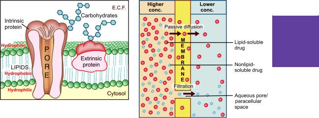
Biological membrane This is a bilayer (about 100 Å thick) of phospholipid and cholesterol molecules, the polar groups (glyceryl phosphate attached to ethanolamine/choline or hydroxyl
group of cholesterol) of these are oriented at the two surfaces and the nonpolar hydrocarbon chains are embedded in the matrix to form a continuous sheet. This imparts high electrical resistance and relative impermeability to the membrane. Extrinsic and intrinsic protein molecules are adsorbed on the lipid bilayer (Fig. 2.2). Glycoproteins or glycolipids are formed on the surface by attachment to polymeric sugars, aminosugars or sialic acids. The specific lipid and protein composition of different membranes differs according to the cell or the organelle type. The proteins are able to freely float through the membrane: associate and organize or vice versa. Some of the intrinsic ones, which extend through the full thickness of the membrane, surround fine aqueous pores. Paracellular spaces or channels also exist between certain epithelial/endothelial
Fig. 2.1: Schematic depiction of pharmacokinetic processes
MEMBRANE TRANSPORT, ABSORPTION AND DISTRIBUTION OF DRUGS |
11 |
|
|
Fig. 2.2: Illustration of the organisation of biological membrane
cells. Other adsorbed proteins have enzymatic, carrier, receptor or signal transduction properties. Lipid molecules also are capable of lateral movement. Thus, biological membranes are highly dynamic structures.
Drugs are transported across the membranes by:
(a)Passive diffusion and filtration
(b)Specialized transport
Passive diffusion
The drug diffuses across the membrane in the direction of its concentration gradient, the membrane playing no active role in the process. This is the most important mechanism for majority of drugs; drugs are foreign substances (xenobiotics), and specialized mechanisms are developed by the body primarily for normal metabolites.
Lipid soluble drugs diffuse by dissolving in the lipoidal matrix of the membrane (Fig. 2.3), the rate of transport being proportional to the lipid : water partition coefficient of the drug. A more lipid-soluble drug attains higher concentration in the membrane and diffuses quickly. Also, greater the difference in the concentration of the drug on the two sides of the membrane, faster is its diffusion.
Influence of pH Most drugs are weak electrolytes, i.e. their ionization is pH dependent (contrast strong electrolytes that are nearly completely
2 CHAPTER
Fig. 2.3: Illustration of passive diffusion and filtration across the lipoidal biological membrane with aqueous pores
ionized at acidic as well as alkaline pH). The ionization of a weak acid HA is given by the equation:
[A¯ ]
pH = pKa + log —–— ...(1) [HA]
pKa is the negative logarithm of acidic dissociation constant of the weak electrolyte. If the concentration of ionized drug [A¯ ] is equal to concentration of unionized drug [HA], then
[A¯ ]
—–— = 1 |
|
[HA] |
|
since log 1 is 0, under this condition |
|
pH = pKa |
...(2) |
Thus, pKa is numerically equal to the pH at which the drug is 50% ionized.
If pH is increased by 1 scale, then—
log [A¯ ]/[HA] = 1 or [A¯ ]/[HA] = 10
Similarly, if pH is reduced by 1 scale, then— [A¯ ]/[HA] = 1/10
Thus, weakly acidic drugs, which form salts with cations, e.g. sod. phenobarbitone, sod. sulfadiazine, pot. penicillin-V, etc. ionize more at

12 |
GENERAL PHARMACOLOGY |
|
|
|
alkaline pH and 1 scale change in pH causes 10 |
|
|
fold change in ionization. |
|
|
Weakly basic drugs, which form salts with |
|
1 |
||
anions, e.g. atropine sulfate, ephedrine HCl, |
||
SECTION |
||
chloroquine phosphate, etc. conversely ionize |
||
more at acidic pH. Ions being lipid insoluble, |
||
do not diffuse and a pH difference across a |
||
membrane can cause differential distribution of |
||
|
weakly acidic and weakly basic drugs on the two |
|
|
||
|
sides (Fig. 2.4). |
Fig. 2.4: Influence of pH difference on two sides of a biological membrane on the steady-state distribution of a weakly acidic drug with pKa = 6
Implications of this consideration are:
(a)Acidic drugs, e.g. aspirin (pKa 3.5) are largely unionized at acid gastric pH and are absorbed from stomach, while bases, e.g. atropine (pKa 10) are largely ionized and are absorbed only when they reach the intestines.
(b)The unionized form of acidic drugs which crosses the surface membrane of gastric mucosal cell, reverts to the ionized form within the cell (pH 7.0) and then only slowly passes to the extracellular fluid. This is called ion trapping, i.e. a weak electrolyte crossing a membrane to encounter a pH from which it is not able to escape easily. This may contribute to gastric mucosal cell damage caused by aspirin.
(c)Basic drugs attain higher concentration intracellularly (pH 7.0 vs 7.4 of plasma).
(d)Acidic drugs are ionized more in alkaline urine—do not back diffuse in the kidney tubules and are excreted faster. Accordingly, basic drugs are excreted faster if urine is acidified.
Lipid-soluble nonelectrolytes (e.g. ethanol, diethyl-ether) readily cross biological membranes and their transport is pH independent.
Filtration
Filtration is passage of drugs through aqueous pores in the membrane or through paracellular spaces. This can be accelerated if hydrodynamic flow of the solvent is occurring under hydrostatic or osmotic pressure gradient, e.g. across most capillaries including glomeruli. Lipid-insoluble drugs cross biological membranes by filtration if their molecular size is smaller than the diameter of the pores (Fig. 2.3). Majority of cells (intestinal mucosa, RBC, etc.) have very small pores (4 Å) and drugs with MW > 100 or 200 are not able to penetrate. However, capillaries (except those in brain) have large paracellular spaces (40 Å) and most drugs (even albumin) can filter through these (Fig. 2.8). As such, diffusion of drugs across capillaries is dependent on rate of blood flow through them rather than on lipid solubility of the drug or pH of the medium.
Specialized transport
This can be carrier mediated or by pinocytosis.
Carrier transport
All cell membranes express a host of transmembrane proteins which serve as carriers or transporters for physiologically important ions, nutrients, metabolites, transmitters, etc. across the membrane. At some sites, certain transporters also translocate xenobiotics, including drugs and their metabolites. In contrast to channels, which open for a finite time and allow passage of specific ions, transporters combine transiently with their substrate (ion or organic compound)—undergo a conformational change carrying the substrate to the other side of the membrane where the substrate dissociates and the transporter returns back to its original state (Fig. 2.5). Carrier transport is specific for the substrate (or the type of substrate, e.g. an organic anion), saturable, competitively inhibited by analogues which utilize the same transporter, and is much slower than

MEMBRANE TRANSPORT, ABSORPTION AND DISTRIBUTION OF DRUGS |
13 |
|
|
2 CHAPTER
Fig. 2.5: Illustration of different types of carrier mediated transport across biological membrane ABC—ATP-binding cassettee transporter; SLC—Solute carrier transporter; M—Membrane
A.Facilitated diffusion: the carrier (SLC) binds and moves the poorly diffusible substrate along its concentration gradient (high to low) and does not require energy
B.Primary active transport: the carrier (ABC) derives energy directly by hydrolysing ATP and moves the substrate against its concentration gradient (low to high)
C.Symport: the carrier moves the substrate ‘A’ against its concentration gradient by utilizing energy from downhill movement of another substrate ‘B’ in the same direction
D.Antiport: the carrier moves the substrate ‘A’ against its concentration gradient and is energized by the downhill movement of another substrate ‘B’ in the opposite direction
the flux through channels. Depending on requirement of energy, carrier transport is of two types:
a.Facilitated diffusion The transporter, belonging to the super-family of solute carrier (SLC) transporters, operates passively without needing energy and translocates the substrate in the direction of its electrochemical gradient, i.e. from higher to lower concentration (Fig. 2.5A). It mearly facilitates permeation of a poorly diffusible substrate, e.g. the entry of glucose into muscle and fat cells by the glucose transporter GLUT 4.
b.Active transport It requires energy, is
inhibited by metabolic poisons, and transports the solute against its electrochemical gradient (low
to high), resulting in selective accumulation of the substance on one side of the membrane. Drugs related to normal metabolites can utilize the transport processes meant for these, e.g. levodopa and methyl dopa are actively absorbed from the gut by the aromatic amino acid transporter. In addition, the body has developed some relatively nonselective transporters, like P-glycoprotein (P-gp), to deal with xenobiotics. Active transport can be primary or secondary depending on the source of the driving force.
i. Primary active transport Energy is obtained directly by the hydrolysis of ATP (Fig. 2.5B). The transporters belong to the superfamily of ATP binding cassettee (ABC) transporters whose intracellular loops have ATPase activity.
14 |
GENERAL PHARMACOLOGY |
|
|
SECTION 1
They mediate only efflux of the solute from the cytoplasm, either to extracellular fluid or into an intracellular organelli (endoplasmic reticulum, mitochondria, etc.)
Encoded by the multidrug resistance 1 (MDR1) gene, P-gp is the most well known primary active transporter expressed in the intestinal mucosa, renal tubules, bile canaliculi, choroidal epithelium, astrocyte foot processes around brain capillaries (the blood-brain barrier), testicular and placental microvessels, which pumps out many drugs/ metabolites and thus limits their intestinal absorption, penetration into brain, testes and foetal tissues as well as promotes biliary and renal elimination. Many xenobiotics which induce or inhibit P-gp also have a similar effect on the drug metabolizing isoenzyme CYP3A4, indicating their synergistic role in detoxification of xenobiotics.
Other primary active transporters of pharmacological significance are multidrug resistance associated protein 2 (MRP 2) and breast cancer resistance protein (BCRP). ii. Secondary active transport In this type of active transport effected by another set of SLC transporters, the energy to pump one solute is derived from the downhill movement of another solute (mostly Na+). When the concentration gradients are such that both the solutes move in the same direction (Fig. 2.5C), it is called symport or cotransport, but when they move in opposite directions (Fig. 2.5D), it is termed antiport or exchange transport. Metabolic energy (from hydrolysis of ATP) is spent in maintaining high transmembrane electrochemical gradient of the second solute (generally Na+). The SLC transporters mediate both uptake and efflux of drugs and metabolites.
The organic anion transporting polypeptide (OATP) and organic cation transporter (OCT), highly expressed in liver canaliculi and renal tubules, are secondary active transporters important in the metabolism and excretion of drugs and metabolites (especially glucuronides). The Na+,Cl– dependent neurotransmitter transporters for norepinephrine, serotonin and dopamine (NET, SERT and DAT) are active SLC transporters that are targets for action of drugs like tricyclic antidepressants, selective serotonin reuptake inhibitors (SSRIs), cocaine, etc. Similarly, the Vesicular monoamine transporter (VMAT-2) of adrenergic and serotonergic storage vesicles transports catecholamines and 5-HT into the vesicles by exchanging with H+ions, and is inhibited by reserpine. The absorption of glucose in intestines and renal tubules is through secondary active transport by sodium-glucose transporters (SGLT1 and SGLT2).
saturable and follows the Michaelis-Menten kinetics. The maximal rate of transport is dependent on the density of the transporter in a particular membrane, and its rate constant (Km), i.e. the substrate concentration at which rate of transport is half maximal, is governed by its affinity for the substrate. Genetic polymorphism can alter both the density and affinity of the transporter protein for different substrates and thus affect the pharmacokinetics of drugs. Moreover, tissue specific drug distribution can occur due to the presence of specific transporters in certain cells.
Pinocytosis It is the process of transport across the cell in particulate form by formation of vesicles. This is applicable to proteins and other big molecules, and contributes little to transportofmostdrugs,barringfewlikevitB12whichisabsorbed from the gut after binding to intrinsic factor (a protein).
ABSORPTION
Absorption is movement of the drug from its site of administration into the circulation. Not only the fraction of the administered dose that gets absorbed, but also the rate of absorption is important. Except when given i.v., the drug has to cross biological membranes; absorption is governed by the above described principles. Other factors affecting absorption are:
Aqueous solubility Drugs given in solid form must dissolve in the aqueous biophase before they are absorbed. For poorly water soluble drugs (aspirin, griseofulvin) rate of dissolution governs rate of absorption. Ketoconazole dissolves at low pH: gastric acid is needed for its absorption. Obviously, a drug given as watery solution is absorbed faster than when the same is given in solid form or as oily solution.
Concentration Passive diffusion depends on concentration gradient; drug given as concentrated solution is absorbed faster than from dilute solution.
As indicated earlier, carrier transport (both facilitated diffusion and active transport) is
Area of absorbing surface Larger is the surface area, faster is the absorption.

MEMBRANE TRANSPORT, ABSORPTION AND DISTRIBUTION OF DRUGS |
15 |
|
|
Vascularity of the absorbing surface Blood circulation removes the drug from the site of absorption and maintains the concentration gradient across the absorbing surface. Increased blood flow hastens drug absorption just as wind hastens drying of clothes.
Route of administration This affects drug absorption, because each route has its own peculiarities.
Oral
The effective barrier to orally administered drugs is the epithelial lining of the gastrointestinal tract, which is lipoidal. Nonionized lipid soluble drugs, e.g. ethanol are readily absorbed from stomach as well as intestine at rates proportional to their lipid : water partition coefficient. Acidic drugs, e.g. salicylates, barbiturates, etc. are predominantly unionized in the acid gastric juice and are absorbed from stomach, while basic drugs, e.g. morphine, quinine, etc. are largely ionized and are absorbed only on reaching the duodenum. However, even for acidic drugs absorption from stomach is slower, because the mucosa is thick, covered with mucus and the surface area is small. Absorbing surface area is much larger in the small intestine due to villi. Thus, faster gastric emptying accelerates drug absorption in general. Dissolution is a surface phenomenon, therefore, particle size of the drug in solid dosage form governs rate of dissolution and in turn rate of absorption.
Presence of food dilutes the drug and retards absorption. Further, certain drugs form poorly absorbed complexes with food constituents, e.g. tetracyclines with calcium present in milk; moreover food delays gastric emptying. Thus, most drugs are absorbed better if taken in empty stomach. However, there are some exceptions, e.g. fatty food greatly enhances lumefantrine absorption. Highly ionized drugs, e.g. gentamicin, neostigmine are poorly absorbed when given orally.
Certain drugs are degraded in the gastrointestinal tract, e.g. penicillin G by acid, insulin by peptidases, and are ineffective orally. Enteric coated tablets (having acid resistant coating) and
sustained release preparations (drug particles coated with slowly dissolving material) can be used to overcome acid lability, gastric irritancy and brief duration of action.
The oral absorption of certain drugs is low because a fraction of the absorbed drug is extruded back into the intestinal lumen by the efflux transporter P-gp located in the gut epithelium. The low oral bioavailability of digoxin and cyclosporine is partly accounted by this mechanism. Inhibitors of P-gp like quinidine, verapamil, erythromycin, etc. enhance, while P-gp inducers like rifampin and phenobarbitone reduce the oral bioavailability of these drugs.
Absorption of a drug can be affected by other concurrently ingested drugs. This may be a luminal effect: formation of insoluble complexes, e.g. tetracyclines and iron preparations with calcium salts and antacids, phenytoin with sucralfate. Such interaction can be minimized by administering the two drugs at 2–3 hr intervals.Alteration of gut flora by antibiotics may disrupt the enterohepatic cycling of oral contraceptives and digoxin. Drugs can also alter absorption by gut wall effects: altering motility (anticholinergics, tricyclic antidepressants, opioids, metoclopramide) or causing mucosal damage (neomycin, methotrexate, vinblastine).
Subcutaneous and Intramuscular
By these routes the drug is deposited directly in the vicinity of the capillaries. Lipid soluble drugs pass readily across the whole surface of the capillary endothelium. Capillaries having large paracellular spaces do not obstruct absorption of even large lipid insoluble molecules or ions (Fig. 2.8A). Very large molecules are absorbed through lymphatics. Thus, many drugs not absorbed orally are absorbed parenterally.Absorption from s.c. site is slower than that from i.m. site, but both are generally faster and more consistent/ predictable than oral absorption. Application of heat and muscular exercise accelerate drug absorption by increasing blood flow, while vasoconstrictors, e.g. adrenaline injected with the drug (local anaesthetic) retard absorption. Incorporation of hyaluronidase facilitates drug absorption from s.c. injection by
2 CHAPTER
16 |
GENERAL PHARMACOLOGY |
|
|
SECTION 1
promoting spread. Many depot preparations, e.g. benzathine penicillin, protamine zinc insulin, depot progestins, etc. can be given by these routes.
Topical sites (skin, cornea, mucous membranes)
Systemic absorption after topical application depends primarily on lipid solubility of drugs. However, only few drugs significantly penetrate intact skin. Hyoscine, fentanyl, GTN, nicotine, testosterone, and estradiol (see p. 8) have been used in this manner. Corticosteroids applied over extensive areas can produce systemic effects and pituitary-adrenal suppression. Absorption can be promoted by rubbing the drug incorporated in an olegenous base or by use of occlusive dressing which increases hydration of the skin. Organophosphate insecticides coming in contact with skin can produce systemic toxicity. Abraded surfaces readily absorb drugs, e.g. tannic acid applied over burnt skin has produced hepatic necrosis.
Cornea is permeable to lipid soluble, unionized physostigmine but not to highly ionized neostigmine. Drugs applied as eye drops may get absorbed through the nasolacrimal duct, e.g. timolol eye drops may produce bradycardia and precipitate asthma. Mucous membranes of mouth, rectum, vagina absorb lipophilic drugs: estrogen cream applied vaginally has produced gynaecomastia in the male partner.
BIOAVAILABILITY
Bioavailability refers to the rate and extent of absorption of a drug from a dosage form as determined by its concentration-time curve in blood or by its excretion in urine (Fig. 2.6). It is a measure of the fraction (F ) of administered dose of a drug that reaches the systemic circulation in the unchanged form. Bioavailability of drug injected i.v. is 100%, but is frequently lower after oral ingestion because—
Fig. 2.6: Plasma concentration-time curves depicting bioavailability differences between three preparations of a drug containing the same amount
Note that formulation B is more slowly absorbed than A, and though ultimately both are absorbed to the same extent (area under the curve same), B may not produce therapeutic effect; C is absorbed to a lesser extent— lower bioavailability
Incomplete bioavailability after s.c. or i.m. injection is less common, but may occur due to local binding of the drug.
Bioequivalence Oral formulations of a drug from different manufacturers or different batches from the same manufacturer may have the same amount of the drug (chemically equivalent) but may not yield the same blood levels—biologically inequivalent. Two preparations of a drug are considered bioequivalent when the rate and extent of bioavailability of the active drug from them is not significantly different under suitable test conditions.
Before a drug administered orally in solid dosage form can be absorbed, it must break into individual particles of the active drug (disintegration). Tablets and capsules contain a number of other materials—diluents, stabilizing agents, binders, lubricants, etc. The nature of these as well as details of the manufacture process, e.g. force used in compressing the tablet, may affect
(a)the drug may be incompletely absorbed. disintegration. The released drug must then
(b)the absorbed drug may undergo first pass dissolve in the aqueous gastrointestinal contents.
metabolism in the intestinal wall/liver or be excreted in bile.
The rate of dissolution is governed by the inherent solubility, particle size, crystal form and other
MEMBRANE TRANSPORT, ABSORPTION AND DISTRIBUTION OF DRUGS |
17 |
|
|
physical properties of the drug. Differences in bioavailability may arise due to variations in disintegration and dissolution rates.
Differences in bioavailability are seen mostly with poorly soluble and slowly absorbed drugs. Reduction in particle size increases the rate of absorption of aspirin (microfine tablets). The amount of griseofulvin and spironolactone in the tablet can be reduced to half if the drug particle is microfined. There is no need to reduce the particle size of freely water soluble drugs, e.g. paracetamol.
Bioavailability variation assumes practical significance for drugs with low safety margin (digoxin) or where dosage needs precise control (oral hypoglycaemics, oral anticoagulants). It may also be responsible for success or failure of an antimicrobial regimen.
However, in the case of a large number of drugs bioavailability differences are negligible and the risks of changing from branded to generic product or to another brand of the same drug have often been exaggerated.
DISTRIBUTION
Once a drug has gained access to the blood stream, it gets distributed to other tissues that initially had no drug, concentration gradient being in the direction of plasma to tissues. The extent and pattern of distribution of a drug depends on its:
•lipid solubility
•ionization at physiological pH (a function of its pKa)
•extent of binding to plasma and tissue proteins
•presence of tissue-specific transporters
•differences in regional blood flow.
Movement of drug proceeds until an equilibrium is established between unbound drug in the plasma and the tissue fluids. Subsequently, there is a parallel decline in both due to elimination.
Apparent volume of distribution (V) Presuming that the body behaves as a single homogeneous compartment with volume V into which the drug gets immediately and uniformly distributed
dose administered i.v. |
...(3) |
V = ————————— |
|
plasma concentration |
|
Since in the example shown in Fig. 2.7, the drug |
CHAPTER |
|
|
||
does not actually distribute into 20 L of body |
|
|
water, with the exclusion of the rest of it, this |
|
|
is only an apparent volume of distribution which |
|
|
can be defined as “the volume that would |
2 |
|
accommodate all the drug in the body, if the |
||
|
||
|
||
concentration throughout was the same as in |
|
|
plasma”. Thus, it describes the amount of drug |
|
|
present in the body as a multiple of that contained |
|
|
in a unit volume of plasma. Considered together |
|
|
with drug clearance, this is a very useful |
|
|
pharmacokinetic concept. |
|
|
Lipid-insoluble drugs do not enter cells— |
|
|
V approximates extracellular fluid volume, e.g. |
|
|
streptomycin, gentamicin 0.25 L/kg. |
|
|
Distribution is not only a matter of dilution, |
|
|
but also binding and sequestration. Drugs |
|
|
extensively bound to plasma proteins are largely |
|
|
restricted to the vascular compartment and have |
|
|
low values, e.g. diclofenac and warfarin (99% |
|
|
bound) V = 0.15 L/kg. |
|
|
A large value of V indicates that larger quantity |
|
|
of drug is present in extravascular tissue. Drugs |
|
|
sequestrated in other tissues may have, V much |
|
|
more than total body water or even body mass, |
|
Fig. 2.7: Illustration of the concept of apparent volume of distribution (V).
In this example, 1000 mg of drug injected i.v. produces steady-state plasma concentration of 50 mg/L, apparent volume of distribution is 20 L

18 |
GENERAL PHARMACOLOGY |
|
|
SECTION 1
e.g. digoxin 6 L/kg, propranolol 4 L/kg, morphine 3.5 L/kg, because most of the drug is present in other tissues, and plasma concentration is low. Therefore, in case of poisoning, drugs with large volumes of distribution are not easily removed by haemodialysis.
Pathological states, e.g. congestive heart failure, uraemia, cirrhosis of liver, etc. can alter the V of many drugs by altering distribution of body water, permeability of membranes, binding proteins or by accumulation of metabolites that displace the drug from binding sites.
More precise multiple compartment models for drug distribution have been worked out, but the single compartment model, described above, is simple and fairly accurate for many drugs.
Redistribution Highly lipid-soluble drugs get initially distributed to organs with high blood flow, i.e. brain, heart, kidney, etc. Later, less vascular but more bulky tissues (muscle, fat) take up the drug—plasma concentration falls and the drug is withdrawn from the highly perfused sites. If the site of action of the drug was in one of the highly perfused organs, redistribution results in termination of drug action. Greater the lipid solubility of the drug, faster is its redistribution.
Factors governing volume of drug distribution
• Lipid: water partition coefficient of the drug
•pKa value of the drug
•Degree of plasma protein binding
• Affinity for different tissues
•Fat: lean body mass ratio, which can vary
with age, sex, obesity, etc.
•Diseases like CHF, uremia, cirrhosis
Anaesthetic action of thiopentone sod. injected i.v. is terminated in few minutes due to redistribution. A relatively short hypnotic action lasting 6–8 hours is exerted by oral diazepam or nitrazepam due to redistribution despite their elimination t ½ of > 30 hr. However, when the same drug is given repeatedly or continuously over long periods, the low perfusion high capacity sites get progressively filled up and the drug becomes longer acting.
Penetration into brain and CSF The capillary endothelial cells in brain have tight junctions and lack large paracellular spaces. Further, an investment of neural tissue (Fig. 2.8B) covers the capillaries. Together they constitute the so called blood-brain barrier (BBB). A similar blood-CSF barrier is located in the choroid plexus: capillaries are lined by choroidal
Fig. 2.8: Passage of drugs across capillaries
A.Usual capillary with large paracellular spaces through which even large lipid-insoluble molecules diffuse
B.Capillary constituting blood brain or blood-CSF barrier. Tight junctions between capillary endothelial cells and investment of glial processes or choroidal epithelium do not allow passage of non lipid-soluble molecules/ions

MEMBRANE TRANSPORT, ABSORPTION AND DISTRIBUTION OF DRUGS |
19 |
|
|
epithelium having tight junctions. Both these barriers are lipoidal and limit the entry of nonlipidsoluble drugs, e.g. streptomycin, neostigmine, etc. Only lipid-soluble drugs, therefore, are able to penetrate and have action on the central nervous system. In addition, efflux transporters like P-gp and anion transporter (OATP) present in brain and choroidal vessels extrude many drugs that enter brain by other processes and serve to augment the protective barrier against potentially harmful xenobiotics. Dopamine does not enter brain but its precursor levodopa does; as such, the latter is used in parkinsonism. Inflammation of meninges or brain increases permeability of these barriers. It has been proposed that some drugs accumulate in the brain by utilizing the transporters for endogenous substances.
There is also an enzymatic BBB: Monoamine oxidase (MAO), cholinesterase and some other enzymes are present in the capillary walls or in the cells lining them. They do not allow catecholamines, 5-HT, acetylcholine, etc. to enter brain in the active form.
The BBB is deficient at the CTZ in the medulla oblongata (even lipid-insoluble drugs are emetic) and at certain periventricular sites—(anterior hypothalamus). Exit of drugs from the CSF and brain, however, is not dependent on lipid-solubility and is rather unrestricted. Bulk flow of CSF (alongwith the drug dissolved in it) occurs through the arachnoid villi. Further, nonspecific organic anion and cation transport processes (similar to those in renal tubule) operate at the choroid plexus.
Passage across placenta Placental membranes are lipoidal and allow free passage of lipophilic drugs, while restricting hydrophilic drugs. The placental efflux P-gp and other transporters like BCRP, MRP3 also serve to limit foetal exposure to maternally administered drugs. Placenta is a site for drug metabolism as well, which may lower/modify exposure of the foetus to the administered drug. However, restricted amounts of nonlipid-soluble drugs, when present in high concentration or for long periods in maternal circulation, gain access to the foetus.
Some influx transporters also operate at the |
|
|
placenta. Thus, it is an incomplete barrier and |
|
|
almost any drug taken by the mother can affect |
|
|
CHAPTER |
||
the foetus or the newborn (drug taken just before |
||
|
||
delivery, e.g. morphine). |
|
|
Plasma protein binding |
|
|
Most drugs possess physicochemical affinity for |
2 |
|
plasma proteins and get reversibly bound to these. |
||
|
||
Acidic drugs generally bind to plasma albumin and |
|
|
basic drugs to α1 acid glycoprotein. Binding to |
|
|
albumin is quantitatively more important. Extent |
|
|
of binding depends on the individual compound; |
|
|
no generalization for a pharmacological or |
|
|
chemical class can be made (even small chemical |
|
|
change can markedly alter protein binding), for |
|
|
example the binding percentage of some benzo- |
|
diazepines is: |
|
|
|
Flurazepam |
10% |
Alprazolam |
70% |
Lorazepam |
90% |
Diazepam |
99% |
Increasing concentrations of the drug can progressively saturate the binding sites: fractional binding may be lower when large amounts of the drug are given. The generally expressed percentage binding refers to the usual therapeutic plasma concentrations of a drug. The clinically significant implications of plasma protein binding are:
(i) Highly plasma protein bound drugs are largely restricted to the vascular compartment because protein bound drug does not cross membranes (except through large paracellular spaces, such
Drugs highly bound to plasma protein
To albumin |
To α1-acid |
|
glycoprotein |
|
|
Barbiturates |
β-blockers |
Benzodiazepines |
Bupivacaine |
NSAIDs |
Lidocaine |
Valproic acid |
Disopyramide |
Phenytoin |
Imipramine |
Penicillins |
Methadone |
Sulfonamides |
Prazosin |
Tetracyclines |
Quinidine |
Tolbutamide |
Verapamil |
Warfarin |
|
|
|

20 |
GENERAL PHARMACOLOGY |
|
|
SECTION 1
as in capillaries). They tend to have smaller volumes of distribution.
(ii)The bound fraction is not available for action. However, it is in equilibrium with the free drug in plasma and dissociates when the concentration of the latter is reduced due to elimination. Plasma protein binding thus tantamounts to temporary storage of the drug.
(iii)High degree of protein binding generally makes the drug long acting, because bound fraction is not available for metabolism or excretion, unless it is actively extracted by liver or by kidney tubules. Glomerular filtration does not reduce the concentration of the free form in the efferent vessels, because water is also filtered. Active tubular secretion, however, removes the drug without the attendant solvent → concentration of free drug falls → bound drug dissociates and is eliminated resulting in a higher renal clearance value of the drug than the total renal blood flow (see Fig. 3.3). The same is true of active transport of highly extracted drugs in liver. Plasma protein binding in this situation acts as a carrier mechanism and hastens drug elimination, e.g. excretion of penicillin (elimination t½ is 30 min); metabolism of lidocaine. Highly protein bound drugs are not removed by haemodialysis and need special techniques for treatment of poisoning.
(iv)The generally expressed plasma concentrations of the drug refer to bound as well as free drug. Degree of protein binding should be taken
into account while relating these to concentrations of the drug that are active in vitro, e.g. MIC of an antimicrobial.
(v) One drug can bind to many sites on the albumin molecule. Conversely, more than one drug can bind to the same site. This can give rise to displacement interactions among drugs bound to the same site(s). The drug bound with higher affinity will displace that bound with lower affinity. If just 1% of a drug that is 99% bound is displaced, the concentration of free form will be doubled. This, however, is often transient because the displaced drug will diffuse into the tissues as well as get metabolized or excreted: the new steadystate free drug concentration is only marginally higher unless the displacement extends to tissue binding or there is concurrent inhibition of metabolism and/or excretion. The overall impact of many displacement interactions is minimal; clinical significance being attained only in case of highly bound drugs with limited volume of distribution (many acidic drugs bound to albumin) and where interaction is more complex. Moreover, two highly bound drugs do not necessarily displace each other—their binding sites may not overlap, e.g. probenecid and indomethacin are highly bound to albumin but do not displace each other. Similarly, acidic drugs do not generally displace basic drugs and vice versa. Some clinically important displacement interactions are:
•Aspirin displaces sulfonylureas.
•Indomethacin, phenytoin displace warfarin.
|
|
Drugs concentrated in tissues |
|
|
|
Skeletal |
muscle, heart |
— digoxin, emetine (bound to muscle proteins). |
Liver |
|
— chloroquine, tetracyclines, emetine, digoxin. |
Kidney |
|
— digoxin, chloroquine, emetine. |
Thyroid |
|
— iodine. |
Brain |
|
— chlorpromazine, acetazolamide, isoniazid. |
Retina |
|
— chloroquine (bound to nucleoproteins). |
Iris |
|
— ephedrine, atropine (bound to melanin). |
Bone and teeth |
— tetracyclines, heavy metals (bound to mucopolysaccharides of |
|
|
|
connective tissue), bisphosphonates (bound to hydroxyapatite) |
Adipose |
tissue |
— thiopentone, ether, minocycline, phenoxybenzamine, DDT |
|
|
dissolve in neutral fat due to high lipid-solubility; remain |
|
|
stored due to poor blood supply of fat. |
|
|
|

MEMBRANE TRANSPORT, ABSORPTION AND DISTRIBUTION OF DRUGS |
21 |
|
|
•Sulfonamides and vit K displace bilirubin (kernicterus in neonates).
•Aspirin displaces methotrexate.
(vi) In hypoalbuminemia, binding may be reduced and high concentrations of free drug may be attained, e.g. phenytoin and furosemide. Other diseases may also alter drug binding, e.g. phenytoin and pethidine binding is reduced in uraemia; propranolol binding is increased in pregnant women and in patients with inflammatory disease (acute phase reactant α1 acid-glycoprotein increases).
Tissue storage Drugs may also accumulate in specific organs by active transport or get bound to specific tissue constituents (see box).
Drugs sequestrated in various tissues are unequally distributed, tend to have larger volume of distribution and longer duration of action. Some may exert local toxicity due to high concentration, e.g. tetracyclines on bone and teeth, chloroquine on retina, streptomycin on vestibular apparatus, emetine on heart and skeletal muscle. Drugs may also selectively bind to specific intracellular organelle, e.g. tetracycline to mitochondria, chloroquine to nuclei.
)PROBLEM DIRECTED STUDY
2.1A 60-year-old woman complained of weakness, lethargy and easy fatigability. Investigation showed that she had iron deficiency anaemia (Hb. 8 g/dl). She was prescribed cap. ferrous fumarate 300 mg twice daily. She returned after one month with no improvement in symptoms. Her Hb. level was unchanged. On enquiry she revealed that she felt epigastric distress after taking the iron capsules, and had started taking antacid tablets along with the capsules.
(a) What could be the possible reason for her failure to respond to the oral iron medication?
2.2 A 50-year-old type-2 diabetes mellitus patient was maintained on tab. glibenclamide (a sulfonylurea) 5 mg twice daily. He developed toothache for which he took tab. aspirin 650 mg 6 hourly. After taking aspirin he experienced anxiety, sweating, palpitation, weakness, ataxia, and was behaving abnormally. These symptoms subsided when he was given a glass of glucose solution.
(a)Waht could be the explanation for his symptoms?
(b)Which alternative analgesic should have been taken? (see Appendix-1 for solutions)
2 CHAPTER

Chapter 3 Pharmacokinetics: Metabolism
and Excretion of Drugs, Kinetics
of Elimination
BIOTRANSFORMATION (Metabolism)
Biotransformation means chemical alteration of the drug in the body. It is needed to render nonpolar (lipid-soluble) compounds polar (lipidinsoluble) so that they are not reabsorbed in the renal tubules and are excreted. Most hydrophilic drugs, e.g. streptomycin, neostigmine, pancuronium, etc. are little biotransformed and are largely excreted unchanged. Mechanisms which metabolize drugs (essentially foreign substances) have developed to protect the body from ingested toxins.
The primary site for drug metabolism is liver; others are—kidney, intestine, lungs and plasma. Biotransformation of drugs may lead to the following.
(i)Inactivation Most drugs and their active metabolites are rendered inactive or less active, e.g. ibuprofen, paracetamol, lidocaine, chloramphenicol, propranolol and its active metabolite 4-hydroxypropranolol.
(ii)Active metabolite from an active drug
Many drugs have been found to be partially converted to one or more active metabolite; the effects observed are the sumtotal of that due to the parent drug and its active metabolite(s) (see box).
(iii) Activation of inactive drug Few drugs are inactive as such and need conversion in the body to one or more active metabolites. Such a drug is called a prodrug (see box). The prodrug may offer advantages over the active form in being more stable, having better bioavailability or other desirable pharmacokinetic properties or less side effects and toxicity. Some prodrugs are activated selectively at the site of action.
Active drug |
|
Active metabolite |
|
|
|
Chloral hydrate |
— |
Trichloroethanol |
Morphine |
— Morphine-6-glucuronide |
|
Cefotaxime |
— |
Desacetyl cefotaxime |
Allopurinol |
— |
Alloxanthine |
Procainamide |
— |
N-acetyl procainamide |
Primidone |
— |
Phenobarbitone, |
|
|
phenylethylmalonamide |
Diazepam |
— |
Desmethyl-diazepam, |
|
|
oxazepam |
Digitoxin |
— |
Digoxin |
Imipramine |
— |
Desipramine |
Amitriptyline |
— |
Nortriptyline |
Codeine |
— |
Morphine |
Spironolactone |
— |
Canrenone |
Losartan |
— |
E 3174 |
|
|
|
Biotransformation reactions can be classified into:
(a)Nonsynthetic/Phase I/Functionalization reactions: a functional group is generated or exposed— metabolite may be active or inactive.
(b)Synthetic/Conjugation/ Phase II reactions: an endogenous radical is conjugated to the drug— metabolite is mostly inactive; except few drugs, e.g. glucuronide conjugate of morphine and sulfate conjugate of minoxidil are active.
Nonsynthetic reactions
(i) Oxidation This reaction involves addition of oxygen/negatively charged radical or removal of hydrogen/positively charged radical. Oxidations are the most important drug metabolizing reactions. Various oxidation reactions are:
hydroxylation; oxygenation at C, N or S atoms; N or O-dealkylation, oxidative deamination, etc.
In many cases the initial insertion of oxygen atom into the drug molecule produces short lived highly reactive quinone/epoxide/superoxide

METABOLISM AND EXCRETION OF DRUGS, KINETICS OF ELIMINATION |
23 |
|
|
Prodrug |
|
Active form |
|
|
|
Levodopa |
— |
Dopamine |
Enalapril |
— |
Enalaprilat |
a-Methyldopa |
— |
a-methylnorepinephrine |
Dipivefrine |
— |
Epinephrine |
Sulindac |
— |
Sulfide metabolite |
Proguanil |
— |
Cycloguanil |
Prednisone |
— |
Prednisolone |
Bacampicillin |
— |
Ampicillin |
Sulfasalazine |
— |
5-Aminosalicylic acid |
Cyclophos- |
— |
Aldophosphamide, |
phamide |
|
phosphoramide mustard, |
|
|
acrolein |
Fluorouracil |
— |
Fluorouridine |
|
|
monophosphate |
Mercaptopurine |
— |
Methylmercaptopurine |
|
|
ribonucleotide |
Acyclovir |
— |
Acyclovir triphosphate |
|
|
|
intermediates which then convert to more stable compounds.
Oxidative reactions are mostly carried out by a group of monooxygenases in the liver, which in the final step involve a cytochrome P-450 haemoprotein, NADPH, cytochrome P-450 reductase and molecular O2. More than 100 cytochrome P-450 isoenzymes differing in their affinity for various substrates (drugs), have been identified.
Depending upon the extent of amino acid sequence homology, the cytochrome P-450 (CYP) isoenzymes are grouped into families designated by numerals (1, 2, 3.....), each having several sub-families designated by capital letters (A, B, C.....), while individual isoenzymes are again alloted numerals (1, 2, 3....). In human beings, only a few members of three isoenzyme families (CYP 1, 2 and 3) carryout metabolism of most of the drugs, and many drugs such as tolbutamide, barbiturates, nifedipine are substrates for more than one isoform. The CYP isoenzymes important in man are:
CYP3A4/5 Carryout biotransformation of largest number (nearly 50%) of drugs. In addition to liver, these isoforms are expressed in intestine (responsible for first pass metabolism at this site) and kidney as well. Inhibition of this isoenzyme by erythromycin, clarithromycin, ketoconazole, itraconazole is responsible for the important drug interaction with terfenadine, astemizole and cisapride (see p. 166) which are its substrates. Losartan, nifedipine hydrocortisone, mifepristone, simvastatin, ritonavir, carbamazepine and cyclosporine are also metabolized by CYP3A4/5. Verapamil, diltiazem, ritonavir and a constituent of grape fruit juice are other important inhibitors, while rifampicin, barbiturates and other anticonvulsants are the important inducers.
CYP2D6 This is the next most important CYP isoform which metabolizes nearly 20% drugs including tricyclic antidepressants, selective serotonin reuptake inhibitors, many neuroleptics, antiarrhythmics, β-blockers and opiates. Inhibition of this enzyme by quinidine results in failure of conversion of codeine to morphine → analgesic effect of codeine is lost. Human subjects can be grouped into ‘extensive’ or ‘poor’ metabolizers of metoprolol and debrisoquin. The poor metabolizers have an altered CYP2D6 enzyme and exhibit low capacity to hydroxylate many drugs.
CYP2C8/9 Important in the biotransformation of >15 commonly used drugs including phenytoin, carbamazepine, warfarin which are narrow safety margin drugs, as well as ibuprofen, tolbutamide, repaglinide, celecoxib and losartan.
CYP2C19 Metabolizes > 12 frequently used drugs including omeprazole, lansoprazole, phenytoin, diazepam, propranolol.
Rifampicin and carbamazepine are potent inducers of the CYP2C subfamily, while omeprazole is an inhibitor.
CYP1A1/2 Though this subfamily participates in the metabolism of only few drugs like theophylline, caffeine, paracetamol, carbamazepine, it is more important for activation of procarcinogens. Apart from rifampicin and carbamazepine, polycyclic hydrocarbons, cigarette smoke and charbroiled meat are its potent inducers.
CYP2E1 It catalyses oxidation of alcohol, holothane, and formation of minor metabolites of few drugs, notably the hepatotoxic N-acetyl benzoquinoneimine from paracetamol; chronic alcoholism induces this isoenzyme.
The relative amount of different cytochrome P-450s differs among species and among individuals of the same species. These differences largely account for the marked interspecies and interindividual differences in rate of metabolism of drugs.
Barbiturates, phenothiazines, imipramine, propranolol, ibuprofen, paracetamol, steroids, phenytoin, benzodiazepines, theophylline and many other drugs are oxidized in this way. Few drugs like cimetidine, ranitidine, clozapine are oxidized at their N, P or S atoms by a group of flavin-monooxygenases that are also located at hepatic endoplasmic reticulum, but are distinct from CYP enzymes. They are not susceptible to induction or inhibition by other drugs, and thus are not involved in drug interactions. Some other drugs, e.g. adrenaline, alcohol, mercaptopurine are oxidized by mitochondrial or cytoplasmic enzymes.
(ii) Reduction This reaction is the converse of oxidation and involves cytochrome P-450 enzymes working in the opposite direction. Alcohols, aldehydes, quinones are reduced. Drugs
3 CHAPTER
24 |
GENERAL PHARMACOLOGY |
|
|
SECTION 1
primarily reduced are chloralhydrate, chloramphenicol, halothane, warfarin.
(iii) Hydrolysis This is cleavage of drug molecule by taking up a molecule of water.
|
esterase |
Ester + H2O |
→ Acid + Alcohol |
Similarly, amides and polypeptides are hydrolysed by amidases and peptidases. In addition, there are epoxide hydrolases which detoxify epoxide metabolites of some drugs generated by CYP oxygenases. Hydrolysis occurs in liver, intestines, plasma and other tissues. Examples of hydrolysed drugs are choline esters, procaine, lidocaine, procainamide, aspirin, carbamazepine-epoxide, pethidine, oxytocin.
(iv)Cyclization This is formation of ring structurefromastraightchaincompound,e.g.proguanil.
(v)Decyclization This is opening up of ring structure of the cyclic drug molecule, e.g. barbiturates, phenytoin. This is generally a minor pathway.
Synthetic reactions
These involve conjugation of the drug or its phase I metabolite with an endogenous substrate, usually derived from carbohydrate or amino acid, to form a polar highly ionized organic acid, which is easily excreted in urine or bile. Conjugation reactions have high energy requirement.
(i) Glucuronide conjugation This is the most important synthetic reaction carriedout by a group of UDP-glucuronosyl transferases (UGTs). Compounds with a hydroxyl or carboxylic acid group are easily conjugated with glucuronic acid which is derived from glucose. Examples are— chloramphenicol, aspirin, paracetamol, diazepam, lorazepam, morphine, metronidazole. Not only drugs but endogenous substrates like bilirubin, steroidal hormones and thyroxine utilize this pathway. Glucuronidation increases the molecular weight of the drug which favours its excretion in bile. Drug glucuronides excreted in bile can be hydrolysed by bacteria in the gut—the liberated drug is reabsorbed and undergoes the same fate.
This enterohepatic cycling (see Fig. 3.2) of the drug prolongs its action, e.g. phenolphthalein, oral contraceptives.
(ii)Acetylation Compounds having amino or hydrazine residues are conjugated with the help of acetyl coenzyme-A, e.g. sulfonamides, isoniazid, PAS, dapsone, hydralazine, clonazepam, procainamide. Multiple genes control the N-acetyl transferases (NATs), and rate of acetylation shows genetic polymorphism (slow and fast acetylators).
(iii)Methylation The amines and phenols can be methylated by methyl transferases (MT); methionine and cysteine acting as methyl donors, e.g. adrenaline, histamine, nicotinic acid, methyldopa, captopril, mercaptopurine.
(iv)Sulfate conjugation The phenolic compounds and steroids are sulfated by sulfotransferases (SULTs), e.g. chloramphenicol, methyldopa, adrenal and sex steroids.
(v) Glycine conjugation Salicylates, nicotinic acid and other drugs having carboxylic acid group are conjugated with glycine, but this is not a major pathway of metabolism.
(vi)Glutathione conjugation This is carried out by glutathione-S-transferase (GST) forming a mercapturate. It is normally a minor pathway. However, it serves to inactivate highly reactive quinone or epoxide intermediates formed during metabolism of certain drugs, e.g. paracetamol. When large amount of such intermediates are formed (in poisoning or after enzyme induction),
glutathione supply falls short—toxic adducts are formed with tissue constituents → tissue damage.
(vii)Ribonucleoside/nucleotide synthesis
This pathway is important for the activation of many purine and pyrimidine antimetabolites used in cancer chemotherapy.
Most drugs are metabolized by many pathways, simultaneously or sequentially as illustrated in Fig. 3.1. Rates of reaction by different pathways often vary considerably. A variety of metabolities (some more, some less) of a drug may be produced. Stereoisomers of a drug may be metabolized differently and at different rates, e.g.

METABOLISM AND EXCRETION OF DRUGS, KINETICS OF ELIMINATION |
25 |
|
|
Fig. 3.1: Simultaneous and/or sequential metabolism of a drug by phase I and phase II reactions
S-warfarin rapidly undergoes ring oxidation, while R-warfarin is slowly degraded by sidechain reduction.
Only a few drugs are metabolized by enzymes of intermediary metabolism, e.g. alcohol by dehydrogenase, allopurinol by xanthine oxidase, succinylcholine and procaine by plasma cholinesterase, adrenaline by monoamine oxidase. Majority of drugs are acted on by relatively nonspecific enzymes which are directed to types of molecules rather than to specific drugs. The same enzyme can metabolize many drugs. The drug metabolising enzymes are divided into two types:
Microsomal enzymes These are located on smooth endoplasmic reticulum (a system of microtubules inside the cell), primarily in liver, also in kidney, intestinal mucosa and lungs. The monooxygenases, cytochrome P450, UGTs, epoxide hydrolases, etc. are microsomal enzymes.
They catalyse most of the oxidations, reductions, hydrolysis and glucuronide conjugation. Microsomal enzymes are inducible by drugs, diet and other agencies.
Nonmicrosomal enzymes These are present in the cytoplasm and mitochondria of hepatic cells as well as in other tissues including plasma. The esterases, amidases, some flavoprotein oxidases and most conjugases are nonmicrosomal. Reactions catalysed are:
Some oxidations and reductions, many hydrolytic reactions and all conjugations except glucuronidation.
The nonmicrosomal enzymes are not inducible but many show genetic polymorphism (acetyl transferase, pseudocholinesterase).
Both microsomal and nonmicrosomal enzymes are deficient in the newborn, especially premature, making them more susceptible to many drugs, e.g. chloramphenicol, opioids. This deficit is made up in the first few months, more quickly in case of oxidation and other phase I reactions than in case of glucuronide and other conjugations which take 3 or more months.
The amount and kind of drug metabolizing enzymes is controlled genetically and is also altered by environmental factors. Thus, marked interspecies and interindividual differences are seen, e.g. cats are deficient in UGTs while dogs are deficient in NATs. Upto 6-fold difference in the rate of metabolism of a drug among normal human adults may be observed. This is one of the major causes of individual variation in drug response.
Hofmann elimination This refers to inactivation of the drug in the body fluids by spontaneous molecular rearrangement without the agency of any enzyme, e.g. atracurium.
INHIBITION OF DRUG METABOLISM
One drug can competitively inhibit the metabolism of another if it utilizes the same enzyme or cofactors. However, such interactions are not as common as one would expect, because often different drugs are substrates for different CY P-450 isoenzymes. It is thus important to know the CYP isoenzyme(s) that carry out the metabolism of a particular drug. A drug may inhibit one isoenzyme while being itself a substrate of another isoenzyme, e.g. quinidine is metabolized mainly by CYP3A4 but inhibits CYP2D6. Also most drugs, at therapeutic concentrations, are metabolized by non-saturation kinetics, i.e. the enzyme is present in excess. Clinically significant inhibition of drug metabolism occurs in case of drugs having affinity for the same isoenzyme, specially if they are metabolized by saturation kinetics or if kinetics changes from first order
3 CHAPTER

26 |
GENERAL PHARMACOLOGY |
|
|
Drugs that inhibit drug metabolizing enzymes
|
|
Allopurinol |
Amiodarone |
|
1 |
||||
|
Omeprazole |
Propoxyphene |
||
SECTION |
|
Erythromycin |
Isoniazid |
|
|
Clarithromycin |
Cimetidine |
||
|
|
|||
|
|
Chloramphenicol |
Quinidine |
|
|
|
Ketoconazole |
Disulfiram |
|
|
|
Itraconazole |
Diltiazem |
|
|
|
Metronidazole |
Verapamil |
|
|
|
Ciprofloxacin |
MAO inhibitors |
|
|
|
Sulfonamides |
Ritonavir (and other |
|
|
|
Fluoxetine (and |
HIV protease |
|
|
|
other SSRIs) |
inhibitors) |
|
|
|
|
|
to zero order over the therapeutic range (capacity limited metabolism). Obviously, inhibition of drug metabolism occurs in a dose related manner and can precipitate toxicity of the object drug (whose metabolism has been inhibited).
Because enzyme inhibition occurs by direct effect on the enzyme, it has a fast time course (within hours) compared to enzyme induction (see below).
Metabolism of drugs with high hepatic extraction is dependent on liver blood flow (blood flow limited metabolism). Propranolol reduces rate of lidocaine metabolism by decreasing hepatic blood flow. Some other drugs whose rate of metabolism is limited by hepatic blood flow are morphine, propranolol, verapamil and imipramine.
MICROSOMAL ENZYME INDUCTION
Many drugs, insecticides and carcinogens interact with DNA and increase the synthesis of microsomal enzyme protein, especially cytochrome P-450 and UGTs As a result rate of metabolism of inducing drug itself and/or other drugs is increased.
Different inducers are relatively selective for certain cytochrome P-450 isoenzyme families, e.g.:
•Anticonvulsants (phenobarbitone, phenytoin, carbamazepine), rifampin, glucocorticoids induce CYP3A isoenzymes.
•Phenobarbitone also induces CYP2B1 and rifampin also induces CYP2D6.
•Isoniazid and chronic alcohol consumption induce CYP2E1.
•Polycyclic hydrocarbons like 3-methylcholan- threne and benzopyrene found in cigarette smoke, charcoalbroiled meat, omeprazole and industrial pollutants induce CYP1A isoenzymes.
•Other important enzyme inducers are: phenylbutazone, griseofulvin, DDT.
Since different CYP isoenzymes are involved in the metabolism of different drugs, every inducer increases biotransformation of certain drugs but not that of others. However, phenobarbitone like inducers of CYP3A and CYP2D6 affect the metabolism of a large number of drugs, because these isoenzymes act on many drugs. On the other hand induction by polycyclic hydrocarbons is limited to few drugs (like theophylline, phenacetin) because CYP1A isoenzyme metabolizes only few drugs.
Induction involves microsomal enzymes in liver as well as other organs and increases the rate of metabolism by 2–4 fold. Induction takes 4–14 days to reach its peak and is maintained till the inducing agent is being given. Thereafter the enzymes return to their original value over 1–3 weeks.
Consequences of microsomal enzyme induction
1.Decreased intensity and/or duration of action of drugs that are inactivated by metabolism, e.g. failure of contraception with oral contraceptives.
2.Increased intensity of action of drugs that are activated by metabolism. Acute paracetamol toxicity is due to one of its metabolites—toxicity occurs at lower doses in patients receiving enzyme inducers.
3.Tolerance—if the drug induces its own metabolism (autoinduction), e.g. carbamazepine, rifampin.
4.Some endogenous substrates (steroids, bilirubin) are also metabolized faster.
5.Precipitation of acute intermittent porphyria: enzyme induction increases porphyrin synthesis

METABOLISM AND EXCRETION OF DRUGS, KINETICS OF ELIMINATION |
27 |
|
|
by derepressing δ-aminolevulenic acid synthetase.
6.Intermittent use of an inducer may interfere with adjustment of dose of another drug prescribed on regular basis, e.g. oral anticoagulants, oral hypoglycaemics, antiepileptics, antihypertensives.
7.Interference with chronic toxicity testing in animals.
in the gut and liver can be avoided by administering the drug through sublingual, transdermal or parenteral routes. However, limited presystemic metabolism can occur in the skin (transdermally administered drug) and in lungs (for drug reaching venous blood through any route). The extent of first pass metabolism differs for different drugs (Table 3.1) and is an important determinant of oral bioavailability.
|
Drugs whose metabolism is significantly |
Attributes of drugs with high first pass |
||||
affected by enzyme induction are—phenytoin, |
metabolism: |
|||||
warfarin, tolbutamide, imipramine, oral contra- |
(a) Oral dose is considerably higher than sub- |
|||||
ceptives, chloramphenicol, doxycycline, theo- |
lingual or |
parenteral dose. |
||||
phylline, griseofulvin, phenylbutazone. |
(b) There is marked individual variation in the |
|||||
|
|
oral dose due to differences in the extent of first |
||||
Possible uses of enzyme induction |
pass metabolism. |
|||||
1. Congenital nonhaemolytic jaundice: It is due |
(c) Oral bioavailability is apparently increased in |
|||||
to deficient glucuronidation of bilirubin; pheno- |
patients with severe liver disease. |
|||||
barbitone hastens clearance of jaundice. |
(d) Oral bioavailability of a drug is increased if |
|||||
2. |
Cushing’s syndrome: phenytoin may reduce |
another drug competing with it in first pass |
||||
the manifestations by enhancing degradation of |
metabolism is given concurrently, e.g. chlorpro- |
|||||
adrenal steroids which are produced in excess. |
mazine and |
propranolol. |
||||
3. Chronic poisonings: by faster metabolism of |
|
|
|
EXCRETION |
||
the accumulated poisonous substance. |
|
|
|
|||
Excretion |
is |
the passage out of systemically |
||||
4. |
Liver disease. |
|||||
absorbed drug. Drugs and their metabolites are |
||||||
|
|
|||||
FIRST PASS (PRESYSTEMIC) |
excreted in: |
|
||||
1. |
Urine |
Through the kidney. It is the most |
||||
METABOLISM |
||||||
This refers to metabolism of a drug during its |
important channel of excretion for majority of |
|||||
drugs (see |
below). |
|||||
passage from the site of absorption into the sys- |
||||||
2. |
|
|
|
|||
temic circulation. All orally administered drugs |
Faeces |
Apart from the unabsorbed frac- |
||||
are exposed to drug metabolizing enzymes in the |
tion, most of the drug present in faeces is derived |
|||||
intestinal wall and liver (where they first reach |
from bile. Liver actively transports into bile orga- |
|||||
through the portal vein). Presystemic metabolism |
nic acids (especially drug glucuronides by OATP |
|||||
TABLE 3.1  Extent of first pass metabolism of some important drugs
Low |
Intermediate |
|
High |
|
|
|
not given orally |
high oral dose |
|
|
|
|
|
|
Phenobarbitone |
Aspirin |
Isoprenaline |
Propranolol |
|
Phenylbutazone |
Quinidine |
Lidocaine |
Alprenolol |
|
Tolbutamide |
Desipramine |
Hydrocortisone |
Verapamil |
|
Theophylline |
Nortriptyline |
Testosterone |
Salbutamol |
|
Pindolol |
Chlorpromazine |
|
Glyceryl trinitrate |
|
Isosorbide |
Pentazocine |
|
Morphine |
|
mononitrate |
Metoprolol |
|
Pethidine |
|
3 CHAPTER

28 |
GENERAL PHARMACOLOGY |
|
|
SECTION 1
and MRP2), organic bases (by OCT), other lipophilic drugs (by P-gp) and steroids by distinct nonspecific active transport mechanisms. Relatively larger molecules (MW > 300) are preferentially eliminated in the bile. Most of the free drug in the gut, including that released by deconjugation of glucuronides by enteric bacteria is reabsorbed (enterohepatic cycling) and ultimate excretion occurs in urine (Fig. 3.2). Only the remaining is excreted in the faeces. Enterohepatic cycling contributes to longer stay of the drug in the body. Drugs that attain high concentrations in bile are erythromycin, ampicillin, rifampin, tetracycline, oral contraceptives, vecuronium, phenolphthalein.
Certain drugs are excreted directly in colon, e.g. anthracene purgatives, heavy metals.
3.Exhaled air Gases and volatile liquids (general anaesthetics, alcohol) are eliminated by lungs, irrespective of their lipid solubility. Alveolar transfer of the gas/vapour depends on its partial pressure in the blood. Lungs also serve to trap and extrude any particulate matter that enters circulation.
4.Saliva and sweat These are of minor importance for drug excretion. Lithium, pot. iodide, rifampin and heavy metals are present in these secretions in significant amounts. Most of the saliva along with the drug in it, is swallowed and meets the same fate as orally taken drug.
5.Milk The excretion of drug in milk is not important for the mother, but the suckling infant inadvertently receives the drug. Most drugs enter
Fig. 3.2: Enterohepatic cycling of drugs
In the liver many drgus (D), including steroids, are conjugated by the enzyme UDP-glucuronosyl transferases (UGTs) to form drug-glucuronide (DG). Part of the DG enters systemic circulation and is excreted into urine by the kidney through both glomerular filtration (GF) as well as active tubular secretion involving renal organic-anion transporting peptide (OATP).
Another part of DG is actively secreted into bile by the hepatic OATP. On reaching the gut lumen via bile, a major part of DG is deconjugated by becterial hydrolytic enzymes (deconjugases) while the remaining is excreted into faeces. The released D is reabsorbed from the gut to again reach the liver through portal circulation and complete the enterohepatic cycle.

METABOLISM AND EXCRETION OF DRUGS, KINETICS OF ELIMINATION |
29 |
|
|
breast milk by passive diffusion. As such, more lipid soluble and less protein bound drugs cross better. Milk has a lower pH (7.0) than plasma, basic drugs are somewhat more concentrated in it. However, the total amount of drug reaching the infant through breast feeding is generally small and majority of drugs can be given to lactating mothers without ill effects on the infant. Nevertheless, it is advisable to administer any drug to a lactating woman only when essential. Drugs that are safe, as well as those contraindicated during breast feeding or need special caution are given in Appendix-4 at the end of the book.
RENAL EXCRETION
The kidney is responsible for excreting all water soluble substances. The amount of drug or its metabolites ultimately present in urine is the sum total of glomerular filtration, tubular reabsorption and tubular secretion (Fig. 3.3).
Net renal |
= |
(Glomerular |
filtration + tubular |
excretion |
secretion) – |
tubular reabsorption |
Fig. 3.3: Schematic depiction of glomerular filtration, tubular reabsorption and tubular secretion of drugs FD—free drug; BD—bound drug; UD—unionized drug; ID—ionized drug; Dx—actively secreted organic acid (or base) drug
Glomerular filtration Glomerular capillaries have pores larger than usual; all nonprotein bound drug (whether lipid-soluble or insoluble) presented to the glomerulus is filtered. Thus, glomerular filtration of a drug depends on its plasma protein binding and renal blood flow. Glomerular filtration rate (g.f.r.), normally ~ 120 ml/min, declines progressively after the age of 50, and is low in renal failure.
Tubular reabsorption This occurs by passive diffusion and depends on lipid solubility and ionization of the drug at the existing urinary pH. Lipid-soluble drugs filtered at the glomerulus back diffuse in the tubules because 99% of glomerular filtrate is reabsorbed, but nonlipid-soluble and highly ionized drugs are unable to do so. Thus, rate of excretion of such drugs, e.g. aminoglycoside antibiotics, quaternary ammonium compounds parallels g.f.r. (or creatinine clearance). Changes in urinary pH affect tubular reabsorption of drugs that are partially ionized—
•Weak bases ionize more and are less reabsorbed in acidic urine.
•Weak acids ionize more and are less reabsorbed in alkaline urine.
This principle is utilized for facilitating elimination of the drug in poisoning, i.e. urine is alkalinized in barbiturate and salicylate poisoning. Though elimination of weak bases (morphine, amphetamine) can be enhanced by acidifying urine, this is not practiced clinically, because acidosis can induce rhabdomyolysis, cardiotoxicity and actually worsen outcome. The effect of changes in urinary pH on drug excretion is greatest for those having pKa values between 5 to 8, because only in their case pH dependent passive reabsorption is significant.
Tubular secretion This is the active transfer of organic acids and bases by two separate classes of relatively nonspecific transporters (OAT and OCT) which operate in the proximal tubules. In addition, efflux transporters P-gp and MRP2 are located in the luminal membrane of proximal tubular cells. If renal clearance of a drug is greater
3 CHAPTER

30 |
GENERAL PHARMACOLOGY |
|
|
SECTION 1
than 120 mL/min (g.f.r.), additional tubular secretion can be assumed to be occurring.
Active transport of the drug across tubules reduces concentration of its free form in the tubular vessels and promotes dissociation of protein bound drug, which then becomes available for secretion (Fig. 3.3). Thus, protein binding, which is a hinderance for glomerular filtration of the drug, is not so (may even be facilitatory) to excretion by tubular secretion.
(a) Organic acid transport (through OATP) operates for penicillin, probenecid, uric acid, salicylates, indomethacin, sulfinpyrazone, nitrofurantoin, methotrexate, drug glucuronides and sulfates, etc.
(b) Organic base transport (through OCT) operates for thiazides, amiloride, triamterene, furosemide, quinine, procainamide, choline, cimetidine, etc.
Inherently both transport processes are bidirectional, i.e. they can transport their substrates from blood to tubular fluid and vice versa. However, for drugs and their metabolites (exogenous substances) secretion into the tubular lumen predominates, whereas an endogenous substrate like uric acid is predominantly reabsorbed.
Drugs utilizing the same active transport compete with each other. Probenecid is an organic acid which has high affinity for the tubular OATP. It blocks the active transport of both penicillin and uric acid, but whereas the net excretion of the former is decreased, that of the latter is increased. This is because penicillin is primarily secreted while uric acid is primarily reabsorbed. Many drug interactions occur due to competition for tubular secretion, e.g.
(i)Salicylates block uricosuric action of probenecid and sulfinpyrazone and decrease tubular secretion of methotrexate.
(ii)Probenecid decreases the concentration of nitrofurantoin in urine, increases the duration of action of penicillin/ampicillin and impairs secretion of methotrexate.
(iii)Sulfinpyrazone inhibits excretion of tolbutamide.
(iv) Quinidine decreases renal and biliary clearance of digoxin by inhibiting efflux carrier P-gp.
Tubular transport mechanisms are not well developed at birth. As a result, duration of action of many drugs, e.g. penicillin, cephalosporins, aspirin is longer in neonates. These systems mature during infancy. Renal function again progressively declines after the age of 50 years; renal clearance of most drugs is substantially lower in the elderly (>75 yr).
KINETICS OF ELIMINATION
The knowledge of kinetics of elimination of a drug provides the basis for, as well as serves to devise rational dosage regimens and to modify them according to individual needs. There are three fundamental pharmacokinetic parameters, viz. bioavailability (F), volume of distribution (V) and clearance (CL) which must be understood. The first two have already been considered.
Drug elimination is the sumtotal of metabolic inactivation and excretion. As depicted in Fig. 2.1, drug is eliminated only from the central compartment (blood) which is in equilibrium with peripheral compartments including the site of action. Depending upon the ability of the body to eliminate a drug, a certain fraction of the central compartment may be considered to be totally ‘cleared’ of that drug in a given period of time to account for elimination over that period.
Clearance (CL) The clearance of a drug is the theoretical volume of plasma from which the drug is completely removed in unit time (analogy creatinine clearance; Fig. 3.4). It can be calculated as
CL = Rate of elimination/C |
...(1) |
where C is the plasma concentration.
For majority of drugs the processes involved in elimination are not saturated over the clinically obtained concentrations, they follow:
First order kinetics The rate of elimination is directly proportional to the drug concentration, CL remains constant; or a constant fraction of

METABOLISM AND EXCRETION OF DRUGS, KINETICS OF ELIMINATION |
31 |
|
|
Fig. 3.4: Illustration of the concept of drug clearance. A fraction of the drug molecules present in plasma are removed on each passage through the organs of elimination. In the case shown, it requires 50 mL of plasma to account for the amount of drug being eliminated every minute: clearance is 50 mL/min
the drug present in the body is eliminated in unit time. This applies to majority of drugs which do not saturate the elimination processes (transporters, enzymes, blood flow, etc.) over the therapeutic concentration range. However, if the dose is high enough, elimination pathways of all drugs will get saturated.
Few drugs normally saturate eliminating mechanisms and are handled by—
Zero order kinetics The rate of elimination remains constant irrespective of drug concentration, CL decreases with increase in concentration; or a constant amount of the drug is eliminated in unit time, e.g. ethyl alcohol. This is also called capacity limited elimination or Michaelis-Menten elimination.
The elimination of some drugs approaches saturation over the therapeutic range, kinetics changes from first order to zero order at higher doses. As a result plasma concentration increases disproportionately with increase in dose (see Fig. 3.6), as occurs in case of phenytoin, tolbutamide, theophylline, warfarin.
Plasma half-life The Plasma half-life (t½) of a drug is the time taken for its plasma concentration to be reduced to half of its original value.
Taking the simplest case of a drug which has rapid one compartment distribution and first order
elimination, and is given i.v. a semilog plasma concentration-time plot as shown in Fig. 3.5 is obtained. The plot has two slopes.
•initial rapidly declining (α) phase—due to distribution.
•later less declined (β) phase—due to elimination.
At least two half-lives (distribution t½ and elimination t½) can be calculated from the two slopes. The elimination half life derived from the β slope is simply called the ‘half life’ of the drug.
Most drugs infact have multicompartment distribution and multiexponential decay of plasma concentration-time plot. Half-lives calculated from the terminal slopes (when plasma concentrations are very low) are exceptionally long, probably due to release of the drug from slow equilibrating tissues, enterohepatic circulation, etc. Only the t½ calculated over the steady-state plasma concentration range is clinically relevant. It is this t½ which is commonly mentioned.
Mathematically, elimination t½ is
ln2
t½ = —— ...(2) k
Where ln2 is the natural logarithm of 2 (or 0.693) and k is the elimination rate constant of the drug, i.e. the fraction of the total amount of drug in the body which is removed per unit time. For
Fig. 3.5: Semilog plasma concentration-time plot of a drug eliminated by first order kinetics after intravenous injection
3 CHAPTER

32 |
GENERAL PHARMACOLOGY |
|
|
SECTION 1
example, if 2 g of the drug is present in the body and 0.1 g is eliminated every hour, then
k = 0.1/2 = 0.05 or 5% per hour. |
|
It is calculated as: |
|
CL |
...(3) |
k = —– |
|
V |
|
V |
...(4) |
therefore t½ = 0.693 × —— |
|
CL |
|
As such, half-life is a derived parameter from two variables V and CL both of which may change independently. It, therefore, is not an exact index of drug elimination. Nevertheless, it is a simple and useful guide to the sojourn of the drug in the body, i.e. after
1 t½–50% |
drug is eliminated. |
2 t½–75% |
(50 + 25) drug is eliminated. |
3 t½ – 87.5% (50 + 25 + 12.5) drug is eliminated. 4 t½–93.75% (50 + 25 + 12.5 + 6.25) drug
is eliminated.
Thus, nearly complete drug elimination occurs in 4–5 half lives.
For drugs eliminated by—
First order kinetics—t½ remains constant because V and CL do not change with dose.
Zero order kinetics—t½ increases with dose because CL progressively decreases as dose is increased.
Half life of some representative drugs
Aspirin |
4 hr |
Digoxin |
40 hr |
Penicillin-G |
30 min |
Digitoxin |
7 days |
Doxycycline |
20 hr |
Phenobarbitone |
90 hr |
Repeated drug administration
When a drug is repeated at relatively short intervals, it accumulates in the body until elimination balances input and a steady state plasma concentration (Cpss) is attained—
dose rate |
...(5) |
Cpss = ————— |
|
CL |
|
From this equation it is implied that doubling the dose rate would double the average Cpss and so on. Further, if the therapeutic plasma concentration
of the drug has been worked out and its CL is known, the dose rate needed to achieve the target Cpss can be determined—
dose rate = target Cpss × CL |
...(6) |
After oral administration, often only a fraction (F) of the dose reaches systemic circulation in the active form. In such a case—
target Cpss × CL
dose rate = ———————— ...(7)
F
The dose rate-Cpss relationship is linear only in case of drugs eliminated by first order kinetics. For drugs (e.g. phenytoin) which follow Michaelis Menten kinetics, elimination changes from first order to zero order kinetics over the therapeutic range. Increase in their dose beyond saturation levels causes an increase in Cpss which is out of proportion to the change in dose rate (Fig. 3.6). In their case:
(Vmax) (C)
Rate of drug elimination = ———— ...(8)
Km + C
where C is the plasma concentration of the drug, Vmax is the maximum rate of drug elimination, and Km is the plasma concentration at which elimination rate is half maximal.
Fig. 3.6: Relationship between dose rate and average steady-state plasma concentration of drugs eliminated by first order and Michaelis Menten (zero order) kinetics
METABOLISM AND EXCRETION OF DRUGS, KINETICS OF ELIMINATION |
33 |
|
|
Plateau principle
When constant dose of a drug is repeated before the expiry of 4 t½, it would achieve higher peak concentration, because some remnant of the previous dose will be present in the body. This continues with every dose until progressively increasing rate of elimination (which increases with increase in concentration) balances the amount administered over the dose interval. Subsequently plasma concentration plateaus and fluctuates about an average steady-state level. This is known as the plateau principle of drug accumulation. Steady-state is reached in 4–5 half lives unless dose interval is very much longer than t½ (Fig. 3.7).
The amplitude of fluctuations in plasma concentration at steady-state depends on the dose interval relative to the t½, i.e. the difference between the maximum and minimum levels is less if smaller doses are repeated more frequently (dose rate remaining constant). Dose intervals are gene-rally a compromise between what amplitude of fluctuations is clinically tolerated (loss of efficacy at troughs and side effects at peaks) and what frequency of dosing is convenient. However, if the dose rate is changed, a new average Cpss is attained over the next 4–5 half lives. When the drug is administered orally (absorption takes
Fig. 3.7: Plateau principle of drug accumulation on repeated oral dosing.
Note. The area of the two shaded portions is equal
some time), average Cpss is approximately 1/3 of the way between the minimal and maximal levels in a dose interval.
Target level strategy For drugs whose effects are not easily quantifiable and safety margin is not big, e.g. anticonvulsants, antidepressants, lithium, antiarrhythmics, theophylline, some antimicrobials, etc. or those given to prevent an event, it is best to aim at achieving a certain plasma concentration which has been defined to be in the therapeutic range; such data are now available for most drugs of this type.
Drugs with short t½ (upto 2–3 hr) administered at conventional intervals (6–12 hr) achieve the target levels only intermittently and fluctuations in plasma concentration are marked. In case of many drugs (penicillin, ampicillin, chloramphenicol, erythromycin, propranolol) this however is therapeutically acceptable.
For drugs with longer t½ a dose that is sufficient to attain the target concentration after single administration, if repeated will accumulate according to plateau principle and produce toxicity later on. On the other hand, if the dosing is such as to attain target level at steady state, the therapeutic effect will be delayed by about 4 half lives (this may be clinically unacceptable). Such drugs are often administered by initial loading and subsequent maintenance doses.
Loading dose This is a single or few quickly repeateddosesgiveninthebeginningtoattaintarget concentration rapidly. It may be calculated as—
target Cp × V
Loading dose = —————— ...(9)
F
Thus, loading dose is governed only by V and not by CL or t½.
Maintenance dose This dose is one that is to be repeated at specified intervals after the attainment of target Cpss so as to maintain the same by balancing elimination. The maintenance dose rate is computed by equation (7) and is governed by CL (or t½) of the drug. If facilities for measurement of drug concentration are available,
3 CHAPTER
34 |
GENERAL PHARMACOLOGY |
|
|
SECTION 1
attainment of target level in a patient can be verified subsequently and dose rate adjusted if required.
Such two phase dosing provides rapid therapeutic effect with long term safety; frequently applied to digoxin, chloroquine, long-acting sulfonamides, doxycycline, amiodarone, etc. However, if there is no urgency, maintenance doses can be given from the beginning. The concept of loading and maintenance dose is valid also forshortt½drugsandi.v.administrationincritically ill patients, e.g. lidocaine (t½ 1.5 hr) used for cardiac arrhythmias is given as an i.v. bolus dose followed by slow i.v. infusion or intermittent fractional dosing.
Monitoring of plasma concentration of drugs It is clear from the above considerations that the Cpss of a drug attained in a given patient depends on its F, V and CL in that patient. Because each of these parameters varies considerably among individuals, the actual Cpss in a patient may be 1/3 to 3 times that calculated on the basis of population data. Measurement of plasma drug concentration can give an estimate of the pharmacokinetic variables in that patient and the magnitude of deviation from the ‘average patient’, so that appropriate adjustments in the dosage regimen can be made.
In case of drugs obeying first order kinetics:
|
Previous dose rate × Target Cpss |
Revised = —————————————— ...(10) |
|
dose rate |
Measured Cpss |
Therapeutic drug monitoring (TDM) is particularly useful in the following situations:
1.Drugs with low safety margin, e.g. —digoxin, anticonvulsants, antiarrhythmics, theophylline, aminoglycoside antibiotics, lithium, tricyclic antidepressants.
2.If individual variations are large, e.g.—anti- depressants, lithium.
3.Potentially toxic drugs used in the presence of renal failure, e.g. —aminoglycoside antibiotics, vancomycin.
4.In case of poisoning.
5.In case of failure of response without any apparent reason, e.g. —antimicrobials.
6.To check patient compliance, e.g. —psycho- pharmacological agents.
Selection of the correct interval between drug administration and drawing of blood sample for TDM is critical, and depends on the purpose of TDM as well as the nature of the drug.
a.When the purpose is dose adjustment: In case of drugs which need to act continuously (relatively long-acting drugs), it is prudent to measure the trough steady-state blood levels, i.e. just prior to the next dose, because this is governed by both V and CL. On the other hand, for short-acting drugs which achieve therapeutic levels only intermittently (e.g. ampicillin, gentamicin), sampling is done in the immediate post-absorptive phase (usually after 1–2 hours of oral/i.m. dosing) to reflect the peak levels.
b.In case of poisoning: Blood for drug level estimation should be taken at the earliest to confirm the poisoning and to gauge its seriousness. It should then be repeated at intervals to monitor the progress.
c.For checking compliance to medication: Even random blood sampling can be informative.
Monitoring of plasma concentration is of no value for
1.Drugs whose response is easily measurable, e.g.— antihypertensives, hypoglycaemics, diuretics, oral anticoagulants, general anaesthetics.
2.Drugs activated in the body, e.g.—levodopa.
3.‘Hit and run drugs’ (whose effect lasts much longer than the drug itself), e.g.—reserpine, guanethidine, MAO inhibitors, omeprazole.
4.Drugs with irreversible action, e.g.—organo- phosphate anticholinesterases, phenoxybenzamine.
PROLONGATION OF DRUG ACTION
It is sometimes advantageous to modify a drug in such a way that it acts for a longer period. By doing so:
(i)Frequency of administration is reduced—more convenient.
(ii)Improved patient compliance—a single morning dose is less likely to be forgotten/omitted than a 6 or 8 hourly regimen; a monthly or quarterly administered contraceptive over one that has to be taken daily.

METABOLISM AND EXCRETION OF DRUGS, KINETICS OF ELIMINATION |
35 |
|
|
(iii)Large fluctuations in plasma concentration are avoided—side effects related to high peak plasma level just after a dose (e.g. nifedipine) would be minimized; better round-the-clock control of blood sugar, etc.
(iv)Drug effect could be maintained overnight without disturbing sleep, e.g. antiasthmatics, anticonvulsants, etc.
However, all drugs do not need to be made long acting, e.g. those used for brief therapeutic effect (sleep-inducing hypnotic, headache remedy) or those with inherently long duration of action (doxycycline, omeprazole, digoxin, amlodipine). Drugs with t½ < 4 hr are suitable for controlled release formulations, while there is no need of such formulations for drugs with t½ >12 hr. Methods utilized for prolonging drug action are summarised below. Some of these have already been described.
1. By prolonging absorption from site of administration
(a) Oral Sustained release tablets, spansule capsules, etc.; drug particles are coated with resins, plastic materials or other substances which temporally disperse release of the active ingredient in the g.i.t. Another technique (controlled release tablet/capsule; Fig. 3.8) utilizes a semipermeable membrane to control the release of drug from the dosage form. Such preparations prolong the action by 4 to 8 hours and no more, because
Fig. 3.8: Pattern of drug release from oral controlled release tablet/capsule; 30% of the dose outside the semipermeable membrane is released immediately, while 70% of the dose is released slowly through the membrane over the next 4–8 hours
in that time drug particles reach the colon. Also, the drug release pattern and consequently the attained blood levels of the drug may be more variable than the regular tablet of the same drug.
(b) Parenteral The s.c. and i.m. injection of drug in insoluble form (benzathine penicillin, lente insulin) or as oily solution (depot progestins); pellet implantation, sialistic and biodegradable implants can provide for its absorption over a couple of days to several months or even years. Inclusion of a vasoconstrictor with the drug also delays absorption (adrenaline with local anaesthetics).
(c) Transdermal drug delivery systems The drug impregnated in adhesive patches, strips or as ointment applied on skin is utilized in some cases to prolong drug action, e.g. GTN (see p. 8).
2. By increasing plasma protein binding
Drug congeners have been prepared which are highly bound to plasma protein and are slowly released in the free active form, e.g. sulfadoxine.
3. By retarding rate of metabolism Small chemical modification can markedly affect the rate of metabolism without affecting the biological action, e.g. addition of ethinyl group to estradiol makes it longer acting and suitable for use as oral contraceptive. Inhibition of specific enzyme by one drug can prolong the action of another drug, e.g. allopurinol inhibits the degradation of 6-mercaptopurine, ritonavir boosts the levels of indinavir, cilastatin protects imipenem from degradation in kidney.
4. By retarding renal excretion The tubular secretion of drug being an active process, can be suppressed by a competing substance, e.g. probenecid prolongs duration of action of penicillin and ampicillin.
Targeted drug delivery devices
Some new devices have been invented (and many are under development) to localise and prolong the delivery of the contained drug to a specific target organ. The ones already in use are:
3 CHAPTER

36 |
GENERAL PHARMACOLOGY |
|
|
SECTION 1
1. Liposomes These are unilamellar or bilamellar nano-vesicles (60–80 nM) produced by sonication of lecithin or other biodegradable phospholipids. Since liposomes injected i.v. are selectively taken up by reticuloendothelial cells, especially liver and spleen, and some malignant cells, the drug incorporated in them gets selectively delivered to these cells. Liposomal amphotericin B is being used in Kala azar and some serious cases of systemic mycosis. Antibody tagging of liposomes is being tried as a means to target other specific tissues.
2. Drug releasing implants The implant is coated with the drug using special techniques and then placed in the target organ to provide prolonged delivery of minute quantities of the drug by slow release. Progestin impregnated intrauterine contraceptive device (IUCD) affords protection for upto 5 years. It is also being tried for other gynaecological problems. Antithrombotic drug coated stents (devices placed in the thrombosed coronary artery after balloon angioplasty to keep it patent) are in use to prevent restenosis and failure of angioplasty.
)PROBLEM DIRECTED STUDY
3.1A 30-year-old mother of 2 children weighing 60 kg was taking combined oral contraceptive pill containing levonorgestrel 0.15 mg + ethinylestradiol 30 μg per day cyclically (3 weeks treatment— 1 week gap). She developed fever with cough and was diagnosed as a case of pulmonary tuberculosis after sputum smear examination. She was put on isoniazid (300 mg) + rifampin (600 mg) + pyrazinamide (1.5 g) + ethambutol (1.0 g) daily for 2 months, followed by isoniazid (600 mg) + rifampin (600 mg) thrice weekly. In the 3rd month she failed to have the usual withdrawal bleeding during the gap period of contraceptive cycle. After 10 days her urinary pregnancy test was found to be positive.
(a)What could be the reason for failure of the oral contraceptive?
(b)What precaution could have prevented the unwanted pregnancy?
3.2 A 20-year-old patient weighing 60 kg has to be prescribed an antiepileptic drug (available as 200 and 400 mg tablets) for generalized tonic-clonic seizures. The pharmacokinetic parameters and therapeutic plasma concentration of the selected drug are:
Target steady-state plasma concentration (Cpss) |
– 6 mg/L |
||
Oral bioavailability (F) |
– 70% |
||
Volume of distribution (V) |
– 1.4 L/kg |
||
Clearance (CL) |
– |
80 |
ml/hr/kg |
Plasma half life (t½) |
– |
15 |
hours |
What should be the loading dose and the daily maintenance dose of the drug for this patient? (see Appendix-1 for solutions)

Chapter 4 Pharmacodynamics:
Mechanism of Drug Action;
Receptor Pharmacology
Pharmacodynamics is the study of drug effects. It starts with describing what the drugs do, and goes on to explain how they do it. Thus, it attempts to elucidate the complete action-effect sequence and the dose-effect relationship. Modification of the action of one drug by another drug is also an aspect of pharmacodynamics.
PRINCIPLES OF DRUG ACTION
Drugs (except those gene based) do not impart new functions to any system, organ or cell; they only alter the pace of ongoing activity. However, this alone can have profound medicinal as well as toxicological impact. The basic types of drug action can be broadly classed as:
1.Stimulation It refers to selective enhancement of the level of activity of specialized cells, e.g. adrenaline stimulates heart, pilocarpine stimulates salivary glands. However, excessive stimulation is often followed by depression of that function, e.g. high dose of picrotoxin, a central nervous system (CNS) stimulant, produces convulsions followed by coma and respiratory depression.
2.Depression It means selective diminution of activity of specialized cells, e.g. barbiturates depress CNS, quinidine depresses heart, omeprazole depresses gastric acid secretion. Certain drugs stimulate one type of cells but depress the other, e.g. acetylcholine stimulates intestinal smooth muscle but depresses SA node in heart. Thus, most drugs cannot be simply classed as stimulants or depressants.
3.Irritation This connotes a nonselective, often noxious effect and is particularly applied to less specialized cells (epithelium, connective tissue). Strong irritation results in inflammation, corrosion, necrosis and morphological damage. This may result in diminution or loss of function.
4.Replacement This refers to the use of natural metabolites, hormones or their congeners in deficiency states, e.g. levodopa in parkinsonism, insulin in diabetes mellitus, iron in anaemia.
5.Cytotoxic action Selective cytotoxic action on invading parasites or cancer cells, attenuating them without significantly affecting the host cells is utilized for cure/
palliation of infections and neoplasms, e.g. penicillin, chloroquine, zidovudine, cyclophosphamide, etc.
MECHANISM OF DRUG ACTION
Only a handful of drugs act by virtue of their simple physical or chemical property; examples are:
•Bulk laxatives (ispaghula)—physical mass
•Dimethicone, petroleum jelly—physical form, opacity
•Paraamino benzoic acid—absorption of UV rays
•Activated charcoal—adsorptive property
•Mannitol, mag. sulfate—osmotic activity
•131I and other radioisotopes—radioactivity
•Antacids—neutralization of gastric HCl
•Pot. permanganate—oxidizing property
•Chelating agents (EDTA, dimercaprol)—che- lation of heavy metals.
•Cholestyramine—sequestration of bile acids and cholesterol in the gut
•Mesna—Scavenging of vasicotoxic reactive metabolites of cyclophosphamide
Majority of drugs produce their effects by
interactingwithadiscretetargetbiomolecule,which usually is a protein. Such mechanism confers selectivity of action to the drug. Functional proteins that are targets of drug action can be grouped into four major categories, viz. enzymes, ion channels, transporters and receptors (see Fig. 4.1). However, a few drugs do act on other proteins (e.g. colchicine, vinca alkaloids, taxanes bind to the structural protein tubulin) or on nucleic acids (alkylating agents).
I. ENZYMES
Almost all biological reactions are carried out under catalytic influence of enzymes; hence,

38 |
GENERAL PHARMACOLOGY |
|
|
SECTION 1
often optimally set. Thus, stimulation of enzymes by drugs, that are truly foreign substances, is unusual. Enzyme stimulation is relevant to some natural metabolites only, e.g. pyridoxine acts as a cofactor and increases decarboxylase activity. Several enzymes are stimulated through receptors and second messengers, e.g. adrenaline stimulates hepatic glycogen phosphorylase through b receptors and cyclic AMP. Stimulation of an enzyme increases its affinity for the substrate so that rate constant (kM) of the reaction is lowered (Fig. 4.2).
Fig. 4.1: Four major types of biomacromolecular targets of drug action.
(A) Enzyme; (B) Transmembrane ion channel;
(C) Membrane bound transporter; (D) Receptor (see text for description)
enzymes are a very important target of drug action. Drugs can either increase or decrease the rate of enzymatically mediated reactions. However, in physiological systems enzyme activities are
Fig. 4.2: Effect of enzyme induction, stimulation and inhibition on kinetics of enzyme reaction Vmax—Maximum velocity of reaction; Vmax (s) of stimulated enzyme; Vmax (i)—in presence of noncompetitive inhibitor; kM—rate constant of the reaction; kM (s)—of stimulated enzyme; kM (i)—in presence of
competitive inhibitor
Note: Enzyme induction and noncompetitive inhibition do not change the affinity of the enzyme (kM is unaltered), whereas enzyme stimulation and competitive inhibition respectively decrease and increase the kM.
Apparent increase in enzyme activity can also occur by enzyme induction, i.e. synthesis of more enzyme protein. This cannot be called stimulation because the kM does not change. Many drugs induce microsomal enzymes (see p. 26).
Enzyme inhibition
Some chemicals (heavy metal salts, strong acids and alkalies, formaldehyde, phenol, etc.) denature proteins and inhibit all enzymes nonselectively. They have limited medicinal value restricted to external application only. However, selective
|
MECHANISM OF DRUG ACTION; RECEPTOR PHARMACOLOGY |
39 |
|
||||
|
|
|
|
|
|
|
|
|
|
|
|
|
|
|
|
|
Enzyme |
Endogenous substrate |
Competitive inhibitor |
|
|
|
|
|
|
|
|
|
|
|
|
|
• Cholinesterase |
Acetylcholine |
Physostigmine, Neostigmine |
|
|
|
|
|
4 CHAPTER |
|
|||||
|
• Monoamine-oxidase A (MAO-A) |
Catecholamines |
Moclobemide |
|
|
|
|
|
• Dopa decarboxylase |
Levodopa |
Carbidopa, Benserazide |
|
|
|
|
|
• Xanthine oxidase |
Hypoxanthine |
Allopurinol |
|
|
|
|
|
• Angiotensin converting enzyme (ACE) |
Angiotensin-1 |
Captopril |
|
|
|
|
|
• 5α-Reductase |
Testosterone |
Finasteride |
|
|
|
|
|
• Aromatase |
Testosterone, Androstenedione |
Letrozole, Anastrozole |
|
|
|
|
|
|
|
|||||
|
• Bacterial folate synthase |
Para-amino benzoic acid (PABA) |
Sulfadiazine |
|
|
|
|
|
|
|
|
|
|
|
|
inhibition of a particular enzyme is a common mode of drug action. Such inhibition is either competitive or noncompetitive.
(i) Competitive (equilibrium type) The drug being structurally similar competes with the normal substrate for the catalytic binding site of the enzyme so that the product is not formed or a nonfunctional product is formed (Fig. 4.1A), and a new equilibrium is achieved in the presence of the drug. Such inhibitors increase the kM but the Vmax remains unchanged (Fig. 4.2), i.e. higher concentration of the substrate is required to achieve ½ maximal reaction velocity, but if substrate concentration is sufficiently increased, it can displace the inhibitor and the same maximal reaction velocity can be attained. Examples are given in the box above.
A nonequilibrium type of enzyme inhibition can also occur with drugs which react with the same catalytic site of the enzyme but either form strong covalent bonds or have such high affinity for the enzyme that the normal substrate is not able to displace the inhibitor, e.g.
•Organophosphates react covalently with the esteretic site of the enzyme cholinesterase.
•Methotrexate has 50,000 times higher affinity for dihydrofolate reductase than the normal substrate DHFA.
In these situations, kM is increased and Vmax is reduced.
(ii) Noncompetitive The inhibitor reacts with an adjacent site and not with the catalytic site, but alters the enzyme in such a way that it loses
its catalytic property. Thus, kM is unchanged but Vmax is reduced. Examples are given in the box.
Noncompetitive |
Enzyme |
inhibitor |
|
|
|
Acetazolamide |
— Carbonic anhydrase |
Aspirin, indomethacin |
— Cyclooxygenase |
Disulfiram |
— Aldehyde |
|
dehydrogenase |
Omeprazole |
— H+ K+ ATPase |
Digoxin |
— Na+ K+ ATPase |
Theophylline |
— Phosphodiesterase |
Propylthiouracil |
— Peroxidase in thyroid |
Lovastatin |
— HMG-CoA reductase |
Sildenafil |
— Phosphodiesterase-5 |
|
|
II. ION CHANNELS
Proteins which act as ion selective channels participate in transmembrane signaling and regulate intracellular ionic composition. This makes them a common target of drug action (Fig. 4.1B). Drugs can affect ion channels, some of which actually are receptors, because they are operated by specific signal molecules either directly and are called ligand gated channels (e.g. nicotinic receptor, see Fig. 4.4) or through G-proteins and are termed G-protein regulated channels (e.g. cardiac β1 adrenergic receptor activated Ca2+ channel, see Table 4.1). Drugs can also act on voltage operated and stretch sensitive channels by directly binding to the channel and affecting ion movement through it, e.g. local anaesthetics which obstruct voltage sensitive Na+ channels (see Ch. 26). In addition, certain drugs modulate opening and closing of the channels, e.g.:

40 |
GENERAL PHARMACOLOGY |
|
|
SECTION 1
•Quinidine blocks myocardial Na+ channels.
•Dofetilide and amiodarone block myocardial delayed rectifier K+ channel.
•Nifedipine blocks L-type of voltage sensitive Ca2+ channel.
•Nicorandil opens ATP-sensitive K+ channels.
•Sulfonylurea hypoglycaemics inhibit pancreatic ATP-sensitive K+ channels.
•Amiloride inhibits renal epithelial Na+ channels.
•Phenytoin modulates (prolongs the inactivated state of) voltage sensitive neuronal Na+ channel.
•Ethosuximide inhibits T-type of Ca2+ channels in thalamic neurones
III. TRANSPORTERS
Several substrates are translocated across membranes by binding to specific transporters (carriers) which either facilitate diffusion in the direction of the concentration gradient or pump the metabolite/ion against the concentration gradient using metabolic energy (see p. 12–14; Fig. 2.5). Many drugs produce their action by directly interacting with the solute carrier (SLC) class of transporter proteins to inhibit the ongoing physiological transport of the metabolite/ion (Fig. 4.1C). Examples are:
•Desipramine and cocaine block neuronal reuptake of noradrenaline by interacting with norepinephrine transporter (NET).
•Furosemide inhibits the Na+K+2Cl¯ cotransporter in the ascending limb of loop of Henle.
•Hydrochlorothiazide inhibits the Na+Cl¯ symporter in the early distal tubule.
•Probenecid inhibits active transport of organic acids (uric acid, penicillin) in renal tubules by interacting with organic anion transporter (OAT).
IV. RECEPTORS
The largest number of drugs do not bind directly to the effectors, viz. enzymes, channels, transporters, structural proteins, template biomolecules, etc. but act through specific regulatory macromolecules which control the above listed effectors. These regulatory macromolecules or the sites on them which bind and interact with the drug are called ‘receptors’.
Receptor: It is defined as a macromolecule or binding site located on the surface or inside the effector cell that serves to recognize the signal molecule/drug and initiate the response to it, but itself has no other function.
Though, in a broad sense all types of target biomolecules, including the effectors (enzymes, channels, transporters, etc.) with which a drug can bind to produce its action have been denoted as ‘receptors’ by some authors, such designation tends to steal the specific meaning of this important term. If so applied, xanthine oxidase would be the ‘receptor’ for allopurinol, L-type Ca2+ channel would be the ‘receptor’ for nifedipine, serotonin transporter (SERT) would be the ‘receptor’ for fluoxetine; a connotation not in consonence
•Fluoxetine (and other SSRIs) inhibit neuronal with the general understanding of the term ‘receptor’. It is reuptake of 5-HT by interacting with serotonin therefore better to reserve the term ‘receptor’ for purely
transporter (SERT).
•Amphetamines selectively block dopamine reuptake in brain neurons by dopamine transporter (DAT).
•Reserpine blocks the vesicular reuptake of noradrenaline and 5-HT by the vesicular mono-amine transporter (VMAT-2).
•Hemicholinium blocks choline uptake into cholinergic neurones and depletes acetylcholine.
•The anticonvulsant tiagabine acts by inhibiting reuptake of GABA into brain neurones by GABA transporter GAT1.
regulatory macromolecules which combine with and mediate the action of signal molecules including drugs.
The following terms are used in describing drug-receptor interaction:
Agonist An agent which activates a receptor to produce an effect similar to that of the physiological signal molecule.
Inverse agonist An agent which activates a receptor to produce an effect in the opposite direction to that of the agonist.
Antagonist An agent which prevents the action of an agonist on a receptor or the subsequent response, but does not have any effect of its own.

MECHANISM OF DRUG ACTION; RECEPTOR PHARMACOLOGY |
41 |
|
|
Partial agonist An agent which activates a receptor to produce submaximal effect but antagonizes the action of a full agonist.
Ligand (Latin: ligare—to bind) Any molecule which attaches selectively to particular receptors or sites. The term only indicates affinity or ability tobindwithoutregardtofunctionalchange:agonists and competitive antagonists are both ligands of the same receptor.
The overall scheme of drug action through receptors is depicted in Fig. 4.1D.
Basic evidences for drug action through receptors
(i) Many drugs exhibit structural specificity of action, i.e. specific chemical configuration is associated with a particular action, e.g. isopropyl substitution on the ethylamine side chain of sympathetic drugs produces compounds with marked cardiac and bronchial activity—most b adrenergic agonists and antagonists have this substitution. A 3 carbon internitrogen separation in the side chain of phenothiazines results in antidopaminergic-antipsychotic compounds, whereas 2 carbon separation produces anticholinergic-antihistaminic compounds. Further, chiral drugs show stereospecificity in action, e.g. levo noradrenaline is 10 times more potent than dextro noradrenaline; d-propranolol is about 100 times less potent in blocking β receptors than the l-isomer, but both are equipotent local anaesthetics.
Thus, the cell must have some mechanism to recognize a particular chemical configuration and three dimensional structure.
(iii) It was calculated by Clark that adrenaline and acetylcholine produce their maximal effect on frog’s heart by occupying only 1/6000th of the cardiac cell surface— thus, special regions of reactivity to such drugs must be present on the cell.
Receptor occupation theory
After studying quantitative aspects of drug action, Clark (1937) propounded a theory of drug action based on occupation of receptors by specific drugs and that the pace of a cellular function can be altered by interaction of these receptors with drugs which, in fact, are small molecular ligands. He perceived the interaction between the two molecular species, viz. drug (D ) and receptor (R) to be governed by the law of mass action, and the effect (E) to be a direct function of the drugreceptor complex (DR) formed:
Subsequently, it has been realized that occupation of the receptor is essential but not itself sufficient to elicit a response; the agonist must also be able to activate (induce a conformational change in) the receptor. The ability to bind with the receptor designated as affinity, and the capacity to induce a functional change in the receptor designated as intrinsic activity (IA) or efficacy are independent properties. Competitive antagonists occupy the receptor but do not activate it. Moreover, certain drugs are partial agonists which occupy and submaximally activate the receptor. An all or none action is not a must at the receptor. A theoretical quantity(S) denoting strength of stimulus imparted to the cell was interposed in the Clark’s equation:
4 CHAPTER
(ii) Competitive antagonism is seen between specific agonists and antagonists. Langley in 1878 was so impressed by the mutual antagonism among two alkaloids pilocarpine and atropine that he proposed that both reacted with the same ‘receptive substance’ on the cell. Ehrlich (1900) observed quantitative neutralization between toxins and antitoxins and designated ‘receptor’ to be the anchoring group of the protoplasmic molecule for the administered compound.
Depending on the agonist, DR could generate a stronger or weaker S, probably as a function of the conformational change brought about by the agonist in the receptor. Accordingly:
Agonists have both affinity and maximal intrinsic activity (IA = 1), e.g. adrenaline, histamine, morphine.

42 |
GENERAL PHARMACOLOGY |
|
|
SECTION 1
Competitive antagonists have affinity but no intrinsic activity (IA = 0), e.g. propranolol, atropine, chlorpheniramine, naloxone.
Partial agonists have affinity and submaximal intrinsic activity (IA between 0 and 1), e.g. dichloroisoproterenol (on β adrenergic receptor), pentazocine (on μ opioid receptor).
Inverse agonists have affinity but intrinsic activity with a minus sign (IA between 0 and –1), e.g. DMCM (on benzodiazepine receptor), chlorpheniramine (on H1 histamine receptor).
It has also been demonstrated that many full agonists can produce maximal response even while occupying <1% of the available receptors. A large receptor reserve exists in their case, or a number of spare receptors are present.
The two-state receptor model
An attractive alternative model for explaining the action of agonists, antagonists, partial agonists and inverse agonists has been proposed.
The receptor is believed to exist in two interchangeable states: Ra (active) and Ri (inactive) which are in equilibrium. In the case of majority of receptors, the Ri state is favoured at equili- brium—no/very weak signal is generated in the absence of the agonist—the receptor exhibits no constitutive activation (Fig. 4.3I). The agonist (A) binds preferentially to the Ra conformation and shifts the equilibrium → Ra predominates and a response is generated (Fig. 4.3II) depending on the concentration of A. The competitive antagonist (B) binds to Ra and Ri with equal affinity → the equilibrium is not altered → no response is generated (Fig. 4.3 III), and when the agonist is applied fewer Ra are available to bind it—response to agonist is decreased. If an agonist has only slightly greater affinity for Ra than for Ri, the equilibrium is only modestly shifted towards Ra (Fig. 4.3 IV) even at saturating concentrations → a submaximal response is produced and the drug is called a partial agonist
(C). The inverse agonist (D) has high affinity for the Ri state (Fig. 4.3V), therefore it can produce an opposite response, provided the resting
Fig. 4.3: Illustration of the two-state receptor model (see text for explanation)
equilibrium was in favour of the Ra state. Certain ion channel receptors such as benzodiazepine receptor and some G-protein coupled receptors like histamine H2, angiotensin AT1, adrenergic β1 and cannabinoid receptors exhibit constitutive activation, i.e. an appreciable intensity signal is generated even in the basal state (no agonist present). In their case the inverse agonist stabilizes the receptor in the inactive conformation resulting in an opposite response. Only few inverse agonists are known at present.
This model provides an explanation for the phenomenon of positive cooperativity often seen with neurotransmitters, and is supported by studies of conformational mutants of the receptor with altered equilibrium. However, receptors are now known to be capable of adopting not just two, but multiple active and inactive conformations favoured by different ligands.

MECHANISM OF DRUG ACTION; RECEPTOR PHARMACOLOGY |
43 |
|
|
Nature of receptors
Receptors are regulatory macromolecules, mostly proteins, though nucleic acids may also serve as receptors. Hundreds of receptor proteins have been isolated, purified, cloned and their primary amino acid (AA) sequence has been worked out. Molecular cloning has also helped in obtaining the receptor protein in larger quantity to study its structure and properties, and in subclassifying receptors. The cell surface receptors with their coupling and effector proteins are considered to be floating in a sea of membrane lipids; the folding, orientation and topography of the system being determined by interactions between the lipophilic and hydrophilic domains of the peptide chains with solvent molecules (water on one side and lipids on the other). Nonpolar portions of the AA chain tend to bury within the membrane, while polar groups tend to come out in the aqueous medium. In such a delicately balanced system, it is not difficult to visualize that a small molecular ligand binding to one site in the receptor molecule could be capable of tripping the balance (by altering distribution of charges, etc.) and bringing about conformational changes at distant sites. Each of the four major families of receptors (described later) have a well defined common structural motif, while the individual receptors differ in the details of amino acid sequencing, length of intra/ extracellular loops, etc. Majority of receptor molecules are made up of several non-identical subunits (heteropolymeric), and agonist binding has been shown to bring about changes in their quaternary structure or relative alignment of the subunits, e.g. on activation the subunits of nicotinic receptor move apart opening a centrally located cation channel.
Many drugs act upon physiological receptors which mediate responses to transmitters, hormones, autacoids and other endogenous signal molecules; examples are cholinergic, adrenergic, histaminergic, steroid, leukotriene, insulin and other such receptors. In addition, now some truly drug receptors have been described for which there are no known physiological ligands, e.g.
benzodiazepine receptor, sulfonylurea receptor. Receptors for which no endogenous mediator or ligand is known have been called ‘Orphan receptors’.
Receptor subtypes
The delineation of multiple types and subtypes of receptors for signal molecules has played an important role in the development of a number of targeted and more selective drugs. Even at an early stage of evolution of receptor pharmacology, it was observed that actions of acetylcholine could be grouped into ‘muscarinic’ and ‘nicotinic’ depending upon whether they were mimicked by the then known alkaloids muscarine or nicotine. Accordingly, they were said to be mediated by two types of cholinergic receptors, viz. muscarinic (M) or nicotinic (N); a concept strengthened by the finding that muscarinic actions were blocked by atropine, while nicotinic actions were blocked by curare. In a landmark study, Ahlquist (1948) divided adrenergic receptors into ‘α’ and ‘β’ on the basis of two distinct rankorder of potencies of adrenergic agonists. These receptors have now been further subdivided (M1,
M2 ….M5), (NM, NN) (α1, α2) (β1, β2, β3). Multiple subtypes of receptors for practically all
transmitters, autacoids, hormones, etc. are now known and have paved the way for introduction of numerous clinically superior drugs. In many cases, receptor classification has provided sound explanation for differences observed in the actions of closely related drugs.
The following criteria have been utilized in classifying receptors:
a.Pharmacological criteria Classification is based on relative potencies of selective agonists and antagonists. This is the classical and oldest approach with direct clinical bearing; was used in delineating M and N cholinergic, α and β adrenergic, H1 and H2 histaminergic receptors, etc.
b.Tissue distribution The relative organ/tissue distribution is the basis for designating the subtype, e.g. the
cardiac β adrenergic receptors as β1, while bronchial as β2. This division was confirmed by selective agonists and antagonists as well as by molecular cloning.
c.Ligand binding Measurement of specific binding of high affinity radio-labelled ligand to cellular fragments
4 CHAPTER
44 |
GENERAL PHARMACOLOGY |
|
|
SECTION 1
(usually membranes) in vitro, and its displacement by various selective agonists/antagonists is used to delineate receptor subtypes. Multiple 5-HT receptors were distinguished by this approach. Autoradiography has helped in mapping distribution of receptor subtypes in the brain and other organs.
d.Transducer pathway Receptor subtypes may be distinguished by the mechanism through which their activation is linked to the response, e.g. M cholinergic
receptor acts through G-proteins, while N cholinergic receptor gates influx of Na+ ions; α adrenergic receptor
acts via IP3-DAG pathway and by decreasing cAMP, while β adrenergic receptor increases cAMP; GABAA receptor is a ligand gated Cl– channel, while GABAB receptor increases K+ conductance through a G-protein.
e.Molecular cloning The receptor protein is cloned and its detailed amino acid sequence as well as three dimentional structure is worked out. Subtypes are designated on the basis of sequence homology. This approach has in the recent years resulted in a flood of receptor subtypes and several isoforms (which do not differ in ligand selectivity) of each subtype. The functional significance of many of these subtypes/ isoforms is dubious. Even receptors without known ligands (orphan receptors) have been described.
Application of so many approaches has thrown up several detailed, confusing and often conflicting classifications of receptors. However, a consensus receptor classification is now decided on a continuing basis by an expert group of the International Union of Pharmacological Sciences (IUPHAR).
Silent receptors These are sites which bind specific drugs but no pharmacological response is elicited. They are better called drug acceptors or sites of loss, e.g. plasma proteins which have binding sites for many drugs. To avoid confusion, the term receptor should be restricted to those regulatory binding sites which are capable of generating a response.
ACTION-EFFECT SEQUENCE
‘Drug action’ and ‘drug effect’ are often loosely used interchangeably, but are not synonymous.
Drug action It is the initial combination of the drug with its receptor resulting in a conformational change in the latter (in case of agonists), or prevention of conformational change through exclusion of the agonist (in case of antagonists).
Drug effect It is the ultimate change in biological function brought about as a consequence
of drug action, through a series of intermediate steps (transducer).
Receptors subserve two essential functions, viz, recognition of the specific ligand molecule and transduction of the signal into a response. Accordingly, the receptor molecule has a ligand bindingdomain (spatiallyandenergeticallysuitable for binding the specific ligand) and an effector domain (Fig. 4.4) which undergoes a functional conformational change. These domains have now actually been identified in some receptors. The perturbation in the receptor molecule is variously translated into the response. The sequential relationship between drug action, transducer and drug effect can be seen in Fig. 4.1D and 4.6.
TRANSDUCER MECHANISMS
Considerable progress has been made in the understanding of transducer mechanisms which in most instances have been found to be highly complex multistep processes that provide for amplification and integration of concurrently received extraand intra-cellular signals at each step. Because only a handful of transducer pathways are shared by a large number of receptors, the cell is able to generate an integrated response reflecting the sum total of diverse signal input. The transducer mechanisms can be grouped into 5 major categories. Receptors falling in one category also possess considerable structural homology, and belong to one super-family of receptors.
1. G-protein coupled receptors (GPCRs)
These are a large family of cell membrane receptors which are linked to the effector (enzyme/ channel/carrier protein) through one or more GTPactivated proteins (G-proteins) for response effectuation. All such receptors have a common pattern of structural organization (Fig. 4.5). The molecule has 7 α-helical membrane spanning hydrophobic amino acid (AA) segments which run into 3 extracellular and 3 intracellular loops. The agonist binding site is located somewhere between the helices on the extracellular face, while

MECHANISM OF DRUG ACTION; RECEPTOR PHARMACOLOGY |
45 |
|
|
4 CHAPTER
Fig. 4.4: Diagrammatic representation of receptor mediated operation of membrane ion channel.
In case of nicotinic cholinergic receptor, the molecule (8 nm in diameter) is composed of 5 subunits (2α +β + γ + δ) enclosing a transmembrane ion channel within the α subunit. Normally the channel is closed (A). When two molecules of acetylcholine bind to the two α subunits (B), all subunits move apart opening the central pore to 0.7 nm, enough to allow passage of partially hydrated Na+ ions. Anions are blocked from passage through the channel by positive charges lining it.
In other cases, K+, Ca2+ or Cl¯ ions move through the channel depending on its ion selectivity.
Fig. 4.5: Diagrammatic representation of G-protein coupled receptor molecule
The receptor consists of 7 membrane spanning helical segments of hydrophobic amino acids. The intervening segments connecting the helices form 3 loops on either side of the membrane. The amino terminus of the chain lies on the extracellular face, while the carboxy terminus is on the cytosolic side. The approximate location of the agonist and G-protein binding sites is indicated
another recognition site formed by cytosolic segments binds the coupling G-protein. The G- proteins float in the membrane with their exposed domain lying in the cytosol, and are heterotrimeric in composition (α, β and γ subunits). In the inactive state GDP is bound to the α subunit at the exposed domain; activation through the receptor leads to displacement of GDP by GTP. The activated α-subunit carrying GTP dissociates from the other two subunits and either activates or inhibits the effector. The βγ diamer has also been shown to activate receptor-operated K+ channels, to inhibit voltage gated Ca2+ channels and to promote GPCR desensitization at higher rates of activation.
A number of G proteins distinguished by their α subunits have been described. The important ones with their action on the effector are:
Gs : Adenylyl cyclase activation, Ca2+ channel opening

46 |
GENERAL PHARMACOLOGY |
|
|
SECTION 1
Gi |
: |
Adenylyl cyclase inhibition, K+ |
|
|
channel opening |
Go |
: |
Ca2+ channel inhibition |
Gq |
: |
Phospholipase C activation |
A limited number of G-proteins are shared between different receptors and one receptor can utilize more than one G-protein (agonist pleotropy), e.g. the following couplers have been associated with different receptors.
Receptor |
Coupler |
Muscarinic M2 |
Gi, Go |
Muscarinic M1, M3 |
Gq |
Dopamine D2 |
Gi, Go |
β-adrenergic |
Gs |
α1-adrenergic |
Gq |
α2-adrenergic |
Gi, Go |
GABAB |
Gi, Go |
Serotonin 5-HT1 |
Gi, Go |
Serotonin 5-HT2 |
Gq |
Prostanoid |
Gs, Gi, Gq |
In addition, Gs is the coupler for histamine H2, serotonin 5HT4-7, glucagon, thyrotropin (TSH) and many other hormones, while Gi is utilized by opioid, cannabinoid and some other receptors. Moreover, a receptor can utilize different biochemical pathways in different tissues.
The α-subunit has GTPase activity: the bound GTP is slowly hydrolysed to GDP: the α-subunit then dissociates from the effector to rejoin its other subunits, but not before the effector has been activated/inhibited for several seconds and the signal has been greatly amplified. The rate of GTP hydrolysis by the α subunit and thus the period for which it remins activated is regulated by another protein called ‘regulator of G protein signaling’ (RGS). The onset time of response through this type of receptors is also in seconds.
There are three major effector pathways (Table 4.1) through which GPCRs function.
(a) Adenylyl cyclase: cAMP pathway
Activation of AC results in intracellular accumulation of second messenger cAMP (Fig. 4.6) which functions mainly through cAMP-dependent protein kinase (PKA). The PKA phosphorylates
and alters the function of many enzymes, ion channels, transporters, transcription factors and structural proteins to manifest as increased contractility/impulse generation (heart), relaxation (smooth muscle), glycogenolysis, lipolysis, inhibition of secretion/mediator release, modulation of junctional transmission, hormone synthesis, etc. In addition, cAMP directly opens a specific type of membrane Ca2+ channel called cyclic nucleotide gated channel (CNG) in the heart, brain and kidney. The other mediators of cellular actions of cAMP are: cAMP response element binding protein (CREB) which is a transcription factor, cAMP regulated guanine nucleotide exchange factors called EPACs and certain transporters. Responses opposite to the above are produced when AC is inhibited through inhibitory Gi-protein.
The action of cAMP is terminated intracellularly by phosphodiesterases (PDEs) which hydrolyse it to 5-AMP. Some isoforms of PDE (PDE3, PDE4) are selective for cAMP, while PDE5 is selective for cGMP.
(b) Phospholipase C: IP3-DAG pathway
Activation of phospholipase Cβ (PLcβ) by the activated GTP carrying α subunit of Gq hydrolyses the membrane phospholipid phosphatidyl inositol 4,5-bisphosphate (PIP2) to generate the second messengers inositol 1,4,5-trisphosphate (IP3) and diacylglycerol (DAG). The IP3 being water soluble diffuses to the cytosol and mobilizes Ca2+ from endoplasmic reticular depots (Fig. 4.7). The lipophilic DAG remains within the membrane, but recruits protein kinase C (PKc) and activates it with the help of Ca2+. The activated PKc phosphorylates many intracellular proteins (depending on the type of effector cell) and mediates various physiological responses. So that it can serve signaling functions, the cytosolic concentration of Ca2+ is kept very low (~ 100 nM) by specific pumps located at the plasma membrane and at the endoplasmic reticulum. Triggered by IP3, the released Ca2+ (third messenger in this setting) acts as a highly versatile regulator acting through calmodulin (CAM), PKc and other effectors—
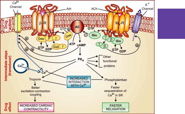
MECHANISM OF DRUG ACTION; RECEPTOR PHARMACOLOGY |
47 |
|
|
4 CHAPTER
Fig. 4.6: The action-effect sequence of two G-protein coupled (β adrenergic and muscarinic M2) receptor activation in myocardial cell
Adrenaline (Adr) binds to β-adrenergic receptor (β-R) on the cell surface inducing a conformational change which permits interaction of the G-protein binding site with the stimulatory G-protein (Gs). The activated α subunit of Gs now binds GTP (in place of GDP), and dissociates from the βγ diamer as well as the receptor. The Gsα carrying bound GTP associates with and activates the enzyme adenylyl cyclase (AC) located on the cytosolic side of the membrane: ATP is hydrolysed to cAMP which then phosphorylates and thus activates cAMP dependent protein kinase (PKA). The PKA in turn phosphorylates many functional proteins including troponin and phospholamban, so that they interact with Ca2+, respectively resulting in increased force of contraction and faster relaxation. Calcium is made available by entry from outside (direct activation of myocardial membrane Ca2+ channels by Gsα and through their phosphorylation by PKA) as well as from intracellular stores.
One of the other proteins phosphorylated by cAMP is phosphorylase kinase which then activates the enzyme phosphorylase resulting in breakdown of glycogen to be utilized as energy source for increased contractility.
Action of acetylcholine (ACh) on muscarinic M2 receptor (M2-R), also located in the myocardial membrane, similarly activates an inhibitory G-protein (Gi). The GTP carrying active Giα subunit inhibits AC, and opposes its activation by Gsα. The βγ diamer of Gi activates membrane K+ channels causing hyperpolarization which depresses impulse generation.
mediates/modulates contraction, secretion/transmitter release, eicosanoid synthesis, neuronal excitability, intracellular movements, membrane function, metabolism, cell proliferation, etc. Like AC, the PLcβ can also be inhibited through inhibitory G-protein when directionally opposite responses would be expected.
Intracellular Ca2+ release has been found to occur in waves (Ca2+ mediated Ca2+ release from successive pools facilitated by inositol 1, 3, 4, 5-tetrakisphosphate—IP4) and exhibits a variety of agonist and concentration dependent oscillatory patterns. The activation of different effectors may depend on the amplitude and pattern of these oscillations. Thus, the same intracellular messenger can trigger different responses depending on the nature and strength of the extracellular signal.

|
48 |
|
|
|
|
|
GENERAL PHARMACOLOGY |
|
|
|
|
|
|
|
|
|
|
|
|
||||
|
|
|
|
|
|
|
|
||||
|
|
|
TABLE 4.1 |
Major functional pathways of G-protein coupled receptor transduction |
|
|
|||||
|
|
|
|
|
|
|
|
|
|
|
|
|
|
|
Adenylyl cyclase: cAMP |
Phospholipase |
|
Channel regulation |
|
||||
|
|
|
|||||||||
|
1 |
|
|
|
|
|
IP3-DAG |
|
|
|
|
|
|
↑ |
↓ |
|
Ca2+↑ |
Ca2+↓ |
K+↑ |
|
|||
|
SECTION |
|
|
|
|||||||
|
|
Glucagon |
5-HT1 |
5-HT2 |
|
Adenosine-A1 |
5-HT1A |
|
|||
|
|
|
Adrenergic-β |
Adrenergic-α2 |
Adrenergic-α1 |
Adrenergic-β1 |
Dopamine-D2 Adrenergic-α2 |
|
|||
|
|
|
Histamine-H2 |
Muscarinic-M2 |
Histamine-H1 |
(Heart, |
GABAB |
Muscarinic-M2 |
|
||
|
|
|
Dopamine-D1 |
Dopamine-D2 |
Muscarinic-M1, M3 |
Sk.muscle) |
Opioid-κ |
Dopamine-D2 |
|
||
|
|
|
FSH & LH |
GABAB |
Vasopressin-Oxytocin |
Somatostatin |
GABAB |
|
|||
|
|
|
ACTH |
Opioid-μ, δ |
Bradykinin-B2 |
|
|
Opioid-μ, δ |
|
||
|
|
|
TSH |
Angiotensin-AT1 |
Angiotensin-AT1 |
|
|
Adenosine-A1 |
|
||
|
|
|
Prostaglandin-EP2 |
Prostaglandin-EP3 Prostaglandin-FP, EP1, EP3 |
|
|
|
||||
|
|
|
Prostacyclin-IP |
Somatostatin |
Thromboxane-TP |
|
|
|
|
||
|
|
|
Adenosine-A2 |
Adenosine-A1 |
Leukotriene BLT, cys LT |
|
|
|
|||
|
|
|
|
|
|
|
Cholecystokinin-Gastrin |
|
|
|
|
|
|
|
|
|
|
|
PAF |
|
|
|
|
|
|
|
|
|
|
|
|
|
|
|
|
Fig. 4.7: The important steps of phospholipase cβ(PLcβ) pathway of response effectuation (in smooth muscle) The agonist, e.g. histamine binds to its H1 receptor (H1 R) and activates the G-protein Gq. Its α subunit binds GTP in place of GDP, dissociates from the receptor as well as from βγ diamer to activate membrane bound PLcβ that hydrolyses phosphatidyl inositol 4, 5-bisphosphate (PIP2), a membrane bound phospholipid. The products inositol 1, 4, 5-trisphosphate (IP3) and diacylglycerol (DAG) act as second messengers. The primary action of IP3 is facilitation of Ca2+ mobilization from intracellular organellar pools, while DAG in conjunction with Ca2+ activates protein kinase C (PKc) which phosphorylates and alters the activity of a number of functional and structural proteins. Cytosolic Ca2+ is a veritable messenger: combines with calmodulin (CAM) to activate myosin light chain kinase (MLCK) inducing contraction, and another important regulator calcium-calmodulin protein kinase (CCPK). Several other effectors are regulated by Ca2+ in a CAM dependent or independent manner. Cytosolic Ca2+ is recycled by uptake into the endoplasmic reticulum as well as effluxed by membrane Ca2+ pump.

MECHANISM OF DRUG ACTION; RECEPTOR PHARMACOLOGY |
49 |
|
|
(c) Channel regulation The activated G- proteins (Gs, Gi, Go) can also open or inhibit ionic channels specific for Ca2+ and K+, without the intervention of any second messenger like cAMP or IP3, and bring about hyperpolarization/ depolarization/changes in intracellular Ca2+. The Gs opens Ca2+ channels in myocardium and skeletal muscles, while Gi and Go open K+ channels in heart and smooth muscle as well as inhibit neuronal Ca2+ channels. Direct channel regulation is mostly the function of the βγ diamer of the dissociated G protein. Physiological responses like changes in inotropy, chronotropy, transmitter release, neuronal activity and smooth muscle relaxation follow. Receptors found to regulate ionic channels through G-proteins are listed in Table 4.1.
2. Ion channel receptor
These cell surface receptors, also called ligand gated ion channels, enclose ion selective channels (for Na+, K+, Ca2+ or Cl¯) within their molecules. Agonist binding opens the channel (Fig. 4.4) and causes depolarization/hyperpolarization/ changes in cytosolic ionic composition, depending on the ion that flows through. The nicotinic cholinergic, GABAA, glycine (inhibitory AA), excitatory AA-glutamate (kainate, NMDA and AMPA) and 5HT3 receptors fall in this category.
The receptor is usually a pentameric protein; all subunits, in addition to large intraand extracellular segments, generally have four membrane spanning helical domains. The subunits are mostly arranged round the channel like a rosette and the α subunits usually bear the agonist binding sites.
Certain receptor-operated (or ligand-gated) ion channels also have secondary ligands which bind to an allosteric site and modulate the gating of the channel by the primary ligand, e.g. the benzodiazepine receptor modulates GABAA gated Cl¯ channel.
Thus, in these receptors the agonist directly operates ion channels, without the intervention of any coupling protein or second messenger. The onset and offset of responses through this class of receptors is the fastest (in milliseconds).
3. Transmembrane enzyme-linked receptors
This class of receptors are utilized primarily by peptide hormones, and are made up of a large
extracellular ligand binding domain connected |
|
through a single transmembrane helical peptide |
|
chain to an intracellular subunit having enzymatic |
CHAPTER |
property. The enzyme at the cytosolic side is |
|
generally a protein kinase, but can also be guanylyl |
|
cyclase in few cases. The commonest protein |
|
kinases are the ones which phosphorylate tyrosine |
|
residues on the substrate proteins and are called |
4 |
‘receptor tyrosine kinases’ (RTKs), see Fig. 4.8. |
|
|
|
Examples are—insulin, epidermal growth factor |
|
(EGF), nerve growth factor (NGF) and many other |
|
growth factor receptors. However, the transfor- |
|
ming growth factor (TGF) receptor and few |
|
others are serine/threonine kinases—which |
|
phosphorylate serine/threonine residues of the |
|
target proteins. |
|
In the unliganded monomeric state, the kinase |
|
remains inactive. Hormone binding induces |
|
dimerization of receptor molecules, brings about |
|
conformation changes which activate the kinase |
|
to autophosphorylate tyrosine residues on each |
|
other, increasing their affinity for binding substrate |
|
proteins which have SH2 domains. These are then |
|
phosphorylated and released to carry forward the |
|
cascade of phosphorylations leading to the |
|
response. |
|
Fig. 4.8: Model of receptor tyrosine kinase, an enzymelinked receptor:
On binding the peptide hormone to the extracellular domains, the monomeric receptors move laterally in the membrane and form diamers. Dimerization activates tyrosine-kinase (RTK) activity of the intracellular domains so that they phosphorylate tyrosine (t) residues on each other, as well as on several SH2 domain substrate proteins (SH2-Pr). The phosphorylated substrate proteins then perform downstream signaling function.
50 |
GENERAL PHARMACOLOGY |
|
|
SECTION 1
Fig. 4.9: Model of transmembrane JAK-STAT binding receptor.
The intracellular domain of these receptors lacks intrinsic protein kinase activity. Cytokines/hormones binding to the extracellular domain induce receptor dimerization which activates the intracellular domain to bind free moving JAK (Janus Kinase) molecules. The activated JAK phosphorylatetyrosineresiduesonthereceptorwhichthen binds another protein STAT (signal transducer and activator of transcription). Tyrosine residues of STAT also get phosphorylated by JAK. The phosphorylated STAT dimerize, dissociate from the receptor and move to the nucleus to regulate transcription of target genes.
A large number of intracellular signaling proteins have SH2 domains. Thus, by controlling phosphorylation of key enzymes, ion channels, transporters, etc. the RTKs are able to regulate diverse cellular functions including metabolic reactions, cell growth and differentiation. One of the SH2 domain enzymes is phospholipase cγ (PLcγ) which is activated by certain RTKs, and which, like PLcβ, generates IP3 and DAG as second messengers for response effectuation.
The transmembrane enzyme-linked receptors transduce responses in a matter of few minutes to few hours.
Another feature of this class of receptors is that their dimerization also promotes receptor internalization, degradation in lysosomes and down regulation if activation is fast enough.
In place of protein kinase the enzyme can also be guanylyl cyclase (GC), as in the case
of atrial natriuretic peptide (ANP). Agonist activation of the receptor generates cGMP in the cytosol as a second messenger, which in turn activates cGMP-dependent protein kinase (PKG) and modulates cellular activity.
4. Transmembrane JAK-STAT binding receptors
These receptors differ from RTKs in not having any intrinsic catalytic domain. Agonist induced dimerization alters the intracellular domain conformation to increase its affinity for a cytosolic tyrosine protein kinase JAK (Janus Kinase). On binding, JAK gets activated and phosphorylates tyrosine residues of the receptor, which now bind another free moving protein STAT (signal transducer and activator of transcription). This is also phosphorylated by JAK. Pairs of phosphorylated STAT dimerize and translocate to the nucleus to regulate gene transcription resulting in a biological response. Many cytokines, growth hormone, prolactin, interferons, etc. act through this type of receptor.
5. Receptors regulating gene expression (Transcription factors, Nuclear receptors)
In contrast to the above 3 classes of receptors, these are intracellular (cytoplasmic or nuclear) soluble proteins which respond to lipid soluble chemical messengers that penetrate the cell (Fig. 4.10). The receptor protein (specific for each hormone/regulator) is inherently capable of binding to specific genes, but its attached proteins HSP-90 and may be some others prevent it from adopting the configuration needed for binding to DNA. When the hormone binds near the carboxy terminus of the receptor, the restricting proteins (HSP-90, etc.) are released, the receptor dimerizes and the DNA binding regulatory segment located in the middle of the molecule folds into the requisite configuration. The liganded receptor diamer moves to the nucleus and binds other co- activator/co-repressor proteins which have a modulatory influence on its capacity to alter gene function. The whole complex then attaches to
MECHANISM OF DRUG ACTION; RECEPTOR PHARMACOLOGY |
51 |
|
|
4 CHAPTER
Fig. 4.10: Operational scheme of intracellular (glucocorticoid) receptor
The glucocorticoid (G) penetrates the cell membrane and binds to the glucocorticoid receptor (GR) protein that normally resides in the cytoplasm in association with heat shock protein 90 (HSP90) + other proteins. The GR has a steroid binding domain near the carboxy terminus and a mid region DNA binding domain joined by a ‘hinge region’. The DNA binding domain has two ‘zinc fingers’, each made up of a loop of amino acids with chelated zinc ion. Binding of the steroid to GR dissociates the complexed proteins (HSP90, etc) removing their inhibitory influence on it. A dimerization region that overlaps the steroid binding domain is exposed, promoting dimerization of the occupied receptor. The steroid bound receptor diamer translocates to the nucleus, binds coactivator/corepressor proteins and interacts with specific DNA sequences called ‘glucocorticoid responsive elements’ (GREs) within the regulatory region of appropriate genes. The expression of these genes is consequently altered resulting in promotion (or suppression) of their transcription. The specific mRNA thus produced is directed to the ribosome where the message is translated into a specific pattern of protein synthesis, which inturn modifies cell function.
52 |
GENERAL PHARMACOLOGY |
|
|
SECTION 1
specific DNA sequences (hormone response elements) of the target genes and facilitates or represses their expression so that specific mRNA is synthesized/repressed on the template of the gene. This mRNA moves to the ribosomes and directs synthesis of specific proteins which regulate activity of the target cells.
All steroidal hormones (glucocorticoids, mineralocorticoids, androgens, estrogens, progesterone), thyroxine, vit D and vit A function in this manner. Different steroidal hormones affect different target cells and produce different effects because each one binds to its own receptor and directs a unique pattern of synthesis of specific proteins. The specificity as to which hormone will be bound is provided by the hormone binding domain, while that as to which gene will be activated or repressed is a function of the DNA binding/ N-terminus domain. Different ligands of the same nuclear receptor have been found to induce ligandspecific conformations of the receptor so that different combinations of co-activators and corepressors may be bound in different target tissues, e.g. selective estrogen receptor modulators (SERMs). Chimeric receptors have also been produced which respond to one hormone, but produce the effects of the other hormone.
This transduction mechanism is the slowest in its time course of action (takes hours) because adequate quantity of the effector protein will have to be produced before the response occurs. The effects also generally out last the signal (hormone), because majority of the generated effector proteins have slow turnover, and persist in the body even after the hormone has been eliminated.
Regulation of receptors
Receptors exist in a dynamic state; their density and efficacy to elicit the response is subject to regulation by the level of on-going activity, feedback from their own signal output and other physiopathological influences, e.g. estrogens increase the density of oxytocin receptors on the myometrium. The sensitivity of uterus to contractile action of oxytocin increases progressively during the third trimester of pregnancy, especially
near term. In tonically active systems, prolonged deprivation of the agonist (by denervation or continued use of an antagonist or a drug which reduces input) results in supersensitivity of the receptor as well as the effector system to the agonist. This has clinical relevance in clonidine/CNS depressant/opioid withdrawal syndromes, sudden discontinuation of propranolol in angina pectoris, etc. The mechanisms involved may be unmasking of receptors or their proliferation (up regulation) or accentuation of signal amplification by the transducer.
Conversely, continued/intense receptor stimulation causes desensitization or refractoriness: the receptor becomes less efficient in transducing response to the agonist. This can be easily demonstrated experimentally (Fig. 4.11); clinical examples are bronchial asthma patients treated continuously with β adrenergic agonists and parkinsonian patients treated with high doses of levodopa gradually become less reponsive. The changes may be brought about by:
(i) Masking or internalization of the receptor (it becomes inaccessible to the agonist) or impaired
Fig. 4.11: Illustration of the phenomenon of desensitization
Contractile responses of frog’s rectus abdominis muscle to acetylcholine. Note that shortly after exposure to a high (100 fold) dose of the agonist, the response is markedly attenuated, but is regained if sufficient time is allowed to elapse.

MECHANISM OF DRUG ACTION; RECEPTOR PHARMACOLOGY |
53 |
|
|
coupling of the transducer to the receptor. In this case refractoriness develops as well as fades quickly.
In the case of β adrenergic receptor, it has been found that agonist binding promotes phosphorylation of its serine residues near the intracellular carboxy terminus by an enzyme β adrenergic receptor kinase (βARK), allowing it to bind a protein called β-arrestin which hinders its interaction with Gs → receptor transduction is impaired. When the β-agonist is removed, the serine residues are dephosphorylated and receptor mediated activation of Gs is restored.
(ii) Decreased synthesis/increased destruction of thereceptor(down regulation):refractorinessdevelops over weeks or months and recedes slowly. Receptor down regulation is particularly exhibited by the tyrosine kinase receptors. Similarly, the transducer and effector proteins are also up or down regulated.
Some times response to all agonists which act through different receptors but produce the same overt effect (e.g. histamine and acetylcholine both contract intestinal smooth muscle) is decreased by exposure to any one of these agonists (heterologous desensitization), showing that mechanisms of response effectuation have become less efficient. However, often desensitization is limited to agonists of the same receptor that is being repeatedly activated (homologous desensitization).
Both homologous and heterologous desensitization has been observed in the case of GPCRs. The BARK-β arrestin mechanism described above produces homologous desensitization. The GPCRs transduce many responses by activating PKA and PKC. These kinases phosphorylate many GPCRs rather nonselectively (at a site different from that of BARK) and hinder their interaction with G-proteins, resulting in heterologous desensitization.
Functions of receptors
These can be summarized as:
(a)To propagate regulatory signals from outside to inside the effector cell when the molecular species carrying the signal cannot itself penetrate the cell membrane.
(b)To amplify the signal.
(c)To integrate various extracellular and intracellular regulatory signals.
(d)To adapt to short term and long term changes in the regulatory melieu and maintain homeostasis.
Nonreceptor-mediated drug action
This refers to drugs which do not act by binding to specific regulatory macromolecules. Drug action by purely physical or chemical means, interactions with small molecules or ions (antacids, chelating agents, cholestyramine, etc.), as well as direct interaction with enzymes, ionic channels and transporters has already been described. In addition, there are drugs like alkylating agents which react covalently with several critical biomolecules, especially nucleic acids, and have cytotoxic property useful in the treatment of cancer.Another important class of drugs are the antimetabolites (purine/pyrimidine analogues) which lead to production of nonfunctional or dysfunctional cellular components that exert antineoplastic, antiviral and immunosuppressant activity.
DOSE-RESPONSE RELATIONSHIP
When a drug is administered systemically, the dose-response relationship has two components: dose-plasma concentration relationship and plasma concentration-response relationship. The former is determined by pharmacokinetic considerations and ordinarily, descriptions of doseresponse relationship refer to the latter, which can be more easily studied in vitro.
Generally, the intensity of response increases with increase in dose (or more precisely concentration at the receptor) and the dose-response curve is a rectangular hyperbola (Fig. 4.12). This is because drug-receptor interaction obeys law of mass action, accordingly—
Emax × [D]
E = —————— ...(3) KD + [D]
Where E is the observed effect at a dose [D] of the drug, Emax is the maximal response, KD is the dissociation constant of the drug-receptor complex, which is equal to the dose of the drug at which half maximal response is produced. If the dose is plotted on a logarithmic scale, the curve becomes sigmoid and a linear relationship between log of dose and the response is seen in the intermediate (30–70% response) zone, as can be predicted from
4 CHAPTER
54 |
GENERAL PHARMACOLOGY |
|
|
SECTION 1
Fig. 4.12: Dose-response and log dose-response curves
equation (3). This is not peculiar to drugs. In fact all stimuli are graded biologically by the fractional change in stimulus intensity, e.g. 1 kg and 2 kg weights held in two hands can be easily differentiated, but not 10 kg and 11 kg weights. Though the absolute difference in both cases remains 1kg, there is a 100% fractional change in the former case but only 10% change in the latter case. In other words, response is proportional to an exponential function (log) of the dose.
Other advantages of plotting log dose-response curves (DRC) are:
(i)A wide range of drug doses can be easily displayed on a graph.
(ii)Comparison between agonists and study of antagonists becomes easier.
The log dose-response curve (DRC) can be characterized by its shape (slope and maxima) and position on the dose axis.
Drug potency and efficacy
The position of DRC on the dose axis is the index of drug potency which refers to the amount of drug needed to produce a certain response. A DRC positioned rightward indicates lower potency (Fig. 4.13). Relative potency is often more meaningful than absolute potency, and is generally defined by comparing the dose (concentration) of the two agonists at which they elicit half maximal response (EC50). Thus, if 10 mg of
Fig. 4.13: Illustration of drug potency and drug efficacy. Dose-response curve of four drugs producing the same qualitative effect
Note:
Drug B is less potent but equally efficacious as drug A. Drug C is less potent and less efficacious than drug A. Drug D is more potent than drugs A, B, & C, but less efficacious than drugs A & B, and equally efficacious as drug C.
morphine = 100 mg of pethidine as analgesic, morphine is 10 times more potent than pethidine. However, a higher potency, in itself, does not confer clinical superiority unless the potency for therapeutic effect is selectively increased over potency for adverse effect. Drug potency is clearly a factor in choosing the dose of a drug.
The upper limit of DRC is the index of drug efficacy and refers to the maximal response that can be elicited by the drug, e.g. morphine produces a degree of analgesia not obtainable with any dose of aspirin—morphine is more efficacious than aspirin. Efficacy is a more decisive factor in the choice of a drug.
Often the terms ‘drug potency’ and ‘drug efficacy’ are used interchangeably, but these are not synonymous and refer to different characteristics of the drug. The two can vary independently:
(a)Aspirin is less potent as well as less efficacious analgesic than morphine.
(b)Pethidine is less potent but equally efficacious analgesic as morphine.

MECHANISM OF DRUG ACTION; RECEPTOR PHARMACOLOGY |
55 |
|
|
(c)Furosemide is less potent but more efficacious diuretic than metolazone.
(d)Diazepam is more potent but less efficacious CNS depressant than pentobarbitone.
Depending on the type of drug, both higher efficacy (as in the case of furosemide confering utility for mobilizing edema fluid and in renal failure) or lower efficacy (as in the case of diazepam confering safety in over-dose) could be clinically advantageous.
The slope of the DRC is also important. A steep slope indicates that a moderate increase in dose will markedly increase the response (dose needs individualization), while a flat one implies that little increase in response will occur over a wide dose range (standard doses can be given to most patients). Hydralazine has a steep, while hydrochlorothiazide has a flat DRC of antihypertensive effect (Fig. 4.14).
expressed in terms of (a) degree of benefit/relief afforded by the drug (in the recommended dose range) or (b) the success rate in achieving a defined therapeutic end point. For example, the degree of relief in parkinsonian symptoms afforded by levodopa-carbidopa is much greater than that possible with trihexyphenidyl: the former has higher therapeutic efficacy than the latter. A drug which makes a higher percentage of epileptic patients totally seizure free than another drug, is the more therapeutically effective antiepileptic.
Drug selectivity
Drugs seldom produce just one action: the DRCs for different effects of a drug may be different. The extent of separation of DRCs of a drug for different effects is a measure of its selectivity, e.g. the DRCs for bronchodilatation and cardiac stimulation (Fig. 4.15) are quite similar in case of isoprenaline, but far apart in case of salbuta- mol—the latter is a more selective bronchodilator drug.
The gap between the therapeutic effect DRC and the adverse effect DRC defines the safety margin or the therapeutic index of a drug. In experimental animals, therapeutic index is often calculated as:
4 CHAPTER
Fig. 4.14: Steep and flat dose-response curves illustrated by antihypertensive effect of hydralazine and hydrochlorothiazide
Therapeutic efficacy
The ‘therapeutic efficacy’ or ‘clinical effectiveness’ is a composite attribute of a drug different from the foregoing pharmacological description of ‘potency’ and ‘efficacy’. It depends not only on the relative potency and efficacy of the drug, but on many pharmacokinetic and pathophysiological variables as well. It is often
Fig. 4.15: Illustration of drug selectivity
Log dose-response curves of salbutamol for bronchodilatation (A) and cardiac stimulation (D)
Log dose-response curves of isoprenaline for bronchodilatation (B) and cardiac stimulation (C)
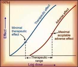
56 |
GENERAL PHARMACOLOGY |
|
|
|
median lethal dose |
|
|
Therapeutic index = —————————– |
|
|
median effective dose |
|
1 |
||
LD50 |
||
SECTION |
or ——– |
|
ED50 |
||
|
||
|
where: Median effective dose (ED50) is the dose |
|
|
which produces the specified effect in 50% |
|
|
individuals and median lethal dose (LD50) is the |
|
|
dose which kills 50% of the recipients. |
|
|
But this is irrelevant in the clinical set up where |
|
|
the therapeutic range, also called the ‘therapeutic |
|
|
window’ is bounded by the dose which produces |
|
|
minimal therapeutic effect and the dose which |
|
|
produces maximal acceptable adverse effect (Fig. |
|
|
4.16). Because of individual variability, the |
|
|
effective dose for some subjects may be toxic |
|
|
for others; defining the therapeutic range for many |
|
|
drugs is a challenging task. A drug may be capable |
|
|
of inducing a higher therapeutic response (have |
|
|
higher efficacy) but development of intolerable |
|
|
adverse effects may preclude use of higher doses, |
|
|
e.g. prednisolone in bronchial asthma. |
|
|
Risk-benefit ratio This term is very frequently |
|
|
used, and conveys a judgement on the estimated |
|
|
harm (adverse effects, cost, inconvenience) vs |
|
|
expected advantages (relief of symptoms, cure, |
|
|
reduction of complications/mortality, improvement |
|
|
in quality of life). A drug should be prescribed |
Fig. 4.16: Illustrative dose-response curves for therapeutic effect and adverse effect of the same drug
only when the benefits outweigh the risks. However, risk-benefit ratio can hardly ever be accurately measured for each instance of drug use, because ‘risk’ is the probability of harm; and harm has to be qualified by its nature, quantum, time-course (transient to life-long) as well as the value that the patient attaches to it. None of these can be precisely ascertained beforehand in an individual patient. As such, the physician has to rely on data from use of drugs in large populations (pharmacoepidemiology) and his own experience of the drug and the patient.
Drug specificity
Specificity of a drug refers to the range of actions produced by it. Certain drugs produce just one or a limited number of actions, while others have widespread effects on many organs of the body. Specificity is governed by:
(a)whether a drug acts on a single receptor/ target or on many targets, and
(b)how widely the target is distributed in the body.
Omeprazole (and other proton pump inhibitors) is an example of a highly selective drug. The singular perceptible action in therapeutic doses is inhibition of gastric acid secretion, because it acts only on one target molecule H+K+ ATPase (proton pump) which is localized to the gastric parietal cells. An example of a drug acting on multiple targets is chlorpromazine which has antagonistic action on dopamine D2, α-adrenergic, muscarinic cholinergic, histamine H1 and some 5-HT receptors. It also has Na+ channel blocking action. As a result, it produces a wide range of actions. Another case is dexamethasone which is an agonist only of glucocorticoid receptor, but produces effects involving many organs and tissues, because the glucocorticoid receptor is expressed by practically every cell of the body. Drugs with all grades of specificity are available.
COMBINED EFFECT OF DRUGS
When two or more drugs are given simultaneously or in quick succession, they may be either indifferent to each other or exhibit synergism or

MECHANISM OF DRUG ACTION; RECEPTOR PHARMACOLOGY |
57 |
|
|
antagonism. The interaction may take place at pharmacokinetic level (see Ch. 2 and 3) or at pharmacodynamic level.
SYNERGISM
(Greek: Syn—together; ergon—work)
When the action of one drug is facilitated or increased by the other, they are said to be synergistic. In a synergistic pair, both the drugs can have action in the same direction or given alone one may be inactive but still enhance the action of the other when given together. Synergism can be:
(a) Additive The effect of the two drugs is in the same direction and simply adds up:
effect of drugs A + B = |
effect of drug A + |
|||
|
|
|
effect of drug B |
|
|
|
|||
|
Additive drug combinations |
|||
|
|
|
|
|
Aspirin |
+ paracetamol |
as |
analgesic/ |
|
|
|
|
antipyretic |
|
Nitrous |
oxide |
+ halothane |
as |
general |
|
|
|
anaesthetic |
|
Amlodipine + |
atenolol |
as |
antihypertensive |
|
Glibenclamide |
+ metformin |
as hypoglycaemic |
||
Ephedrine + theophylline |
as |
bronchodilator |
||
|
|
|
|
|
Side effects of the components of an additive pair may be different—do not add up. Thus, the combination is better tolerated than higher dose of one component.
Supraadditive drug combinations
Drug pair |
|
Basis of |
potentiation |
|
Acetylcholine |
+ |
Inhibition |
of |
break |
physostigmine |
down |
|
|
|
Levodopa + |
carbidopa/ |
Inhibition |
of |
peripheral |
benserazide |
|
metabolism |
|
|
Adrenaline + |
cocaine/ |
Inhibition |
of |
|
desipramine |
|
neuronal |
uptake |
|
Sulfamethoxazole + |
Sequential blockade |
|||
trimethoprim |
|
|
|
|
Antihypertensives |
Tackling two contributory |
|||
(enalapril+ |
|
factors |
|
|
hydrochlorothiazide) |
|
|
|
|
Tyramine + MAO |
Increasing releaseable |
|||
inhibitors |
|
CA store |
|
|
4 CHAPTER

Fig. 4.17: Log dose-response curves of a drug ‘A’ depicting additive synergism (in purple) and potentiation (Supra-additive synergism) in blue.
A:An agonist drug.
B:Another agonist in a fixed submaximal dose producing 40% response.
C:A potentiating drug which itself has no agonistic activity.
(b) Supraadditive (potentiation) The effect of combination is greater than the individual effects of the components:
effect of drug A + B > effect of drug A + effect of drug B
This is always the case when one component given alone produces no effect, but enhances the effect of the other (potentiation). Examples are given in the box. Additive synergism and potentiation are depicted diagrammatically in Fig. 4.17.
ANTAGONISM
When one drug decreases or abolishes the action of another, they are said to be antagonistic: effect of drugs A + B < effect of drug A +
effect of drug B Usually in an antagonistic pair one drug is inactive as such but decreases the effect of the other. Depending on the mechanism involved, antagonism may be:
(a) Physical antagonism
Based on the physical property of the drugs, e.g.
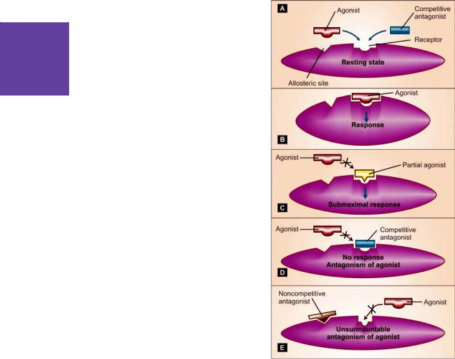
58 |
GENERAL PHARMACOLOGY |
|
|
SECTION 1
charcoal adsorbs alkaloids and can prevent their absorption—used in alkaloidal poisonings.
(b) Chemical antagonism
The two drugs react chemically and form an inactive product, e.g.
•KMnO4 oxidizes alkaloids—used for gastric lavage in poisoning.
•Tannins + alkaloids—insoluble alkaloidal tannate is formed.
•Chelating agents (BAL, Cal. disod. edetate) complex toxic metals (As, Pb).
•Nitrites form methaemoglobin which reacts with cyanide radical.
Drugs may react when mixed in the same syringe or infusion bottle:
•Thiopentone sod. + succinylcholine chloride
•Penicillin-G sod. + succinylcholine chloride
•Heparin + penicillin/tetracyclines/streptomycin/ hydrocortisone
(c) Physiological/functional antagonism
The two drugs act on different receptors or by different mechanisms, but have opposite overt effects on the same physiological function, i.e. have pharmacological effects in opposite direction, e.g.
•Histamine and adrenaline on bronchial muscles and BP.
•Hydrochlorothiazide and triamterene on urinary K+ excretion.
•Glucagon and insulin on blood sugar level. Fig. 4.18: Illustration of sites of action of agonists and
(d) Receptor antagonism
One drug (antagonist) blocks the receptor action of the other (agonist). This is a very important mechanism of drug action, because physiological signal molecules act through their receptors, blockade of which can produce specific and often profound pharmacological effects. Receptor antagonists are selective (relatively), i.e. an anticholinergic will oppose contraction of intestinal smooth muscle induced by cholinergic agonists, but not that induced by histamine or 5-HT (they act through a different set of receptors).
antagonists (A), and the action of full agonist (B), partial agonist (C), competitive antagonist (D) and noncompetitive antagonist (E) on the receptor
Receptor antagonism can be competitive or noncompetitive.
Competitive antagonism (equilibrium type)
The antagonist is chemically similar to the agonist, competes with it (Fig. 4.18 A, D) and binds to the same site to the exclusion of the agonist molecules. Because the antagonist has affinity but no intrinsic activity (see p. 42), no response is produced and the log DRC of the agonist is shifted
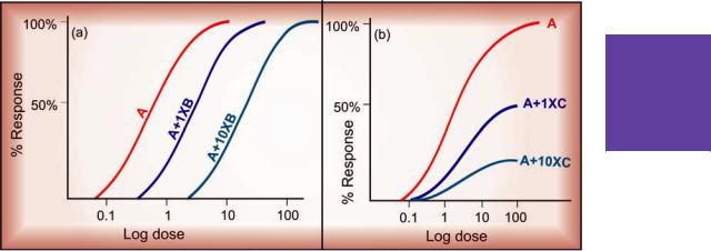
MECHANISM OF DRUG ACTION; RECEPTOR PHARMACOLOGY |
59 |
|
|
4 CHAPTER
Fig. 4.19: Dose-response curves showing competitive (a) and noncompetitive (b) antagonism A—agonist, B—competitive antagonist, C—noncompetitive antagonist
to the right. Since antagonist binding is reversible and depends on the relative concentration of the agonist and antagonist molecules, higher concentration of the agonist progressively overcomes the block—a parallel shift of the agonist DRC with no suppression of maximal response is obtained (Fig. 4.19a). The extent of shift is dependent on the affinity and concentration of the antagonist.
A partial agonist (Fig. 4.18 C), having affinity for the same receptor, also competes with and antagonizes a full agonist, while producing a submaximal response of its own.
Noncompetitive antagonism The antagonist is chemically unrelated to the agonist, binds to a different allosteric site altering the receptor in
such a way that it is unable to combine with the agonist (Fig. 4.18E), or is unable to transduce the response, so that the downstream chain of events are uncoupled. This is also called allosteric antagonism. Because the agonist and the antagonist are combining with different sites, there is no competition between them—even high agonist concentration is unable to reverse the block completely. Increasing concentrations of the antagonist progressively flatten the agonist DRC (Fig. 4.19b). Noncompetitive antagonists have been produced experimentally, but are not in clinical use.
Nonequilibrium antagonism Certain antagonists bind to the receptor with strong (covalent) bonds or dissociate from it slowly (due to very high affinity) so that agonist molecules are unable
|
Competitive (equilibrium type) |
Noncompetitive |
|
|
|
1. |
Antagonist binds with the same receptor as the agonist |
Binds to another site of receptor |
2. |
Antagonist resembles chemically with the agonist |
Does not resemble |
3. |
Parallel rightward shift of agonist DRC |
Flattening of agonist DRC |
4. |
The same maximal response can be |
Maximal response is suppressed |
|
attained by increasing dose of agonist |
(unsurmountable antagonism) |
|
(surmountable antagonism) |
|
5. |
Intensity of response depends on the concen- |
Maximal response depends only on |
|
tration of both agonist and antagonist |
the concentration of antagonist |
6. |
Examples: ACh—Atropine |
Diazepam—Bicuculline |
|
Morphine—Naloxone |
|
|
|
|

60 |
GENERAL PHARMACOLOGY |
|
|
SECTION 1
to reduce receptor occupancy of the antagonist molecules—law of mass action cannot apply— an irreversible or nonequilibrium antagonism is produced. The agonist DRC is shifted to the right and the maximal response is lowered (if spare receptors are few). Since in this situation the agonist molecules are not able to compete with the antagonist molecules and flattening of
agonist DRC is a feature of noncompetitive antagonism; nonequilibrium antagonism has also been called ‘a type of noncompetitive antagonism’. Phenoxybenzamine is a nonequilibrium antagonist of adrenaline at the α adrenergic receptors.
Features of competitive and noncompetitive antagonism are compared on previous page.
)PROBLEM DIRECTED STUDY
4.1A patient being treated with methotrexate (Mtx) developed oral ulceration, megaloblastic anaemia and other toxic symptoms. Given that (i) Mtx acts by inhibiting the enzyme dihydrofolate reductase (DHFRase) which generates the essential coenzyme tetrahydrofolic acid (THFA) from dihydrofolic acid (DHFA) needed for one carbon transfer reactions, (ii) Mtx binds to the catalytic site of DHFRase with an affinity 50,000 times greater than the natural substrate DHFA, and that (iii) two forms of folate viz. folic acid and folinic acid (N5 formyl THFA) are available for therapeutic use:
(a)Which type of enzyme inhibition will be produced by Mtx?
(b)Which form of folate should be used to treat Mtx toxicity? (see Appendix-1 for solution)

Chapter 5 Aspects of Pharmacotherapy,
Clinical Pharmacology and
Drug Development
Pharmaco- (drug) therapy is dynamic and an ever evolving science. It requires understanding of the drug, the disease, the patient and the milieu in which it is undertaken. As such, in addition to knowledge of drug action, mechanisms and pharmacokinetics, several aspects like drug dosage, sources of variability in drug response, pharmacogenetics, influence of disease on drug action, etc. are important for otimum drug therapy.
DRUG DOSAGE
‘Dose’ is the appropriate amount of a drug needed to produce a certain degree of response in a given patient. Accordingly, dose of a drug has to be qualified in terms of the chosen response, e.g. the analgesic dose of aspirin for headache is 0.3–0.6 g, its antiplatelet dose is 60–150 mg/ day, while its antiinflammatory dose for rheumatoid arthritis is 3–5 g per day. Similarly there could be a prophylactic dose, a therapeutic dose or a toxic dose of the same drug.
The dose of a drug is governed by its inherent potency, i.e. the concentration at which it should be present at the target site, and its pharmacokinetic characteristics. The recommended doses are based on population data and cater to an ‘average’ patient. However, individual patients may not be ‘average’ in respect to a number of pharmacokinetic and pharmacodynamic parameters, emphasizing the need for individualizing drug dose. The strategies adopted for different types of drugs and conditions are:
1. Standard dose The same dose is appropriate for most patients—individual variations are minor or the drug has a wide safety margin so that a large enough dose can be given to cover them, e.g. oral contraceptives, penicillin, chloroquine, mebendazole, hydrochlorothiazide.
2.Regulated dose The drug modifies a finely regulated body function which can be easily measured. The dosage is accurately adjusted by repeated measurement of the affected physiological parameter, e.g. antihypertensives, hypoglycaemics, anticoagulants, diuretics, general anaesthetics. In their case, measurement of plasma drug concentration is not needed.
3.Target level dose (see p. 33) The response is not easily measurable but has been demonstrated to be obtained at a certain range of drug concentration in plasma. An empirical dose aimed at attaining the target level is given in the beginning and adjustments are made later by actual monitoring of plasma concentrations. When facilities for drug level monitoring are not available, crude adjustments are made by observing the patient at relatively long intervals, e.g. antidepressants, antiepileptics, digoxin, lithium, theophylline.
4.Titrated dose The dose needed to produce maximal therapeutic effect cannot be given because of intolerable adverse effects. Optimal dose is arrived at by titrating it with an acceptable level of adverse effect. Low initial dose and upward titration (in most non-critical situations) or high initial dose and downward titration
(in critical situations) can be practised. Often a compromise between submaximal therapeutic effect but tolerable side effects can be struck, e.g. anticancer drugs, corticosteroids, levodopa.
Fixed dose combinations (FDCs) of drugs
A large number of pharmaceutical preparations contain two or more drugs in a fixed dose ratio. Advantages offered by these are:

62 |
GENERAL PHARMACOLOGY |
|
|
SECTION 1
1.Convenience and better patient compliance— when all the components present in the FDC are actually needed by the patient and their amounts are appropriate. It may also be cost saving compared to both/all the components administered separately.
2.Certain drug combinations are synergistic, e.g. sulfamethoxazole + trimethoprim; levodopa + carbidopa/benserazide; combination oral contraceptives, isoniazid + rifampin.
3.The therapeutic effect of two components being same may add up while the side effects being different may not. For this the components of the FDC should act by different mechanisms, e.g. amlodipine + atenolol as antihypertensive.
4.The side effect of one component may be counteracted by the other, e.g. a thiazide + a potassium sparing diuretic. However, the amount of the latter may not be sufficient in all cases.
5.Combined formulation ensures that a single drug will not be administered. This is important in the treatment of tuberculosis, HIV-AIDS and falciparum malaria.
Before prescribing a combination, the physician must consider whether any of the ingredients is unnecessary; if it is, the combination should not be prescribed. It can never be justified that a drug is given to a patient who does not need it in order to provide him another one that he needs.
There are many inbuilt disadvantages of FDCs:
1.The patient may not actually need all the drugs present in a combination: he is subjected to additional side effects and expense (often due to ignorance of the physician about the exact composition of the combined formulations).
2.The dose of most drugs needs to be adjusted and individualised. When a combined formulation is used, this cannot be done without altering the dose of the other component(s). However, few combinations are available at more than one dose ratios, e.g. levodopa (100 mg) + Carbidopa (10 mg or 25 mg),
amoxicillin (250 mg or 500 mg) + clavulanic acid (125 mg).
3.The time course of action of the components may be different: administering them at the same intervals may be inappropriate.
4.Altered renal or hepatic function of the patient may differently affect the pharmacokinetics of the components.
5.Adverse effect, when it occurs, cannot be easily ascribed to the particular drug causing it.
6.Contraindication to one component (allergy, other conditions) contraindicates the whole product.
7.Confusion of therapeutic aims and false sense of superiority of two drugs over one is fostered, specially in case of antimicrobials whose combinations should be avoided. Corticosteroids should never be combined with any other drug meant for internal use. Drug combinations that are banned in India are listed in Appendix-5.
Thus, only a handful of FDCs are rational and justified, while far too many are produced and vigorously promoted by the pharmaceutical industry. In fact, the latest WHO essential medicines list incorporates only 23 FDCs, and the NLEM (2011) of India has only 12 FDCs (see Appendix-2).
FACTORS MODIFYING DRUG ACTION
Variation in response to the same dose of a drug between different patients and even in the same patient on different occasions is a rule rather than exception. One or more of the following categories of differences among individuals are responsible for the variations in drug response:
(1)Individuals differ in pharmacokinetic handling of drugs: attain varying plasma/target site concentration of the drug. This is more marked for drugs disposed by metabolism (e.g. propranolol) than for drugs excreted unchanged (e.g. atenolol).
(2)Variations in number or state of receptors, coupling proteins or other components of response
effectuation.

PHARMACOTHERAPY, CLINICAL PHARMACOLOGY AND DRUG DEVELOPMENT |
63 |
|
|
(3) Variations in neurogenic/hormonal tone or concentrations of specific constituents, e.g. atropine tachycardia depends on vagal tone, propranolol bradycardia depends on sympathetic tone, captopril hypotension depends on body Na+ status.
A multitude of host and external factors influence drug response. They fall in two categories viz genetic and nongenetic including all environmental, circumstantial and personal variables. Though individual variation cannot be totally accounted for by these factors, their understanding can guide the choice of appropriate drug and dose for an individual patient. However, final adjustments have to be made by observing the response in a given patient on a given occasion.
The factors modify drug action either:
(a)Quantitatively The plasma concentration and/or the action of the drug is increased or decreased. Most of the factors introduce this type of change and can be dealt with by adjustment of drug dosage.
(b)Qualitatively The type of response is altered, e.g. drug allergy or idiosyncrasy. This is less common but often precludes further use of that drug in the affected patient.
The various factors are discussed below—
1. Body size It influences the concentration of the drug attained at the site of action. The average adult dose refers to individuals of medium built. For exceptionally obese or lean individuals and for children dose may be calculated on body weight (BW) basis:
BW (kg)
Individual dose = ———— × average adult dose 70
It has been argued that body surface area (BSA) provides a more accurate basis for dose calculation, because total body water, extracellular fluid volume and metabolic activity are better paralleled by BSA.
BSA (m2)
Individual dose = ———— × average adult dose 1.7
The BSA of an individual can be calculated from Dubois formula:
BSA (m2) =
BW (kg)0.425 × Height (cm)0.725 × 0.007184
or obtained from chart-form or slide-rule nomograms based on BW and height.
However, dose recommendations in terms of BSA are available only for anticancer and a handful of other drugs: for the rest BW has been used as the index. Thus, prescribing on BSA basis suffers from lack of data base, is more cumbersome and has not thrived, except in few cases.
2. Age The dose of a drug for children is often calculated from the adult dose
Age |
|
Child dose = ———— × adult dose ... (Young’s |
|
Age + 12 |
formula) |
Age |
(Dilling’s |
Child dose = –—— × adult dose ... |
|
20 |
formula) |
|
|
It can also be calculated (more accurately) on BW or BSA basis (see above), and for many drugs, manufacturers give dosage recommendations on mg/kg basis. Average figures for children are given below.
|
Ideal |
BSA |
% of |
Age |
BW (Kg) |
(m2) |
Adult dose |
|
|
|
|
Newborn |
3.2 |
0.23 |
12.5 |
1 month |
4.0 |
0.26 |
15 |
3 months |
5.5 |
0.32 |
18 |
6 months |
7.5 |
0.4 |
22 |
1 year |
10 |
0.47 |
25 |
3 years |
14 |
0.62 |
33 |
5 years |
18 |
0.73 |
40 |
7 years |
23 |
0.88 |
50 |
12 years |
37 |
1.25 |
75 |
|
|
|
|
However, infants and children are not small adults. They have important physiological differences from adults. The newborn has low g.f.r. and tubular transport is immature. As such, the t½ of drugs excreted by glomerular filtration (gentamicin) and tubular secretion (penicillin) is prolonged by 3 to 5 times. Glomerular filtration reaches adult rates by 5 month of age and tubular
5 CHAPTER
64 |
GENERAL PHARMACOLOGY |
|
|
SECTION 1
secretion takes about 7 months to mature. Similarly, hepatic drug metabolizing system is inadequate in newborns —chloramphenicol can produce gray baby syndrome. Blood-brain barrier is more permeable—drugs attain higher concentration in the CNS (accumulation of unconjugated bilirubin causes kernicterus). These defects are exaggerated in the premature infant. Drug absorption may also be altered in infants because of lower gastric acidity and slower intestinal transit. Transdermal absorption however, is faster because their skin is thin and more permeable. Rectal absorption is fast and more predictable in infants and young children; diazepam solution is given rectally to control febrile seizures in children < 5 years. Therefore, infant doses must be learned as such and not derived from any formula.
After the first year of life, drug metabolism is often faster than in adults, e.g. theophylline, phenytoin, carbamazepine t½ is shorter in children. Also, higher per kg dose is needed for drugs which are primarily excreted unchanged by kidney, e.g. daily dose of digoxin is about 8–12 µg/kg compared to adult dose of 3–5 µg/kg.
Solid dosage forms and metered dose inhalers are difficult to administer to young children.
Children are growing and are susceptible to special adverse effects of drugs, e.g. suppression of growth can occur with corticosteroids; androgens may promote early fusion of epiphysis resulting in stunting of stature; tetracyclines get deposited in growing teeth and discolour/deform them. Dystonic reactions to phenothiazines are more common in children.
Elderly In the elderly, renal function progressively declines (intact nephron loss) so that g.f.r. is ~ 75% at 50 years and ~ 50% at 75 years age compared to young adults. Drug doses have to be reduced, e.g. daily dose of streptomycin is 0.75 g after 50 years and 0.5 g after 70 years of age compared to 1 g for young adults. There is also a reduction in the hepatic microsomal drug metabolizing activity and liver blood flow: oral bioavailability of drugs with high hepatic extraction is generally increased, but the overall effects
on drug metabolism are not uniform. Due to lower renal as well as metabolic clearance, the elderly are prone to develop cumulative toxicity while receiving prolonged medication. Other affected aspects of drug handling are:
• slower absorption due to reduced gut motility as well as blood flow to intestines,
•lesser plasma protein binding due to lower plasma albumin,
•increased or decreased volume of distribution of lipophilic and hydrophilic drugs respectively.
Aged are relatively intolerant to digitalis. The responsiveness of β adrenergic receptors to both agonists and antagonists is reduced in the elderly and sensitivity to other drugs also may be altered. Due to prostatism in elderly males, even mild anticholinergic activity of the drug can accentuate bladder voiding difficulty. Elderly are also likely to be on multiple drug therapy for hypertension, ischaemic heart disease, diabetes, arthritis, etc. which increases many fold the chances of drug interactions. They are more prone to develop postural instability, giddiness and mental confusion. In general, the incidence of adverse drug reactions is much higher in the elderly.
3. Sex Females have smaller body size and require doses that are on the lower side of the range. Subjective effects of drugs may differ in females because of their mental makeup. Maintenance treatment of heart failure with digoxin is reported to be associated with higher mortality among women than among men. A number of antihypertensives (clonidine, methyldopa, β-blockers, diuretics) have potential to interfere with sexual function in males but not in females. Gynaecomastia is a side effect (of ketoconazole, metoclopramide, chlorpromazine, digitalis) that can occur only in men. Ketoconazole causes loss of libido in men but not in women. Obviously androgens are unacceptable to women and estrogens to men. In women consideration must also be given to menstruation, pregnancy and lactation.
Drugs given during pregnancy can affect the foetus (see Ch. 6 and Appendix-3). There are

PHARMACOTHERAPY, CLINICAL PHARMACOLOGY AND DRUG DEVELOPMENT |
65 |
|
|
marked and progressive physiological changes during pregnancy, especially in the third trimester, which can alter drug disposition.
(i)Gastrointestinal motility is reduced → delayed absorption of orally administered drug.
(ii)Plasma and extracellular fluid volume expands—volume of drug distribution may increase.
(iii)While plasma albumin level falls, that of α1 acid glycoprotein increases—the unbound fraction of acidic drugs increases but that of basic drugs decreases.
(iv)Renal blood flow increases markedly—polar drugs are eliminated faster.
(v)Hepatic microsomal enzymes undergo induction—many drugs are metabolized faster.
Thus, the overall effect on drug disposition is complex and often difficult to predict.
4.Species and race There are many examples of differences in responsiveness to drugs among different species; rabbits are resistant to atropine, rats and mice are resistant to digitalis and rat is more sensitive to curare than cat. These differences are important while extrapolating results from experimental animals to man.
Among human beings some racial differences have been observed, e.g. blacks require higher
and mongols require lower concentrations of atropine and ephedrine to dilate their pupil. β- blockers are less effective as antihypertensive in Afro-Caribbeans. Indians tolerate thiacetazone better than whites. Considering the widespread use of chloramphenicol in India and Hong Kong, relatively few cases of aplastic anaemia have been reported compared to its incidence in the west. Similarly, quiniodochlor related cases of subacute myelooptic neuropathy (SMON) occurred in epidemic proportion in Japan, but there is no confirmed report of its occurrence in India despite extensive use.
5.Genetics The dose of a drug to produce the same effect may vary by 4–6 fold among different individuals. All key determinants of drug response, viz. transporters, metabolizing enzymes,
ion channels, receptors with their couplers and effectors are controlled genetically. Hence, a great deal of individual variability can be traced to the genetic composition of the subject.
Pharmacogenetics The study of genetic basis for variability in drug response is called
‘Pharmacogenetics’. It deals with genetic influences on drug action as well as on drug handling by the body. As the genomic technology has advanced and the human genome project has been undertaken, gene libraries and huge data bases (like ‘pharmacogenetics and pharmacogenomics knowledge base’, ‘Human genome variation database’, etc.) have been created aiming at improving precision in drug therapy.
Pharmacogenomics is the use of genetic information to guide the choice of drug and dose on an individual basis. It intends to identify individuals who are either more likely or less likely to respond to a drug, as well as those who require altered dose of certain drugs. Attempt is made to define the genetic basis of an individual’s profile of drug response and to predict the best treatment option for him/her. So far, this has been applied largely to patients with known genetic abnormalities, but the goal is ‘personalized medicine’ on a wide scale. However, a large proportion of genetic variability still remains unaccounted for.
A continuous variation with bell-shaped Gaussian frequency distribution is seen in the case of most drugs. In addition, there are some specific genetic defects which lead to discontinuous variation in drug responses, e.g.—
1.Atypical pseudocholinesterase results in prolonged succinylcholine apnoea.
2.G-6PD deficiency is responsible for haemolysis with primaquine and other oxidizing drugs. This X-linked monogenic trait is more common in the Mediterranean, African and Southeast Asian races. The haemolysis is largely dose related. Several variants of the G-6PD gene occur in the population resulting in differing severity of haemolysis triggered by different oxidizing drugs.
5 CHAPTER

66 |
GENERAL PHARMACOLOGY |
|
|
|
|
Haemolysis is severe in homozygous |
|||
|
|
deficient individuals of certain genotypes. |
|||
|
|
Important drugs reported to cause haemo- |
|||
1 |
|||||
|
lysis in G-6PD deficient subjects are listed |
||||
SECTION |
|
||||
|
in the box. |
|
|||
|
|
|
|||
|
|
|
|||
|
|
Drugs with potential* to cause haemolysis |
|||
|
|
|
in G-6PD deficient individuals |
||
|
|
|
|
|
|
|
|
Primaquine |
Nalidixic acid |
||
|
|||||
|
|
Chloroquine |
Fluoroquinolones |
||
|
|
Quinine/ |
Quinidine |
Chloramphenicol |
|
|
|
Proguanil |
|
Nitrofurantoin |
|
|
|
Pyrimethamine |
Furazolidone |
||
|
|
Dapsone |
|
Methyldopa |
|
|
|
Sulfonamides |
Hydralazine |
||
|
|
Cotrimoxazole |
Procainamide |
||
|
|
Sulfasalazine |
Probenecid |
||
|
|
Thiazide |
diuretics |
Colchicine |
|
|
|
Aspirin |
|
Methylene blue |
|
*Drugs carrying higher risk are italicized.
3.The low activity CYP2C9 variants metabolize warfarin at a slow rate and are at higher risk of bleeding.
4.Thiopurine methyl transferase (TPMT) deficiency increases risk of severe bone marrow toxicity of 6-mercaptopurine and azathioprine.
5.Irinotecan induced neutropenia and diarrhoea is more in patients with UGT1A1 *28 allele of glucuronyl transferase.
6.Severe 5-fluorouracil toxicity occurs in patients with dihydropyrimidine dehydrogenase (DPD) deficiency.
7.Over expression of P-gp results in tumour resistance to many cancer chemotherapeutic drugs, because it pumps out the drug from the tumour cells.
8.Polymorphism of N-acetyl transferase 2 (NAT2) gene results in rapid and slow acetylator status. Isoniazid neuropathy, procainamide and hydralazine induced lupus occurs mainly in slow acetylators.
9.Acute intermittent porphyria—precipitated by barbiturates is due to genetic defect in repression of porphyrin synthesis.
10.CYP2D6 abnormality causes poor metoprolol/debrisoquin metabolizer status. Since several antidepressants and antipsychotics
also are substrates of CYP2D6, deficient patients are more likely to experience their toxicity. Codeine fails to produce analgesia in CYP2D6 deficient, because this enzyme generates morphine from codeine.
11.Malignant hyperthermia after halothane is due to abnormal Ca2+ release channel (ryanodine receptor) in the sarcoplasmic reticulum of skeletal muscles.
12.Inability to hydroxylate phenytoin results in toxicity at usual doses.
13.Resistance to coumarin anticoagulants is due to an abnormal enzyme VKOR (that regenerates the reduced form of vit. K) which has low affinity for the coumarins.
14.Attack of angle closure glaucoma is precipitated by mydriatics in individuals with narrow iridocorneal angle.
Genetic variability in drug response could be due to single gene mutation or polygenic. Genotype to phenotype predictability is much better in monogenic phenotypic traits such as G-6PD, CYP2D6, TPMT, etc., than for multigenic traits, which are clinically less significant. Majority of gene polymorphisms are due to substitution of a single base pair by another. When found in the population at a frequency of >1%, these are called ‘Single neucleotide polymorphisms’ (SNPs). Gene polymorphisms are often encountered at different frequencies among different ethnic/geographical groups.
Despite accumulation of considerable pharmacogenomic data and the fact that genotyping of the individual needs to be done only once, its practical application in routine patient care is at present limited due to prerequirement of multiple drug specific genotypic screening. Simple spot tests for some, e.g. G-6 PD deficiency are currently in use.
6. Route of administration Route of administration governs the speed and intensity of drug response (see Ch. 1). Parenteral administration is often resorted to for more rapid, more pronounced and more predictable drug action. A drug may have entirely different uses through different routes, e.g. magnesium sulfate given

PHARMACOTHERAPY, CLINICAL PHARMACOLOGY AND DRUG DEVELOPMENT |
67 |
|
|
orally causes purgation, applied on sprained joints—decreases swelling, while intravenously it produces CNS depression and hypotension.
7. Environmental factors and time of administration Several environmental factors affect drug responses. Exposure to insecticides, carcinogens, tobacco smoke and consumption of charcoal broiled meat are well known to induce drug metabolism. Type of diet and temporal relation between drug ingestion and meals can alter drug absorption, e.g. food interferes with absorption of ampicillin, but a fatty meal enhances absorption of griseofulvin and lumefantrine. Subjective effects of a drug may be markedly influenced by the setup in which it is taken. Hypnotics taken at night and in quiet, familiar surroundings may work more easily. It has been shown that corticosteroids taken as a single morning dose cause less pituitary-adrenal suppression.
8. Psychological factor Efficacy of a drug can be affected by patient’s beliefs, attitudes and expectations. This is particularly applicable to centrally acting drugs, e.g. a nervous and anxious patient requires more general anaesthetic; alcohol generally impairs performance but if punishment (which induces anxiety) is introduced, it may actually improve performance by relieving anxiety.
Placebo This is an inert substance which is given in the garb of a medicine. It works by psychodynamic rather than pharmacodynamic means and often produces responses equivalent to the active drug. Some individuals are more suggestible and easily respond to a placebo: and are called ‘placebo reactors’. Placebos are used in two situations:
1.As a control device in clinical trial of drugs (dummy medication).
2.To treat a patient who, in the opinion of the
physician, does not require an active drug. Placebo is a Latin word meaning ‘I shall please’. A patient responds to the whole therapeutic setting; placebo-effect largely depends on the physicianpatient relationship.
Placebos do induce physiological responses, e.g. they can release endorphins in brain—caus- ing analgesia. Naloxone, an opioid antagonist, blocks placebo analgesia. When an active drug is administered, it produces effects both due to its pharmacodynamic action as well as the psychodynamic effect of the act of medication. Placebo effects can thus supplement pharmacological effects of active medicines. However, placebo effects are highly variable even in the same individual, e.g. a placebo may induce sleep on the first night, but not subsequently. Thus, it has a very limited role in practical therapeutics. Substances commonly used as placebo are lactose tablets/capsules and distilled water injection.
Nocebo It is the converse of placebo, and refers to negative psychodynamic effect evoked by the pessimistic attitude of the patient, or by loss of faith in the medication and/or the physician. Nocebo effect can oppose the therapeutic effect of active medication.
9. Pathological states Not only drugs modify disease processes, several diseases can influence drug disposition and drug action:
Gastrointestinal (g.i.) diseases Certain g.i. diseases can alter absorption of orally administered drugs. The changes are complex and drug absorption can increase or decrease, e.g. in coeliac disease absorption of amoxicillin is decreased but that of cephalexin and cotrimoxazole is increased. Thus, malabsorption syndrome does not necessarily reduce absorption of all drugs. Gastric stasis occurring during migraine attack retards the absorption of ingested drugs. Achlorhydria decreases aspirin absorption by favouring its ionization. NSAIDs can aggravate peptic ulcer disease.
Liver disease Liver disease (especially cirrhosis) can influence drug disposition in several ways:
•Bioavailability of drugs having high first pass metabolism (see Ch. 3) is increased due to loss of hepatocellular function and portocaval shunting.
•Serum albumin is reduced—protein binding of acidic drugs (diclofenac, warfarin, etc.) is
5 CHAPTER

68 |
GENERAL PHARMACOLOGY |
|
|
SECTION 1
reduced and more drug is present in the free form.
•Metabolism and elimination of some drugs (morphine, lidocaine, propranolol) is decreased—their dose should be reduced. Alternative drugs that do not depend on hepatic metabolism for elimination and/or have shorter t½ should be preferred, e.g. oxazepam or
lorazepam in place of diazepam; atenolol as β-blocker.
•Prodrugs needing hepatic metabolism for activation, e.g. bacampicillin are less effective and should be avoided.
The changes are complex and there is no
simple test (like creatinine clearance for renal disease) to guide the extent of alteration in drug disposition; kinetics of different drugs is affected to different extents.
Not only disposition, but action as well of certain drugs may be altered in liver disease, e.g.
•The sensitivity of brain to depressant action of morphine and barbiturates is markedly increased in cirrhotics—normal doses can produce coma.
•Brisk diuresis can precipitate mental changes in patients with impending hepatic encephalopathy, because diuretics cause hypo-
kalemic alkalosis which favours conversion of NH+4 to NH3. Ammonia enters brain easily and causes mental derangement.
•Oral anticoagulants can markedly increase prothrombin time, because clotting factors are already low.
•Fluid retaining action of phenylbutazone (also
other NSAIDs) and lactic acidosis due to metformin are accentuated.
Hepatotoxic drugs should be avoided in liver disease.
Kidney disease It markedly affects pharmacokinetics of many drugs as well as alters the effects of some drugs.
Clearance of drugs that are primarily excreted unchanged (aminoglycosides, digoxin, phenobarbitone) is reduced parallel to decrease in creatinine clearance (CLcr). Loading dose of such a drug
is not altered (unless edema is present), but maintenance doses should be reduced or dose interval prolonged proportionately. A rough guideline is given in the box:
CLcr (patient) |
Dose rate to be |
|
reduced to |
|
|
50–70 ml/min |
70% |
30–50 ml/min |
50% |
10–30 ml/min |
30% |
5–10 ml/min |
20% |
Dose rate of drugs only partly excreted unchanged in urine also needs reduction, but to lesser extents. If the t½ of the drug is prolonged, attainment of steady-state plasma concentration with maintenance doses is delayed proportionately.
Plasma proteins, especially albumin, are often low or altered in structure in patients with renal disease—binding of acidic drugs is reduced, but that of basic drugs is not much affected.
The permeability of blood-brain barrier is increased in renal failure; opiates, barbiturates, phenothiazines, benzodiazepines, etc. produce more CNS depression. Pethidine should be avoided because its metabolite nor-pethidine can accumulate on repeated dosing and cause seizures. The target organ sensitivity may also be increased. Antihypertensive drugs produce more postural hypotension in patients with renal insufficiency.
Certain drugs worsen the existing clinical condition in renal failure, e.g.
•Tetracyclines have an anti-anabolic effect and accentuate uraemia.
•NSAIDs cause more fluid retention.
Antimicrobials needing dose reduction in renal failure
Even in mild |
failure |
Only in severe failure |
Aminoglycosides |
Cotrimoxazole |
|
Cephalexin |
|
Carbenicillin |
Ethambutol |
|
Cefotaxime |
Vancomycin |
|
Norfloxacin |
Amphotericin |
B |
Ciprofloxacin |
Acyclovir |
|
Metronidazole |
|
|
|

PHARMACOTHERAPY, CLINICAL PHARMACOLOGY AND DRUG DEVELOPMENT |
69 |
|
|
•Potentially nephrotoxic drugs, e.g. aminodigoxin is roughly proportional to thyroid function,
glycosides, tetracyclines (except doxycycline), sulfonamides (crystalluria), vancomycin, nitrofurantoin, cyclosporine, amphotericin B should be avoided.
Thiazide diuretics tend to reduce g.f.r. They are ineffective in renal failure and can worsen uraemia; furosemide should be used. Potassium sparing diuretics are contraindicated; can cause hyperkalemia → cardiac depression. Repeated doses of pethidine are likely to cause muscle twitching and seizures due to accumulation of its excitatory metabolite norpethidine.
Urinary antiseptics like nalidixic acid, nitrofurantoin and methenamine mandelate fail to achieve high concentration in urine and are likely to produce systemic toxicity.
Congestive heart failure It can alter drug kinetics by—
(i)Decreasing drug absorption from g.i.t. due to mucosal edema and splanchnic vasoconstriction. Adefinite reduction in procainamide and hydrochlorothiazide absorption has been documented.
(ii)Modifying volume of distribution which can increase for some drugs due to expansion of extracellular fluid volume or decrease for others as a result of decreased tissue per- fusion—loadingdosesofdrugslikelidocaine and procainamide should be lowered.
(iii)Retarding drug elimination as a result of decreased perfusion and congestion of liver, reduced glomerular filtration rate and increased tubular reabsorption; dosing rate of drugs may need reduction, as for lidocaine, procainamide, theophylline.
(iv)The decompensated heart is more sensitive to digitalis.
Thyroid disease The hypothyroid patients are more sensitive to digoxin, morphine and CNS depressants. Hyperthyroid patients are relatively resistant to inotropic action but more prone to arrhythmic action of digoxin. The clearance of
but this only partially accounts for the observed changes in sensitivity.
Other examples of modification of drug response by pathological states are:
• Antipyretics lower body temperature only when it is raised (fever).
•Thiazides induce more marked diuresis in edematous patients.
•Myocardial infarction patients are more prone to adrenaline and digitalis induced cardiac arrhythmias.
•Myasthenics are very sensitive to curare, and in them weakness is aggravated by quinine.
•Schizophrenics tolerate large doses of phenothiazines.
•Head injury patients are prone to go into respiratory failure with normal doses of morphine.
•Atropine, imipramine, furosemide can cause urinary retention in individuals with prostatic hypertrophy.
•Hypnotics given to a patient in severe pain may cause mental confusion and delirium.
•Cotrimoxazole produces a higher incidence of adverse reactions in AIDS patients.
10.Other drugs Drugs can modify the response to each other by pharmacokinetic or pharmacodynamic interaction between them. Many ways in which drugs can interact have already been considered (see Ch. 2, 3, 4), and a comprehensive account of clinically important drug interactions is presented in Ch. 69.
11.Cumulation Any drug will cumulate in the body if rate of administration is more than the rate of elimination. However, slowly eliminated drugs are particularly liable to cause cumulative toxicity, e.g. prolonged use of chloroquine causes retinal damage.
• Full loading dose of digoxin should not be given if patient has received it within the past week.
• A course of emetine should not be repeated within 6 weeks.
5 CHAPTER

70 |
GENERAL PHARMACOLOGY |
|
|
SECTION 1
12. Tolerance It refers to the requirement of higher dose of a drug to produce a given response. Loss of therapeutic efficacy (e.g. of sulfonylureas in type 2 diabetes, or of β2 agonists in bronchial asthma), which is a form of tolerance, is often called ‘refractoriness’. Tolerance is a widely occurring adaptive biological phenomenon. Drug tolerance may be:
Natural The species/individual is inherently less sensitive to the drug, e.g. rabbits are tolerant to atropine; black races are tolerant to mydriatics. Certain individuals in any population are hyporesponders to certain drugs, e.g. to β adrenergic blockers or to alcohol.
Acquired This occurs by repeated use of a drug in an individual who was initially responsive. Body is capable of developing tolerance to most drugs, but the phenomenon is very easily recognized in the case of CNS depressants. An uninterrupted presence of the drug in the body favours development of tolerance. However, significant tolerance does not develop to atropine, digoxin, cocaine, sodium nitroprusside, etc. Tolerance need not develop equally to all actions of a drug, consequently therapeutic index of a drug may increase or decrease with prolonged use, e.g.:
•Tolerance develops to the sedative action of chlorpromazine but not to its antipsychotic action.
•Tolerance occurs to the sedative action of phenobarbitone but not as much to its antiepileptic action.
•Tolerance occurs to analgesic and euphoric action of morphine, but not as much to its constipating and miotic actions.
Cross tolerance It is the development of tolerance to pharmacologically related drugs, e.g. alcoholics are relatively tolerant to barbiturates and general anaesthetics. Closer the two drugs are, more complete is the cross tolerance between them, e.g.—
There is partial cross tolerance between morphine and barbiturates but complete cross tolerance between morphine and pethidine.
Mechanisms of tolerance The mechanisms responsible for development of tolerance are incompletely understood. However, tolerance may be:
(i)Pharmacokinetic/drug disposition tole- rance—the effective concentration of the drug at the site of action is decreased, mostly due to enhancement of drug elimination on chronic use, e.g. barbiturates and carbamazepine induce thier own metabolism, while renal excretion of amphetamine is accelerated after regular intake.
(ii)Pharmacodynamic/cellular tolerance—drug action is lessened; cells of the target organ become less responsive, e.g. morphine, barbiturates, nitrates. This may be due to desensitization/down regulation of receptors (see p. 52, 53), or weakening of response effectuation.
Tachyphylaxis (Tachy-fast, phylaxis-protection) It refers to rapid development of tolerance when doses of a drug repeated in quick succession result in marked reduction in response. This is usually seen with indirectly acting drugs, such as ephedrine, tyramine, nicotine. These drugs act by releasing catecholamines in the body, synthesis of which is unable to match the rate of release: stores get depleted. Other mechanisms like slow dissociation of the drug from its receptor, desensitization/internalization or down regulation of receptor, etc. (see p. 52, 53) and/or compensatory homeostatic adaptation.
Drug resistance It refers to tolerance of microorganisms to inhibitory action of antimicrobials, e.g. Staphylococci to penicillin (see Ch. 49).
RATIONAL USE OF MEDICINES
It is widely assumed that use of drugs by qualified doctors of modern medicine would be rational. However, in reality, irrationality abounds in almost every aspect of drug use. Medically inappropriate, ineffective and economically inefficient use of drugs occurs all over the world, more so in the developing countries. As per the WHO — ‘rational
PHARMACOTHERAPY, CLINICAL PHARMACOLOGY AND DRUG DEVELOPMENT |
71 |
|
|
use of medicines requires that the patients receive medication appropriate to their clinical needs in doses that meet their own individual requirements for an adequate period of time, and at the lowest cost to them and to their community’.
Rational use of medicines addresses every step in the supply-use chain of drugs, i.e. selection, procurement, storage, prescribing, dispensing, monitoring and feedback. However, only rational prescribing and related aspects are dealt here.
Rational prescribing
Rational prescribing is not just the choice of a correct drug for a disease, or mere matching of drugs with diseases, but also the appropriateness of the whole therapeutic set up along with follow up of the outcome. The criteria to evaluate rational prescribing are:
•Appropriate indication: the reason to prescribe the medicine is based on sound medical considerations.
•Appropriate drug in efficacy, tolerability, safety,
and suitability for the patient.
• Appropriate dose, route and duration according to specific features of the patient.
•Appropriate patient: no contraindications exist; drug acceptable to the patient; likelihood of adverse effect is minimal and less than the expected benefit.
•Correct dispensing with appropriate information/instruction to the patient.
•Adequate monitoring of patient’s adherence to medication, as well as of anticipated beneficial and untoward effects of the medication.
There is no doubt that knowledge of the prescriber about drugs and disease is the most important determinant of his/her prescribing pattern, but it has been demonstrated time and again that simply improving knowledge has failed to promote rational drug use. A variety of other factors influence prescribing as summarized in the box.
Irrationalities in prescribing It is helpful to know the commonly encountered irrationalities in prescribing so that a conscious effort is made to avoid them.
|
Factors influencing prescribing |
|
|
||||
|
|
|
|
|
|
|
|
• |
Knowledge |
of the |
prescriber. |
|
|
||
CHAPTER |
|||||||
• |
Role models: one tends to follow prescribing |
|
|||||
• |
Attitude to afford prompt symptomatic relief at all |
|
|||||
|
practices of one’s teachers or senior/popular |
|
|
||||
|
physicians. |
|
|
|
|
|
|
• |
Patient load: heavy load tends to foster routinized |
|
|
||||
|
symptom based prescribing. |
|
|
||||
|
cost. |
|
|
|
|
5 |
|
• |
Imprecise diagnosis: medication is given to cover |
|
|
||||
|
|||||||
|
all possible causes of the illness. |
|
|
||||
• Drug promotion |
and |
unrealistic claims by |
|
|
|||
|
manufacturers. |
|
|
|
|
||
• Unethical |
inducements |
(gifts, dinner parties, |
|
|
|||
|
conference |
delegation, etc.). |
|
|
|||
• Patient’s demands: many patients are not satisfied |
|
|
|||||
|
unless medication is prescribed; misconceptions, |
|
|
||||
|
unrealistic |
expectations, |
‘pill for every ill’ belief. |
|
|
||
•Use of drug when none is needed; e.g. antibiotics for viral fevers and nonspecific diarrhoeas.
•Compulsive coprescription of vitamins/tonics.
•Use of drugs not related to the diagnosis, e.g. chloroquine/ciprofloxacin for any fever, proton pump inhibitors for any abdominal symptom.
•Selection of wrong drug, e.g. tetracycline/ ciprofloxacin for pharyngitis, β blocker as antihypertensive for asthmatic patient.
•Prescribing ineffective/doubtful efficacy drugs, e.g. serratiopeptidase for injuries/ swellings, antioxidants, cough mixtures, memory enhancers, etc.
•Incorrect route of administration: injection when the drug can be given orally.
•Incorrect dose: either underdosing or overdosing; especially occurs in children.
•Incorrect duration of treatment, e.g. prolonged postsurgical use of antibiotics or stoppage of antibiotics as soon as relief is obtained, such as in tuberculosis.
•Unnecessary use of drug combinations, e.g. ciprofloxacin + tinidazole for diarrhoea, ampicillin + cloxacillin for staphylococcal infection, ibuprofen + paracetamol as analgesic.
•Unnecessary use of expensive medicines when cheaper drugs are equally effective; craze for latest drugs, e.g. routine use of newer antibiotics.

72 |
GENERAL PHARMACOLOGY |
|
|
SECTION 1
•Unsafe use of drugs, e.g. corticosteroids for fever, anabolic steroids in children, use of single antitubercular drug.
•Polypharmacy without regard to drug interactions: each prescription on an average has 3–4 drugs, some may have as many as 10–12 drugs, of which many are combinations.
Irrational prescribing has a number of adverse consequences for the patient as well as the community. The important ones are:
Impact of irrational prescribing
• Delay/inability in affording relief/cure of disease.
•More adverse drug effects.
•Prolongation of hospitalization; loss of man days.
•Increased morbidity and mortality.
•Emergence of microbial resistance.
• |
Financial loss to the patient/community. |
• |
Loss of patient’s confidence in the doctor. |
•Lowering of health standards of patients/community.
•Perpetuation of public health problem.
Rational prescribing is a stepwise process of scientifically analyzing the therapeutic set up based on relevant inputs about the patient as well as the drug, and then taking appropriate decisions. It does not end with handing over the prescription to the patient, but extends to subsequent monitoring, periodic evaluations and modifications as and when needed, till the therapeutic goals are achieved. The important steps are summarized in the box.
Information/instructions to the patient
Rational prescribing also includes giving relevant and adequate information to the patient about the drug(s) and disease, as well as necessary instructions to be followed.
Effects of the drug Which symptoms will disappear and when (e.g. antidepressant will take weeks to act); whether disease will be cured or not (e.g. diabetes, parkinsonism can only be ameliorated, but not cured), what happens if the drug is not taken as advised (e.g. tuberculosis will worsen and may prove fatal).
Process of rational prescribing
• Establish a diagnosis (at least provisional).
•Define therapeutic problem(s), e.g. pain, infection, etc.
•Define therapeutic goals to be achieved, e.g. symptom relief, cure, prevention of complications, etc.
•Select the class of drug capable of achieving each goal.
•Identify the drug (from the class selected) based
on:
Efficacy
Safety Suitability Cost
•Decide the route, dose, duration of treatment, considering patient’s condition.
•Provide proper information and instructions about the medication.
•Monitor adherence to the medication (compliance).
•Monitor the extent to which therapeutic goal is achieved, e.g. BP lowering, peptic ulcer healing, etc.
•Modify therapy if needed.
•Monitor any adverse drug events that occur, and modify therapy if needed.
Side effects There is considerable debate as to how much the patient should be told about the side effects. Detailed descriptions may have a suggestive effect or may scare the patient and dissuade him from taking the drug, while not informing tantamounts to negligence, and the side effect, when it occurs, may upset the uninformed patient. Communicating the common side effects without discouraging the patient is a skill to be developed.
Instructions How and when to take the drug (special dosage forms like inhalers, transdermal patches, etc. may need demonstration); how long to take the drug; when to come back to the doctor; instructions about diet and exercise if needed; what laboratory tests are needed, e.g. prothrombin time with oral anticoagulants, leucocyte count with anticancer drugs.
Precautions/warnings What precautions to take; what not to do, e.g. driving (with conventional antihistaminics) or drinking (with metronidazole), or standing still (after sublingual

PHARMACOTHERAPY, CLINICAL PHARMACOLOGY AND DRUG DEVELOPMENT |
73 |
|
|
glyceryl trinitrate); risk of allergy or any serious reaction, etc.
In the end it should be ensured that the instructions have been properly understood by the patient. Rational prescribing, thus, is a comprehensive process.
EXPIRY DATE OF PHARMACEUTICALS
It is a legal requirement that all pharmaceutical products must carry the date of manufacture and date of expiry on their label. The period between the two dates is called the ‘life period’ or ‘shelf-life’ of the drug. Under specified storage conditions, the product is expected to remain stable (retain >95% potency) during this period. In India, the schedule P (Rule 96) of Drugs and Cosmetics Act (1940) specifies the life period (mostly 1–5 years) of drugs and the conditions of storage. The expiry of other medicines has to be specified by the manufacturer, but cannot exceed 5 years, unless permitted by the licencing authority on the basis of satisfactory stability proof.
The shelf-life of a medicine is determined by real time stability studies or by extrapolation from accelerated degradation studies. The expiry date does not mean that the medicine has actually been found to lose potency or become toxic after it, but simply that quality of the medicine is not assured beyond the expiry date, and the manufacturer is not liable if any harm arises from the use of the product. Infact, studies have shown that majority of solid oral dosage forms (tablets/capsules, etc.) stored under ordinary conditions in unopened containers remained stable for 1–5 years (some even up to 25 years) after the expiry date. Liquid formulations (oral and parenteral) are less stable. Suspensions clump by freezing. Injectable solutions may develop precipitates, become cloudy or discoloured by prolonged storage. Adrenaline injection (in ampoules) has been found to lose potency few months after the expiry date of 1 year (it gets oxidized).
There is hardly any report of toxicity of expired medicines. The degradation product of only one drug (tetracycline) has caused toxicity in man. Outdated tetracycline capsules produced renal tubular damage resembling Fanconi syndrome in the early 1960s. The capsules have now been reformulated to minimize degradation.
Loss of potency beyond the ‘life period’ of the formulation depends on the drug as well as the storage conditions. High humidity and temperature accelerate degradation of many drugs. Though, majority of medicines, especially solid oral dosage forms, remain safe and active years after the stated expiry date, their use cannot be legally allowed beyond this date.
EVIDENCE-BASED MEDICINE
Extensive scientific investigation of drugs in man and introduction of numerous new drugs over the
past few decades is gradually transforming the practice of medicine from ‘experience based’ wherein clinical decisions are made based on the experience (or rather impression) of the physician to ‘evidence-based’ wherein the same are guided by scientifically credible evidence from well designed clinical studies. Evidence-based medicine is the process of systematically finding, evaluating and using contemporary research findings as the basis of clinical decisions. Results of well designed multicentric interventional trials are forming the basis of constantly evolving guidelines for disease management. Today’s physician has to be skilled in the new techniques of searching and evaluating the literature on efficacy, safety and appropriateness of a particular therapeutic measure (drug). Therapeutic evaluation
of |
a drug includes: |
• |
Quantitation of benefit afforded by it. |
• |
The best way (dosage, duration, patient |
|
selection, etc.) to use it. |
•How it compares with other available drugs.
•Surveillance of adverse effects produced by it.
Clinical studies are basically of the following three types:
a.Clinical trials
b.Cohort studies
c.Case control studies
Clinical trial
It is a prospective ethically designed investigation in human subjects to objectively discover/verify/ compare the results of two or more therapeutic measures (drugs). Clinical trials are designed to answer one or more precisely framed questions about the value of treating equivalent groups of patients by two or more modalities (drugs, dosage regimens, other interventions). Depending on the objective of the study, clinical trial may be conducted in healthy volunteers or in volunteer patients. Healthy volunteers may be used to determine pharmacokinetic characteristics, tolerability, safety and for certain type of drugs (e.g. hypoglycaemic, hypnotic, diuretic) even efficacy. For majority of drugs (e.g. antiepileptic,
5 CHAPTER
74 |
GENERAL PHARMACOLOGY |
|
|
SECTION 1
antipsychotic, antiinflammatory, antitubercular, etc.) therapeutic efficacy can only be assessed in patients.
Ethical considerations All clinical trials must be conducted only after scrutiny and approval by an independent ethics committee as per the ‘Good Clinical Practice’ (GCP) guidelines. In India, the ICMR (2006) has brought out ‘Ethical guidelines, for biomedical research on human participants: A proper written Informed consent of the patient/trial subject must be obtained. The ethics committee has to ensure that the study does not breach the ethical principles of:
Autonomy: Freedom, dignity and confidentiality of the subject; right to choose whether or not to participate in the trial or to continue with it. Beneficence: Motive to do good to the subject and/or the society at large.
Non-maleficence: Not to do harm or put the participant at undue risk/disadvantage. Justice: Observence of fairness, honesty and impartiality in obtaining, analysing and communicating the data.
Controlled trial The inclusion of a proper comparator (control) group in clinical trials is crucial. The control group, which should be as similar to the test group as possible, receives either a placebo (if ethically permissible) or the existing standard treatment (active control). Separate test and control groups may run simultaneously
(parallel group design), or all the subjects may be treated by the two options one after the other (cross over design) so that the same subjects serve as their own controls. Individual variation in response is thus avoided and sample size may be reduced. In the cross over design, some patients are treated first by drug ‘A’ followed by drug ‘B’, while in others the order is reversed. This nullifies the effect (if any) of order of treatment. However, there may still be ‘carry over’ effects. This design is applicable only to certain chronic diseases which remain stable over long periods.
When one drug is compared to another drug or to a placebo, the dosage regimen (dose, frequency, duration) of the drug is decided in
advance. The trial results are applicable only to this chosen regimen. No conclusions can be drawn about a higher or lower dose. To determine the most appropriate dose, separate dose-ranging studies (trials) have to be performed.
It is well known that both the participants and the investigators of the trial are susceptible to conscious as well as unconscious bias in favour of or against the test drug. The greatest challenge in the conduct of clinical trial is the elimination of bias. The credibility of the trial depends on the measures that are taken to minimize bias. The two basic strategies for minimizing bias are ‘randomization’ and concealment or ‘blinding’.
Randomization The subjects are allocated to either group using a preselected random number table or computer programme so that any subject has equal chance of being assigned to the test or the control group. Discretion (and likely bias) of the investigator/subject in treatment allocation is thus avoided. If considered necessary, stratified randomization according to age/sex/disease severity/other patient variable may be adopted.
Blinding (masking) This refers to concealment of the nature of treatment (test or control) from the subject (single blind) or both the subject as well as the investigator (double blind). For this purpose the two medications have to appear similar in looks, number, weight, taste, etc. and are to be supplied in unlabelled packets marked for each patient. In double blind, the key/code to treatment allocation is kept by a third ‘data management’ party who is not involved in treating or recording observations. The code is broken at the completion of the trial and the results are analysed according to prespecified statistical method. However, all clinical trials need not be blinded. Those in which the nature of treatment is not concealed are called ‘open’ trials.
Randomized controlled double blind trial is the most credible method of obtaining evidence of efficacy, safety or comparative value of treatments.
Inclusion/exclusion criteria The characteristics of the subject/patient (age, sex, disease/symptom,

PHARMACOTHERAPY, CLINICAL PHARMACOLOGY AND DRUG DEVELOPMENT |
75 |
|
|
severity and/or duration of illness, coexistant and past diseases, concurrent/preceeding drug therapy, etc.) who are to be recruited in the trial or excluded from it must be decided in advance. The trial results are applicable only to the population specified by these criteria.
End point The primary and secondary (if any) end points (cure, degree of improvement, symptom relief, surrogate marker, avoidance of complication, curtailment of hospitalization, survival, quality of life, etc.) of the trial must be specified in advance. The results are analysed in relation to the specified end points.
Higher efficacy may not always be the aim of a clinical trial. A trial may be designed to prove ‘non inferiority’ (of the new drug) to the existing treatment, and possibly afford advantages in terms of tolerability, safety, convenience, cost or applicability to special patient subgroup(s).
Sample size: Both financial and ethical considerations demand that the number of subjects in the trial be the minimum needed for a valid result. The minimum number of subjects for obtaining a decisive conclusion (test better than control/ control better than test/no difference between the two) must be calculated statistically beforehand. Because the trial is conducted on a sample of the whole patient population, there is always a chance that the sample was not representative of the population. Thus, the results cannot be absolutely conclusive.
Two types of errors are possible:
Type I (α) error: a difference is found between the two groups while none exists. Its possibility is called ‘significance’ of the result, e.g. if test drug is found to be better than control at a significance level of 0.05, it means that there is 5% chance that this is not real.
Type II (β) error: no difference is found while it really exists. The probability of failing to detect an actual difference is expressed by the ‘power’ of the trial. A power of 0.9 means that there is 10% chance of missing a real difference.
The sample size of the trial depends on the desired level of significance and power. The other input needed for calculation of sample size is the magnitude of difference between the two groups that is expected or is considered clinically significant, e.g. a 10% reduction in pain intensity may not be considered clinically significant, while a 10%
reduction in mortality may be worthwhile. Larger sample size is required to detect smaller difference. Also, higher the significance and power level desired, greater is the number of subjects. The variability of response in terms of the primary end point also affects the sample size. Responses that show greater individual variation need larger number of subjects to achieve the desired significance and power levels.
Many large scale trials are subjected to interim analysis from time to time as the trial progresses by an independent committee which can order an early termination if a decisive result (positive or negative) is obtained; because it would be unethical to subject some of the remaining patients to a treatment (test or control) which has been found inferior.
Multicentric trial Many large trials are conducted at more than one centre by as many teams of investigators, sometimes spread over several countries. The advantages are:
• Larger number of patients can be recruited in a shorter period of time.
•Results are applicable to a wider population base which may cover several countries/ethnic groups.
•Regulatory requirements of several countries may be satisfied.
•Credibility of the trial is enhanced.
The phase III trials are generally multicentric.
Sequential trial
This design attempts to detect a significant result as soon as it is achieved, minimizing the number of subjects. The trial is conducted on matched pairs of subjects and is scored as ‘A’ treatment better than ‘B’ or ‘B’ better than ‘A’ or no difference. This is plotted continuously as the trial proceeds till the boundries of predetermined level of significant superiority/inferiority/no difference are touched. The trial is then terminated. This design is applicable only to certain types of drugs and diseases for which clinical end points are achieved quickly and paired comparisons are possible. Moreover, it may not always be practicable to recruit matching pairs of trial subjects.
Meta-analysis
This is an exercise in which data from several similarly conducted randomized controlled clinical trials with the same drug (or class of drugs) examining the same clinical end point(s) is pooled to bring out the overall balance of evidence by enlarging the number of test and control subjects and increasing the significance and power of the conclusions. Because individual trials are often
5 CHAPTER

76 |
GENERAL PHARMACOLOGY |
|
|
SECTION 1
conducted on relatively smaller number of patients, some may fail to detect a significant difference, while others may find it. Discordant results are published which confuse the medical practitioner. There are many criticisms of meta-analysis, such as:
a.bias in the selection of trials for analysis (selection bias);
b.unintentional exclusion of negative results which are less likely to be published (publication bias);
c.nonuniformity of the trials in details of methodology and conduct.
Nevertheless, meta-analysis is a useful tool to arrive at conclusions that may influence medical practice. For example, meta-analysis of trials has strongly supported the use of β-adrenergic blockers in heart failure and use of statins to reduce risk of coronary artery disease.
To be reliable, the meta-analysis should observe the following:
•Comprehensive search of the literature to identify all eligible trials.
•Use objective criteria in selecting the trials for inclusion.
• Include only randomized trials of assured quality.
•Employ proper statistical methods in pooling and treating the data from individual trials.
Meta-analysis are now frequently published on contemporary therapeutic issues.
Cohort study
This is a type of observational study in which no intervention for the sake of the study is done. ‘Cohort’ is a group of individuals having some common feature. In the context of drug research, the common feature is that all study subjects have taken a particular drug. Occurrence of events (beneficial or adverse) in users and nonusers of the drug is compared, i.e. prescription event monitoring. It can be a prospective or a retrospective study. In the prospective design, all patients who receive the study drug are followed up for therapeutic outcomes or adverse effects. A matching group of patients who have not received the drug is identified and followed up to serve as control. Cohort studies are primarily used to discover uncommon adverse effects that may be missed during formal therapeutic trials which
involve fewer patients and often exclude certain type of patients who may be susceptible to that adverse effect. Its value for defining therapeutic outcomes is less credible. The limitations of cohort studies are that controls included may not be appropriate, and relatively long period of follow up is needed.
In the retrospective cohort study, health records of a population are scrutinized for exposure to the study drug and the subsequent beneficial/adverse events. Its value is questionable because many events may have been missed in the records and several unknown factors may have contributed to the findings. However, it may serve as pointer, or to arouse suspicion.
Case control study
This type of observational study is used mainly to reveal association of a suspected rare adverse event with the use of a particular drug. Cases of the suspected adverse event (e.g. agranulocytosis) are collected from hospital records or disease registries, etc. A matched control group similar in other respects but not having the adverse event is selected. Drug histories of both groups are traced backwards to compare exposure to the indicted drug (e.g. phenylbutazone) among patients with the adverse event to those without it. The suspicion is strengthened if high association is found. Though case control studies can be performed rather quickly because the number of patients analysed is small compared to the cohort design, they do not prove causality. Also, the causative drug and the adverse event have to be suspected first to plan the study, whereas cohort study can reveal unsuspected adverse events. Variable accuracy of retrospective records, non randomly selected control group, chances of bias and a variety of unknown factors make the case control study a weak instrument for affording convincing evidence.
Grading strength of evidence
The strength of evidence from the conclusions of various kinds of trials, studies and reports has

|
|
PHARMACOTHERAPY, CLINICAL PHARMACOLOGY AND DRUG DEVELOPMENT |
77 |
|
|||||||
|
|
|
|
|
|
|
|
|
|
|
|
|
|
|
|
|
|
|
|
|
|
|
|
|
|
|
|
Grades of strength of evidence |
|
|
|
|
|
||
|
|
|
|
|
|
|
|
|
|
|
|
|
Grade |
I |
Systematic reviews/Meta-analysis |
Most reliable, may form the basis of |
|
|
|
|
|||
|
CHAPTER |
|
|||||||||
|
|
|
|
|
|
clinical decisions |
|
|
|
|
|
|
|
|
|
|
|
|
|
|
|
|
|
|
Grade |
II |
Well powered randomized |
Reliable, but may be supported or |
|
|
|
||||
|
|
|
|
|
|||||||
|
|
|
controlled trial/more than one trials |
refuted by similar |
studies |
|
|
|
|
||
|
|
|
|
|
|
|
|
|
|
||
|
Grade |
III |
Open label trials/pilot |
studies/ |
Less reliable, need more rigorous testing, |
|
|
|
|
||
|
|
|
observational (cohort |
and case-control) |
may indicate further investigation |
|
|
5 |
|
||
|
|
|
studies (prospective or |
retrospective) |
|
|
|
|
|
||
|
|
|
|
|
|
|
|
|
|||
|
Grade |
IV |
Case reports/anecdotal |
reports/ |
Least reliable; may serve as pointers |
|
|
|
|
||
|
|
|
clinical experience |
|
|
to initiate formal |
studies |
|
|
|
|
been graded from strong to weak as presented in the box.
NEW DRUG DEVELOPMENT
In this era of bewildering new drug introduction and rapid attrition of older drugs, the doctor needs to have an overall idea of the manner in which new drugs are developed and marketed. Drug development now is a highly complex, tedious, competitive, costly and commercially risky process. From the synthesis/identification of the molecule to marketing, a new drug takes at least 10 years and costs 500–1000 million US$. As such, invention and development of new drugs is now possible only in the set up of big pharmaceutical houses that alone have the resources, infrastructure and dedicated teams of scientists to carry out the multiple specialized stages of the process. Though the pharmaceutical industry is often regarded cunning, greedy, unscrupulous and deceptive, there is no denying that it is responsible for most of the progress in therapeutics as well as pharmacological knowledge of today.
The major steps/stages in the development of a new drug are given in the box.
Approaches to drug discovery/ invention
Exploration of natural sources Plants are the oldest source of medicines. Clues about these have been obtained from traditional systems of medicine prevalent in various parts of the world; Opium (morphine), Ephedra (ephedrine), Cinchona (quinine), curare (tubocurarine), belladonna (atropine), Quinghaosu (artemisinin) are the outstanding examples. Though animal parts have been used as cures
Stages in new drug development
• Synthesis/isolation of the compound: (1–2 years)
•Preclinical studies: screening, evaluation, pharmacokinetic and short-term toxicity testing in
animals: (2–4 years)
• Scrutiny and grant of permission for clinical trials: (3–6 months)
•Pharmaceutical formulation, standardization of chemical/biological/immuno-assay of the compound:
|
(0.5–1 year) |
• |
Clinical studies: phase I, phase II, phase III trials; |
|
long-term animal toxicity testing: (3–10 years) |
• |
Review and grant of marketing permission: |
|
(0.5–2 years) |
• |
Postmarketing surveillance: (phase IV studies) |
since early times, it was physiological experiments performed in the 19th and early 20th century that led to introduction of some animal products into medicine, e.g. adrenaline, thyroxine, insulin, liver extract, antisera, etc. Few minerals (iron/calcium salts, etc.) are the other natural medicinal substances. The discovery of penicillin (1941) opened the flood-gates of a vast source—microorganisms—of a new kind of drugs (antibiotics). The use of microbes for production of vaccines is older than their use to produce antibiotics.
Though few drugs are now produced from plants, animals or microbes, these sources of medicines are by no means exhausted. However, they mostly serve as lead compounds.
Random or targeted chemical synthesis Synthetic chemistry made its debut in the 19th century and is now the largest source of medicines. Randomly synthesized compounds can be tested for a variety of pharmacological activities. Though some useful drugs (barbiturates, chlorpromazine) have been produced serendipitously by this approach, it has very low probability of hitting at the right activity in the right compound; rarely employed now.
Lead optimization A more practical approach is to synthesize chemical congeners of natural products/synthetic compounds with known pharmacological activity in the hope of producing more selective/superior drugs. Many families

78 |
GENERAL PHARMACOLOGY |
|
|
SECTION 1
of clinically useful drugs have been fathered by a ‘lead compound’. Often only ‘me too’ drugs are produced, but sometimes breakthroughs are achieved, e.g. thiazide diuretics from acetazolamide, tricyclic antidepressants from phenothiazines.
Study of several congeners of the lead compound can delineate molecular features responsible for a particular property. Application of this structure-activity relationship information has proven useful on many occasions, e.g. selective β2 agonists (salbutamol) and β blockers (propranolol, etc.) have been produced by modifying the structure of isoprenaline, H2 blockers by modifying the side chain of histamine, ethinyl-estradiol by introducing a substitution that resists metabolic degradation, mesoprostol (more stable) by esterifying PGE1.
More commonly now, as described later in the rational approach, identification of the target biomolecule is the starting point for new drug invention. A lead compound capable of interacting with the target is searched by applying diverse approaches described above and below. The affinity and selectivity of the lead compound for the target is determined. It is then chemically modified to optimise these parameters as well as pharmacokinetic, pharmaceutical, toxicological and other characteristics. More suitable candidate drug(s) may thus emerge for further development.
Single enantiomers Many drugs are chiral compounds. Because pharmacological activity depends on three dimensional interaction of drug molecules with their target biomolecules, the enantiomers (R and S forms or d and l isomers) of chiral drugs differ in biological activity, metabolic degradation, etc. Often only one of the enantiomers is active. Single enantiomer drug could be superior to its racemate, because the additional enantiomer may not only be a ‘silent passenger’ but contribute to side effects, toxicity (dextro-dopa is more toxic than levo-dopa), load on metabolism or even antagonize the active enantiomer. Regulatory authorities in many countries, led by US-FDA, have mandated separate investigation of the enantiomers in case the new drug is a chiral molecule. Approval is withheld unless the pure enantiomers are shown to be no better than the racemate. Several drugs, originally introduced as racemates, have now been made available as single enantiomer preparations as well (see box).
Rational approach This depends on sound physiological, biochemical, pathological knowledge and identification of specific target for drug action, such as H+K+ATPase for gastric acid suppression or glycoprotein IIa/IIIb receptor for platelet function inhibition.The drug is aimed at mitigating the derangement caused by the disease, e.g. levodopa was tried in parkinsonism based on the finding that the condition resulted from deficiency of dopamine in the striatum. The purine, pyrimidine, folate antimetabolites were introduced in cancer chemotherapy after elucidation of key role of these metabolites in cell proliferation. Because virus directed reverse transcriptase is unique to retroviruses, its inhibitors have been developed as anti-HIV drugs. This approach is very attractive but requires a lot of basic research.
Drugs marketed as single enantiomers
Enantiomer |
Advantage claimed |
||||||
(S) |
atenolol |
half |
dose, |
better |
tolerated |
||
(S) |
metoprolol |
half |
dose |
|
|
|
|
(S) |
amlodipine |
half |
dose, |
less peripheral |
|||
|
|
edema |
|
|
|
||
(S) |
omeprazole |
better |
oral |
bioavailability |
|||
|
(esomeprazole) |
|
|
|
|
|
|
(S) |
pantoprazole |
more |
potent |
|
|||
(R) |
salbutamol |
more |
active, (S) |
may |
|||
|
|
antagonize |
(R) |
|
|||
(S) |
citalopram |
lower |
dose, less |
side |
|||
|
(escitalopram) |
effects |
|
|
|
||
(S) |
naproxen |
less burden on kidney (but |
|||||
|
|
inversion occurs in vivo) |
|||||
cisatracurium |
4× |
more |
potent, |
less |
|||
|
|
histamine |
release |
|
|||
levofloxacin |
more active, slower elimination |
||||||
levocetirizine (R) |
half dose, only (R) form active |
||||||
desloratadine |
half |
dose |
|
|
|
||
Molecular modelling Advances in protein chemistry and computer aided elucidation of three dimensional structure of key receptors, enzymes, etc. has permitted designing of targeted compounds, e.g. designing of selective COX-2 inhibitors was prompted by the comparative configuration of COX-1 and COX-2 isoenzyme molecules. Study of drug binding to mutated receptors and elucidation of configuration of drug-receptor complexes is now guiding production of improved drugs. Attempts are being made to produce individualized drugs according to pharmacogenomic suitability.
Combinatorial chemistry Chemical groups are combined in a random manner to yield innumerable compounds and subjected to high-throughput screening on cells, genetically engineered microbes, receptors, enzymes, etc. in robotically controlled automated assay systems. Computerized analysis is used to identify the so called ‘hits.’ These compounds are then subjected to conventional tests. This new approach has vast potentials, but failure rates are high.
Biotechnology Several drugs are now being produced by recombinant DNA technology, e.g. human growth hormone, human insulin, interferon, etc. Some monoclonal and chimeral antibodies have been introduced as drugs.
New molecules, especially antibiotics, regulatory peptides, growth factors, cytokines, etc. produced by biotechnological methods can be evaluated as putative drugs. Other experimental approaches in new drug development are antisense oligonucleotides and gene therapy.
Preclinical studies
After synthesizing/identifying a prospective compound, it is tested on animals to expose the whole pharmacological profile. Experiments are

PHARMACOTHERAPY, CLINICAL PHARMACOLOGY AND DRUG DEVELOPMENT |
79 |
|
|
generally performed on a rodent (mouse, rat, guinea pig, hamster, rabbit) and then on a larger animal (cat, dog, monkey). As the evaluation progresses unfavourable compounds get rejected at each step, so that only a few out of thousands reach the stage when administration to man is considered.
The following types of tests are performed.
1. Screening tests These are simple and rapidly performed tests to indicate presence or absence of a particular pharmacodynamic activity that is sought for, e.g. analgesic or hypoglycaemic activity.
2.Tests on isolated organs, bacterial cultures, etc.
These also are preliminary tests to detect specific activity, such as antihistaminic, antisecretory, vasodilator, antibacterial, etc.
3.Tests on animal models of human disease Such as kindled seizures in rats, spontaneously (genetically) hypertensive rats, experimental tuberculosis in mouse, alloxan induced diabetes in rat or dog, etc.
4.Confirmatory tests and analogous activities
Compounds found active are taken up for detailed study by more elaborate tests which confirm and characterize the activity. Other related activities, e.g. antipyretic and
anti-inflammatory activity in an analgesic are tested. 5. Systemic pharmacology Irrespective of the primary
action of the drug, its effects on major organ systems such as nervous, cardiovascular, respiratory, renal, g.i.t are worked out. Mechanism of action, including additional mechanisms, e.g. α adrenergic blockade, calcium channel blockade, nitro-vasodilatation, etc. in a
β adrenergic blocker antihypertensive, are elucidated.
6.Quantitative tests The dose-response relationship, maximal effect and comparative potency/efficacy with existing drugs is ascertained.
7.Pharmacokinetics The absorption, tissue distribution, metabolism, excretion, volume of distribution and halflife of the drug are quantified.
8.Toxicity tests
The aim is to determine safety of the compound in at least 2 animal species, mostly mouse/rat and dog by oral and parenteral routes.
Acute toxicity: Single escalating doses are given to small groups of animals that are observed for overt effects and mortality for 1–3 days. The dose which kills 50% animals (LD50) is calculated. Organ toxicity is examined by histopathology on all animals.
Subacute toxicity: Repeated doses are given for 2–12 weeks depending on the duration of intended treatment in man. Doses are selected on the basis of ED50 and LD50. Animals are examined for overt effects, food intake, body weight, haematology, etc. and organ toxicity.
Chronic toxicity: The drug is given for 6–12 months and effects are studied as in subacute toxicity. This is generally undertaken concurrently with early clinical trials.
Reproduction and teratogenicity: Effects on spermatogenesis, ovulation, fertility and developing foetus are studied.
Mutagenicity: Ability of the drug to induce genetic damage is assessed in bacteria (Ames test), mammalian cell cultures and in intact rodents.
Carcinogenicity: Drug is given for long-term, even the whole life of the animal and they are watched for development of tumours.
Standardised procedures under ‘Good Laboratory Practices’ (GLP) have been laid down for the conduct of animal experiments, especially toxicity testing.
Clinical trials
When a compound deserving trial in man is identified by animal studies, the regulatory authorities are approached who on satisfaction issue an ‘investigational new drug’ (IND) licence. The drug is formulated into a suitable dosage form and clinical trials are conducted in a logical phased manner. To minimize any risk, initially few subjects receive the drug under close supervision. Later, larger numbers are treated with only relevant monitoring. Standards for the design, ethics, conduct, monitoring, auditing, recording and analyzing data and reporting of clinical trials have been laid down in the form of ‘Good Clinical Practice’ (GCP) guidelines by an International Conference on Harmonization (ICH). National agencies in most countries, including ICMR in India, have also framed ethical guidelines for clinical trials. Adherence to these provides assurance that the data and reported results are credible and accurate, and that the rights, integrity and confidentiality of trial subjects are protected as enunciated in the Helsinki Declaration of the World Medical Association. The requirements and regulations for the conduct of clinical trials on a new drug in India have been laid down in the schedule Y of the Drugs and Cosmetics Rules.
The clinical studies are conventionally divided into 4 phases.
Phase I: Human pharmacology and safety
The first human administration of the drug is carried out by qualified clinical pharmacologists/ trained physicians in a setting where all vital functions are monitored and emergency/
5 CHAPTER

80 |
GENERAL PHARMACOLOGY |
|
|
SECTION 1
resuscitative facilities are available. Subjects (mostly healthy volunteers, sometimes patients) are exposed to the drug one by one (total 20– 80 subjects), starting with the lowest estimated dose and increasing stepwise to achieve the effective dose. The emphasis is on safety, tolerability, and to detect any potentially dangerous effects on vital functions, such as precipitous fall/ rise in bloof pressure or heart rate, arrhythmias, bronchospasm, seizures, kidney/liver damage, etc. Unpleasant side effects are noted and an attempt is made to observe the pharmacodynamic effects in man. The human pharmacokinetic parameters of the drug are measured for the first time. No blinding is done: the study is open label.
Phase 0: Microdosing study
This is a new strategy being developed to reduce the cost and time of the drug development process. The rate of rejection of candidate drugs at various stages of clinical development has progressively increased recently, discouraging pharmaceutical companies to venture into the risky business of new drug invention. This has alarmed the FDA (USA) and the European Medicines Agency to encourage novel cost-cutting approaches in drug development. One such tool is the microdosing human study undertaken before phase-1 trial, and is also called phase ‘0’ study.
Many candidate drugs fail during clinical trials due to sub-optimal human pharmacokinetics. Very low doses, generally about 1/100th of the estimated human dose, or a maximum of 100 μg total dose of candidate drug, are administered to healthy volunteers and pharmacokinetics is worked out using highly sophisticated instrumentation, such as Accelerator mass spectrometry (AMS) with radiolabelled drug, or LC-Tandem mass spectrometry (LC-MS-MS) to measure ultra low drug levels. These subpharmacological doses are not expected to produce any therapeutic or toxic effects, but yield human pharmacokinetic information. These studies may obviate the need for animal pharmacokinetic studies and can be undertaken before extensive animal toxicity tests. Thus, elaborate animal studies and costly phase 1 human trials could be avoided for candidate drugs which would have later failed due to unsuitable human pharmacokinetics. Moreover, the pharmacokinetic 0 phase data could be useful in more precise selection of doses for phase 1 study.
The major objection against phase ‘0’ study is that the microdose pharmacokinetics may be quite different from that at pharmacological doses, since body may handle such divergent doses in different ways. The phase 0 studies have not yet been technically fully developed or adequately evaluated. They are neither established nor mandatory. However, they are promising, and most regulatory authorities are willing to allow and consider them.
Phase II: Therapeutic exploration and dose ranging
This is conducted by physicians who are trained as clinical investigators, and involve 100–500 patients selected according to specific inclusion and exclusion criteria. The primary aim is establishment of therapeutic efficacy, dose range and ceiling effect in a controlled setting. Tolerability and pharmacokinetics are studied as extension of phase I. The study is mostly controlled and randomized, and may be blinded or open label. It is generally carried out at 2–4 centres. The candidate drug may get dropped at this stage if the desired level of clinical efficacy is not obtained.
Phase III: Therapeutic confirmation/ comparison
Generally these are randomized double blind comparative trials conducted on a larger patient population (500–3000) by several physicians (usually specialists in treating the target disease) at many centres. The aim is to establish the value of the drug in relation to existing therapy. Safety and tolerability are assessed on a wider scale, while pharmacokinetic studies may be conducted on some of the participants to enlarge the population base of pharmacokinetic data. Indications are finalized and guidelines for therapeutic use are formulated. A ‘new drug application’ (NDA) is submitted to the licencing authority, who if convinced give marketing permission.
Phase IV: Postmarketing surveillance/ studies
After the drug has been marketed for general use, practicing physicians are identified through whom data are collected on a structured proforma about the efficacy, acceptability and adverse effects of the drug (similar to prescription event monitoring). Patients treated in the normal course form the study population: numbers therefore are much larger. Uncommon/idiosyncratic adverse effects, or those that occur only after long-term use and unsuspected drug interactions are detected at this stage. Patterns of drug utilization and

PHARMACOTHERAPY, CLINICAL PHARMACOLOGY AND DRUG DEVELOPMENT |
81 |
|
|
additional indications may emerge from the surveillance data.
Further therapeutic trials involving special groups like children, elderly, pregnant/lactating women, patients with renal/hepatic disease, etc. (which are generally excluded during clinical
trials) may be undertaken at this stage. Modified release dosage forms, additional routes of administration, fixed dose drug combinations, etc. may be explored.
As such, most drugs continue their development even after marketing.
)PROBLEM DIRECTED STUDY
5.1A 65-year-old male hepatic cirrhosis patient was admitted to the hospital for treatment of gross ascites. He was administered inj. furosemide 40 mg i.m. three times a day to excrete the ascitic fluid. He responded with brisk diuresis, but on the 3rd day he was found to be talking irrelevant, was weak and partly disoriented. He had a fainting episode on getting up from the bed. His serum K+ was 2.8 mEq/L (low) and blood pH was 7.6 (raised).
(a)What is the likely cause of his condition on the 3rd day?
(b)What should be the principles of management of this complication? (see Appendix-1 for solution)
5 CHAPTER