

PS-2020a / part17
.pdfDICOM PS3.17 2020a - Explanatory Information |
Page 341 |
NN.3.2 Relationship With The Laboratory Information System
The Laboratory Information System (LIS) is critical to management of workflow and processes in the pathology lab. It is ultimately the source of the identifiers applied to specimens and containers, and is responsible for recording the processes that were applied to specimens.
An important purpose of the Specimen Module is to store specimen information necessary to understand and interpret an image withintheimageinformationobject,asimagesmaybedisplayedincontextswheretheLaboratoryInformationSystemisnotavailable. Implementation of the Specimen Module therefore requires close, dynamic integration between the LIS and imaging systems in the laboratory workflow.
It is expected that the Laboratory Information Systems will participate in the population of the Specimen Module by passing the appro- priate information to a DICOM compliant imaging system in the Modality Worklist, or by processing the image objects itself and pop- ulating the Specimen Module Attributes.
The nature of the LIS processing for imaging in the workflow will vary by product implementation. For example, an image of a gross specimen may be taken before a gross description is transcribed. A LIS might provide short term storage for images and update the description Attributes in the module after a particular event (such as sign out). The DICOM Standard is silent on such implementation issues, and only discusses the Attributes defined for the information objects exchanged between systems.
NN.3.3 Case Level Information and The Accession Number
A pathology "case" is a unit of work resulting in a report with associated codified, billable acts. Case Level Attributes are generally outside the scope of the Specimen Module. However, a case is equivalent to a DICOM Requested Procedure, for which Attributes are specified in the DICOM Study level modules.
DICOM has existing methods to handle most "case level" issues, including accepting cases referred for other institutions, clinical history, status codes, etc. These methods are considered sufficient to support DICOM imaging in Pathology.
The concept of an "Accession Number" in Pathology has been determined to be sufficiently equivalent to an "Accession Number" in Radiology that the DICOM data element "Accession Number" at the Study level at the DICOM information model may be used for the Pathology Accession Number with essentially the existing definition.
It is understood that the value of the laboratory accession number is often incorporated as part of a Specimen ID. However, there is no presumption that this is always true, and the Specimen ID should not be parsed to determine an accession number. The accession number will always be sent in its own discrete Attribute.
NN.3.4 Laboratory Workflows and Specimen Types
Whilecreatedwithanatomicpathologyinmind,theDICOMSpecimenModuleisdesignedtosupportspecimenidentification,collection, sampling and processing Attributes for a wide range of laboratory workflows. The Module is designed in a general way so not to limit the nature, scope, scale or complexity of laboratory (diagnostic) workflow that may generate DICOM images.
To provide specificity on the general process, the Module provides extendable lists of Container Types, Container Component Types, Specimen Types, Specimen Collection Types, Specimen Process Types and Staining Types. It is expected that the value sets for these "types" can be specialized to describe a wide range of laboratory procedures.
In typical anatomic pathology practice, and in Laboratory Information Systems, there are conventionally three identified levels of specimen preparation - part, block, and slide. These terms are actually conflations of the concepts of specimen and container. Not all processing can be described by only these three levels.
Apartistheuniquelyidentifiedtissueormaterialcollectedfromthepatientanddeliveredtothepathologydepartmentforexamination. Examples of parts would include a lung resection, colon biopsy at 20 cm, colon biopsy at 30 cm, peripheral blood sample, cervical cells obtained via scraping or brush, etc. A part can be delivered in a wide range of containers, usually labeled with the patients name, medical record number, and a short description of the specimen such as "colon biopsy at 20 cm". At accession, the lab creates a part identifier and writes it on the container. The container therefore conveys the part's identifier in the lab.
A block is a uniquely identified container, typically a cassette, containing one or more pieces of tissue dissected from the part (tissue dice).Thetissuepiecesmaybeconsidered,bysomelaboratories,asseparatespecimens.Howeverinmostlabs,allthetissuepieces in a block are considered a single specimen.
- Standard -

Page 342 |
DICOM PS3.17 2020a - Explanatory Information |
A slide is a uniquely identified container, typically a glass microscope slide, containing tissue or other material. Common slide prepar- ations include:
•"Tissue sections" created from tissue embedded in blocks. (1 slide typically contains one or more tissue sections coming from one block)
•"Touch preps" prepared by placing a slide into contact with unprocessed tissue.
•"Liquid preparations" are a thin layer of cells created from a suspension.
NN.3.5 Relationship Between Specimens and Containers
Virtually all specimens in a clinical laboratory are associated with a container, and specimens and containers are both important in imaging (see "Definitions", above). In most clinical laboratory situations there is a one to one relationship between specimens and containers. In fact, pathologists and LIS systems routinely consider a specimen and its container as single entity; e.g., the slide (a container) and the tissue sections (the specimen) are considered a single unit.
However, there are legitimate use cases in which a laboratory may place two or more specimens in the same container (see Sec- tionNN.4forexamples).Therefore,theDICOMSpecimenModuledistinguishesbetweenaSpecimenIDandaContainerID.However, in situations where there is only one specimen per container, the value of the Specimen ID and Container ID may be the same (as assigned by the LIS).
Some Laboratory Information System may, in fact, not support multiple specimens in a container, i.e., they manage only a single identifier used for the combination of specimen and container. This is not contrary to the DICOM Standard; images produced under such a system will simply always assert that there is only one specimen in each container. However, a pathology image display ap- plication that shows images from a variety of sources must be able to distinguish between container and specimen IDs, and handle the 1:N relationship.
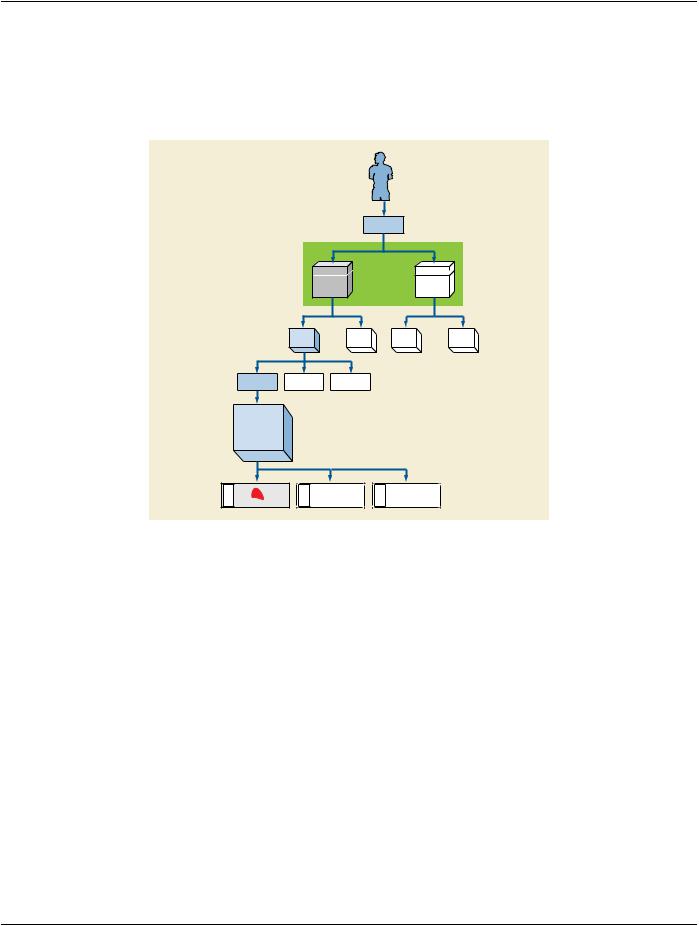
In allowing for one container to have multiple specimens, the Specimen Module asserts that it is the Container, not the Specimen, that is the unique target of the image. In other words, one Container ID is required in the Specimen Module, and multiple Specimen IDs are allowed in the Specimen Sequence. See Figure NN.3-1.
- Standard -

DICOM PS3.17 2020a - Explanatory Information |
Page 343 |
Patient
1 |
1 |
|
has |
is source of |
|
1-n |
||
1-n |
||
|
Equipment
Modality Study
1 |
1 |
creates |
contains |
1-n |
1-n |
Series |
|
|
|
1 |
|
contains |
|
|
1-n |
|
|
Image |
|
|
|
1 |
|
is acquired on |
|
|
|
1 |
|
Container |
|
|
Box, Block, Slide, etc. |
|
|
1 |
1 |
|
has |
contains |
|
1-n |
1-n |
|
Component |
Specimen |
|
Base, Coverslip |
Physical Object |
|
|
1 |
1 |
|
has |
is child of |
|
1-n |
1-n |
|
Preparation Step |
|
|
Collect, Sample, |
|
|
Stain, Process |
|
Figure NN.3-1. Extension of DICOM E-R Model for Specimens
If there is more than one specimen in a container, there must be a mechanism to identify and locate each specimen. When there is more than one specimen in a container, the Module allows various approaches to specify their locations. The Specimen Localization Content Item Sequence (0040,0620), through its associated TID 8004 “Specimen Localization”, allows the specimen to be localized by a distance in three dimensions from a reference point on the container, by a textual description of a location or physical Attribute such as a colored ink, or by its location as shown in a referenced image of the container. The referenced image may use an overlay, burned-in annotation, or an associated Presentation State SOP Instance to specify the location of the specimen.
NN.3.6 Relationship Between Specimens and Images
Because the Module supports one container with multiple specimens, the Module can be used with an image of:
•A single specimen associated with a container
•One or more specimens out of several in the same container
•All specimens in the same container
However the Module is not designed for use with an image of:
•Multiple specimens that are not associated with the same container, e.g., two gross specimens (two Parts) on a photography table, each with a little plastic label with their specimen number.
•Multiple containers that hold specimens (e.g., eight cassettes containing breast tissue being X-Rayed for calcium).
Such images may be included in the Study, but would not use the Specimen Module; they would, for instance, be general Visible Light Photographic images. Note, however, that the LIS might identify a "virtual container" that contains such multiple real containers, and manage that virtual container in the laboratory workflow.
- Standard -
Page 344 |
DICOM PS3.17 2020a - Explanatory Information |
NN.4 Specimen Identification Examples
NN.4.1 One Specimen Per Container
In normal clinical practice, when there is one specimen per container, the value of the specimen identifier and the value of the con- tainer identifier will be the same. In Figure NN.4-1, each slide is prepared from a single tissue sample from a single block (cassette).
|
|
|
OP-Material |
|
|
Case |
|
|
|
|
|
Box |
|
Box |
|
(Transport) |
|
(Transport) |
|
|
Part |
Part |
Part |
Part |
Tissue dice |
Tissue dice |
Tissue dice |
|
|
Cassette |
|
|
|
|
(Tissue dice |
|
|
|
|
embedded in a |
|
|
|
|
Block) |
|
|
|
|
Cassette ID |
|
|
|
|
Slide ID |
Slide ID |
|
Slide ID |
|
Figure NN.4-1. Sampling for one specimen per container
NN.4.2 Multiple Items From Same Block
Figure NN.4-2 shows more than one tissue item on the same slide coming from the same block (but cut from different levels). The laboratory information system considers two tissue sections (on the same slide) to be separate specimens.
TwoSpecimenIDswillbeassigned,differentfromtheContainer(Slide)ID.Thespecimensmaybelocalized,forexample,bydescriptive text "Left" and "Right".
If the slide is imaged, a single image with more than one specimen may be created. In this case, both specimens must be identified in the Specimen Sequence of the Specimen Module. If only one specimen is imaged, only its Specimen ID must be included in the Specimen Sequence; however, both IDs may be included (e.g., if the image acquisition system cannot determine which specimens in/on the container are in the field of view).
- Standard -

DICOM PS3.17 2020a - Explanatory Information |
Page 345 |
Slide ID
Figure NN.4-2. Container with two specimens from same parent
NN.4.3 Items From Different Parts in The Same Block
Figure NN.4-3 shows processing where more than one tissue item is embedded in the same block within the same Cassette, but coming from different clinical specimens (parts). This may represent different lymph nodes embedded into one cassette, or different tissue dice coming from different parts in a frozen section examination, or tissue from the proximal margin and from the distal margin, and both were placed in the same cassette. Because the laboratory wanted to maintain the sample as separate specimens (to maintain their identity), the LIS gave them different IDs and the tissue from Part A was inked blue and the tissue from Part B was inked red.
ThespecimenIDsmustbedifferentfromeachotherandfromthecontainer(cassette)ID.Thespecimensmaybelocalized,forexample, by descriptive text "Red" and "Blue" for Visual Coding of Specimen.
Ifasectionismadefromtheblock,eachtissuesectionwillincludefragmentsfromtwospecimens(redandblue).Theslide(container) ID will be different from the section id (which will be different form each other).
If the slide is imaged, a single image with more than one specimen may be created but the different specimens must be identified and unambiguously localized within the container.
Part |
Part |
Tissue dice |
Tissue dice |
|
Cassette |
|
(Tissue dice |
|
embedded in a |
|
Block) |
|
Cassette ID |
Slide ID
Tissue Section Item ID |
Tissue Section Item ID |
Figure NN.4-3. Sampling for two specimens from different ancestors
NN.4.4 Items From Different Parts On The Same Slide
Figure NN.4-4 shows the result of two tissue collections placed on the same slide by the surgeon. E.g., in gynecological smears the different directions of smears might represent different parts (portio, cervix).
- Standard -

Page 346 |
DICOM PS3.17 2020a - Explanatory Information |
The specimen IDs must be different from each other and from the container (slide) ID. The specimens may be localized, for example, by descriptive text "Short direction smear" and "Long direction smear".
Slide ID
Tissue Section Item ID
Tissue Section Item ID
Figure NN.4-4. Two specimens smears on one slide
NN.4.5 Tissue Micro Array
Slides created from a TMA block have small fragments of many different tissues coming from different patients, all of which may be processedatthesametime,underthesameconditionsbyadesiredtechnique.Thesearetypicallyutilizedinresearch.SeeFigureNN.4- 5. Tissue items (spots) on the TMA slide come from different tissue items (cores) in TMA blocks (from different donor blocks, different parts and different patients).
Each Specimen (spot) must have its own ID. The specimens may be localized, for example, by X-Y coordinates, or by a textual column-row identifier for the spot (e.g., "E3" for fifth column, third row).
If the TMA slide is imaged as a whole, e.g., at low resolution as an index, it must be given a "pseudo-patient" identifier (since it does not relate to a single patient). Images created for each spot should be assigned to the real patients.
Cassette |
|
Cassette |
|
Cassette |
|
Cassette |
|
Cassette |
|
Cassette |
(Tissue dice |
|
(Tissue dice |
|
(Tissue dice |
|
(Tissue dice |
|
(Tissue dice |
|
(Tissue dice |
embedded in a |
|
embedded in a |
|
embedded in a |
|
embedded in a |
|
embedded in a |
|
embedded in a |
Block) |
|
Block) |
|
Block) |
|
Block) |
|
Block) |
|
Block) |
|
|
|
|
|
|
|
|
|
|
|
Cassette (Tissue dice embedded in a Block) Cassette ID
Slide ID
Cassette (Tissue dice embedded in a Block) Cassette ID
Tissue Section Item IDs |
Tissue Section Item IDs |
Figure NN.4-5. Sampling for TMA Slide
NN.5 Structure of The Specimen Module
The Specimen Module content is specified as a Macro as an editorial convention to facilitate its use in both Composite IODs and in the Modality Worklist Information Model.
The Module has two main sections. The first deals with the specimen container. The second deals with the specimens within that container. Because more than one specimen may reside in single container, the specimen section is set up as a sequence.
The Container section is divided two "sub-sections":
•One deals with the Specimen Container ID and the Container Type. Note that the "Container Identifier" is a required field.
- Standard -

DICOM PS3.17 2020a - Explanatory Information |
Page 347 |
•One deals with Container Components. Because there may be more than one component, this section is set up as a sequence.
The Specimen Description Sequence contains five "sub-sections"
•One deals with the Specimen ID
•One deals with descriptions of the specimen
•One deals with preparation of the specimen and its ancestor specimens (including sampling, processing and staining). Because of its importance in interpreting slide images, staining is distinguished from other processing. Specimen preparation is set up as sequence of process steps (multiple steps are possible); each step is in turn a sequence of content items (Attributes using coded vocabularies). This is the most complex part of the module.
•One deals with the original anatomic location of the specimen in the patient.
•One deals with specimen localization within a container. This is used to identify specimens when there is more than one in a con- tainer. It is set up as sequence.
NN.6 Examples of Specimen Module Use
This section includes examples of the use of the Specimen Module. Each example has two tables.
The first table contains the majority of the container and specimen elements of the Specimen Module. The second includes the Spe- cimen Preparation Sequence (which documents the sampling, processing and staining steps).
In the first table, invocations of Macros have been expanded to their constituent Attributes. The Table does not include Type 3 (op- tional) Attributes that are not used for the example case.
The second table shows the Items of the Specimen Preparation Sequence and its subsidiary Specimen Preparation Step Content ItemSequence.ThatlattersequenceitselfhassubsidiaryCodeSequenceItems,buttheseareshowninthecanonicalDICOM"triplet" format (see PS3.16), e.g., (44714003, SCT, "Left Upper Lobe of Lung"). In the table, inclusions of subsidiary templates have been expanded to their constituent Content Items. The Table does not include Type U (optional) Content Items that are not used for the example case.
Values in the colored columns of the two tables actually appear in the image object.
NN.6.1 Gross Specimen
ThisisanexampleofhowtheSpecimenModulecanbepopulatedforagrossspecimen(alungloberesectionreceivedfromsurgery). The associated image would be a gross image taken in gross room.
Table NN.6-1. Specimen Module for Gross Specimen
Attribute Name |
Tag |
Attribute Description |
Example Value |
Container Identifier(0040,0512)The identifier for the container S07-100 A that contains the specimen(s)
being imaged.
Issuer of the |
(0040,0513)Organization that assigned the |
|
Container Identifier |
Container Identifier |
|
Sequence |
|
|
>Local Namespace(0040,0031)IdentifiesanentitywithinthelocalCase Medical Center |
||
Entity ID |
|
namespace or domain. |
Comments
Note that the container ID is required, even though the container itself does not figure in the image.
- Standard -

Page 348 |
|
DICOM PS3.17 2020a - Explanatory Information |
|
|
Attribute Name |
Tag |
Attribute Description |
Example Value |
Comments |
Container Type |
(0040,0518)Type of container that contains |
|
This would likely be a |
|
Code Sequence |
|
the specimen(s) being imaged. |
|
default container value |
|
|
Zero or one items shall be |
|
for all gross specimens. |
|
|
permitted in this sequence |
|
The LIS does not keep |
|
|
|
|
information on the gross |
|
|
|
|
container type, so this is |
|
|
|
|
an empty sequence. |
Specimen |
(0040,0560)Sequence of identifiers and |
|
|
|
Description |
|
detailed description of the |
|
|
Sequence |
|
specimen(s) being imaged. One |
|
|
|
|
or more Items shall be included |
|
|
|
|
in this Sequence. |
|
|
>SpecimenIdentifier(0040,0551)A departmental information |
S07-100 A |
SpecimenandContainer |
||
|
|
system identifier for the |
|
have same ID |
|
|
Specimen. |
|
|
>Issuer of the |
(0040,0562)The name or code for the |
|
|
|
Specimen Identifier |
institution that has assigned the |
|
||
Sequence |
|
Specimen Identifier. |
|
|
>> Local |
(0040,0031)IdentifiesanentitywithinthelocalCase Medical Center |
|
||
Namespace Entity |
|
namespace or domain. |
|
|
ID |
|
|
|
|
>Specimen UID |
(0040,0554)Unique Identifier for Specimen 1.2.840.99790.986.33.1677.1.1.17.1 |
|||
>Specimen Short |
(0040,0600)Short textual specimen |
Part A: LEFT UPPER LOBE |
The LIS "Specimen |
|
Description |
|
description |
|
Received" field is |
|
|
|
|
mapped to this DICOM |
|
|
|
|
field |
>SpecimenDetailed(0040,0602)Detailed textual specimen |
A: Received fresh for intraoperativeThis is a mapping from |
|||
Description |
|
description |
consultation,labeledwiththepatient'sthe LIS "Gross |
|
|
|
|
name, number and "left upper lobe,"Description" field. Note |
|
|
|
|
isapink-tan,wedge-shapedsegmentthat in Case S07-100 |
|
|
|
|
of soft tissue, 6.9 x 4.2 x 1.0 cm. Thethereweresixparts.This |
|
|
|
|
pleural surface is pink-tan and |
means the LIS gross |
|
|
|
glistening with a stapled line |
descriptionfieldwillhave |
|
|
|
measuring 12.0 cm. in length. The six sections (A - F). We |
|
|
|
|
pleural surface shows a 0.5 cm. areawould have to parse the |
|
|
|
|
of puckering. The pleural surface isgross description field |
|
|
|
|
inked black. The cut surface revealsintothoseparts(A-F)and |
|
|
|
|
a 1.2 x 1.1 cm, white-gray, irregularthen only incorporate |
|
|
|
|
massabuttingthepleuralsurfaceandsection "A" into this |
|
|
|
|
deep to the puckered area. The |
Attribute. NOTE: One |
|
|
|
remainder of the cut surface is |
could consider listing all |
|
|
|
red-brown and congested. No otherthe Blocks associated |
|
|
|
|
lesionsareidentified.Representativewith Part A. It would be |
|
|
|
|
sections are submitted. |
easytodoandmightgive |
|
|
|
|
useful information. |
>Specimen |
(0040,0610)SequenceofItemsidentifyingthe(see Table NN.6-2) |
|
||
Preparation |
|
process steps used to prepare |
|
|
Sequence |
|
the specimen for image |
|
|
|
|
acquisition. One or more Items |
|
|
may be present. This Sequence includes description of the specimen sampling step from a parentspecimen,potentiallyback to the original part collection.
- Standard -

|
|
DICOM PS3.17 2020a - Explanatory Information |
|
Attribute Name |
Tag |
Attribute Description |
Example Value |
>>Specimen |
(0040,0612)Sequence of Content Items |
|
|
Preparation Step |
|
identifying the processes used in |
|
Content Item |
|
one preparation step to prepare |
|
Sequence |
|
the specimen for image |
|
|
|
acquisition. One or more Items |
|
|
|
may be present. |
|
>Primary Anatomic(0008,2228)Original anatomic location in Structure Sequence patientofspecimen.Thislocation
may be inherited from the parent specimen, or further refined by modifiers depending on the sampling procedure for this specimen.
>>Code Value |
(0008,0100) |
44714003 |
>>Coding Scheme(0008,0102) |
SCT |
|
Designator |
|
|
>>Code Meaning |
(0008,0104) |
Left Upper Lobe of Lung |
Page 349
Comments
This is a code sequence item
Table NN.6-2. Specimen Preparation Sequence for Gross Specimen
Specimen SpecimenTemplate/Row Value Type ConceptNameCodeSequence Example Value |
Comments |
|||||
PreparationPrep. Step |
(0040,A040) |
(0040,A043) |
|
|
||
Sequence - Content |
|
|
|
|
||
Item # |
|
Item |
|
|
|
|
|
Sequence - |
|
|
|
|
|
|
Item # |
|
|
|
|
|
1 |
1 |
8001 / 1 |
TEXT |
(121041, DCM, "Specimen |
S07-100 A |
Collection |
|
|
|
|
Identifier") |
|
in OR |
|
2 |
8001 / 2 |
TEXT |
(111724, DCM, "Issuer of |
Case Medical Center |
|
|
|
|
|
Specimen Identifier") |
|
|
|
1 |
8001 / 3 |
CODE |
(111701, DCM, "Processing |
(17636008, SCT, |
|
|
|
|
|
type") |
"Specimen collection") |
|
|
2 |
8001 / 4 |
DATETIME |
(111702, DCM, "DateTime of |
200703230827 |
|
|
|
|
|
processing") |
|
|
|
3 |
8001 / 5 |
TEXT |
(111703, DCM, "Processing stepTaken |
|
|
|
|
|
description") |
|
|
4 |
8001 / 8 |
CODE |
(111704, DCM, "Sampling |
(65801008, SCT, |
|
|
8002 / 1 |
|
Method") |
"Excision") |
|
|
|
|
|
|
2 |
1 |
8001 / 1 |
TEXT |
(121041, DCM, "Specimen |
S07-100 A |
|
|
|
|
Identifier") |
|
|
2 |
8001 / 2 |
TEXT |
(111724, DCM, "Issuer of |
Case Medical Center |
|
|
|
|
Specimen Identifier") |
|
Specimen received in Pathology department
1 |
8001 / 3 |
CODE |
(111701, DCM, "Processing |
(428995007, SCT, |
|
|
|
type") |
"Specimen Receiving") |
2 |
8001 / 4 |
DATETIME |
(111702, DCM, "DateTime of |
200703230943 |
|
|
|
processing") |
|
NN.6.2 Slide
This is an example of how the Specimen Module can be populated for a slide (from a lung lobe resection received from surgery). The associated image would be a whole slide image.
- Standard -

Page 350 |
DICOM PS3.17 2020a - Explanatory Information |
Table NN.6-3. Specimen Module for a Slide
Attribute Name |
Tag |
Attribute Description |
Example Value |
Container Identifier |
(0040,0512)The identifier for the containerS07-100 A 5 1 |
||
|
|
that contains the specimen(s) |
|
|
|
being imaged. |
|
Issuer of the |
(0040,0513)Organization that assigned the |
||
Container Identifier |
|
Container Identifier |
|
Sequence |
|
|
|
>Local Namespace |
(0040,0031)Identifies an entity within the |
Case Medical Center |
|
Entity ID |
|
local namespace or domain. |
|
Container Type Code(0040,0518)Type of container that contains |
|||
Sequence |
|
the specimen(s) being imaged. |
|
|
|
Only a single item shall be |
|
|
|
permitted in this sequence |
|
>Code Value |
(0008,0100) |
258661006 |
|
>Coding Scheme |
(0008,0102) |
SCT |
|
Designator |
|
|
|
>Code Meaning |
(0008,0104) |
Slide |
|
ContainerComponent(0040,0520)Description of one or more Sequence components of the container
(e.g.,descriptionoftheslideand of the coverslip). One or more Items may be included in this Sequence.
>Container |
(0050,0012)Type of container component. |
|
Component Type |
One Item shall be included in |
|
Code Sequence |
this Sequence. |
|
>>Code Value |
(0008,0100) |
433472003 |
>>Coding Scheme |
(0008,0102) |
SCT |
Designator |
|
|
>>Code Meaning |
(0008,0104) |
Microscope slide coverslip |
>Container |
(0050,001A)Materialofcontainercomponent.GLASS |
|
Component Material |
|
|
SpecimenDescription(0040,0560)Sequence of identifiers and |
|
|
Sequence |
detailed description of the |
|
|
specimen(s)beingimaged.One |
|
|
or more Items shall be included |
|
|
in this Sequence. |
|
Comments
This would likely be a default container value for all slide specimens.
This is a code sequence item
This is a code sequence item
>Specimen Identifier(0040,0551)A departmental information |
S07-100 A 5 1 |
Specimen and |
|
|
system identifier for the |
|
Container have same |
|
Specimen. |
|
ID |
>Issuer of the |
(0040,0562)The name or code for the |
|
|
Specimen Identifier |
institution that has assigned the |
|
|
Sequence |
Specimen Identifier. |
|
|
>>Local Namespace(0040,0031)Identifies an entity within the |
Case Medical Center |
|
|
Entity ID |
local namespace or domain. |
|
|
>Specimen UID |
(0040,0554)Unique Identifier for Specimen1.2.840.99790.986.33.1677.1.1.19.5 |
||
- Standard -