


312 |
BRS Cell Biology and Histology |
Juxtaglomerular cells (modified smooth muscle)
Bowman capsule (visceral layer podocytes)
|
The parietal layer of Bowman capsule is |
|
composed of simple squamous |
|
epithelium, whereas its visceral layer is |
|
modified to form podocytes. The |
|
ultrafiltrate enters Bowman (urinary) |
|
space and leaves the renal corpuscle at |
|
its urinary pole, via the proximal |
|
convoluted tubule. The afferent |
|
glomerular arteriole enters and the |
|
efferent glomerular arteriole leaves the |
|
renal corpuscle at its vascular pole, the |
Urinary space |
former supplying and the latter draining |
the glomerulus. The macula densa |
|
|
component of the distal tubule comes in |
|
close proximity to the juxtaglomerular |
|
cells of the afferent (and efferent) |
|
glomerular arterioles. |
|
Brush border (microvilli) |
Proximal convoluted tubule |
Endothelium |
|
Primary |
|
Basal lamina |
Endothelium |
|
|
The fenestrated capillaries constituting |
Filtration |
|
slit |
||
the glomerulus are invested by pedicels |
||
|
||
arising from the primary processes of |
|
|
podocytes. Filtration slits between |
|
|
adjoining pedicles are bridged by thin |
|
|
diaphragms that, in association with the |
|
|
fused basal laminae of the capillary |
Podocyte |
|
endothelium and podocyte, contribute to |
||
the formation of the filtration barrier. |
cell body |
|
process |
Primary |
|
(pedicel) |
||
|
process |
FIGURE 18.2. A diagram illustrating& components of the renal corpuscle. !From Gartner LP. Hiatt JL. Color Atlas of Histology. 5th ed. Baltimore. MD: Lippincott William Wilkins; 2009:337.)

l!1llll"!ttfflThe Urinary System 313
(2)The visceral layer (glomerular epithelium) is the modified simple squamous epithelium composed of podocytes that lines the inner wall of the Bowman capsule and envelops the glomerular capillaries.
(3)Bowman space (also known as capsular space or urinary space) is the narrow chalice shaped cavity between the visceral and parietal layers into which the ultrafiltrate passes.
(4)The vascular pole is the site on Bowman capsule where the afferent glomerular arteriole enters and the efferent glomerular arteriole leaves the glomerulus.
(5)The urinary pole is the site on Bowman capsule where the capsular space becomes continuous with the lumen ofthe proximal convoluted tubule.
b.Podocytes are highly modified epithelial cells that form the visceral layer of Bowman capsule and synthesize glomerular endothelial growth factor, a signaling molecule that facilitates the formation and maintenance of the glomerular endothelial cells. Podocytes have complex shapes and possess several primary processes that give rise to many secondary processes called pedicels.
(1 ) Pedicels
(a)Pedicels embrace the glomerular capillaries and interdigitate with pedicels arising from other primary processes.
(b)Their surfaces facing Bowman space are coated with podocalyxin, a protein that is thought to assist in maintaining their organization and shape.
(c)Pedicels possess :XJ 1 integrin molecules that cause them to adhere to the basal lamina.
(2)Filtration slits are elongated spaces about 40 nm in width between adjacent pedicels. A filtration slit diaphragm bridges each filtration slit and is the principal structure that is the barrier responsible for the filtration of proteins.
(a)Each slit diaphragm is composed ofthe extracellular portion ofthe transmembrane protein nephrin of one pedicel that contacts the extracellular portion of nephrin from the adjacent pedicel.
(b)Within the cytoplasm of the pedicel, the intracellular moiety of nephrin binds to podocin as well as to CD2-associated proteins that in turn bind to actin filaments and in that manner stabilize the nephrin molecule.
c.The renal glomerulus is the tuft of capillaries that extends into the Bowman capsule. (1 ) Glomerular endothelial cells
(a)form the inner layer of the capillary walls.
(b)have a thin cytoplasm that is thicker around the nucleus, where most organelles are located.
(c)possess large fenestrae (60-90 nm in diameter) but lack the thin diaphragms that typically span the openings in other fenestrated capillaries.
(2)The basal lamina is between the podocytes and the glomerular endothelial cells and is manufactured by both cell populations. It is unusually thick (0. 15-0.5 J..m)L and contains three distinct zones:
(a)The lamina rara externa, an electron-lucent zone adjacent to the podocyte epithelium.
(b)The lamina densa, a thicker, electron-dense intermediate zone ofamorphous material.
(c)The lamina rara interna, an electron-lucent zone adjacent to the capillary endothelium.
(3)The mesangium is the interstitial tissue between glomerular capillaries. It is composed of mesangial cells and an amorphous extracellular matrix elaborated by these cells.
(a)Mesangial cells
1.phagocytose large protein molecules and debris, which may accumulate during filtration or in certain disease states.
2.can also contract, thereby decreasing the surface area available for filtration.
3.possess receptors for angiotensin II and atrial natriuretic peptide.
4.manufacture platelet-derived growth factor, endothelins, inteleukin-1, and prostaglandin E2•
(b)The mesangial matrix, manufactured by mesangial cells, is composed of type IV collagen, laminin, and proteoglycans and helps support glomerular capillaries.


316 |
BRS Cell Biology and Histology |
Ultrafiltration |
Selective resorption of |
|
|
Na+ |
Amino acids |
|
Cl- |
Small proteins |
|
Water |
Ascorbic acid |
|
Glucose |
HC03- |
Connecting tubule
Water resorption regulated by ADH
Collecting tubule
Loop of Henle and vasa recta
Thick ascending segment
Thin limb of loop of Henle
Fenestrated capillary
FIGURE 18.5. A uriniferous tubule showing its major structural and functional features and its vascular associations. ADH, antidiuretic hormone. (Adapted with permission from Williams PL. Warwick R. eds. 36th British ed. London: Churchill Livingstone; 1 980 1 393.)
(2)Prominent interdigitations along their lateral borders, which interlock adjacent cells with one another.
(3)Numerous mitochondria compartmentalized in the basal region by extensive infoldings ofthe basal plasma membrane, which supply energy for the active transport of Na+ out of the tubule.
(4)Apically situated occluding junctions that block the paracellular pathway. And the apical cell membrane has glucose transporters, Na+K+-ATPase pump, and an apically positioned tubulovesicular system designed to endocytose small proteins and peptides that escaped into the ultrafiltrate.
(5)The basolateral cell membrane possesses Na+K+-ATPase pump, and, additionally, the basal cell membrane has glucose and amino acid transporters.
(6)Each cell also possesses a primary cilium that functions in monitoring the flow and composition of the ultrafiltrate.
b.Function
(1 ) The proximal convoluted tubule drains the Bowman space at the urinary pole of the renal corpuscle.
(2)It resorbs from the glomerular filtrate all ofthe glucose, amino acids, and small proteins and 60% to 80% of the sodium chloride and water and returns it into the peritubular capillary system to be distributed from there into the remainder of the body.
(3)It exchanges H+ in the interstitium for HC03in the filtrate.
(4)It secretes organic acids (e.g., creatinine) and bases and certain foreign substances into the filtrate.
4.Loop of Henle (Figure 18.5)
a. Descending thick limb of the Henle loop
(1)The descending limb ofthe Henle loop is also known as the straight portion (pars recta) of the proximal tubule.

l!1llll"!ttfflThe Urinary System 317
FIGURE 18.6. A light micrograph of components in the medulla of the kidney. Collecting tubules (CTl are lined by a simple columnar epithelium composed of cells displaying distinct lateral surfaces. The thin limbs of the loops of Henle (LHl are lined by a simple squamous epithelium whose cell nuclei bulge into the lumen, and capillaries ( arrowheads) may be identi fied by the numerous red blood cells filling their lumens ( x 1 50).
(2)It is lined by a simple cuboidal epithelium that has a prominent brush border and is similar to that lining the proximal convoluted tubule.
(3)Its function is to resorb, exchange, and secrete in a manner similar to that of the proximal convoluted tubule.
b.Thin limb of the Henle loop (Figure 18.6)
(1 ) The thin limb of the Henle loop is composed of a descending segment, a loop, and an ascending segment, all of which are lined by simple squamous epithelial cells possessing a few short microvilli. The nuclei of these cells bulge into the lumen.
(2)In juxtamedullary nephrons, the thin limb can be divided into three distinct portions on the basis of the four different types of simple squamous lepithelial cells that form it, their organelle content, the depth of their tight junctions, and their permeability to water.
(a)Cells ofthe descending thin limb possess many aquaporin channels and, therefore,
this segment is very permeable to water as well as being somewhat permeable to ions such as sodium and chloride.
(b)Henle loop itself is similar to the ascending thin limb and is mostly impermeable to water.
(c)The ascending thin limb is almost completely impermeable to water but possesses many sodium and chloride channels, which permit these ions to enter the cell from the lumen of the tubule and exit the cell into the renal interstitium. Additionally, urea enters the lumen of the ascending thin limb.
c.Ascending thick limb of the Henle loop
(1 ) The ascending thick limb of the Henle loop is also known as the straight portion (pars recta) of the distal tubule.
(2)It is lined by cuboidal epithelial cells that possess only a few microvilli, an apical nucleus, and mitochondria compartmentalized within basal plasma membrane infoldings. These cells manufacture and release a glycoprotein known as uromodulin (Tamm-Horsfall glycoprotein) that reduces the ability of the kidney to form kidney

318 |
BRS Cell Biology and Histology |
stones and in some fashion reduces the possibility ofurinary tract infections. Moreover, uromodulin also modulates the mechanism of urine concentration.
(3)It establishes a gradient of osmolarity in the medulla (see Section V).
(4)The ascending thick limb returns to the renal corpuscle of origin, where it is in close association with the afferent and efferent glomerular arterioles. In this region, the wall of the tubule is modified, forming the macula densa, which is part of the juxtaglomerular apparatus (JG apparatus).
5.The JG apparatus (juxtaglomerular apparatus) is located at the vascular pole of the renal corpuscle.
a.Components of the JG apparatus
(1)JG cells (juxtaglomerular cells)
(a)are modified smooth muscle cells that exhibit some characteristics of protein secreting cells.
(b)are located primarily in the wall of the afferent arteriole, but a few may also be present in the wall ofthe efferent arteriole.
(c)synthesize renin (a proteolytic enzyme) and store it in secretory granules.
(2)Macula densa cells (Figure 18.4)
(a)are tall, narrow, closely packed epithelial cells of the distal tubule.
(b)have elongated, closely packed nuclei that appear as a dense spot (macula densa) by light microscopy.
(c)may monitor the osmolarity and volume ofthe fluid in the distal tubule and transmit this information to JG cells via the gap junctions between the two cell types. When the sodium concentration or the volume of the ultrafiltrate is reduced, the macula densa cells direct the JG cells to release their renin.
(3)Extraglomerular mesangial cells
(a)are also known as polkissen (pole cushion) or lacis cells.
(b)lie between the afferent and efferent glomerular arterioles, but their functions are not understood.
b.Function. The JG apparatus maintains blood pressure bythe following mechanism:
(1 ) A decrease in extracellular fluid volume (perhaps detected by the macula densa as
decreased ultrafiltrate volume), a decrease in blood pressure at the afferentglomerular arteriole, or a decrease in sodium concentration ofthe ultrafiltrate stimulates JG cells to release renin into the bloodstream.
(2)Renin acts on angiotensinogen (a large protein manufactured by hepatocytes of the liver) in the plasma. Renin cleaves the first 10 amino acids from angiotensinogen, converting it to the decapeptide angiotensin I. In capillaries of the lung and elsewhere, angiotensin-converting enzyme cleaves two amino acids from angiotensin I, converting it to angiotensin II, a potent vasoconstrictor that stimulates release of aldosterone in the adrenal cortex and the release of antidiuretic hormone (ADH) by the neurohypophysis.
(3)Aldosteronecr. stimulates the epithelial cells ofthe distal convolutedtubule to remove Na+ and Water follows the ions, thereby increasing the fluid volume in the extracellular
compartment, which leads to an increase in blood pressure.
(4) ADH causes the epithelial cells (mainly the principal cells) of the collecting tubule to add aquaporin 2 (AOP-2) channels (as well as AQP-3 and AQP-4 channels) to their cell membrane and thus become permeable to water, releasing H20 into the renal interstitium. As with the aldosterone mechanism discussed above, the increased extracellular fluidvolume leads to elevation ofthe blood pressure.
6.Distal convoluted tubule (Figure 18.7)
a. The distal convoluted tubule is continuous with the macula densa and is similar histologically to the ascending thick limb of the Henle loop.
b.It is much shorter and has a wider lumen than the proximal convoluted tubule and lacks a brush border.
c.Function. The distal convoluted tubule resorbs Na+ from the filtrate and actively transports it into the renal interstitium; this process is stimulated by aldosterone. It also transfers K+, NH4+, and H+ into the filtrate from the interstitium.

l!1llll"!ttfflThe Urinary System 319
FIGURE 18.7. An electron micrograph of a cell in the distal convoluted tubule of the kidney. Elongated mitochondria (M) are located (arrowhead) within cytoplasmic compartments (CC) formed by deep in foldings of the basal plasma membrane where enzymes associated with ion transport are located. In between these extensive infoldings is extracellular space (arrows). These cells resorb Na+ from the filtrate and actively transport it into the renal interstitiumW (aldosterone stimulates this process) and also trans fer K+, N H4+, and into the filtrate from the interstitium. Note the lamina densa (LD) and lamina Iucida (LL), which form the basal lamina, the endothelial cell (E) lining a fenestrated capillary, and the apical nucleus (N) ( x 6,000).
7.The connecting tubule is a short segment between the distal convoluted tubule and the collecting tubule into which it drains. It is lined by the following two types of epithelial cells:
a.Principal cells have many infoldings of the basal plasma membrane. These cells remove Na+ from the filtrate and secrete K+ into it.
b.Intercalated cells have many apical vesicles and mitochondria. These cells remove K+ from the filtrate and secrete H+ into it.
Nephrotoxic acute tubular necrosis is the death of kidney tubule cells caused by their exposure to a toxic drug or molecule, rather than a lack of
oxygen (ischemic acute tubular necrosis). When a person suffers a crush injury causing significant muscle trauma, myoglobin is released from the muscle and enters the bloodstream. The myoglobin is filtered through the glomeruli, but it is toxic to cells of the kidney tubules, causing nephrotoxic acute tubular necrosis. If the damage is not too severe, the kidney tubule cells may be able to replace themselves, but in severe cases, they cannot and the kidney may not completely recover.
B.Collecting tubules (Figure 18.5) have an embryological origin different from that of nephrons. They have segments in both the cortex and the medulla and converge to form larger and larger tubules.
1.Cortical collecting tubules are located primarily within medullary rays, although a few are interspersed among the convoluted tubules in the cortex (cortical labyrinth). They are lined by a simple epithelium containing two types of cuboidal cells.
a.Principal (light) cells possess a round central nucleus and a single primary cilium. It is these cells that are responsible for the ability of the collecting tubules to concentrate urine.
320BRS Cell Biology and Histology
b.Intercalated (dark) cells are less numerous than principal cells and possess microplicae
(folds) on their apical surface and numerous apical cytoplasmic vesicles. There are two types of intercalated cells: a-intercalated cells that have the ability to release H+ ions into the tubular lumen, thus acidifying urine, and -intercalated cells that have the ability to release HC03ions into the tubular lumen, thus causing the urine to be more alkaline. a-Intercalated cells possess hydrogen pumps, and -intercalated cells possess HC03pumps to fulfill their function.
2.Medullary collecting tubules. In the outer medulla, medullary collecting tubules are similar in structure to cortical collecting tubules and contain both principal and intercalated cells in their lining epithelium. In the inner medulla, the collecting tubules are lined only by principal cells (Figure 18.6).
3.Papillary collecting tubules (ducts of Bellini)
a.Papillary collecting tubules are large collecting tubules (200-300 Jlm in diameter) formed from converging smaller tubules.
b.They are lined by a simple epithelium composed of columnar cells that have a single primary cilium.
c.They empty at the area cribrosa, a region at the apex of each renal pyramid that has 10 to 25 openings through which the urine exits into a minor calyx.
The renal blood circulation is extensive, with total blood flow through both kidneys of about 1200 mL/min. At this rate, all of the circulating blood in the body passes through the kidneys every 4 to 5 minutes.
A.Arterial supply to the kidney (Figure 18.8)
1.Branches of the renal artery enter each kidney at the hilum and give rise to interlobar arteries.
2.Interlobar arteriestravel between the renal pyramids and divide into several arcuate arteries,
which run along the corticomedullary junction parallel to the kidney's surface.
3.Interlobular arteries
a.Interlobular arteries are smaller vessels that arise from the arcuate arteries.
b.They enter the cortical tissue and travel outward between adjacent medullary rays. Adjacent interlobular arteries delimit a renal lobule.
c.They give rise to afferent (glomerular) arterioles and send branches to the interstitium just deep to the renal capsule.
4.Afferent arterioles are branches of the interlobular arteries that supply the glomerular capillaries.
5.Efferent arterioles arise from the glomerular capillaries and are associated with cortical, midcortical, and juxtaglomerular nephrons. They leave the glomerulus of cortical and midcortical nephrons and give rise to an extensive peritubular capillary network that supplies the cortical labyrinth, whereas those that leave the juxtaglomerular nephrons give rise to the vasa recta.
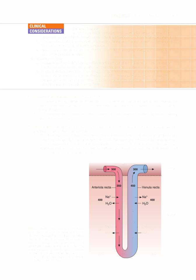
6.Vasa recta
a.The vasa recta arise from the efferent arterioles supplying juxtamedullary nephrons.
b.These long, thin vessels (arteriolae rectae) follow a straight path into the medulla and renal papilla, where they form capillaries and then loop back and increase in diameter toward the corticomedullary boundary (venulae rectae).
c.They are closely associated with the descending and ascending limbs of Henle loops and collecting ducts, to which they supply nutrients and oxygen.
d.These vessels play a critical role in countercurrent exchanges with the interstitium.

l!1llll"!ttfflThe Urinary System 321
Stellate |
Efferent |
Medullary vein |
arteriole |
Interlobular artery
Interlobar artery
Vasa recta
FIGURE 18.8. Blood circulation in the kidney. Arteries are shown in |
red and veins in |
blue. Adjacent interlobular arteries, |
which extend outward from the arcuate artery, define the boundaries of a renal |
lobule. (Reprinted with permission from |
|
Junqueira LC, Carneiro J. Kelley RO. Basic Histology. 9th ed. Stamford, CT: Appleton |
& Lange; 1 998:375.) |
|
B.Venous drainage of the kidney (Figure 18.8)
1.Stellate veins are formed by convergence of superficial cortical veins, which drain the outermost layers of the cortex.
2.Deep cortical veins drain the deeper regions of the cortex.
3.Interlobular veins
a.Interlobular veins receive both stellate and deep cortical veins.
b.They join arcuate veins, which empty into interlobar veins. These then converge to form a branch of the renal vein, which exits the kidney at the hilum.
A.Overview
1.The regulation of urine concentration results in the excretion of large amounts of dilute (hypotonic) urine when water intake is high (diuresis) and of concentrated (hypertonic) urine when body water needs to be conserved (antidiuresis).

322 |
BRS Cell Biology and Histology |
|
- H20 impermeable |
Urea |
1200 |
|
- H20 permeable |
1200 |
|
Diuresis (hypotonic urine) |
Antidiuresis (hypertonic urine, ADH present) |
||
FIGURE 18.9. Summary of ion and water exchanges that occur in the uriniferous tubule in the absence (left) and presence (right'l of antidiuretic hormone. The countercurrent multiplier system involving the loop of Henle produces an osmotic gradient in the medullary interstitium. Numbers refer to the local concentration in milliosmoles per liter. Segments of the tubule freely permeable to water are drawn with a thin line; impermeable segments are drawn with a thick line. In the distal convoluted tubule, some water follows sodium into the interstitium; sodium transport here is regulated by aldosterone. Note thatthe pars recta of the distal tubules actually contact their own renal corpuscles butthey have been rotated in this diagram to facilitate easier identification.
2.This regulation depends on events that occur in the loops of Henle, vasa recta, and collecting tubules.
3.It is affected bythepresence or absence ofADH (antidiuretic hormone), which is secreted from the pars nervosa of the pituitary gland (neurohypophysis) when water must be conserved.
B. The countercurrent multiplier system (Figure 18.9) refers to the establishment of an increasing osmotic concentration gradient in the renal interstitium that extends from the outer medulla to the renal papillae. It is produced by ion and water exchanges between the filtrate in different parts of the loop of Henle and the renal interstitium:
1. In the descending limb of the loop of Henle,
a. the isotonic filtrate coming from the proximal convoluted tubules loses water to the interstitium and gains Na+ and Cl-.
b.the filtrate becomes hypertonic.
2.In the ascending thin and thick limbs ofthe loop of Henle,
a.no water is lost from the filtrate because this part of the nephron is impermeable to water whether or not ADH is present.
b.Cl- is actively transported from the filtrate (in the ascending thick limb but passively in the ascending thin limb) into the interstitium, and Na+ follows.
c.an osmotic gradient is thus established in the interstitium of the outer medulla.
d.the filtrate becomes hypotonic.
3.In the distal convoluted tubule, active resorption ofNa+ from the filtrate may occur in response to aldosterone, resulting in some water loss as well.
C.Role of collecting tubules
1 . In the absence of ADH, the collecting tubules are impermeable to water. Thus, the hypotonic filtrate coming from the ascending limb of the loop of Henle is not changed, and hypotonic urine is excreted.
2.In the presence of ADH, the collecting tubules addAQP-2 channels (aswell as AQP-3 andAQP-4 channels) into their cell membranes of their principal cells and thus become permeable to water. Thus, the isotonic filtrate entering them from the distal convoluted tubules lose water, and hypertonic (concentrated) urine is produced.


l!1llll"!ttfflThe Urinary System 325
2.These structures generally possess a three-layer wall composed of a mucosa of transitional epithelium (except in the urethra) lying on a lamina propria of connective tissue, a muscularis (smooth muscle), and an adventitia.
B.Ureter
1.The ureter conveys urine from the renal pelvis of each kidney to the urinary bladder.
2.It has a transitional epithelium that is thicker and contains more cell layers than that of the renal calyces.
3.It possesses a two-layer muscularis (an inner longitudinal and outer circular layer of smooth muscle) in its upper two-thirds. The lowest third possesses an additional outer longitudinal layer of smooth muscle.
4.It contracts its muscle layers, producing peristaltic waves that propel the urine, so that it enters the bladder in spurts.
C. Urinary bladder. (Figure 18. 11) The urinary bladder possesses a transitional epithelium with a morphology that differs in the relaxed (empty) and distended states, a thin lamina propria of fibroelastic connective tissue, and a three-layer muscularis.
1.Epithelium of the relaxed bladder is five to six cell layers thick and has rounded superficial dome-shaped cells that bulge into the lumen. These cells contain unique plaques (having a highly ordered substructure) in their thick luminal plasma membrane and flattened elliptical vesicles in their cytoplasm.
2.Epithelium of the distended bladder
a.The epithelium of the distended bladder is only three to four cell layers thick.
b.It has squamous superficial cells.
c.It is much thinner and has a larger luminal surface than the relaxed bladder; this results from insertion of the elliptical vesicles into the luminal plasma membrane of the surface cells.
FIGURE 18.1 1 . A light micrograph of the urinary bladder in a relaxed (empty) state. It is lined by transitional epithelium (TEl with dome-shaped surface cells and is separated from the underly ing connective tissue by a basal lamina (arrows). A lamina propria (LPl of cellular, loose connec tive tissue may be distinguished from the submu cosa (Sml of dense connective tissue possessing many large collagen fibers. Note the venules (Vl
and an arteriole (Al present in the |
lamina propria |
|||
( x 1 321. Inset. The |
boxed |
region |
of |
transitional |
epithelium is here |
shown |
at higher |
magnifica |
|
tion to demonstrate the large, dome-shaped sur
face cells (arrow). one of which is |
binucleated |
( x 5401. The transitional epithelium |
undergoes |
marked changes. In contrast to the relaxed state of the bladder shown here, when the bladder is distended and full of urine, the dome-shaped sur face cells become squamous due to the insertion of unusual "elliptical-shaped vesicles" into their plasma membrane, and the entire epithelium is of ten reduced to only three cell layers in thickness.

326 |
BRS Cell Biology and Histology |
Bladder cancer is two to three times more common in men than in women. More than 90% of bladder cancers originate in the transitional epithelium
lining the organ. The most common sign of bladder cancer is blood in the urine (hematuria), which may not be visible to the naked eye. Frequent urination and/or pain during urination are sometimes present, but often the disease is symptomless. Cystoscopy is used to examine the lining of the blad der, to take tissue samples in order to characterize the tumor, and to determine the extent to which it has penetrated into the bladder wall. Superficial bladder cancer (limited to the epithelial layer) has a 5-year survival rate of about 85%, but invasive bladder cancer has a less favorable prognosis.
D. Urethra
1 . Overview
a.The urethra conveys urine from the bladder to outside the body. In men, the urethra also carries semen during ejaculation.
b.It has a two-layer muscularis consisting of an inner longitudinal and an outer circular layer of smooth muscle.
c.It is surrounded at some point by an external sphincter of skeletal muscle, which permits its voluntary closure.
2.Male urethra
a.The male urethra is about 20 em long and is divided into prostatic, membranous, and cavernous portions.
b.It is lined by transitional epithelium in the prostatic portion and by pseudostratified or
stratified columnar epithelium in the other two portions. The fossa navicularis, located at the distal end of the cavernous urethra,( is lined by stratified squamous epithelium.
c.It contains mucus-secreting glands of Litlre in the lamina propria.
3.Female urethra
a.The female urethra is much shorter 4-5 em long) than the male urethra.
b.It is lined primarily by stratified squamous epithelium, although patches ofpseudostratified columnar epithelium are present.
c.It may contain glands of Litlre in the lamina propria.


332 |
BRS Cell Biology and Histology |
FIGURE 19.2. The cortex of this monkey ovary dis plays the presence of primordial follicles (double arrows) as well as several multilaminar primary follicles, one of whose constituent structures is labeled. Nucleus (arrow). nucleolus (arrowhead), zona pellucida (ZP). granulosa cells (G). and theca
(T) (x270).
d.The oogonia that are surrounded by follicular cells begin their first meiotic division, and are known as primary oocy1es. In response to the meiotic division, the follicular cells manufacture and release a meiosis-preventing factor known as oocy1e maturation inhibitor (OM I) that arrests the primary oocyte in the diplotene stage of the first meiotic division, and the primary oocytes remain in that arrested development until they are ovulated. The primary oocyte and its sheath of follicular cells are known as the primordial follicle.
e.Between birth and the 1 1th year oflife, more than 60% ofthe primordial follicles degenerate, and each ovarywill be leftwith approximately 80,000 primordial follicles.
B.The ovarian cortex consists of ovarian follicles in various stages of development and a connective tissue stroma, which contains cells that respond in unique ways to hormonal stimuli.
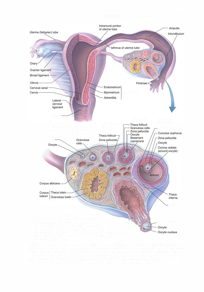
1 . Ovarian follicles (Figures 19.1 and 19.2; Table 19.1)
a. Primordial follicles are the ovary's basic reproductive units and are composed ofa primary oocy1e enveloped by a single layer of flat follicular cells (Figure 19.2). All primordial follicles are in a state ofarrested developmentuntil in some unknown fashion, but probably involving members of the transforming growth factor superfamily, some primordial follicles are "recruited" (activated) to begin to form a group of growing follicles. There appears to be an interaction involving follicular cells, primary oocytes, and mesenchymal cells of the ovarian cortex and, possibly, insulin and follicle-stimulating hormone (FSH) may also have a role in the recruitment ofprimordial follicles.
(1)Primary oocytes
(a)display a prominent, acentric, vesicular-appearing nucleus (germinal vesicle) possessing a single nucleolus.
(b)have many Golgi complexes, mitochondria, profiles of rough endoplasmic reticulum (RER), well-developed annulate lamellae, as well as cortical granules

|
19.1 |
|
l!il!ll'tttD]jFemale Reproductive System |
333 |
|||
t a b I e |
Stages in the Development of Ovarian Follicles |
|
|
|
|||
|
|
|
Follicular Cell |
|
|
Hormone |
|
Stage |
Zona Pellucida |
Layer (Granulosa) |
Liquor Folliculi |
Theca |
Dependency |
|
|
Primordial follicle |
Not present |
Single layer of flat |
Not present |
Not present |
Dependent on |
|
|
|
|
|
cells |
|
|
local factors |
|
Unilaminar |
Present |
Single layer of |
Not present |
Not present |
Dependent on |
|
|
primary follicle |
|
|
cuboidal cells |
|
|
local factors |
|
Multilaminar |
Present |
Multiple layers of |
Not present |
Intern a, externa |
Dependent on |
|
|
primary follicle |
|
|
granulosa cells |
|
present |
local factors |
|
Secondary follicle |
Present |
Spaces among |
Accumulates in |
Interna, externa |
FSH dependent |
||
|
|
|
granulosa cells |
spaces among |
present |
|
|
|
|
|
|
granulosa cells |
|
|
|
Graafian follicle |
Present |
Forms membrana |
Fills the antrum |
Interna, externa |
FSH dependent |
||
|
|
|
granulosa, |
|
present |
until becomes |
|
|
|
|
cumulus |
|
|
dominant |
|
|
|
|
oophorus |
|
|
follicle |
|
|
|
|
|
|
|
|
|
FSH. follicle-stimulating hormone.
located just beneath the oocyte plasmalemma that contain enzymes that cleave proteins.
(c)as stated above, become arrested in prophase (dictyate stage) of meiosis I by paracrine factors OMI produced by the follicular cells during fetal life and remain in this stage until ovulation (perhaps for as long as 40 years).
(2)Follicular cells are
(a)attached to one another by desmosomes.
(b)separated from the surrounding stroma by a basal lamina. b. Growing follicles
(1)Primordial and primary follicles are not dependent on FSH for their development;
instead, local factors such as epidermal growth factor, insulin-like growth factor (IGF), activin, and Ca2+ ions, stimulate the development of primordial and primary follicles. However, some authors are suggesting that even these early follicles may be FSH dependent.
(2)Primary follicles are of two types, unilaminar and multilaminar. They possess an amorphous layer (zona pellucida) surrounding and produced by the primary oocyte (although some authors suggest that the follicular cells also make a contribution to its formation). The zona pellucida (ZP) separates the follicular cells from the primary oocyte. Follicular cells begin to express FSH receptors on their plasmalemma surface, and they also form gap junctions with each other as well as with the microvilli of the primary oocyte. A basal lamina is present at the interface of the follicular cells with the stroma.
• |
The zona pellucida is a glycoprotein covering of the primary oocyte. |
• |
It is composed of four glycoproteins, known as ZP 1, ZP 2, ZP 3, and ZP 4. |
•ZP 3 permits the binding of spermatozoon and facilitates the occurrence of the acrosome reaction.
•ZP 2 is also responsible for binding spermatozoon. After fertilization (discussed in
Section VI below) has occurred, the ovum secretes the enzyme ovastacin, which degrades ZP 2, preventing the binding ofadditional spermatozoa to the zona pellucida.
•ZP 1 functions in cross-linking ZP 2 with ZP 3, thus preventing more spermatozoa from binding.
•The function of ZP 4 is not known.
(a)Unilaminar primary follicles
1.develop from primordial follicles.
2.are composed of a single layer of cuboidal follicular cells surrounding the primary oocyte.


l!il!ll'tttD]jFemale Reproductive System |
335 |
(2)It measures approximately 2.5 em in diameter and is evident as a large bulge on the surface of the ovary.
(3)The primary oocyte is positioned off center on a small mound of granulosa cells
(cumulus oophorus) that projects into the hyaluronic acid-rich liquor folliculi containing antrum of the follicle. Granulosa cells surround the zona pellucida. Those contacting the zona pellucida are known as the corona radiata. Other granulosa cells line the antrum, forming the membrana granulosa.
(4)Theca interna cells, in response to luteinizing hormone (LH) binding to their LH receptors, manufacture androstenedione (androgen), which is transferred to granulosa cells. The granulosa cells, under the influence of FSH binding to their FSH receptors, transform androstenedione into testosterone and, by the use of the enzyme aromatase, convert the testosterone into estradiol (estrogen).
(5)The theca externa is composed of collagenous connective tissue enriched with a plethora of smooth muscle cells. It is also richly supplied with many blood vessels, which provide nourishment to the theca interna.
(6)Ovulation
(a)An LH surge from the pituitary gland, along with the local factor manufactured by the primary oocyte, maturation promoting factor (a complex of cyclin-dependent kinase and cyclin B), triggers the primary oocyte to complete its first meiotic divisionjust prior to ovulation, forming a secondary oocyte and the first polar body.
The second meiotic division is triggered by the presence of local meiosis-inducing factors but is blocked at metaphase.
(b)Ovulation also occurs in response to the LH surge. The secondary oocyte and its corona radiata cells leave the ruptured follicle at the ovarian surface to enter the fimbriated end of the oviduct.
(c)The remnant of the graafian follicle becomes the corpus hemorrhagicum that is transformed into the corpus luteum of menstruation, a temporary structure that may remain functional for a couple of weeks and then degenerates into the corpus albicans. If the woman is pregnant, then the corpus hemorrhagicum becomes transformed into the corpus luteum of pregnancy, which remains functional for several months, and then it also degenerates into the corpus albicans.
(7)Time Period of Folliculogenesis. Although from the prior description offolliculogenesis it appears as if the entire process occurs within a single menstrual cycle, that is not the case. In fact, once a primordial follicle is recruited for development, almost a year is required before ovulation can occur, and it takes approximately 290 days to go from primordial to a completely developed secondary follicle and another 60 days or so for that follicle to become ovulated.
Some women experience a sudden abdominal (or pelvic) pain in the middle of their menstrual cycle that lasts 2 or 3 hours (although it may
last as long as 48-72 hours or even longer), and they have attributed thatto the process of ovula tion. Although this attribution was not universally accepted, it is now known that the sudden surge of LH is followed by a similar surge in prostaglandin F2a, a pharmacologic agent that causes sudden, cramp-like contraction of the ovarian, fallopian tube, or uterine smooth muscle fibers. It is this violent contraction that is responsible for the pain experienced by some women. Other causes, such as painful distension of the ovarian follicles or the rupture of the tunica albuginea of the ovary, may also contribute to the ovulation pain.
2. The corpus hemorrhagicum is formed from the remnants of the graafian follicle. After the cumulus oophorus leaves the ovary, a little blood enters and forms a clot in the former antrum, and within a short period of time macrophages and connective tissue elements enter the region, and the macrophages dispose of the blood clot and other cellular debris. Once the macrophages have performed their function, the corpus hemorrhagicum, under the influence of various hormones, is converted into the corpus luteum. The hormones responsible for this

336 |
BRS Cell Biology and Histology |
transformation are: estrogens, prolactin, LH, human chorionic gonadotrophin (hCG), and insulin-like growth factors I and II (IGF-I and IGF-11).
3.Corpus luteum
a.Overview
(1 1 The corpus luteum is formed from the corpus hemorrhagicum. As the corpus hemorrhagicum collapses upon itself, its cellular components become greatly modified.
(21 The corpus luteum is composed of granulosa lutein cells (modified granulosa cells) and theca lutein cells (modified theca interna cells).
(31 |
The basement membrane between the theca interna and the former membrana |
|
granulosa disintegrates, and blood and lymph vessels arising from the theca interna |
|
invade the former membrana granulosa. |
(41 |
The formation ofthis richly vascularized temporary endocrine gland is dependent on LH. |
b. Granulosa lutein cells |
|
(1 I |
Granulosa lutein cells are large (30 11m in diameter), pale cells that possess an |
|
abundance of smooth endoplasmic reticulum (SER), RER, many mitochondria, a well |
|
developed Golgi complex, and lipid droplets. |
(21 |
They are derived from cells of the membrana granulosa. |
(31 |
Function. Granulosa lutein cells manufacture progesterone and convert androgens |
|
formed by the theca lutein cells into estrogens. |
c. Theca lutein cells |
|
(1 I |
These small (15 11m in diameter) cells are concentrated mainly along the periphery of |
|
the corpus luteum. |
(21 |
They are derived from cells of the theca interna. |
(31 |
Function. Theca lutein cells manufacture progesterone and androgens and small |
|
amounts of estrogen. |
4.The corpus albicans is the remnant ofthe degenerated corpus luteum. Its formation is due to the
hypoxic conditions present in the corpus luteum as fibroblasts manufacture an overabundance of collagen. The fibrotic event elicits the arrival of T cells that releasea, interferon-y, a chemoattractant for macrophages, which release tumor necrosis factor a cytokine that
drives both granulosa lutein and theca lutein cells into apoptosis. As the cell death and fibrosis progresses, the corpus albicans contracts and becomes a small scar on the surface of the ovary.
5.Atretic follicles
a.Atretic follicles are follicles (in various stages of maturation) that are undergoing degeneration.
b.They are commonly present in the ovary.
c.After a dominant graafian follicle ovulates, the remaining graafian and secondary follicles degenerate because the dominant follicle releases inhibin that shuts offFSH production by the basophils of the anterior pituitary gland.
d.Theyoftenshowpyknotic changes inthe nuclei ofthe granulosa cells and other degenerative changes.
C.The ovarian medulla contains large blood vessels, lymphatic vessels, and nerve fibers in a loose connective tissue stroma. They also possess a small number ofestrogen-secreting interstitial cells and a few androgen-secreting hilar cells.
D. Hormonal regulation (Figure 19.3)
1.Control of follicle maturation and ovulation
a.The primary oocyte of unilaminar primary follicles secretes activin, which facilitates proliferation of granulosa cells. Granulosa cells secrete stem cell factor (kit-ligand) that binds to kit ligand receptors on the surface of the primary oocyte plasmalemma as well as to kit ligand receptors on the surface of the theca interna cell membranes. The binding to primary oocyte kit ligand receptors is responsible for growth of the primary oocyte, and binding to kit ligand receptors on the theca interna cells facilitates their organization around the developing follicles.
b.Both the normal development of the primary follicle and the conversion from primary to secondary follicles is greatly dependent on BMP-15 (bone morphogenic protein 15) and GDF-9 (growth differentiation factor 9).


l!il!ll'tttD]jFemale Reproductive System |
339 |
(3)Their cytoplasm contains abundant RER, a well-developed Golgi complex, and many apical electron-dense secretory granules.
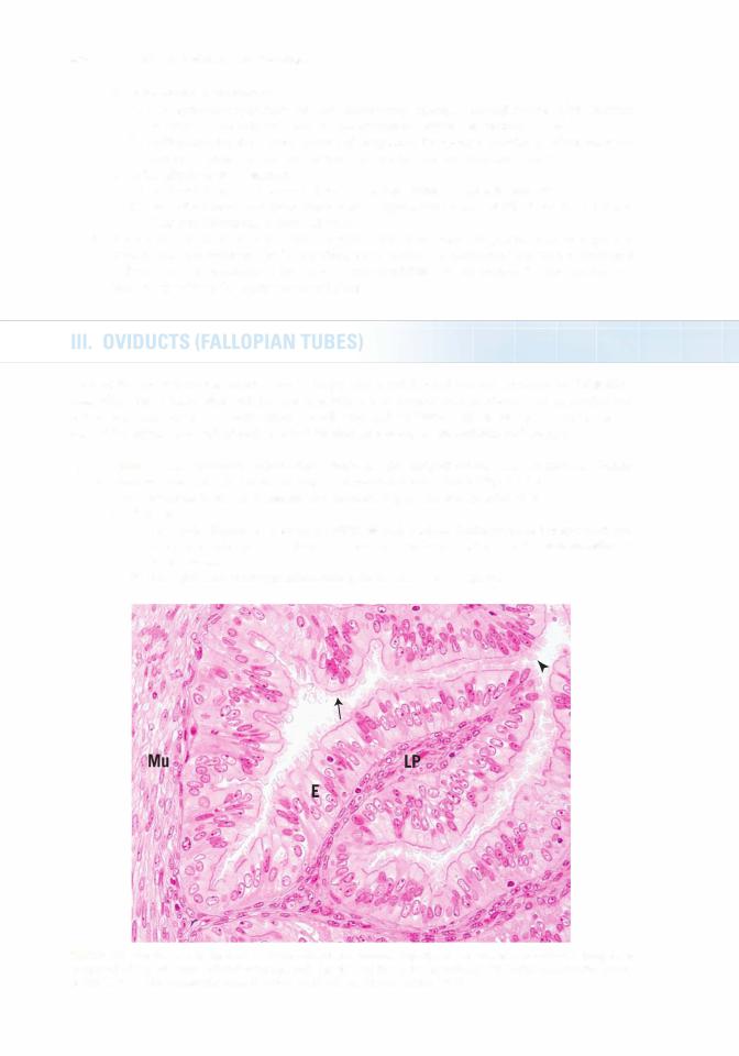
b.Ciliated cells
(1 ) Ciliated cells (Figure 19.4) possess many cilia, which beat mostly toward the lumen of
the uterus.
(2)The number of cilia and the intensity of their motion are increased by the presence of estrogen.
(3)Function. Ciliated cells aid in the transport of the developing embryo to the uterus.
2.The lamina propria (Figure 19.4) consists ofloose connective tissue containing reticular fibers, fibroblasts, mast cells, and lymphoid cells.
B.Muscularis
1.The muscularis is composed of an ill-defined inner circular and an outer longitudinal layer of smooth muscle.
2.Function. By contracting rhythmically, the muscularis probably assists in moving the developing embryo toward the uterus.
C.The serosa, which is composed of a simple squamous epithelium overlying a thin connective tissue layer, covers the outer surface of the oviduct.
The uterus has three regions, the fundus, body (corpus), and cervix.
A.The uterine wall consists ofthe endometrium, myometrium, and adventitia (or serosa). 1 . Endometrium
a.Overview
(1 ) The endometrium (Figure 19.5), composed of an epithelial lining and a gland-rich connective tissue stroma, undergoes hormone-modulated cyclic alterations during the menstrual cycle, varying in thickness from less than 1 mm to 7 mm.
(2)It is lined by a simple columnar epithelium containing secretory and ciliated cells.
(3)Its stroma resembles mesenchymal connective tissue, with stellate cells and an abundance of reticular fibers. Macrophages and leukocytes are also present. The stroma houses the simple tubular glands of the endometrium.
b.Layers of the endometrium
(1 ) The functional layer (functionalis) is the thick superficial layer of the endometrium that is sloughed and reestablished monthly as a result of hormonal changes during the menstrual cycle.
(2)The basal layer (basalis) is the deeper layer of the endometrium that is about 1 mm or less in thickness and is preserved during menstruation. It has endometrial glands, which have basal cells that provide a source for reepithelialization ofthe endometrium after the functional layer is shed.
c.The endometrial vascular supply consists of two types of arteries derived from vessels in the stratum vasculare of the myometrium.
(1 ) Coiled arteries extend into the functional layer and undergo pronounced changes
during various stages of the menstrual cycle.
(2) Straight arteries do not undergo cyclic changes and terminate in the basal layer.
Endometriosis is a condition in which the pelvic peritoneal cavity con tains uterine endometrial tissue. It is associated with hormone-induced
changes in the ectopic endometrium during the menstrual cycle. As the endometrium is shed, bleed ing occurs in the peritoneal cavity, causing severe pain and the formation of cysts and adhesions. It may lead to sterility because the ovaries and oviducts become deformed and embedded in scar tis sue. The factors that contribute to the occurrence of endometriosis are not known.
340 |
BRS Cell Biology and Histology |
FIGURE 19.5. The uterine mucosa is in the process of being rebuilt during the follicular phase. The connective tissue stroma (Stl is well developed, and uter ine glands (Gil are being formed and are beginning to become coiled. A simple columnar epithelium (E) lines the lumen Ill of the uterus (x53l.
2.Myometrium
a.The myometrium is the thick smooth muscle tunic ofthe uterus.
b.It is composed of inner and outer longitudinal layers and a thick middle circular layer. The circular layer is richly vascularized and is often referred to as the stratum vasculare.
c.The myometrium thickens during pregnancy because of the hypertrophy and hyperplasia of individual smooth muscle cells.
d.Nearthe end ofpregnancy, the myometrium developsmanygapjunctions betweenits smooth muscle cells. These junctions coordinate contraction ofthe muscle cells during parturition.
e.At parturition, the myometrium undergoes powerful contractions triggered by the hormone oxytocin and by prostaglandins (both ofwhich are increased at term).
f.After parturition, the myometrium shrinks because many of the smooth muscle cells become deprived of estrogen and, therefore, undergo apoptosis.
3.External covering
a.Serosa is present over surfaces of the uterus bulging into the peritoneal cavity. b. Adventitia is present along the retroperitoneal surfaces of the uterus.
B.The menstrual cycle begins on the day menstrual bleeding appears. Atthistime, the blood levels of estrogen, progesterone, FSH, and LH are very low.
1 . Menstrual phase (days 1-4) is characterized by a hemorrhagic discharge (menses) of the
functional layer of the endometrium.
a.It is triggered by spasms of contraction and relaxation of the coiled arteries (caused by low levels of progesterone and estrogen). Long-term (2-3 days) vasoconstriction of these arteries causes ischemia and eventual necrosis.
b.Vasoconstriction is followed by sudden, intermittent vasodilation of the coiled arteries, which ruptures their walls, flooding the stroma with blood, detaching the functional layer, and dislodging the necrotic tissue.

l!il!ll'tttD]jFemale Reproductive System |
341 |
c.Because the basal layer is supplied by short straight vessels that do not undergo prolonged vasoconstriction, it is not sloughed and does not become necrotic.
d.During the menstrual phase of the menstrual cycle, blood clotting is inhibited.
2.The proliferative (follicular) phase (Figure 19.5), days 4 to 14, follows the menstrual phase and involves renewal of the entire functional layer, including the repair ofglands, connective tissue, and vascular elements (specifically, the coiled arteries). During the proliferative phase, the levels of estrogen in the blood continue to rise, and this is the principal hormone responsible for the stage of proliferation.
a.The epithelium that lines the luminal surface ofthe uterus is renewed by mitotic activity of cells remaining in the uterine glands of the basal layer of the endometrium.
b.Glands are straight and lined by a simple columnar epithelium.
c.Stromal cells divide, accumulate glycogen, and enlarge.
d.Coiled arteries extend approximately two-thirds of the way into the endometrium.
e.At the end ofthe proliferative phase, the endometrium is approximately 3 mm in thickness.
3.The secretory (luteal) phase (days 15-28) begins shortly after ovulation and is characterized by a thickening of the endometrium, resulting from edema and secretion by the endometrial glands. Progesterone, and to a certain degree estrogen, are the principal hormones driving the secretory phase.
a.Glands become coiled; theirlumina become filled with a secretion of glycoprotein material and glycogen; and their cells accumulate large amounts of glycogen, in the basal aspect of their cytoplasm.
b.Coiled arteries become not only more highly coiled but also longer, extending into the superficial aspects ofthe functional layer.
c.At the end of the secretory phase, the endometrium is somewhat edematous and is approximately 6 to 7 mm in thickness.
Uterine prolapse is a condition in which the uterine ligaments, namely the round ligament, broad ligament, uterosacral ligaments, and the ligament
of the ovary, are no longer able to maintain the normal anatomical position of this organ, and the uterus slides down and frequently bulges into the vagina. The uterus may occupy only the upper one third of the vagina or may slide all the way down to protrude through the vaginal orifice between the labia minora and majora. Uterine prolapse usually occurs in older women, but may also be present in younger women who had very large babies and a prolonged, difficult delivery. Extensive prolapse of the uterus may interfere with urination as well as with defecation. The usual treatment for uterine prolapse is surgical reattachment or, in more extreme cases, hysterectomy.
A.The cervix does not participate in menstruation, but its secretions change during various stages of the menstrual cycle.
B.The cervical wall is composed mainly of dense collagenous connective tissue interspersed with numerous elastic fibers and a few smooth muscle cells.
C.The cervix has a simple columnar (mucus-secreting) epithelium except for the inferior portion (continuous with the lining ofthe vagina), which is covered by a stratified squamous nonkeratinized epithelium.
D.Branched cervical glands secrete a serous fluid near the time of ovulation that facilitates the entry ofspermatozoa into the uterine lumen. During pregnancy, cervical glands produce a thick, viscous secretion that hinders the entry of spermatozoa (and microorganisms) into the uterus.
E.Prior to parturition, the cervix dilates and softens as a result ofthe lysis ofthe collagen fiber bundles in response to the hormone relaxin.

342 BRS Cell Biology and Histology
1. In a Papanicolaou (Pap) smear, epithelial cells a re scra ped from the lining of the cervix (or vagina) and a re examined to detect
cervical cancer. A Pap smear shows variation in cell populations with stages of the men strual cycle.
2. Carcinoma of the cervix originates from stratified squamous nonkeratinized epithelial cells. It may be contained within the epithelium and not invade the underlying stroma (carcinoma in situ), or it may penetrate the basal lamina and metastasize to other parts of the body (invasive carcinoma). It occurs at a relatively high frequency, but may be cured by surgery if discovered early (by Pap smear), before it becomes invasive.
A.Fertilization
1.Fertilization usually takes place within the ampulla of the oviduct, and occurs when a spermatozoon penetrates the corona radiata and the zona pellucida and pierces the plasma membrane of a secondary oocyte.
a. Before a spermatozoon is capable of fertilizing, it must undergo maturation, and capacitation, and hyperactivity.
(1 ) Maturation occurs in the male reproductive tract. Prior to maturation, the spermatozoon cannot travel in a forward direction; instead, it travels only in a circular fashion.
(2)Capacitation occurs in the female reproductive tract. While the sperm is in the male reproductive tract, it is unable to undergo capacitation because the concentration of the prostate-manufactured fertilization-promoting peptide (FPP) is too high. Once the sperm arrives in the female reproductive tract, the concentration of FPP is reduced to a suitable level (because it becomes diluted by the vaginal secretions), and the sperm is prompted to begin the process of capacitation.
(a)Cholesterol molecules are removed from the acrosomal plasmalemma, making the membrane less rigid.
(b)Special Ca2+ ion channels open in the plasma membrane of the spermatozoon flagella, permitting the influx ofcalcium ions.
(c)Elevated levels of intraflagellar calcium ions induce the formation of cAMP, which causes the spermatozoon to become hyperactive.
(3)Hyperactivity is merely the ability of the spermatozoon to swim faster and more vigorously, making it easier for the sperm to penetrate the zona pellucida to contact the oocyte.
b.Once the spermatozoon has undergone capacitation, it is capable of fertilizing the egg, but in order to do so it must undergo the acrosome reaction, bind to ZP3, and fuse to the plasma membrane of the secondary oocyte.
(1)The acrosome reaction and binding to ZP3 occur when the spermatozoon contacts the zona pellucida. Hydrolytic enzymes, especially acrosin, are released from the acrosome, reduce the viscosity of the zona pellucida just ahead of the spermatozoon, permitting it to reach the secondary oocyte.
(2)Fusion ofthe spermatozoon membrane and the secondary oocyte plasmalemma occurs due to the presence offertilin, an integral protein in the spermatozoon membrane with CD9 molecules and integrins of the secondary oocyte's plasmalemma.
c.The secondary oocyte responds to the contact by the spermatozoon by undergoing the cortical reaction, resuming the second meiotic division, and forming the female pronucleus.
(1)The cortical granules ofthe secondary oocyte, located just deep to the cell membrane, fuse with the cell membrane, releasing their enzymes into the zona pellucida. The ZP proteins form a complex with each other, which transforms the gelatinous nature of the zona pellucida into a highly viscous substance that stops other spermatozoa from reaching the secondary oocyte, thus preventing polyspermy.

l!il!ll'tttD]jFemale Reproductive System |
343 |
(2)The contact of spermatozoon with the secondary oocyte permits the entry of the spermatozoon's nucleus (male pronucleus) and centrosome to enter the secondary oocyte's cytoplasm, triggering the oocyte to complete its second meiotic division, forming the second polar body and thus transforming the secondary oocyte into the ovum.
(3)Thenowhaploidnucleus ofthe ovum is known as thefemale pronucleus. The two haploid pronuclei travel toward each other, shed their nuclear envelopes, their chromosomes intermingle as they form a diploid cell, known as the zygote. Immediately after the intermingling of the chromosomes, the centrosome, contributed by the spermatozoon, forms a mitotic spindle apparatus, and the zygote undergoes its first mitotic division. The two newly formed daughter cells are the firsttwo cells ofthe new embryo. It should be noted that all centrosomes of the new individual are derived from the father, and all of the mitochondria are derived from the mother.
B.Implantation
1.The zygote undergoes mitotic cell division (known as cleavage during the early stages of embryogenesis) and is transformed into a multicellular structure called a morula, which requires about 3 days to travel through the oviduct and enter the uterus.
2.The conceptus (the preimplantation embryo and its surrounding membranes) acquires a fluid-filled cavity and becomes a blastocyst.
3.The blastocyst implants in the endometrium of the uterus and is surrounded by an inner cellular layer, the cytotrophoblast, and an outer multinucleated layer, the syncytiotrophoblast.
4.The syncytiotrophoblast further invades the endometrium in the wall ofthe uterus by the sixth day after fertilization. Formation of the placenta then begins.
1. Ectopic (tubal) pregnancy is the implantation of the early embryo in an abnormal site (e.g., wall of the oviduct). It can be fatal without immedi ate medical intervention.
2. Teratomas are germ cell tumors that fall into three groups: mature, monodermal, and immature.
a.Mature teratomas are benign (although occasionally they may become malignant) and are usually present in young women. These are cysts with walls that frequently contain hair and other epidermal structures such as sebaceous glands, as well as bone, tooth, and cartilage fragments.
b.Monodermal teratomas are rare tumors that are also known as specialized teratomas. The two most frequent types of these tumors are struma ovarii and ovarian carcinoid. Struma ovarii is an ovarian tumor composed of well-developed thyroid follicles that produce thyroid hormone and may be responsible for hyperthyroidism. Ovarian carcinoid is a tumor that usually produces serotonin (5-0H-tryptamine).
c.Immature teratomas are fast-growing malignant tumors with a histology that resembles that of fetal rather than mature tissues. They are usually present in adolescents and very young women.
Theplacenta is a transient structure, consisting of a maternal portion and a fetal portion.
A.Structure
1.At birth it is approximately 18 em in diameter, 2.5 em thick, and weighs about 600 g.
2.As the placenta begins its development, the cytotrophoblasts and syncytiotrophoblasts form the chorion, and in response, the endometrium of the uterus forms the decidua.
3.The chorion develops into the chorionic plate, which gives rise to the chorionic villi.


l 345
factor, platelet-derived growth factor, tumor necrosis growth factor, relaxin, leptin, and interleukins and 3.
3.It also produces estrogen with the assistance of the liver and the adrenal cortex of the fetus.
4.Decidual cells of the stroma produce prostaglandins and prolactin.
The polypeptide hormone prolactin, manufactured by basophils of the adenohypophysis (anterior pituitary), causes an increase in breast size
in the pregnant female and induces milk formation as well as lactation in the mammary glands of the postpartum female. Interestingly, it also causes the feeling of sexual gratification subsequent to the sexual act. Because progesterone has been shown to alleviate the symptoms of multiple sclerosis (MS) in the pregnant female, recent investigations using a mouse model demonstrated that treatment of MS-afflicted mice with prolactin resulted in remyelination of nerve fibers, whose myelin was de stroyed by the disease. It is believed that prolactin stimulates the formation of prooligodendrocytes, which differentiate into oligodendrocytes, cells that are responsible for myelination of axons in the central nervous system.
A. Overview
1.The vagina is a fibromuscular canal with a wall that is composed of three layers: an inner mucosa, a middle muscularis, and an external adventitia.
2.It is circumscribed by a skeletal muscle sphincter at its external orifice.
3.It lacks glands throughout its length and is lubricated by secretions from the cervix and by seepage of the extracellular fluid from the vascular supply of the lamina propria.
B.The mucosa is composed of a thick, stratified squamous nonkeratinized epithelium and a fibroelastic connective tissue, the lamina propria.
1.The epithelium contains glycogen, which is used by the vaginal bacterial flora to produce lactic acid, an acid that lowers the pH during the follicular phase of the menstrual cycle and inhibits invasion by pathogens.
2.The lamina propria is a fibroelastic connective tissue thatis highly vascular in its deeper aspect (which is possibly analogous to a submucosa).
C.The muscularis is composed of irregularly arranged layers of smooth muscle (thin inner circular layer and a thicker outer longitudinal layer) interspersed with elastic fibers.
D.The adventitia is composed of fibroelastic connective tissue. It attaches the vagina to the surrounding structures.
A.The labia majora are fat-laden folds of skin; hair and secretions of sebaceous glands and sweat glands are present on their external surfaces.
B.Labia minora
1.The labia minora are folds of skin that possess a core of highly vascular connective tissue containing elastic fibers.
2.They lack hair follicles, but their dermis contains numerous sebaceous glands, which open directly onto the epithelial surface.

346 |
BRS Cell Biology and Histology |
C.The vestibule is the space between the two labia minora. Glands of Bartholin (mucus-secreting glands) and numerous smaller mucus-secreting glands around the urethra and clitoris (minor vestibular glands) open into this space.
D. Clitoris
1.The clitoris is composed of two small, cylindrical erectile bodies, which terminate in the prepuce-covered glans clitoridis.
2.It contains many sensory nerve fibers and specialized nerve endings (e.g., Meissner corpuscles and pacinian corpuscles).
Mammary glands of both genders are identical for the first decade or so of life, but when the female reaches puberty, the flow of estrogens and progesterone as well as lactogenic hormone induces the mammary gland to enlarge and develop a system of lobules and terminal ductules as well as an increase in the connective tissue mass and a deposit of adipose tissue. Each mammary gland of the postpubertal female is composed of numerous compound tubuloalveolar glands, each with its own lactiferous sinus and a duct that opens at the apex of the nipple.
The use of lavender oil and tea tree oil-based products in prepubes cent males has been shown to cause gynecomastia, the development
of breasts in these individuals. The presence of the breast tissue persisted for several months after the boys stopped using these products, and then slowly regressed to its normal condition. It appears that these oil products are able to imitate estrogens and even inhibit the effects of androgens.
A. Resting (nonlactating) mammary glands (in adult, nonpregnant women)
1 . Resting mammary glands are composed of lactiferous sinuses and ducts lined in most areas by a stratified cuboidal epithelium, with a basal layer consisting of scattered myoepithelial cells.
2. A basal lamina separates the epithelial components from the underlying stroma.
B.Active (lactating) mammary glands are enlarged during pregnancy by the development of alveoli. 1 . Alveolar cells (secretory cells) (Figure 19.6)
a. Alveolar cells line the alveoli of active mammary glands and are surrounded by an incomplete layer of myoepithelial cells.
b.They are richly endowed with RER and contain several Golgi complexes, numerous mitochondria, lipid droplets, and vesicles containing milk protein (caseins) and lactose.
2.Secretion by alveolar cells
a.lipids are released into the lumen, perhaps, via the apocrine mode of secretion.
b.Proteins and sugars are released into the alveolar lumen via the merocrine mode of secretion (exocytosis).
C.Nipple
1.The nipple is composed of dense, irregular collagenous connective tissue interlaced with smooth muscle fibers that act as a sphincter.
2.It contains the openings of the lactiferous ducts.
3.It is surrounded by pigmented skin (areola) that is more deeply pigmented during and subsequent to pregnancy and contains the areolar glands (of Montgomery).

l!il!ll'tttD]jFemale Reproductive System |
347 |
FIGURE 19.6. Transmission electron micrograph showing alveolar epithelial cell (A) from lactating mammary gland and an underlying myoepithelial cell (M). CAP, capillary; L, lumen of alveolus containing milk; F, fat droplet; C, casein. (Reprinted with permission from Strum J. 3rd ed. Baltimore, MD: University of Maryland School of Medicine; 1 992:1 05.)
1. Breast cancer may originate from the epithelium lining the ducts
(ductal carcinoma) or the terminal ductules (lobular carcinoma). If breast cancer is not treated early, the tumor cells metastasize via lymphatic vessels to the axil lary nodes near the affected breast and later via the bloodstream to the lungs, bone, and brain. In the United States, 1 80,000 new cases of breast cancer are diagnosed annually, and every year 43,000 women die of this disease. Early detection by self-examination, mammography, or ultrasound has led to a reduction in the mortality rate associated with breast cancer.
2.Deficiency or mutation in the gene BRCA1 has been shown to decrease the stability or elevate the incidence of the mutation rate of tumor suppressor genes such as p53. It appears that muta tions in the BRCA1 gene result in incapacitation of the checkpoint at G2-M of the cell cycle, and concurrently, the number of centrosomes of these cells is increased. Therefore, these mutated cells have the capability to proliferate unchecked.
D. Secretions ofthe mammary glands
1.Colostrum (protein-rich yellowish fluid)
a.Colostrum is produced during the first few days after birth.
b.It is rich in cells (lymphocytes, monocytes), lactalbumin, fat-soluble vitamins, and minerals and contains immunoglobulin A (lgA).
2.Milk
a.Milk begins to be secreted by the third or fourth day after birth.
b.Milk consists of proteins (caseins, IgA, lactalbumin), many lipid droplets, and lactose.
c.It is released from the mammary glands viathe milk ejection reflex in response to a variety of external stimuli related to suckling. The milk ejection reflex involves release of oxytocin (from axons in the pars nervosa of the pituitary gland), which induces contraction of the myoepithelial cells, forcing milk into the larger ducts and out of the breast.


l!1lllJ1ttlfl!lMale Reproductive System 351
FIGURE 20.1 . The testis and epididymis. (Reprinted with permission from Gartner LP. Hiatt JL. Color Textbook ofHistology. 2nd ed. Philadelphia. PA: Saunders; 2001 :488.)
A.Testicular tunicae (covering of the testes) are applied to the testes as they descend through the abdominal wall to enter the scrotum.
1 . The tunica vaginalis is a serous sac derived from the peritoneum that partially covers the anterior and lateral surfaces of each testis.
2.Tunica albuginea
a.Tunica albuginea is the thick, fibrous connective tissue capsule ofthe testis.
b.It is lined by a highly vascular layer of loose connective tissue, the tunica vasculosa.
c.Itis thickened posteriorlyto formthe mediastinum testis, fromwhich incomplete connective tissue septa arise to divide the organ into approximately 250 compartments (lobuli testis). Additionally, the mediastinum testis is the passage way for blood and lymphatic vessels in and out of the testes.
B.Lobuli testes (Figure 20.1)
1 . The lobuli testes are pyramidal intercommunicating compartments that are separated by incomplete septa.
2.Each contains one to four seminiferous tubules where spermatozoa are produced. These highly convoluted tubules are embedded in a meshwork ofloose connective tissue containing blood and lymphatic vessels, nerves, and interstitial cells of Leydig.
C.Interstitial cells of Leydig
1.Interstitial cells of Leydig are round to polygonal cells in the interstitial regions between seminiferous tubules.
2.They possess a large central nucleus, numerous mitochondria, a well-developed Golgi complex, and many lipid droplets. The lipid droplets contain cholesterol esters, precursors of testosterone.
3.They are richly supplied with capillaries and lymphatic vessels.
4.Function. Interstitial cells ofLeydig are endocrine cells that produce and secrete testosterone. Secretion is stimulated by luteinizing hormone (LH; interstitial cell-stimulating hormone) produced in the pituitary gland. These cells mature and begin to secrete during puberty.
D.Seminiferous tubules
1.Overview (Figure 20.2)
a.Seminiferous tubules are 30 to 70 em long, with a diameter of 150 to 250 J.Lm.
b.They are enveloped by a fibrous connective tissue tunic composed of several layers of fibroblasts and extensive capillary beds.
c.They form tortuous pathways through the testicular lobules and then narrow into short, straight segments, the tubuli recti, which connect with the rete testis.

352 |
BRS Cell Biology and Histology |
FIGURE 20.2. Light micrograph of the seminiferous tubules in the testis j x 132). Observe myoid cells (Me) and fibroblasts
(F) composing the wall of the seminiferous tubules. Spermatogonia (Sg) and the Sertoli cells (SC) lie in the basal compart ment. Just superior to this are primary spermatocytes (P). Note that spermatids (S) are located near the lumen.
d.The lumina of the seminiferous tubules are lined by a thick complex epithelium (seminiferous or germinal epithelium). This epithelium consists of four to eight cell layers and contains spermatogenic cells, from which the germ cells eventually develop (spermatogenesis), and Sertoli cells, which have several functions.
2.Sertoli cells (Figures 20.2 and 20.3)
a.Structure
(1)Sertoli cells are columnar cells that extend from the basal lamina to the lumen of the seminiferous tubule. Theyhave a pale, oval nucleusthat displays frequent indentations; they are highly infolded and possess a large nucleolus. Sertoli cells no longer undergo mitosis after puberty.
(2)Their lateral plasma membranes have long processes that interdigitate with those of neighboring Sertoli cells enfolding small clusters of developing spermatogenic cells. Their apical cell membranes are highly convoluted, forming finger-like processes where the interstices between these processes are occupied by developing spermatozoa.
(3)They have a well-developed smooth endoplasmic reticulum (SER), some rough endoplasmic reticulum (RER), an abundance of mitochondria and lysosomes, and an extensive Golgi complex.
(4)Receptors for follicle-stimulating hormone (FSH) are present on their basal plasma membranes.
(5)They form zonulae occludentes (tight junctions) with adjacent Sertoli cells near their bases, thus dividing the lumen ofthe seminiferous tubule into a basal and an adluminal compartment. These tight junctions are responsible for the blood-testis barrier, which protects developing sperm cells from autoimmune reactions.

l!1lllJ1ttlfl!lMale Reproductive System 353
Testis |
Seminiferous tubule |
Adluminal compartment
--}Junctional complex Basal compartment
Junctional complex
FIGURE 20.3. The seminiferous (germinal) epithelium. Note the intercellular bridges between spermatocytes and the junc tional complexes near the&bases of adjacent Sertoli cells. These junctional complexes of the Sertoli cells divide the epithelium into an adluminal and a basal compartment. !Reprinted with permission from Krause WJ. Cutts JH.
2nd ed. Baltimore. MD: Williams Wilkins; 1 986:414 )
b.Function
(1 ) Sertoli cells support, protect, and nourish the spermatogenic cells.
(2)They phagocytose excess cytoplasm discarded by maturing spermatids.
(3)They secrete a fructose-rich fluid into the lumen that nourishes and facilitates the transport ofspermatozoa through the seminiferous tubules to the genital ducts.
(4)They synthesize androgen-binding protein (ABP) under the influence of FSH. ABP assists in maintaining the necessary concentration of testosterone in the seminiferous tubule so that spermatogenesis can progress.
(5)They secrete inhibin, a hormone that inhibits the synthesis and release of FSH by the anterior pituitary, as well as the hormone activin which boosts FSH release from the anterior pituitary.
(6)They establish a blood-testis barrier.
(7)During fetal development they synthesize and release antimiillerian hormone, which determines maleness.
(8)They manufacture and release testicular transferrin, a protein that facilitates the transfer ofiron from serum transferrin to maturing spermatogenic cells.
3.Spermatogenesis
a.Shortly before puberty, the rise in gonadotrophins initiates spermatogenesis, the process of spermatozoon (sperm) formation. It is divided into three phases:
(1 ) Spermatocytogenesis-differentiation of spermatogonia into primary spermatocytes.
(2)Meiosis-reduction division to reduce the diploid chromosomal complement of primary spermatocytes to form haploid spermatids (see Chapter 2 XI).
(3)Spermiogenesis-transformation of spermatids into spermatozoa.

354BRS Cell Biology and Histology
b.Spermatogenesis does not occur simultaneously or synchronously in all seminiferous tubules, but rather in wavelike sequences of maturation, referred to as cycles of the seminiferous epithelium.
c.During spermatogenesis, daughter cells remain connected to each other via intercellular bridges. The resultant syncytium may be responsible for the synchronous development of germ cells along any one seminiferous tubule.
4.Spermatogenic cells (Figure 20.2)
a. Spermatogonia are diploid germ cells adjacent to the basal lamina of the seminiferous epithelium. At puberty, testosterone influences them to enter the cell cycle.
(1 ) Pale type A spermatogonia possess a pale-staining nucleus, spherical mitochondria, a small Golgi complex, and abundant, free ribosomes. They are mitotically active (starting at puberty) and give rise either to more cells ofthe same type (to maintain the supply) or to type B spermatogonia.
(2)Dark type A spermatogonia represent mitotically inactive (reserve) cells (in the G0 phase of the cell cycle, see Figure 2.6), with dark nuclei; they have the potential to resume mitosis and produce pale type A cells.
(3)Type B spermatogonia undergo mitosis and give rise to primary spermatocytes.
b.Spermatocytes
(1 ) Primary spermatocytes are large diploid cells with 4cDNA content. They undergo the first meiotic division (reductional division) to form secondary spermatocytes (see
Chapter 2).
(2)Secondary spermatocytes are haploid cells with 2cDNA that quickly undergo the second meiotic division (equatorial division), without an intervening S phase, to form spermatids.
c.Spermatids
(1)Spermatids are small haploid cells containing only 1cDNA.
(2)They are located near the lumen ofthe seminiferous tubule.
(3)Their nuclei often display regions of condensed chromatin.
(4)They possess a pair of centrioles, mitochondria, free ribosomes, SER, and a well developed Golgi complex.
5.Spermiogenesis is a unique process of cytodifferentiation, whereby spermatids shed much of their cytoplasm and transform into spermatozoa, which are released into the lumen of the seminiferous tubule. Spermiogenesis is divided into four phases.
a. Golgi phase
(1)The Golgi phase is characterized by the formation of an acrosomal granule, enclosed within an acrosomal vesicle, which becomes attached to the anterior end ofthe nuclear envelope of a spermatid.
(2)In this phase, centrioles migrate away from the nucleus to form the flagellar axoneme. Centrioles then migrate back toward the nucleus to assist in forming the connecting piece associated with the tail.
b.The cap phase is characterized by expansion of the acrosomal vesicle over much of the nucleus, forming the acrosomal cap.
c.Acrosomal phase is characterized bythe following:
(1)The nucleus becomes condensed, flattened, and located in the head region.
(2)Mitochondria aggregate around the proximal portion ofthe flagellum, which develops into the middle piece of the tail.
(3)The spermatid elongates; this process is aided by a temporary cylinder of microtubules called the manchette.
(4)The acrosomal phase ends as the spermatid is oriented with its acrosome pointing toward the base of the seminiferous tubule.
d.Maturation phase is characterized by the following:
(1)Loss of excess cytoplasm and intercellular bridges connecting spermatids into a syncytium. The discarded cytoplasm is phagocytosed by Sertoli cells.
(2)Maturation phase ends when the nonmotile spermatozoa are released (tail first) into the lumen of the seminiferous tubule. Spermatozoa remain immotile until they leave the epididymis. They become capacitated (capable of fertilizing) in the female reproductive system.


l!1lllJ1ttlfl!lMale Reproductive System 359
(b)Principal cells function, in concert with the ductuli efferentes, to absorb nearly 90% ofthe testicular fluid.
(c)They secrete various factors that facilitate sperm maturation; however, they also produce glycerophosphocholine, whichinhibits capacitation (the process whereby a sperm becomes capable of fertilizing an oocyte). Thus, capacitation occurs only after the sperm enters the female genital tract.
3.Ductus (vas) deferens
a.The ductus deferens, which begins at the tail of the epididymis, has a thick muscular wall with inner and outer layers of longitudinal smooth muscle, which are separated from one another by a middle circular layer.
b.It possesses a narrow, irregular lumen lined by pseudostratified columnar epithelium similar to that of the ductus epididymis.
4.Ejaculatory duct
a.The ejaculatory duct is the straight continuation of the ductus deferens beyond where it receives the duct of the seminal vesicle.
b.It lacks a muscular wall.
c.It enters the prostate gland and terminates in a slit on the colliculus seminalis located in the prostatic urethra.
A normal sperm count for a single ejaculate is approximately 50 to
1 00 million spermatozoa/ml. An individual with a sperm count of less than 2 0 million spermatozoa/ml. of ejaculate is considered to be sterile.
Include the paired seminal vesicles, prostate gland, and the paired bulbourethral glands
A.Seminal vesicles
1.Epithelium
a.Epithelium of the seminal vesicles is pseudostratified columnar, with a height that varies with testosterone levels; it lines the extensively folded mucosa.
b.It contains many yellow lipochrome pigment granules and secretory granules, a large Golgi complex, many mitochondria, and abundant RER.
2.The lamina propria consists of fibroelastic connective tissue surrounded by an inner circular and outer longitudinal layer of smooth muscle.
3.The adventitia is composed of fibroelastic connective tissue.
4.The seminal vesicles secrete a yellowish, viscous fluid containing substances that activate sperm (e.g., fructose providing the energy source for the sperm); this fluid constitutes about 70% ofthe human ejaculate.
5.The duct of the seminal vesicle joins the ductus deferens at its ampulla (the enlarged terminal end), thus forming the right and left ejaculatory ducts that enter the prostate gland continuing to the urethra.
B.Prostate gland (Figure 20.7)
1.Overview
a.The prostate gland surrounds the urethra as it exits the urinary bladder.
b.It consists of30 to 50 discrete branched tubuloalveolar glands that empty their contents via excretory ducts into the prostatic urethra. These glands are arranged in three concentric layers (mucosal, submucosal, and main) around the urethra.
c.The gland is covered by a fibroelastic capsule that contains smooth muscle. Septa from the capsule penetrate the gland and divide it into lobes.
360 |
BRS Cell Biology and Histology |
FIGURE 20.7. Light micrograph of the prostate gland ( x 1 32). Note the prostrate concretion (Pel. smooth muscle (arrow , |
|
and the compressed columnar cells (CC) forming the glandular parenchyma. |
) |
2. Epithelium
a.The epithelium of the prostate gland is simple or pseudostratified columnar and lines the individual glands that constitute the prostate.
b.It is composed of cells that contain abundant RER, a well-developed Golgi complex, numerous lysosomes, and many secretory granules.
3.Corpora amylacea are prostatic concretions, composed of glycoprotein, which may become calcified; their numbers increase with age.
4.The prostate secretes a thin whitish fluid, a part of the semen containing proteolytic enzymes, citric acid, acid phosphatase, fibrinolysin, and lipids. The prostatic secretion serves to liquefy the coagulated semen after it is deposited in the female genital tract. Its synthesis and release are regulated by dihydrotestosterone.
CLINICAL |
Benign prostatic hypertrophy (BPH) |
|
CONSIDERATIONS |
||
|
1 . BPH most commonly involves only an enlargement of the mucosal glands.
2.It is frequently associated with an inability to begin and cease urination because the urethra is partially strangulated by the enlarged prostate.
3.It leads to nocturia (urination at night) and sensory urgency (the desire to urinate without having to void).
4.This disease is common in older men, occurring in about 50% of men older than 50 years of age and in 95% of those older than 80 years of age. BPH can be confirmed by a digital rectal exami nation; however, a biopsy is required to rule out prostatic cancer.

|
l!1lllJ1ttlfl!lMale Reproductive System |
361 |
Adenocarcinoma of the prostate gland |
|
|
1 . Adenocarcinoma of the prostate gland may be indicated by palpation through the rectum. |
|
|
|
However, a biopsy is required for positive identification of the disease. |
|
2. |
Although this cancer grows slowly, it commonly metastasizes to bone via the circulatory |
|
|
system. |
|
3. |
It occurs in about one-third of men older than 75 years of age and is the second most common |
|
|
form of cancer in men. |
|
4. It is associated with an elevated prostate-specific antigen (PSA) level in blood. Men with |
|
|
|
elevated PSA values are candidates for more aggressive diagnostic tests for prostatic cancer. |
|
Surgical removal with or without radiation therapy is the usual treatment. although there may be complications of impotence and/or incontinence. Newer procedures (e.g., brachythera py) include radioactive seeding within the gland.
C. Bulbourethral (Cowper) glands
1.Bulbourethral glands are adjacent to the membranous urethra.
2.They empty their viscous, slippery secretions into the lumen of the membranous urethra to lubricate it just prior to the emission of semen.
3.The glands are lined by a simple cuboidal or columnar epithelium.
4.They are surrounded by a fibroelastic capsule containing smooth and skeletal muscle.
Vasectomy is the chosen means of permanent contraception by more than 600,000 US males each year. A physician may perform the procedure
in the office, where the scrotum is slit open, a portion of the spermatic cord is pulled out, cut, cauterized, and returned to the scrotum. At the conclusion of 6 weeks or so, after the procedure, semen is collected and examined to assure that no spermatozoa are ejaculated. This procedure is nearly 1 00% effective. The procedure may have a few complications, but they usually dissipate after a short period of time.
The urethra conveys urine from the bladder to outside the body. In males, the urethra also carries semen during ejaculation. It has a two-layered muscularis consisting of an inner longitudinal and an outer circular layer of smooth muscle. It is surrounded at some point by an external sphincter of skeletal muscle, which permits its voluntary closure.
A.The male urethra is about 20 em long and is divided into prostatic, membranous, and cavernous portions.
B. It is lined by transitional epithelium in the prostatic portion and by pseudostratified or stratified columnar epithelium in the other two portions. The fossa navicularis, located at the distal end of the cavernous urethra, is lined by stratified squamous epithelium.
C. It contains mucus-secreting glands of Littre in the lamina propria.

362BRS Cell Biology and Histology
A.Corpora cavernosa
1.Corpora cavernosa are paired masses of erectile tissue that contain irregular vascular spaces lined by a continuous layer of endothelial cells. These spaces are separated from each other by trabeculae of connective tissue and smooth muscle.
2.The vascular spaces decrease in size toward the periphery of the corpora cavernosa.
3.During erection, thevascular spaces become engorgedwith blood as a resultofparasympathetic impulses, which constrict arteriovenous shunts and dilate the helicine arteries, thus increasing flow to the vascular spaces of the two corpora cavernosa and the single corpus spongiosum.
B.Corpus spongiosum
1.The corpus spongiosum is a single mass of erectile tissue that contains vascular spaces of uniform size.
2.It possesses trabeculae that contain more elastic fibers and less smooth muscle than those of the corpora cavernosa.
3.The penile portion ofthe urethra passes through the length ofthe corpus spongiosum, opening to the exterior at the tip of the glans penis.
C.Connective tissue and skin
1.The tunica albuginea is a thick, fibrous connective sheath that surrounds the paired corpora cavernosa and the corpus spongiosum. The arrangement of dense collagen bundles permits extension of the penis during erection.
2.Glans penis
a.The glans penis is the dilated distal end of the corpus spongiosum.
b.It contains dense connective tissue and longitudinal muscle fibers.
c.It is covered by retractable skin, the prepuce, which is lined by stratified squamous lightly keratinized epithelium.
3.Glands of Littre are mucus-secreting glands present throughout the length of the penile urethra.


