


222 |
BRS Cell Biology and Histology |
|
|
The pituitary gland lies below the hypothalamus in a bony housing known as the hypophyseal fossa, a depression in the sella turcica of the sphenoid bone located in the base of the middle cranial fossa of the skull.
The hypothalamus is a region of the diencephalon of the brain; it possesses nuclei that are struc turally and functionally linked to the pituitary gland. The structural link is via a series of axons whose cell bodies are located in the supraoptic and paraventricular nuclei ofthe hypothalamus. These axons form the hypothalamo-hypophyseal tract and terminate in the pars nervosa of the pituitary gland, where they store and, when needed, release their hormones. The functional connection is via releas ing hormones that are synthesized in the arcuate, paraventricular (and medial paraventricular), peri ventricular, ventromedial, and dorsal nuclei of the hypothalamus. These hormones are released by the neurons located in these nuclei, enter a capillary bed, and make their way, via the hypophyseal portal system to a second capillary bed in the anterior lobe of the pituitary gland, leave the capillary bed, and bind to their respective target cells in the anterior pituitary.
The pituitary gland is a relatively small gland, weighing only about 0.6 g in men and 1.2 g in women who are pregnant or who have given birth to two or more children. The pituitary is divided into two major subdivisions, the adenohypophysis and the neurohypophysis (Figure 13.1). Each sub division is derived from a distinct embryonic analog, which is reflected in its unique cellular constitu ents and functions.
A.The adenohypophysis is also called the anterior pituitary gland (Figures 13.1 and 13.2). It originates from an ectodermal diverticulum of the stomodeum (Rathke pouch). The adenohypophysis is subdivided into the pars distalis, pars intermedia, and pars tuberalis.
1.The pars distalis is supported by a connective tissue capsule and framework. It consists of irregular cords composed of two types of parenchymal cells, chromophils and chromophobes, lying adjacent to fenestrated capillaries.
a. Chromophils (Figures 13.1 and 13.3)
(1 ) Overview. Chromophils are parenchymal cells that stain intensely because of their hormone-containing secretory granules. They synthesize, store, and release several hormones. They are regulated by specific stimulatory and inhibitory hormones produced by neurons, referred to as neurosecretory cells, in the hypothalamus. These hormones are conveyed to the pars distalis via a system of portal blood vessels originating in the median eminence.
(2)Types. Chromophils are classified into two types, acidophils and basophils, depending on the dyes they bind using special histological stains. With hematoxylin-eosin stain, the distinction between the two cell types is much less obvious.
(a)Acidophils (Tables 13.1 and 13.2) bind acidic dyes and often stain orange or red. They are small cells of two subtypes: somatotrophs and mammotrophs.
1 . Somatotrophs constitute about 50% of the chromophils and produce somatotropin (growth hormone). They are stimulated by somatotropin-releasing
hormone and are inhibited by somatostatin.
2.Mammotrophs (lactotrophs) constitute about 10% ofthe chromophil population, except in multiparous women, where they may be as high as 30%. Mammotrophs produce prolactin, which is stored in small secretory granules. They are stimulated by prolactin-releasing hormone and thyrotropin-releasing hormone (TRH) andare inhibited by dopamine (until re-identified that itwas designated as prolactin-inhibiting hormone).
(b)Basophils (Tables 13.1 and 13.2) bind basic dyes and typically stain blue. They include three subtypes: corticotrophs, thyrotrophs, and gonadotrophs.
1 . Corticotrophs constitute about 10% ofthe chromophil population. They produce pro-opiomelanocortin (POMC) whose by-products are adrenocorticotropic hormone (ACTH), melanocyte-stimulating hormone (MSH), and lipotropic


l!iitJ'!1tilUEndocrine System 225
FIGURE 13.3. A light micrograph of cells in the pars distalis of the adenohypophysis. The two types of chromophil cells( are easily identified using the trichrome stain. Basophils (B) stain blue, and acidophils (A) stain red. Chromophobes (C) are smaller and show little affinity for the stain. Many erythrocytes (red blood cells [RBCs]) are present in the capillaries X300).
2.The pars intermedia lies between the pars distalis and pars nervosa.
a.It contains many colloid-containing cysts (Rathke cysts) that are lined by cuboidal cells.
b.It also possesses basophilic cells, which sometimes extend into the pars nervosa. These cells secrete the prohormone POMC, which is cleaved to form ACTH, lipotropin, and MSH. In humans, MSH acts to induce melanocytes to produce melanin and may act in various ways to modulate inflammatory responses throughout the body, and it may play a role in controlling stores of body fat.
3.The pars tuberalis surrounds the cranial part of the infundibulum (hypophyseal stalk).
a.It is composed of cuboidal basophilic cells, arranged in cords along an abundant capillary network.
b.Its cells may secrete FSH and LH, but this has not been confirmed.
CLINICAL |
Pituitary adenomas are common tumors of the anterior pituitary. |
|
CONSIDERATIONS |
||
1 . They enlarge and often suppress secretions by the remaining pars |
||
|
||
|
distalis cells. |
2.These tumors frequently destroy surrounding bone and neural tissues and are treated by surgical removal.
B.The neurohypophysis (Figures 13 . 1 and 13.2; Table 13.1) is also called the posterior pituitary gland. It originates from an evagination ofthe hypothalamus and is divided into the infundibulum, which is continuous with the hypothalamus, and the pars nervosa, or main body ofthe neurohypophysis.
1.Hypothalamo-hypophyseal tract
a. contains the unmyelinated axons of neurosecretory cells whose cell bodies are located in
the supraoptic and paraventricular nuclei of the hypothalamus.
b. transports oxytocin and antidiuretic hormone (ADH; vasopressin), each bound to neurophysin (a binding protein specific for each hormone) to the pars nervosa (see Table 13.2). Oxytocin binds to neurophysin I, whereas ADH binds to neurophysin II. Additionally, acetylcholine and adenosine triphosphate (ATP) are transported to the pars nervosa by the axons that compose the hypothalamo-hypophyseal tract.


|
13.2 |
|
l!iitJ'!1tilUEndocrine System |
227 |
|
t a b I e |
Hormones of the Hypothalamus |
|
|
||
Hormone |
|
|
Nucleus |
Primary Functions |
|
Oxytocin |
|
|
Primarily the paraventricular |
Induces contraction of smooth muscle in wall |
|
|
|
|
nucleus |
of uterus at parturition and in myoepithelial |
|
|
|
|
|
cells of mammary gland during nursing |
|
AD H; vasopressin |
|
|
Primarily the supraoptic nucleus |
Renders kidney collecting tubules permeable |
|
|
|
|
|
to water, which is resorbed to produce |
|
|
|
|
|
a concentrated urine; constricts smooth |
|
|
|
|
|
muscle in wall of arterioles |
|
CRH |
|
|
Arcuate, medial paraventricular, |
Induces the release of POMC by the |
|
|
|
|
and periventricular nuclei |
corticotrophs of the anterior pituitary |
|
Dopamine |
|
|
Arcuate nucleus |
Inhibits prolactin release by mammotrophs |
|
|
|
|
|
(lactotrophs) of the anterior pituitary |
|
GnRH |
|
|
Arcuate, dorsal, paraventricular, |
Induces the release of LH and FSH by |
|
|
|
|
and ventromedial nuclei |
gonadotrophs of the anterior pituitary |
|
Somatostatin |
|
|
Arcuate nucleus |
Inhibits somatotropin release by the |
|
|
|
|
|
somatotrophs of the anterior pituitary |
|
Somatotropin-releasing factor (SRH) |
Arcuate nucleus |
Induces the release of somatotropin (growth |
|||
(also known as growth hormone |
|
hormone) by the somatotrophs of the |
|
||
releasing factor, GHRH) |
|
|
anterior pituitary |
|
|
TRH |
|
|
Dorsal, paraventricular, |
Induces the release of TSH by the thyrotrophs |
|
|
|
|
and ventromedial nuclei |
as well as prolactin by the mammotrophs |
|
|
|
|
|
(lactotrophs) of the anterior pituitary |
|
|
|
|
|
|
|
ADH, antidiuretic hormone; CRH, corticotropin-releasing hormone; PDMC, pro-opiomelanocortin; GnRH, gonadotropin-releasing hormone; LH, luteinizing hormone; FSH, follicle-stimulating hormone; SRH, somatotropin-releasing hormone; GHRH, growth hormone-releasing hormone; TRH, thyrotropin releasing hormone; TSH, thyroid-stimulating hormone.
2.Hypophyseal portal system (Figures 13.1 and 13.2)
a. The primary capillary plexus consists of fenestrated capillaries coming off the superior
hypophyseal arteries.
(1 ) This plexus is located in the median eminence, where stored hypothalamic neurosecretory hormones enter the blood.
(2)It is drained by hypophyseal portal veins, which descend through the infundibulum into the adenohypophysis.
b.The secondary capillary plexus consists of fenestrated capillaries derived from the hypophyseal portal veins. This plexus is located in the pars distalis, where neurosecretory
hormones leave the blood to stimulate or inhibit the parenchymal cells.
Sheehan Syndrome
Sheehan syndrome is necrosis of the anterior pituitary gland due to a sudden reduction in blood pressure of the newborn as a result of post
partum hemorrhage. The bulk of the anterior pituitary becomes necrotic and only the peripheral parenchymal cells remain healthy and viable. The functionality of the adenohypophysis depends on the severity of the necrotic event; the wider the parenchymal destruction, the less function remains. Interestingly, the neurohypophysis is usually unaffected because it has a different blood supply.
Hemosiderosis
Patients afflicted with hemochromatosis (iron overload), whether as a function of heredity or acquired due to multiple transfusions, present with iron deposits in the pituitary gland, especially in the gonadotrophs. This condition is known as hemosiderosis and is fortunately treatable either by phlebotomy or by chelating the iron with one of the available chelating agents in the pharmaceutical armamentarium.

228 |
BRS Cell Biology and Histology |
D. Regulation of the pars distalis (Figures 13.1 and 13.2)
1.Neurosecretory cells in the hypothalamus synthesize specific hormones that enter the hypophyseal portal system and stimulate or inhibit the parenchymal cells of the pars distalis (see Table 13.2).
2.The hypothalamic neurosecretory cells in turn are regulated by the level of hormones in the blood (negative feedback) or by other physiological (or psychological) factors.
3.Some hormones (e.g., thyroid hormones, cortisol) exert negative feedback on the pars distalis directly.
The thyroid gland is composed of two lobes connected by an isthmus. It is surrounded by a dense irregular collagenous connective tissue capsule, in which (posteriorly) the parathyroid glands are embedded. The thyroid gland is subdivided by capsular septa into lobules containing follicles. These septa also serve as conduits for blood vessels, lymphatic vessels, and nerves.
A.Thyroid follicles are spherical structures filled with colloid, a viscous gel consisting mostly of iodinated thyroglobulin (Figure 13.5)
1.Surrounding the colloid within each follicle is a single layer of epithelial cells, called follicular cells. In addition, one or more parafollicular cells occasionally lie sandwiched between the follicular cells. Both of these parenchymal cell types rest upon the basal lamina surrounding the follicle, which separates them from the abundant network offenestrated capillaries in the connective tissue.
2.Function. Thyroid follicles synthesize, store, and release thyroid hormones.
B.Follicular cells (Figure 13.6)
1.Structure
a.Follicular cells are normally cuboidal, but they become columnar when stimulated and squamous when inactive.
b.They possess a distended rough endoplasmic reticulum (RER) with many ribosome free regions, a supranuclear Golgi complex, many lysosomes, and rod-shaped mitochondria.
c.Follicular cells also contain many small apical vesicles, which are involved in the transport and release of thyroglobulin and enzymes into the colloid.
d.They possess short, blunt microvilli that extend into the colloid.
2.Synthesis and release of the thyroid hormones thyroxine (T4) and triiodothyronine (T3) occur by the sequence of events illustrated in Figure 13.7. These processes are evoked by TSH, which binds to G protein-linked receptors on the basal surface of follicular cells.
CLINICAL |
Graves disease is characterized by a diffuse enlargement ofthe thyroid |
|
CONSIDERATIONS |
||
gland and protrusion of the eyeballs (exophthalmic goiter). |
||
|
1.This disease is associated with the presence of columnar-shaped thyroid follicular cells, excessive production of thyroid hormones, and decreased amounts of follicular colloid.
2.It is caused by the binding of autoimmune immunoglobulin G (lgG) antibodies to TSH receptors, which stimulates the thyroid follicular cells. Additionally, inflammatory cells, such as T cells, neutrophils, and macrophages, invade the connective tissues of the retro-orbital region and release cytokines that activate fibroblasts not only to increase their production of proteoglycans but also to differentiate into fat cells. Since proteoglycans readily attract Na+ ions which attract water molecules, the connective tissue volume increases; moreover, the additional number of fat cells also acts to increase the volume of the retro-orbital connective tissue, putting increased pressure on the back of the eyeball, pushing it forward, resulting in protrusion of the eye.


232 |
BRS Cell Biology and Histology |
FIGURE 13.8. Electron micrograph of a parafollicular cell (clear cell, C cell) in the thyroid gland. This cell lies( between) the follicular cells (F) within the basal lamina (BL) enveloping the follicle. Its nucleus (N) displays a nucleolus arrow , and its cytoplasm possesses elongated mitochondria (M). In response to high levels of calcium in the blood, the parafollicular cell releases the hormone calcitonin by exocytosis of the dense granules (G) in its cytoplasm. The calcitonin enters nearby fenestrated capillaries and lowers blood calcium levels by inhibiting osteoclast bone resorption throughout the body ( X7,000).
A.Overview
1.The parathyroid glands are four small glands that lie on the posterior surface of the thyroid gland, embedded in its connective tissue capsule.
2.They have a parenchyma composed of two types of cells, chief cells and oxyphil cells.
3.They are supported by septa from the capsule, which penetrate each gland and also convey blood vessels (Figure 13.4) into its interior.
4.They become infiltrated with fat cells in older persons, and the number of oxyphil cells also increases.
B.Chief cells are small basophilic cells arranged in clusters (Figure 13.9).
1.Chief cells form anastomosing cords, surrounded by a rich, fenestrated capillary network.
2.These cells possess a central nucleus, a well-developed Golgi complex, abundant RER, small mitochondria, glycogen, and secretory granules ofvariable size.

l!iitJ'!1tilUEndocrine System 233
FIGURE 13.9. A light micrograph of the parathyroid gland. Chief cells (C) are small basophilic cells arranged in cords along capillaries. They synthesize and secrete parathyroid hormone that raises blood calcium levels primarily by mobilizing calcium from the bone. Oxyphil cells (0) are also present in the parathyroid gland. They are acidophilic, much larger than the chief cells, and few in number, but they increase in number with age. Oxyphils contain many large elongated mitochondria, but the function of these cells is not known (X 1 50).
3.Function. They synthesize and secrete parathyroid hormone (PTH, or parathormone), which raises blood calcium levels. High blood calcium levels inhibit the production of PTH. The hormone acts on osteoclasts (see Chapter 7 II C 4 and II J) and also induces the decrease in calcium excretion by the thick ascending limb of Henle loop.
4.Mechanism. The cell membrane of chief cells possesses a transmembrane Ca2+ receptor (CaSR) that binds calcium ions. In the presence of calcium ions, CaSR activates G proteins that shut off the release of parathormone, whereas if calcium ions do not bind to CaSR, the inhibitory activity of the G protein is suppressed and the chief cell releases parathormone.
C.Oxyphil cells are large, eosinophilic cells that are present singly or in small clusters within the parenchyma of the gland (Figure 13.9).
1.Oxyphil cells possess many large, elongated mitochondria, a poorly developed Golgi complex, and only a limited amount of RER.
2.Their function is not known.
D.PTH functions primarily to increase blood calcium levels by indirectly stimulating osteoclasts to resorb bone. In concert with calcitonin, the hormone produced and released by the C cells of the thyroid gland, PTH provides a dual mechanism for regulating blood calcium levels. A near absence of PTH (hypoparathyroidism) may be caused by accidental surgical removal of the parathyroid glands, which leads to tetany, characterized by hyperexcitability and spastic skeletal muscle contractions throughout the body.
CLINICAL
Hyperparathyroidism is overactivity of the parathyroid glands, CONSIDERATIONS resulting in excess secretion of PTH and consequent bone resorption
(see Chapter 7 II J 1 ).
1 . Hyperparathyroidism is associated with high blood calcium levels, which may lead to deposition of calcium salts in the kidneys and the walls of blood vessels.
2. It may be caused by a benign tumor of the parathyroid glands.

234 |
BRS Cell Biology and Histology |
|
|
Adrenal glands lie embedded in fat at the superior pole of each kidney. They are derived from two embryonic sources: the ectodermal neural crest, which gives rise to the adrenal medulla, and the mesoderm, which gives( rise to the adrenal cortex. The adrenal glands are invested by their own collagenous capsule.
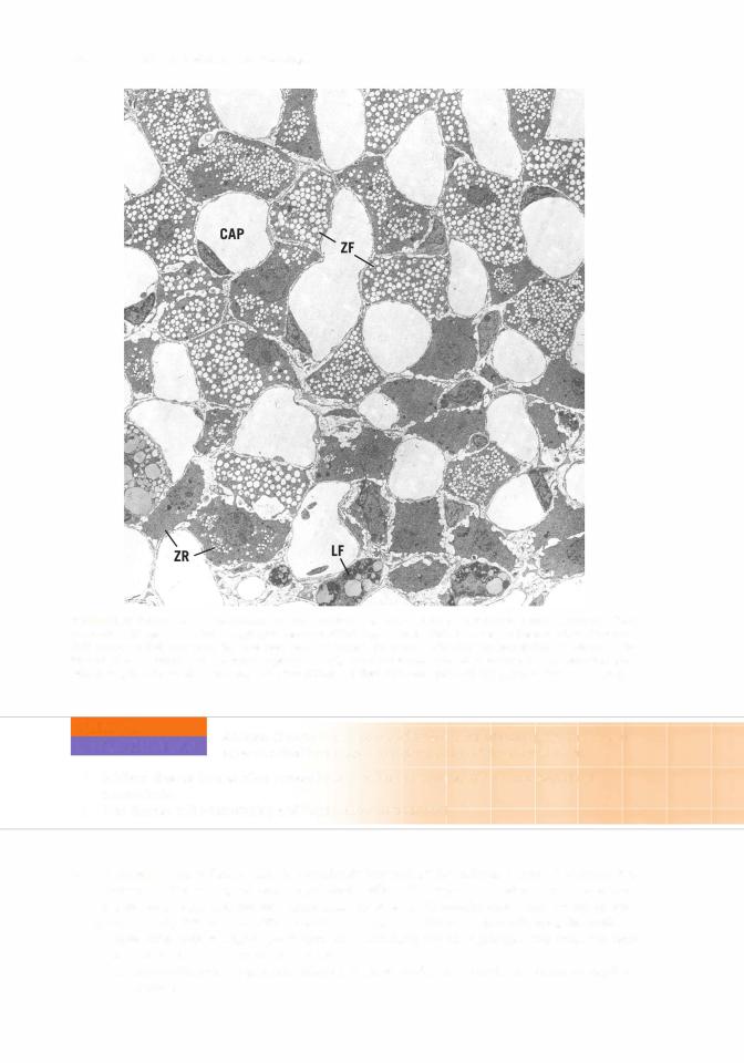
A. The adrenal cortex Table 13.3) contains parenchymal cells that synthesize and secrete but do not store various steroid hormones. The production of steroid hormones is dependent on a specific protein,( steroidogenic acute regulatory protein (StAR) that facilitates the transport of cholesterol across the outer membranes of mitochondria. The adrenal cortex is divided into three concentric histologically recognizable regions: the zona glomerulosa, zona fasciculata, and zona reticularis
Figure 13. 10).
1 . Zona glomerulosa
a.synthesizes and secretes mineralocorticoids, mostly aldosterone and some deoxycortico sterone. Hormone production is stimulated by angiotensin II and ACTH.
b.is composed of small cells arranged in arch-like cords and clusters. These cells have a few
small lipid droplets, an extensive network of smooth endoplasmic reticulum (SER), and mitochondria with shelf-like cristae( .
2.Zona fasciculata
a.synthesizes and secretes glucocorticoids, namely cortisol and corticosterone. Hormone production is stimulated by ACTH Figure( 13. 1 1). )
b.is composed ofcolumns( of cells and sinusoidal capillaries oriented perpendicularly to the capsule.
c.cells contain many lipid droplets and in tissue sections appear so vacuolated thatthey are called spongiocytes Figure 13. 12). These cells( also possess spherical mitochondria with tubular and vesicular cristae, SER, RER, lysosomes, and lipofuscin pigment granules.
3.Zona reticularis
a.synthesizes and secretes weak androgens mostly dehydroepiandrosterone and some
androstenedione) and perhaps small amounts( of glucocorticoids. Hormone production is stimulated by ACTH.
b. is composed of cells, arranged in anastomosing cords. Many large lipofuscin pigment granules are common in these cells Figure 13. 12) and are believed to represent lipid containing13.3 residues oflysosomal digestion.
t a b I e
Cell |
Hormone |
Function |
|
|
|
Adrenal cortex |
|
|
Zona glomerulosa |
Mineralocorticoids (mostly aldosterone) |
Regulate electrolyte, water balance via effect |
|
|
on cells of renal tubules |
Zona fasciculata |
Glucocorticoids (cortisol, corticosterone) |
Regulate carbohydrate metabolism by |
|
|
promoting gluconeogenesis; promote |
|
|
breakdown of proteins, fat; anti·inflammatory |
|
|
properties; suppress immune response |
Zona reticularis |
Weak androgens (dehydroepiandrosterone, |
Promote masculine characteristics |
|
androstenedione) |
|
Adrenal medulla |
|
|
Chromaffin cells |
Epinephrine |
Fight·or·flight response; increases heart rate |
|
|
and force of contraction; relaxes bronchiolar |
|
|
smooth muscle; promotes glycogenolysis |
|
|
and lipolysis |
|
Norepinephrine |
Little effect on cardiac output, rarely used |
|
|
clinically |
|
|
|

l!iitJ'!1tilUEndocrine System 237
b.They are innervated by preganglionic sympathetic (cholinergic) fibers, making these cells analogous in function to postganglionic sympathetic neurons.
c.They possess a well-developed Golgi complex, isolated regions of RER, and numerous mitochondria.
d.They also contain large numbers of membrane-bound granules containing one of the catecholamines, ATP, enkephalins, and chromogranins, which may function as binding proteins for epinephrine and norepinephrine.
2.Catecholamine release occurs in response to intense emotional stimuli and is mediated by the preganglionic sympathetic fibers that innervate the chromaffin cells.
A pheochromocytoma is a tumor arising in catecholamine-secreting chromaffin cells of the adrenal medulla. The tumor is rare; it is found in
both sexes, and 90% of the time it is benign. However, its secretion of excessive amounts of epi nephrine and norepinephrine leads to hypertension (episodic or sustained), although the patient may remain asymptomatic. Increased levels of catecholamines and their metabolites in the urine are diagnostic of pheochromocytoma. If the tumor is detected early and is surgically removed, the hypertension is correctable, but if not, prolonged and sustained hypertension may prove fatal.
C. Blood supplytothe adrenal glands is derived from the superior, middle, and inferior adrenal arteries, which form three groups ofvessels: to the capsule, to parenchymal cells of the cortex, and directly to the medulla.
1. Cortical blood supply
a.A fenestrated capillary network bathes cells of the zona glomerulosa.
b.Straight, discontinuous, fenestrated capillaries supply the zona fasciculata and zona reticularis.
2.Medullary blood supply
a.Venous blood rich in hormones reaches the medulla via the discontinuous fenestrated capillaries that pass through the cortex.
b.Arterial blood from direct branches of capsular arteries forms an extensive fenestrated capillarynetwork among the chromaffin cells of the medulla.
c.Medullary veins join to form the suprarenal vein, which exits the gland.
A. Overview (Figure 13.4)
1.The pineal gland projects from the roof of the diencephalon.
2.Its secretions vary with the light and dark cycles of the day, thereby regulating the individual's circadian rhythm. Although the pineal gland is buried deep within the head, it receives information about the light and dark conditions from special ganglion cells in the retina of the eye. These ganglion cells send their information about the presence of daylight via the retinohypothalamic tract that projects to the suprachiasmatic nucleus of the hypothalamus, from where information reaches the superior cervical sympathetic ganglion whose postganglionic sympathetic fibers reach the pineal gland by riding on the tunica adventitia of blood vessels that supply the pineal.
3.This gland has a capsule formed of the pia mater, from which septa (containing blood vessels and unmyelinated nerve fibers) extend to subdivide it into incomplete lobules.
4.It is composed primarily of pinealocytes, which constitute approximately 95% ofthe cells, and neuroglial cells (interstitial cells), which constitute about 5% ofthe cells.
5.It also contains calcified concretions (brain sand) in its interstitium. The function of these concretions is unknown, but they increase during short light cycles and decrease during periods of darkness.

238 |
BRS Cell Biology and Histology |
B.Pinealocytes are pale-staining cells with numerous long processes that end in dilations near capillaries.
1.Pinealocytes contain many secretory granules, microtubules, microfilaments, and unusual structures called synaptic ribbons.
2.These cells synthesize and immediately secrete melatonin but almost only at night. During the day, melatonin synthesis is mostly inhibited.
3.Pinealocytes may also produce arginine vasotocin, a peptide that appears to be an antagonist ofLH and FSH; they also secrete small quantities of serotonin, histamine, and dopamine. Most
of the serotonin manufactured by the pinealocytes is converted to melatonin in a two-step reaction, the first of which is catalyzed by the enzyme N-acetyltransferase. It is the activity of this particular enzyme that is inhibited during daylight conditions, thus preventing the formation of melatonin during daylight.
C.Neuroglial (interstitial) cells resemble astrocytes, with elongated processes and a small, dense nucleus. They contain microtubules and many microfilaments and intermediate filaments.
CLINICAL |
Melatonin is used to treat jet lag and seasonal affective disorder (SAD), |
|
CONSIDERATIONS |
||
an emotional response to shorter daylight hours during the winter. |
||
|

