Добавил:
Опубликованный материал нарушает ваши авторские права? Сообщите нам.
Вуз: Предмет: Файл:
BRS_Cell_Biology_and_Histology_7th_Edition_2015.pdf
Скачиваний:
138
Добавлен:
03.11.2017
Размер:
96.13 Mб
Скачать

Circulatory System

I. OVERVIEW-BLOOD VASCULAR SYSTEM

The blood vascular system consists ofthe heart, arteries, veins, and capillaries. This system transports oxygen and nutrients to tissues, carries carbon dioxide and waste products from the tissues, and circulates hormones from the site of synthesis to their target cells.

A.The heart is a four-chambered pump composed oftwo atria and two ventricles and is surrounded by a fibroserous sac called the pericardium.

Tetralogy of Fallot

Tetralogy of Fallot is a congenital malformation consisting of a defec­ tive interventricular septum, hypertrophy of the right ventricle (due to a narrow pulmonary artery or valve), and transposed (dextroposed) aorta. It should be repaired surgically early in life, before the pulmonary constriction becomes exacerbated.

B.Atria are receiving chambers, whereas ventricles are discharging chambers. Moreover, the heart is divided functionally into the right heart (pulmonary system) and the left heart (systemic system). The right atrium receives venous blood from the body and discharges it into the right ventricle. From here the blood is pumped into the pulmonary trunk to be distributed to the lungs. Venous blood from the pulmonary system is received in the left atrium and then discharged into the left ventricle to be pumped out through the aorta into the systemic circulation.

The heart receives sympathetic innervation via upper thoracic levels of the spinal cord and parasympathetic innervation via vagus nerves, which modulate the rate of the heartbeat but do not initiate it. The heart (mostly the atria) also produces atrial natriuretic peptide, and B-type natriuretic peptide, hormones that increases secretion of sodium and water bythe kidneys, inhib­ its renin release, and decreases blood pressure.

Sympathetic innervation accelerates the heart rate, increases the force of the heartbeat, and dilates the coronary vessels.

Parasympathetic innervation slows the heart rate, reduces the force of the heartbeat, and con­ stricts the coronary vessels.

1 . Cardiac layers (from internal to external)

a.Endocardium lines the lumen ofthe heart and is composed of simple squamous epithelium (endothelium) and a thin layer of loose connective tissue. Subendocardium, a connective tissue layer that contains veins, nerves, and Purkinje fibers, underlies it.

184

CLINICAL CONSIDERATIONS

l!1lEJ1l!DijCirculatory System

1 85

b.Myocardium consists of layers of striated cardiac muscle cells arranged in a spiral fashion about the heart's chambers and inserted into the fibrous skeleton. The myocardium contracts to propel blood into the arteries for distribution to the body.

c.Epicardium, the outermost layer of the heart, constitutes the visceral layer of the pericardium. It is composed of simple squamous epithelium (mesothelium) on the external surface. Beneath the mesothelium lies fibroelastic connective tissue, containing nerves, the coronary vessels, and adipose tissue.

2.The fibrous skeleton of the heart consists of thick bundles of collagen fibers oriented in various directions and especially oriented to support the fourvalve rings of the heart valves. It also contains occasional foci of fibrocartilage.

3.Heartvalves

a.Atrioventricular (AV) valves are composed of a skeleton of fibrous connective tissue, arranged like an aponeurosis, and lined on both sides by endothelium. They are attached to the annuli fibrosi of the fibrous skeleton. The right AV valve is formed of three interlocking cusps (tricuspid valve), whereas the left AV valve is formed of two interlocking cusps (bicuspid or mitral valve). These valves prevent regurgitation of ventricular blood into the atria.

b.Semilunar valves in the pulmonary and aortic trunks are each composed ofthree cusps that approximate each other as they fill with arterial blood. They are lined with endothelium on both sides separated by sparse strands of connective tissue. These valves prevent regurgitation of pulmonary and aortic blood into the respective ventricles.

CLINICAL

Rheumatic heartvalve disease is a sequel to childhood rheumatic fever (sub­

CONSIDERATIONS

sequent to streptococcal infection), which causes scarring of the heartvalves.


1.The disease is characterized by reduced elasticity of the heart valves, making them unable to close (incompetence) or unable open (stenosis) properly.

2.It most commonly affects the mitral valve, followed by the aortic valve.

4.The impulse-generating and impulse-conducting system of the heart comprises several specialized structures with coordinated functions that act to initiate and regulate the heartbeat.

a.The sinoatrial (SA) node, the pacemaker of the heart, is composed of specialized cardiac cells located within the wall of the right atrium adjacent to the entry of the superior vena cava into the heart. It generates impulses that initiate contraction of atrial muscle cells; which are then conducted to the AV node.

b.The AV node is located in the wall of the right atrium, adjacent to the tricuspid valve.

c.The AV bundle of His is a band of conducting tissue radiating from the AV node into the interventricular septum, where it divides into two branches and continues as Purkinje fibers.

d.Purkinje fibers are large, modified cardiac muscle cells (see Chapter 8 V B 9) that make contact with cardiac muscle cells at the apex of the heart via gap junctions, desmosomes, and fasciae adherentes.

e.As indicated (in Section I B of the current chapter), the autonomic nervous system modulates the heart rate and stroke volume.

Cardiac arrest occurs when the heart ceases to beat, resulting in cessation of blood flow in the entire circulatory system. Cardiac death

is imminenet unless cardiopulmonary resuscitation and defibrillation are conducted successfully and the electrical system of the heart is revived . Health conditions that may lead to cardiac arrest include, but are not limited to, tachycardia, fibrillation, and bradycardia.

t a b l e

11.1

1 86

BRS Cell Biology and Histology

Comparison of Tunicae in Different Types of Arteries

Tunica Components

Elastic Arteries

Muscular Arteries

Arterioles

Metarterioles

Intima


Endothelium

+

+

+

+

Factor VIII in endothelium

+

+

+


Basal lamina

+

+

+

+

Subendothelial layer*

+

+

±


Internal elastic lamina

Incomplete

Thick, complete

Some elastic fibers


Media


Fenestrated elastic membranes

40-70


Smooth muscle cells

Interspersed between

:s:40 layers

1 or 2 1ayers

Discontinuous


elastic membranes


layer

External elastic lamina

Thin

Thick


Vasa vasorum

±


Adventitia


Fibroelastic connective tissue

Thin layer

Thin layer


Loose connective tissue


+


Vasa vasorum

+


Lymphatic vessels

+

+


Nerve fibers

+

+

+


+ , present and prominent; ± , present but not prominent; -, absent.

*In elastic arteries. the subendothelial layer is composed of loose connective tissue containing fibroblasts. collagen. and elastic fibers. In arterioles. this layer is less prominent; the connective tissue is sparse and contains a few reticular fibers.

C.Arteries conduct blood away from the heart to the organs and tissues. Arterial walls are composed of three layers (tunicae): the tunica intima (inner), tunica media (middle), and tunica adventitia (outer). Components of these layers and variations among types of arteries are summarized in

Table 11 . 1 .

1 . TypesI of arteries

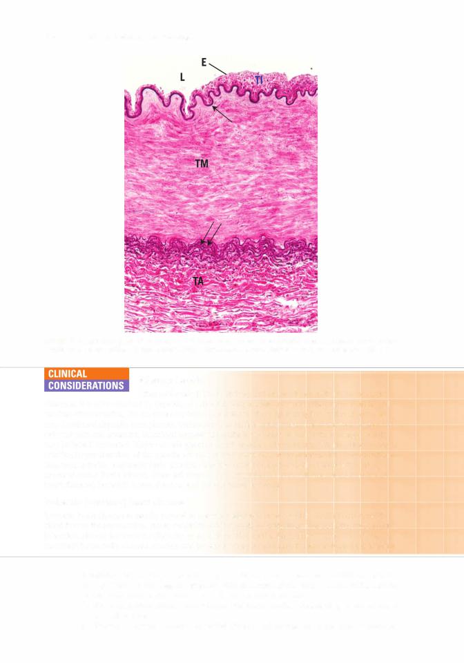
a.Elastic arteries (conducting arteries)are large and include the aorta andits major branches

(Figures 1 1 . 1 and 1 1 .2).

(1 Elastic arteries help reduce changes in blood pressure associated with the heartbeat.

(2)Small vessels (vasa vasorum) and nerves are located in their tunicae adventitia and

media. The vasa vasorum vascularizes the walls of the elastic arteries.

IThick, concentric sheaths of elastic membranes, known as fenestrated membranes, are located in the tunica media.

b.Muscular arteries (distributing arteries) distribute blood to various organs. (1 They include most of the named arteries of the human body.

(2) These medium-sized arteries are smaller than elastic arteries but larger than arterioles.

(3) The tunica adventitia contains vasa vasorum.

(4) The tunica media is thick, composed oflayers of smooth muscle cells. Larger muscular

arteries possess an external elastic lamina separating their boundary with the tunica adventitia (Figure 1 1 .3).

(51 The tunica intima is characterized by its endothelium and a prominent subendothelial internal elastic lamina.

FIGURE 11.1. Light micrograph of the aorta (elastic stain) ( X 132). Observe the wavy elastic fibers (arrows) located in the

tunica media (TM). Note the lumen (L) Iocated at the topL of the micrograph.

E

FIGURE 11 .2. A light micrograph of the aorta (with elastic stain) at a higher magnification ( X 270). Observe the endothelial layer (E) adjacent to the lumen (L), Note that the tunica media (TM) contains an abundance of elastic fibers (arrows).

1 87

Sinusoidal (Discontinuous) Capillary
Continuous Capillary
Fenestrated Capillary

1 90

BRS Cell Biology and Histology

Capillaries consists of a simple squamous epithelium rolled into a narrow cylinder 8 -1 0 11m in diameter. Continuous (somatic) capillaries have no fenestrae; material transverses the endothelial cell in either direction via pinocytotic vesicles. Fenestrated (visceral) capillaries are characterized by the presence of perforations, fenestrae, 60-80 11m in diameter, which may or may not be bridged by a diaphragm. Sinusoidal capillaries have a large lumen (30-40 11m in diameter), possess numerous fenestrae, have discontinuous basal lamina, and lack pinocytotic vesicles. Frequently, adjacent endothelial cells of sinusoidal capillaries overlap one another in an incomplete fashion.

FIGURE 1 1 .4. The& three types of capillaries. !Reprinted with permission from Gartner LP. Hiatt JL. Color Atlas of Histology. Baltimore. MD: Lippincott Williams Wilkins; 2009:1 63.)

l!1lEJ1l!DijCirculatory System

1 91

a.Continuous (somatic) capillaries contain numerous pinocytotic vesicles except in the central nervous system (CNS), where they contain only a limited number of pinocytotic vesicles (a property that is partly responsible for the blood-brain barrier).

(1 ) Continuous capillaries lack fenestrae and have a continuous basal lamina.

(2)They are located in nervous tissue, muscle, connective tissue, exocrine glands, and the lungs.

b.Fenestrated (visceral) capillaries are formed from endothelial cells that are perforated withfenestrae. These openings are 60 to 80 nm in diameter and are bridged by a diaphragm thinner than a cell membrane; in the renal glomerulus, the fenestrae are larger and lack a diaphragm.

(1 ) Fenestrated capillaries have a continuous basal lamina and few pinocytotic vesicles.

(2)They are located in endocrine glands, the intestine, the pancreas, and the glomeruli of kidneys.

c.Sinusoidal capillaries (sinusoids) possess many large fenestrae that lack diaphragms.

(1 ) Sinusoidal capillaries are 30 to 40 IJ,m in diameter, much larger than continuous and fenestrated capillaries.

(2)Sinusoidal capillaries have a discontinuous basal lamina and lack pinocytotic vesicles.

(3)Gaps maybe present at the cell junctions, permitting leakage between endothelial cells.

(4)They are located in the liver, spleen, bone marrow, lymph nodes, and adrenal cortex.

3.Permeability of capillaries is dependent on the morphology of their endothelial cells and on the size, charge, and shape of the traversing molecules. Permeability is altered during the inflammatory response by histamine and bradykinin.

a.Some substances diffuse, whereas others are actively transported across the plasma membrane of capillary endothelial cells.

b.Other substances move across capillary walls via small pores (intercellular junctions) or large pores (fenestrae and pinocytotic vesicles).

c.Leukocytes leave the bloodstream to enter the tissue spaces by penetrating intercellular junctions. This process is called diapedesis.

4.Metabolic functions of capillaries are carried out by the endothelial cells, and include the following (see Table 1 1 .2):

a.Conversion of inactive angiotensin I to active angiotensin II (especially in the lung). This powerful vasoconstrictor also stimulates secretion of aldosterone, a hormone that promotes water retention.

b.Deactivation of various pharmacologically active substances (e.g., bradykinin, serotonin, thrombin, norepinephrine, prostaglandins).

c.Breakdown of lipoproteins to yield triglycerides and cholesterol.

d.Release of prostacyclin, a potent vasodilator and inhibitor of intravascular platelet


aggregation.

e.

Release of the relaxing factor NO (nitric oxide) and contraction factor (endothelin 1).

f.

Regulation oftransendothelial migration ofinflammatory cells (neutrophils).

g.Release of tissue factors responsible for blood coagulation.

5.Blood flow to capillary beds occurs either from metarterioles (with precapillary sphincters) or from terminal arterioles (Figure 1 1 .5).

a.Central channels are vessels that traverse a capillary bed and connect arterioles to small venules. Their proximal portion is the metarteriole (possessing precapillary sphincters), and their distal portion is the thoroughfare channel (with no precapillary sphincter) (see Figure 1 1 .5).

b.Metarterioles supply blood to the capillary bed, whereas thoroughfare channels receive blood from capillary beds.

6.Arteriovenous shunt (bypassing a capillary bed)

a. Contraction of precapillary sphincters forces the blood flow from the metarteriole directly

into the thoroughfare channel, thus bypassing the capillary bed and draining into a postcapillary venule.

b.AV anastomoses are small vessels that directly connect arterioles to venules, bypassing the capillary bed. They function in thermoregulation, especially in the skin where they are abundant. These anastomoses also control blood pressure and flow.

1 92

BRS Cell Biology and Histology

t a b I e

11.2

Endothelial Cell Functions

Function

Principal Locations

Mode of Action

Permeability

Capillaries and

Paracellular;


postcapillary venules

Transcellular

Hemostasis

Throughout the

Release of tissue factor, von Willebrand

promotion

vascular system

factor, plasminogen-activator inhibitor,


and protease activated receptors

Hemostasis

Throughout the

Endothelial cells secrete and/or

inhibition

vascular system

express:Tissue pathway factor inhibitor;


thrombomodulin; heparin; plasminogen


activator; prostacyclin; nitric oxide; and


endothelial protein C receptor

Vasomotor tone

Arterioles

Vasodilatation: nitric oxide (NO').


prostacyclin


Vasoconstriction: endothelins 1, 2, and


3; prostaglandins; thromboxane A2; and


angiotensin I I

Thermoregulation

Microcirculation

The close proximity of capillary beds to


in the dermis and

the surface of the skin and the luminal


bronchial tree

bronchial surface permits heat exchange


with the external environment

Leukocyte

Postcapillary (high

L-selectins, chemokine receptor 7, and

trafficking

endothelial venules

lymphocyte-associated antigens of


[HEVs])

lymphocytes interact with peripheral


node addressin, chemokine ligand 21,


and intercellular adhesion molecules of


endothelial cells of HEVs which facilitate


the migration of lymphocytes into the


stroma of secondary lymphoid organs

Angiogenesis

Throughout the

Vascular endothelial growth factors


vascular system

released by endothelial cells and


angiogenic factors released by tumor cells


prompt other endothelial cells as well as


pericytes to proliferate and form endothelial


tubes thatwill eventually develop their own


tunicae media and adventitia

Angiotensin I I

Mostly in lung

ACE converts angiotensin I to the potent

formation

capillary beds

vascular smooth muscle contractant


angiotensin I I

Notations

Between cells;

Caveolae, VVO*, diapedesis

Weibei-Palade bodies house both von Willebrand Factor and tissue factor. If the integrity of the vascular endothelium is compromised, these factors are released in order to form a blood clot and stop the escape of blood from the vessel

These anticoagulants are always present in the normal, intact endothelium and prevent coagulation to ensure that blood remains in a fluid state

Vasodilator and vasoconstrictor molecules are released in response to local or systemic factors; angiotensin I I is produced by the conversion of angiotensin I by the endothelial cell membrane-bound ACE

Vasoconstriction reduces blood flow into the capillary beds, thus conserving body heat. Vasodilatation increases blood flow into capillary beds, thus releasing body heat

Similar method permits the migration of other leukocytes out of postcapillary venules (non- HEVs) during the inflammatory process

Repairs vascular damage during wound healing as well as providing for vascularization of growing tumors

Angiotensin I I elevates blood pressure, thereby increasing glomerular filtration rate and facilitates additional resorption of sodium from the ultrafiltrate

ACE. angiotensin-converting enzyme.

*WO-Vesicular-vacuolar organelles are believed to be aggregates of fused caveoli.

tNitric oxide (NO) was known as endothelial-derived relaxing factor (EORF); according to some authors, prostacyclin is also considered to be EDRF.

E.Veins conduct blood away from the organs and tissues and return it to the heart. Veins contain about 70% of the body's total blood volume at any given time. Their walls are composed of three layers: the tunica intima (inner), tunica media (middle), and tunica adventitia (outer), the thickest and most prominent. Vasa vasorum are more numerous in veins than in arteries. A distinct internal elastic lamina is also absent in veins. The components of these layers and the variations among different types ofveins are summarized in Table 1 1 .3.

1.Comparison with arteries. Veins have thinner walls and larger, more irregular lumina than the companion arteries. They may have valves in their lumina that prevent retrograde flow of the blood (Figure 1 1 .6).

CLINICAL CONSIDERATIONS

1 94

BRS Cell Biology and Histology

FIGURE 11 .6. Light micrograph of an artery and vein ( X 270l. Vein-lumen (LVI, tunica media (TMVI, and tunica adventitia (TAVI. Artery-tunica adventitia (TAAI, tunica media (TMAI. and lumen (LAI.

b. Small and mediuml -sized veins includel the external jugular vein. These veins have a diameter of to 9 mm.

c. Venules have a diameter of 0.2 to mm and are involved in exchange of metabolites with tissues and in diapedesis (exiting of blood cells through vessel walls).

Varicose veins are abnormally tortuous dilated veins, usually of the leg. 1. They are caused by a decline in muscle tone, degenerative alteration

of the vessel wall, and valvular incompetence. They generally occur in older people, being most prevalent in women. Pregnant women are also susceptible to varicose veins.

2.When they occur in the region of the anorectal junction, they are known as hemorrhoids.

F.Specialized sensory mechanisms within arteries include three types of sensors: the carotid sinus, the carotid body, and the aortic bodies.

1.Carotid sinus is a baroreceptor in the wall ofthe internal carotid arteryjust as it begins at the common carotid artery. Sensory endings of the glossopharyngeal nerve embedded with the wall of the artery are sensitive to changes in pressure that distend the vessel, thus initiating a signal to the vasomotor center of the brain. The resultant response triggers adjustments to the tension on the arterial wall via the smooth muscles of the tunica media, effecting changes in

2.Carotid body is a chemoreceptor at the bifurcation of the commonl)carotid artery. Specialized nerve endings of the vagus and glossopharyngeal cranial nerves are sensitive to oxygen and carbon dioxide levels as well as H+ concentration. Glomus (type cells and sheath (type 2)

l!1lEJ1l!DijCirculatory System

1 95

cells, which envelop the glomus cells, constitute the parenchyma. Nerve endings lose their Schwann cells upon entering the parenchyma, becoming covered instead by sheath cells. Impulses are shuttled to the brain by these two cranial nerves for processing.

3.Aortic bodies are located in the wall of the arch of the aorta at the junction of the common carotid and subclavian arteries. Their structure and function are similar to those of the carotid body.

4.Hormonal control of low blood pressure starts with the kidney:

a.Kidney produces renin

b.Renin cleaves angiotensinogen circulating in the blood, forming angiotensin I, a mild vasoconstrictor.

c.Angiotensin I is converted into angiotensin II by angiotensin-converting enzyme (ACE), located on the luminal plasmalemmas of capillary endothelia (especially capillaries of the lungs).

d.Angiotensin II, a potent vasoconstrictor, causes the walls of arterioles to contract, which raises blood pressure.

e.Antidiuretic hormone, or vasopressin, secreted by the pituitary gland, is another potent vasoconstrictor employed after severe hemorrhage.

This system consists of peripheral lymphatic capillaries, lymphatic vessels of gradually increasing size, and lymphatic ducts. The lymphatic vascular system collects excess tissue fluid (lymph) and returns it to the venous portion ofthe cardiovascular system. It drains most tissues with the exception of the nervous system and bone marrow.

A.Lymphatic capillaries are thin-walled vessels that begin as blind-ended channels (e.g., lacteals) adjacent to capillary beds where they collect lymph.

1.They are composed of a single layer of attenuated endothelial cells that lack fenestrae and fasciae occludentes. They possess a sparse basal lamina.

2.Lymph enters these leaky capillaries via spaces between overlapping endothelial cells.

3.Small lymphatic anchoring filaments between the surrounding connective tissue and the abluminal plasma membrane assist in maintaining luminal patency in these delicate vessels.

B.Large lymphatic vessels possess valves and are similar in structure to small veins, except that they have larger lumina and thinner walls.

1.Lymph nodes that filter the lymph are interposed along their routes.

2.These vessels converge to form the thoracic duct and right lymphatic duct. The thoracic duct empties into the venous system at the junction of the left internal jugular vein with the subclavian vein, whereas the right lymphatic duct empties into the venous system at a similar location on the right side of the neck.

CLINICAL

Edema is a pathologic process resulting in an increased volume of tissue

CONSIDERATIONS fluid.

Edema may be caused by venous obstruction or decreased venous blood flow (as in conges­ tive heart failure), increased capillary permeability (due to injury), starvation, excessive release of histamine, and obstruction of lymphatic vessels. It is common during pregnancy and in older persons. Edema that is responsive to localized pressure (i.e., depressions persist after release of pressure)

is called pitting edema. Edema can be a symptom of a serious underlying disorder, including heart disease; liver disease; or diseases of the thyroid, lymphatic system, or the kidneys, with serious consequences.

Review Test

Directions: Each of the numbered items or incomplete statements in this section is followed by answers or completions of the statement. Select the ONE lettered answer that is BEST in each case.

1 . The epicardium is one ofthe three layers of the heart. It is

(A)continuous with the endocardium.

(B)also known as the visceral pericardium.

(C)composed of modified cardiac muscle cells.

(D)capable of increasing intraventricular pressure.

(E)capable of decreasing the rate of contraction.

2.The atrial muscle ofthe heart produces a hormone that

(A)decreases blood pressure.

(B)increases blood pressure.

(C)causes vasoconstriction.

(D)facilitates the release of renin.

(E)facilitates sodium resorption in the kidneys.

3.The generation of impulses in the normal heart is the responsibility ofwhich of the fol­ lowing structures?

(A)Atrioventricular (AV) node

(B)AV bundle of His

(C)Sympathetic nerves

(D)Sinoatrial (SA) node

(E)Purkinje fibers

4.Metarterioles, vessels interposed between arterioles and capillary beds,

(A)function to control blood flow into arterioles.

(B)possess a complete layer of smooth muscle cells in their tunica media.

(C)possess precapillary sphincters.

(D)receive blood from thoroughfare channels.

(E)possess valves to regulate the direction of blood flow.

5. Which of the following statements concern­ ing innervation of blood vessels is true?

(A)Vasoconstriction is controlled by parasym­ pathetic nerve fibers.

(B)Acetylcholine acts directly on smooth muscle cells.

(C)Acetylcholine acts directly on endothelial cells.

(D)Vasodilation is controlled by sympathetic nerve fibers.

(E)Nitric oxide acts as a vasoconstrictor.

6.Which of the following characteristics dis­ tinguishes somatic capillaries from visceral capillaries?

(A)Presence or absence of fenestrae

(B)Size of the lumen

(C)Thickness of the vessel wall

(D)Presence or absence ofpericytes

(E)Thickness of the basal lamina

7.The blood-brain barrier is thought to

exist because capillaries in the central ner­ vous system have which of the following characteristics?

(A)Discontinuous basal lamina

(B)Fenestrae with diaphragms

(C)Fenestrae without diaphragms

(D)A few pinocytic vesicles

(E)No basement membrane

8.Which of the following statements about healthy, intact capillaries is true?

(A)They control blood pressure.

(B)They are lined by a simple columnar epithelium.

(C)They have a smooth muscle coat.

(D)They inhibit clot formation.

(E)Satellite cells share their basal lamina.

1 96

9.A patient complains of shortness of breath even after only mild exercise. She states that she has had this condition for 2 years but re­ cently has noticed that it has become more pronounced. Her medical history indicates that she had rheumatic fever when she was a child. Auscultation indicates an enlarged heart. What may the physician expect to find with other diagnostic tests?

(A)Mitral valve stenosis

(B)Tetralogy of Fallot

(C)Pulmonary artery aneurysm

(D)Coronary heart disease

(E)Ischemic heart disease

10.Diagnostic tests for ischemic heart disease usually reveal

(A)malformed heart valves.

(B)atherosclerosis of coronary arteries.

(C)irregular heartbeat.

(D)faulty SA valve.

(E)arteriosclerosis of coronary arteries.

l!iltl'115IICirculatory System

197

11 . Vasa vasorum function in a way that is similar to

(A)AV valves.

(B)semilunar valves.

(C)coronary arteries.

(D)elastic arteries.

(E)metarterioles.

12. Which one of the following possesses a distinct internal elastic lamina?

(A)Capillary

(B)Metarteriole

(C)Arteriole

(D)Muscular artery

(E)Vein

13. Ischemic heart disease is the usual sequel to

(A)arteriosclerosis.

(B)abdominal aortic aneurysm.

(C)rheumatic fever.

(D)varicose veins.

(E)atherosclerosis.

Answers and Explanations

1 . B. The pericardium is a fibroserous sac that encloses the heart. The innermost layer of the peri­ cardium, the epicardium, is also known as the visceral pericardium (see Chapter 1 1 I A 1).

2.A. Atrial natriuretic peptide, which decreases blood pressure, is produced mainly by car­ diac muscle cells of the right atrium. It inhibits the release of renin and causes the kidneys to decrease the resorption of sodium and water (see Chapter 1 1 I A 1 b).

3.D. Impulses are generated in the sinoatrial node, which is the pacemaker of the heart. They are then conducted to the AV node. The bundle of His and Purkinje fibers conduct impulses from the AV node to the cardiac muscle cells of the ventricles. Sympathetic nerves can increase the rate of the heartbeat but do not originate it (see Chapter 1 1 I A 4 a).

4.C. The proximal portion of a central channel is known as a metarteriole, whereas its distal portion is the thoroughfare channel. Blood from metarterioles may enter the capillary bed if their precapillary sphincters are relaxed. If the precapillary sphincters of metarterioles are con­ stricted, blood bypasses the capillary bed and flows directly into thoroughfare channels and from there into a venule (see Chapter 11 I B 1 d).

5.C. Acetylcholine stimulates the endothelial cells of a vessel to release nitric oxide (EDRF), which causes relaxation of smooth muscle cells. Thus, acetylcholine does not act directly on smooth muscle cells (see Chapter 1 1 I B 2).

6.A. Somatic (continuous) capillaries lack fenestrae, whereas visceral (fenestrated) capillaries are characterized by their presence. Both types of capillary possess a continuous basal lamina and are surrounded by occasional pericytes (see Chapter 1 1 I C 2).

7.D. Capillaries in the CNS are ofthe continuous type and thus lack fenestrae but have a continu­ ous basal lamina. In contrast to continuous capillaries in other parts of the body, they contain only a few pinocytic vesicles; this characteristic is thought to be partly responsible for the blood-brain barrier (see Chapter 1 1 I C 2).

8.D. The smooth endothelial lining of intact, healthy capillaries inhibits clot formation. Capillaries do not control blood pressure (see Chapter 11 I C 2).

9.A. A person who has had rheumatic fever as a child may develop heart valve disease later in life. Although the mitral valve is the one most commonly affected, the other valves may also be involved. The mitral valve becomes inflamed, fibrotic, and eventually incompetent or ste­ notic. This condition leads to respiratory hypertension and edema, which restricts respiratory function. The key is a history of rheumatic fever, because those individuals are predisposed to developing heart valve diseases (see Chapter 1 1 I A 3 Clinical Considerations).

10.B. Diagnostic tests for ischemic heart disease reveal atherosclerosis ofthe coronary vessels. Over time, excessive plaque composed of cholesterol and fats is layered beneath the intima of these vessels, thus restricting blood flow to the myocardium ofthe heart, leading to angina pec­ toris, an infarction, or perhaps sudden death (see Chapter 1 1 I B 1 Clinical Considerations).

11 . C. Vasa vasorum are the small blood vessels that serve the walls of elastic and muscular arter­ ies with oxygen and nutrients just as the coronary arteries provide for the walls of the heart (see Chapter 11 I B 1).

12.D. The muscular artery possesses a distinct internal elastic lamina. Elastic arteries possess an incomplete internal elastic lamina, whereas capillaries, arterioles, metarterioles, and veins do not possess an internal elastic lamina (see Table 1 1 . 1 and Chapter 1 1 I D).

13.E. Ischemic heart disease is usually caused by coronary atherosclerosis, resulting in decreased blood flowto the myocardium. It may develop into angina pectoris, myocardial infarction, chronic ischemic cardiopathy, or sudden cardiac death. On the other hand, arteriosclerosis (hardening ofthe arteries) usually attacks renal arteries and is related to hypertension and diabetes mellitus (see Chapter 1 1 I B Clinical Considerations).

198

Соседние файлы в предмете Гистология