


Advanced Dissections |
79 |
D. Orbital Decompression (Figures 48–50)
Orbital decompression may be indicated for a patient with an orbital abscess, periorbital or orbital hematoma, or severe Graves’ ophthalmopathy with exposure keratitis and threatened visual loss (69–79). When a subperiosteal abscess is present, only the lamina papyracea needs partial or complete removal to ensure adequate drainage of the abscess loculations into the nose. This may require exposing the periorbita over the superomedial or inferomedial orbital walls to ensure adequate drainage of all potential abscess loculations. Nasal packing is usually avoided. The periorbita is left intact.
For patients with Graves’ ophthalmopathy, the lamina papyracea and MOF are removed medial to the infraorbital nerve through a wide antrostomy. Postoperative diplopia is possible, but may be minimized by preserving the horizontal ridge of the antrostomy (80). The periorbita is incised to allow herniation of orbital fat into the ethmoid and maxillary sinus cavities. The latter generally allows for approximately 4–5 mm of orbital decompression. This may have to be combined with a lateral orbital decompression through an external approach. Care is taken not to occlude the maxillary, frontal, or sphenoid ostia with orbital fat, as this may result in secondary ostial obstruction and rhinosinusitis (81,82). In these situations an extended middle meatal, frontal, and/or sphenoid sinusotomy may be prudent.
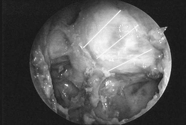
Figure 48 Endoscopic view showing careful elevation and piecemeal removal of lamina papyracea (arrows) away from the periorbita (P). Periorbital abscesses and hematomas occur in this space.

80 |
Chapter 4 |
(a)
Figure 49 Sagittal (a) and endoscopic (b) views denoting longitudinal incisions for orbital fat decompression as performed for Graves’ ophthalmopathy.
Advanced Dissections |
81 |
(b)
82 |
Chapter 4 |
Figure 50 Endoscopic view with orbital fat extending into the ethmoid cavity.
Advanced Dissections |
83 |
E.Optic Nerve Decompression and the Carotid Artery (Figure 51)
In patients with worsening visual acuity due to traumatic neuropathy or neoplastic compression, an optic nerve decompression may be indicated (83–86). The orbital apex may be found by following a line from the superior vertical ridge of the antrostomy to the roof of the posterior ethmoid sinus adjacent to the orbital wall. It is located at the same level as the posterior wall of the maxillary sinus in the coronal plane, and approximately 7 cm from the columnella. The canalicular portion of the optic nerve is identified as it takes an abrupt turn medially at this point as it courses toward the optic chiasm. The thicker bone in this area is carefully thinned with a diamond bur and removed with a periosteal elevator. In the laboratory this can be carefully performed utilizing a bone curette. The length of the canalicular portion is approximately 8–12 mm. The optic nerve sheath is continuous with the dura mater in this area. Incision of this thick sheath reveals the optic nerve. The space around the nerve is continuous with the subdural space and results in a CSF leak if left open to the nasal cavity. Therefore, if the optic nerve sheath is opened, one must be prepared to close the CSF leak with a small mucoperichondrial graft.
The intrasphenoid carotid artery runs in a posteroinferior to anterosuperior direction, giving it the appearance of an inverted S. Its most anterior (cavernous) segment runs immediately inferior to the canalicular portion of the optic nerve as it courses intracranially, creating a small triangular recess (opticocarotid recess). In some patients with a well-pneumatized sphenoid, the carotid projects into the lumen of the sphenoid and is prone to inadvertent injury if one enters too far laterally through the posterior wall of the posterior ethmoid sinus. For this reason the sphenoid is generally entered medially adjacent to the septum, as previously described.

84 |
Chapter 4 |
(a)
Figure 51 Sagittal (a) and endoscopic (b) views showing the canalicular portion of the optic nerve (ON) and intrasphenoid carotid artery (CA) after bone removal. Asterisks denotes the opticocarotid recess. The area of the anulus of Zinn (dotted oval) and the antrostomy ridge (small arrows) are shown.

Advanced Dissections |
85 |
(b)

86 |
Chapter 4 |
F.Orbital Dissection (Figures 52 and 53)
Removal of the periorbita and fat reveals the medial rectus muscle coursing along the medial wall of the orbit. The medial aspects of the inferior rectus muscle may be seen running adjacent to the horizontal ridge of the antrostomy. Further removal of fat between the two muscles reveals the orbital segment of the optic nerve and globe. The anulus of Zinn, representing the thick fibrotic insertion point for all the extraocular muscles, can be identified after removing bone at the orbital apex. By longitudinally transecting the anulus of Zinn, between the medial and inferior rectus muscles, one can expose the entire course of the intraorbital and canalicular segments of the optic nerve.
(a)
Figure 52 Sagittal (a) and endoscopic (b) views after removal of the periorbita and fat, showing the medial rectus muscle (MR), inferior rectus muscle (IR), and globe (G). The anulus of Zinn is circled. The optic nerve sheath has been opened to reveal the canalicular portion of the optic nerve (O). C = intrasphenoid carotid artery.

Advanced Dissections |
87 |
(b)

88 |
Chapter 4 |
Figure 53 Sagittal view after transection of the anulus of Zinn, revealing the orbital (small arrows) and canalicular (large arrows) segments of the optic nerve.