Методичні розробки 3 модуль
.pdfMinistry of health Ukraine
Higher state educational establishment of Ukraine
«Ukrainian medical stomatological academy»
It is «ratified» at meeting of chair of surgical stomatology and maxillofacial surgery with plastic and reconstructive surgery of the head and neck
The Head of the chair doctor of medicine Aveticov D. S. 
METHODICAL INSTRUCTION
FOR INDEPENDENT WORK OF STUDENTS DURING PREPARATION FOR PRACTICAL
(SEMINAR) LESSON
Names of the discipline |
Surgical stomatology |
||
|
Module № |
|
3 |
Thematic module № |
3 |
||
Theme of lesson |
Odontogenic epithelial cysts of the jaws: follicular cyst, radicular |
||
|
|
|
cyst, paradental, eruption cyst, primordial cyst, gingival cyst of |
|
|
|
adults, tooth-containing.Clinical presentation, diagnostics, |
|
|
|
differential diagnostics, treatment‖. |
Course |
IV |
||
Faculty |
Stomatological |
||
Poltava – 2012
1. SUBJECT URGENCY.
Odontogenic epithelial cysts of the jaws borrow one of the big problems in stomatology. For research of this problem it is important to analyse supervision which concern not rare cases of such diseases, and to trace behind their features on a plenty of patients which were on treatment in stomatologic clinics.
2.SPECIFIC GOALS:
2.1.To analyze etiological factors of odontogenic cysts of the jaws.
2.2.To explain a clinical picture of odontogenic cysts of the jaws.
2.3.To offer ways of avoidance of complications during treatment of odontogenic cysts of the
jaws.
2.4.To classify of odontogenic cysts of the jaws.
2.5.To treat radiological researches of patients with odontogenic cysts of the jaws.
2.6.To draw circuits of a radiological picture and localization dontogenic cysts of the jaws, operative interventions are cystostomy and cystectomy.
2.7.To analyse advantages and lacks different osteoplastic materials for filling defects of jaws after removal of odontogenic cysts.
2.8.To make the plan of examination and treatment of the patient with odontogenic cysts of jaws.
3. BASIC LEVEL OF PREPARATION.
Names of the previous disciplines |
The received skills |
|
1. |
Human anatomy. |
To know anatomy of jaws, blood supply and how innervate |
|
|
muscles of a head and a neck. To describe the anatomic region |
|
|
(localization) in maxillofacial area. |
2. |
Histology and |
To know a histologic structure and morphological structure of the |
pathomorphology. |
pathogenesis changed tissues. To distinguish pathological tissues. |
|
|
|
To be able to take a material for morphological researches. |
3. |
Pathological physiology |
To know aetiology and pathogenesis of diseases, a metabolism in |
|
|
pathological tissues. To define aetiology and pathogenesis of |
|
|
odontogenic epithelial tumours. |
4. |
General surgery |
To know methods of processing of hands of the surgeon. To be |
|
|
able to suture of soft tissues. |
4. TASKS FOR INDEPENDENT WORK DURING PREPARATION FOR LESSON.
4.1. The list of the main terms, parameters, characteristics which the student should know during preparation for lesson:
|
The term |
Definition |
1. |
Cyst. |
This formation with capsule and a liquid inside. |
2. |
Cystostomy |
Section of the cyst. |
3. |
Cystectomy |
Removal of the cyst. |
4.2. Theoretical questions for lesson:
1.To give a general characteristic odontogenic epithelial cysts.
2.What kind of tumours are odontogenic epithelial cysts of the jaws?
3.Complications which arise when we have odontogenic epithelial cysts. To list them.
4.1. Practical task which we make during lesson:
1.To make examination of the patient with odontogenic cysts of jaws.
2.To pick out the toolkit for operative intervention cystostomy and Cystectomy.
3.To choose the optimum way of anesthesia.
5. ORGANIZATION OF THE MAINTENANCE OF THE TRAINING MATERIAL.
Cyst – a hollow in the formation of bone with a dense shell and filled with fluid. With the penetration of infection into the bone, damaged cells die and the cavity is formed. To prevent further

spread of the inflammatory process, the shell is formed around it, which is essentially a protective mechanism. Unfortunately, it does not stop the cyst from further growth. Cyst filled with inflammatory exudate – exudate of various constituent elements of the blood vessel walls due to violations of their permeability. It contains a large number of white blood cells (which are responsible for inflammation) and dead cells of the bone.
Size of cysts can vary from 0.5 cm to 4.3 cm and a cyst diameter of less than 0.5-0.7 cm kistogranulemami called, they are a consequence of chronic granulomatous periodontitis. According to X- ray study of the cyst is a hotbed of enlightenment, with clear margins, round or oval in shape with a diameter greater than 0.5-1 cm This focus has always darken the rim bordering the contours of the cyst – a bone, limiting the inflammatory focus. In festering cysts contours become ―blurred.‖
On the localization of the cyst differentiate anterior teeth, wisdom teeth and dental cyst in the maxillary sinus.
For reasons of odontogenic (related to the teeth) cysts can be inflammatory and inflammatory.
Inflammation:
Radicular (periradicular). The most common are located at the top of the causative tooth. Is the latest step in the development of chronic periodontitis.
Residual cysts: radicular cysts remaining after the tooth they involved has been removed, thus resulting isolated in the bone.Make up about 30% of radicular cysts, they remain after the removal or loss of the causal teeth. Parodentalnye (retromolar). Cysts arise in the complicated eruption of wisdom teeth, with low intensity inflammation.
No inflammatory:
Follicular. The cysts contain a germ of a permanent tooth is not infected with the treatment of inflammatory diseases of the teeth. Sometimes a distinction has zubosoderzhaschie cysts, which contain the germ of the tooth supercomplete
Keratokisty (primary) – are formed from zuboobrazovatelnoy tissue is a rare type of cyst, its formation is characterized by multiple, often on the lower jaw. Detected by chance. Diagnosis can be made on the basis of histological examination.
Cysts of the eruption. In children with erupting permanent teeth, gums appear bulging above the crown of the tooth with a cavity formation and accumulation of serous fluid, or it hemorrhagic (exudate can also be purulent).
The main reason for the formation of cysts – a late and inadequate treatment of dental caries is not complicated and complicated forms, to the tooth trauma, periodontal disease, and nasopharynx, malformations of the teeth.
When one of these factors, or when a combination of several, begins to develop cysts. The danger of cysts in the fact that it grows slowly, imperceptibly. Usually there are no complaints of pain. There may be a causative tooth discoloration, percussion it is usually painless but can cause discomfort. With a significant amount of cyst patients complain of a person deformation, displacement of teeth. On palpation of the alveolar process of deformation is detected symptom of ―parchment crunch‖ and the spring cyst wall. The reaction of regional lymph nodes are usually absent, and manifests itself with festering cysts. That festering cysts with fistulas appear that tell cyst cavity with the oral cavity. Despite the pronounced manifestations of intoxication and subjective deterioration of general condition of the patient with festering cysts, constant level of intoxication at the same festering and nenagnoivshihsya cysts. That speaks about the dangers of both.
By reducing the general or local immunity of the oral cavity can accelerate the growth of cysts. If you do not immediately remove the cyst, it begins to grow, or suppurate, as the body can not cope with inflammation. To diagnose a cyst can only be X-ray study, so you should visit the dentist regularly. Puncture of the cyst makes for suspected malignancy of her.
With increasing growth of the cyst may appear general and local complications.
Heavy common complication – this intoxication of the organism, it is caused by the fact that the fall in blood products of microorganisms, so that may be deterioration of the general condition of the patient – pyrexia, headache. All this can lead to sepsis (blood poisoning overall).
What is a Cystectomy?A cystectomy is one possible therapy of a cyst. Cystectomy means the removal of a cyst, i.e. the scraping out of the cyst cavity.
A cyst is a tissue cavity that is enclosed by a membrane (epithelium) and may consist of several chambers, usually containing a liquid/mushy content.

In general, the cystectomy is the therapy of choice for cysts in the area of the head/neck. With the exception of very large cysts or if, for example, important anatomical structures are located in close proximity of the cyst, which could be damaged during the removal of a cyst, a so-called, "Cystostomy".
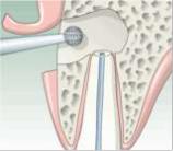
The root canal treatment was renewed prior to the operation in order to prevent any further cyst formation after the cystectomy. The mucous membrane has been opened up and the thin bone lamella located above the cyst is removed. Now, the cyst is scraped with a sharp spoon. After the scraping of the cyst cavity, the wound is primarily closed.
Folicular Cyst
Normally, the resulting bone defect fills with blood. Within the course of the healing of the wound, vessels grow back into the developing blood clot, followed by the subsequent development of new bone. In general, the removal of larger cysts carries a certain risk of wound healing disorders, because the removal of the cyst creates a large bone defect that normally fills with blood immediately after the surgery.
As the blood dries, the clot shrinks. A large blood clot contracts more than a small one; it may contract to such a degree that it no longer touches the walls of the wound.In such a case, it is not possible for blood vessels to grow from the walls into the blood clot. This prevents the clot from being supplied with oxygen, nutrients, and finally, with bone cells – which are an important prerequisite for the regeneration of the bone.
Consequently, the blood clot disintegrates – pus develops and a wound infection results. In order to avoid these complications in large cysts, one could attempt to stabilize the blood clot and to reduce its contraction, for example, by filling the bone defect with a granulate made of bone substitute materials. This prevents a contraction of the blood clot, allowing vessels to grow in from the walls – the basis for a subsequent bone regeneration.
Cystectomy
An alternative to the planned surgery would be the initial performance of a cystostomy followed by a cystectomy. If left untreated, cysts in the area of the mouth, jaw, and face usually grow in size over the years, sooner or later leading to the corresponding local complications.
The risks of the surgery are negligible when performed by an experienced surgeon; nevertheless, complications may occur in individual cases, possibly requiring additional measures. Every additional measure may in turn lead to further complications which, in the course of the surgery, could become lifethreatening. At this point, we will only discuss the specific complications encountered in the cystectomy. These are, for example:
Injury to surrounding structures such as nerves, cheeks, blood vessels, dental roots, and teeth with the respective consequences
An accidental cystectomy of malignant tumors that should be removed while employing a safety margin, wound infections, a fracture of the jaw, leaving parts of a cyst behind, which may result in a recurrence of the cyst.
Luckily, such complications have become very rare due to the positive developments in medicine in the last decades.
Cystectomy

Cyst removal
Wound closure
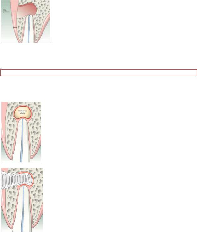
A cystectomy is the complete surgical removal of a small cyst with subsequent wound closure. The cyst is completely removed after a mucosa incision. This operation can also be combined with a root tip resection. The mucosa above the bone defect is then tightly sealed. New cysts may arise from any cyst residues.
Cystostomy
A cystostomy as a treatment option for larger cysts enables sparing of adjacent roots and nerves. Mucosa and bone are opened above the cyst. A window is cut along the bone opening, the actual cyst remains in the bone. This cavity is tamponaded to remain open and is thus formed to create an adjacent oral cavity bay. As the opening does no longer allow the cyst to grow, this bone cavity becomes ever smaller with the passing of time.
Tooth with radicular cyst
Tamponaded open cavity after cyst removal (cystostomy)

Gradual flattening of the bone defect
Follicular cyst of 38
Complete bone regeneration following cystostomy with extraction of 36, 37 and the crowded 38
Of the total cases of maxillary sinusitis, approximately 10–12% is exclusively sinusitis of the home tooth (Brook, 2006; Costa et al. 2007). The close relationship between the roots of the maxillary posterior teeth and the floor of the sinus makes the infection in these pieces directly affect the integrity sinus. Here are five ways that allow the injury of the maxillary sinus:
1)Periapical granuloma, which produces the effect of infectious core areas of the sinus;
2)Instruments beyond the apex via dental canal;
3)Marginal, by periodontal disease;
4)Apical lesion, granuloma, osteitis, or cyst; and
5)Surgery, for drilling and bucosinusal communication.
6.MATERIALS FOR SELF-CHECKING:
A. Tasks for self-checking ( diagrams, figures, the schedule):
1.Scheme of the operative interventions cystostomy and cystectomy.
2.X-rays of patients with odontogenic cysts.
3.Photos of patients with odontogenic cysts of the jaws.
B. Tasks for self-checking:
1. At X-rays examination in a projection of a top of a root 27 teeth is observed destruction a bone tissue of the round form with precise equal edges in the size 0,7х0,7 sm.
Diagnose.
(Answer: Cystogranuloma).
2.At the review of the patient deformation of the alveolar shoot of the maxilla in the region between 22,
24teeth. 23 tooth is absent. Transitive fold within the limits of these teeth changed, a mucous membrane of light pink color, palpation - a dense consistence, not painful. X-rays diagnostic - 22, 24 teeth is marked destruction a bone tissues roundish forms with precise equal borders. In this projection of destruction is crown of the tooth.
Diagnose.
(Answer: Follicular cyst)
3.The man of 35 years has addressed with complaints to a thickening of an alveolar shoot of the maxilla. The previous diagnosis: radicular cyst of the maxilla.
What will be revealed during a puncture of the alveolar shoot in area of "thickening"? (Answer: An yellowish liquid)
C. Materials for test control. Tasks with an individual right answer (ɑ = ІІ):
1.What name has alveolar cyst in other sources?
A.Pearl ‗Епштейна ‗.
B. Adamantinoma. C. Subperipstal cyst. D. Paradental cyst. E. Follicular киста.
(the right answer: А)
2.When appears paradental cyst?
A.In old age.
B.At young age.
C.On a toothless jaw.
D.At babies.
E.At teenagers.
(the right answer: В)
3. Eruption cyst clinically always:
A.On аpical parts of a tooth.
B.Between a teeth.
C.Under a tooth.
D.In a body of a jaw.
E.In a branch of a jaw.
(the right answer: С)
D. Educational tasks of 3-rd level (atypical tasks):
1.At patient К., 34 years, the diagnosis radicular cyst of the maxilla which intergrow in sinus. The name of operative intervention with this pathology is?
(the answer: sinusotomy, maxillary (antrotomy); radical (Caldwell-Luc) with removal of antrochoanal polyps).
2.In clinic mum of the baby of 6 months has addressed. At the child the diagnosis - eruption cyst is established. What tactics of the doctor.
(the answer: To explain to mum, that the pathology does not demand treatment).
3.At the patient, 34 years, it is revealed paradental cyst in the corner of the mandible at the left side up to
5sm in diameter. What possible complication during its removal may be? (the answer: fracture of the jaw.).
7. Literature:
7.1. Basic literature:
1.Бернадський Ю.Й. Основи щелепно-лицевої хірургії і хірургічної стоматології. К.
Спалах, 2003.- 512 с.
2.Маланчук В.О., Копчак А.В. Доброякісні пухлини та пухлиноподібні ураження щелепнолицевої ділянки та шиї / Навчальний посібник. – К.: Видавничий дім «Асканія», 2008. – 320 с.
3.Руководство по хирургической стоматологии и челюстно-лицевой хирургии: В 2-х темах. / Под ред. В.М.Безрукова, Т.Г.Робустовой. - Изд. 2-е, перераб. и доп. - М.: Медицина, 2000. – 776 с.
4.Тимофеев А.А. Руководство по челюстно-лицевой хирургии и хирургической стоматологии. - К.: Червона Рута-Турс, 2004. - 1061 с.
5.Хирургическая стоматология в схемах и таблицах: Учеб. пособие для студентов и врачей-интернов / Г.П.Рузин, А.А. Дмитриева - Харьков: ХГМУ, 2001. - 108 с.
6.Barzalai, G.; Greenberg, E. & Uri, N. Indications for the Caldwell-Luc approach in the endoscopic era. Otolaryngol. Head Neck Surg., 132:219-20, 2005.
7.Bravetti, P.; Membre, H.; Marchal, L. & Jankowski, R. Histologic Changes in the Sinus Membrane After Maxillary Sinus Augmentation in Goats. J. Oral Maxillofac. Surg., 56:1170-6, 1998.
8.Brock, I. Microbiology of Acute and Chronic Maxillary Sinusitis Associated with an Odontogenic Origin. Laryngoscope, 115:823-5, 2005.
9.Brock, I. Sinusitis of odontogenic origin. Otolaryngol.Head Neck Surg., 135:349-55, 2006. 10.Cohen, B. & Rockway, F. The prevention of maxillary sinus disease of dental origin. Oral
Surg. Oral Med. Oral Pathol., 10(7):696-714, 1957.
11.Costa, F.; Robiony, M. & Polini, F. Endoscopic Surgical Treatmentof Chronic Maxillary Sinusitisof Dental Origin. J. Oral Maxillofac. Surg., 65:223-8, 2007.
7.2. Additional literature:
1.Guiliand, L. & Laurent, S. Sinusites maxillaires. EMCOto-rhino-laryngologie, 2:160-73, 2005.
2.Kretzschmar, P. & Kretzschmar, J. Rhinosinusitis: Review from a dental perspective. Oral Surg. Oral Med. Oral Pathol., 96:128-35, 2003.
3.Selden, H. Endo-Antral Syndrome and Various Endodontic Complications. J. Endod., 25:389-
93, 1999.
4.Губайдулина Е.Я., Цегельник Л.Н. Опухоли, опухолеподобные поражения и кисты лица, органов полости рта, челюстей и шеи // Хирургическая стоматология . - М.: Медицина, 1996. –
С.512-624.
5.Корытный Д.Л. Зубные кисты: 'Казахстан, Алма-Ата, 1972. - 141 с. Солнцев А.М., Колесов В.С. Кисты челюстно-лицевой области и шеи. – К., 1982. - 96 с.

Ministry of health Ukraine
Higher state educational establishment of Ukraine
«Ukrainian medical stomatological academy»
It is «ratified» at meeting of chair of surgical stomatology and maxillofacial surgery with plastic and reconstructive surgery of the head and neck
The Head of the chair
doctor of medicine Aveticov D. S.
METHODICAL INSTRUCTION
FOR INDEPENDENT WORK OF STUDENTS DURING PREPARATION FOR PRACTICAL
(SEMINAR) LESSON
Names of the discipline |
Surgical stomatology |
||
|
Module № |
|
3 |
Thematic module № |
3 |
||
Theme of lesson |
Primary bone osteogenic tumour. Osteoblastoma |
||
|
|
|
(osteoblastoclasoma, Giant Cell Reparative Granuloma). |
|
|
|
Osteogenic bone tumours: osteoma, osteoid osteoma, chondroma, |
|
|
|
osteochondroma, Osteofibrous Dysplasia (fibroosteoma, Ossifying |
|
|
|
fibroma). Clinic, diagnostic, differential diagnostic, treatment. |
Course |
IV |
||
Faculty |
Stomatological |
||
Poltava – 2012
1. SUBJECT URGENCY.
Benign osteogenic tumours are often extended pathology of maxillofacial area. Localization, form, structure and the sizes are various. Depending on their structural features, localization, the form, age of patients this or that treatment is recommended. The knowledge of all kinds and morphological forms оsteogenic tumours will allow students to diagnose correctly them and to appoint corresponding treatment.
2.SPECIFIC GOALS:
2.1.To analyze prevalence of bone tumours of the person.
2.2.To explain the reasons of occurrence остеогенных osteogenic formations of maxillofacial
area.
2.3.To offer new approaches in diagnostics of benign tumours of the head and neck.
2.4.To classify оsteogenic benign tumours of maxillofacial area.
2.5.To interpret the data of radiological examination, morphological and pathological researches of оdontogenic tumours of the head and neck.
2.6.To draw diagram, scheme of examination of patients with оdontogenic tumours of maxillofacial area.
2.7.To analyse reliability of malignancy of оsteogenic tumours of the head and neck.
2.8.To make the plan of examination and treatment of patients with odontogenic tumours of maxillofacial area.
3. BASIC LEVEL OF PREPARATION.
Names of the previous disciplines |
The received skills |
|
|
|
|
|
|
1. |
Human anatomy. |
To know anatomy of maxillofacial area, blood supply and nerves |
|
|
|
of a head and a neck. To define the anatomic region of |
|
|
|
maxillofacial region. |
|
2.Histology and pathomorphology. |
To know morphological structure of the pathological changed |
|
|
|
|
tissues. To distinguish pathological changed tissues. To be able to |
|
|
|
take a material for pathomorphological examination. |
|
3.Pathological physiology. |
To know aetiology and pathogenesis of these diseases, a |
|
|
|
|
metabolism in the changed tissues. To define aetiology and |
|
|
|
pathogenesis of оsteogenic tumours of the head and neck. |
|
4. |
General surgery. |
To know methods of processing of hands of the surgeon. To be |
|
|
|
able to make seams on the tissues. |
|
|
4. TASKS FOR INDEPENDENT WORK DURING PREPARATION FOR LESSON. |
|
|
|
4.1. The list of the main terms, parameters, characteristics which the student should know |
|
|
during preparation for lesson: |
|
|
|
|
The term |
Definition |
|
1. |
Оsteogenic |
That which occurs from a bone fissue. |
|
2. |
Cartilaginous |
That whict occurs from cartilaginous (gristly) tissue. |
|
3. |
A brown tumour. |
Оsteoblastoma. |
|
4.2. Theoretical questions for the lesson:
1.Aetiology and pathogenesis of оsteogenic tumours of the head and neck.
2.Classification of оsteogenic tumours of the head and neck.
3.Clinical picture оf osteoblastoma and оsteogenic bone tumours.
4.Diagnostics and differential diagnostics оf osteoblastoma and оsteogenic bone tumours. 5.Treatment оf osteoblastoma and оsteogenic bone tumours in maxillofacial region.
4.3. Practical works (tasks) which are carried out on lesson:
1.Examination of patients with оsteogenic tumours of maxillofacial area.
2.Describe of X-ray picture and results of pathomorphological examination researches of patients with оsteogenic tumours of maxillofacial area.
