

Методичні розробки 3 модуль
.pdfMost very small cancers of the oral tongue can be quickly and successfully treated by surgical removal leaving behind little cosmetic or functional change. THIS IS NOT ALWAYS TRUE, HOWEVER, AS THERE CAN BE MANY VARIABLES AND FACTORS THAT CAN SERIOUSLY IMPACT SPEECH AND SWALLOWING. This can only be assessed by a face to face Surgeon/Patient meeting and examination.
Larger cancers may indeed have some effect on speech and on swallowing, but one must remember that not treating this problem would cause far more significant problems, up to, and including death. If one thinks about that for a moment; a few changes in speech or swallowing seem like a pretty good swap.
There is a school of thought that small oral tongue cancers can be better managed by radiation therapy alone, and this is indeed true in some cases, especially where the patient has serious heart and/or lung disease that might make anesthesia risky. Fortunately, this is a rare occurrence.
The main reason for treating small sqamous cancer of the oral tongue with surgery is that it is at least as curative as radiation, possibly better, it is over with quickly, oftentimes done as an out patient procedure instead of 5 - 6 weeks of daily therapy, it may be significantly less expensive, and finally, and most importantly, it means that if a patient were to later present with a 2nd or 3rd Squamous Cell Cancer of the mouth/throat/or voice box area, you would still have radiation therapy as a treatment option, perhaps then being able to avoid a significant and disfiguring operation. There is a limit as to how much radiation normal tissue can take before it dies.
Some cases of Oral Tongue Cancer can be treated with just removal of the primary tumor in the tongue. But as the size of the primary tumor increases the statistical possibility of some cancer cells spreading through lymphatic vessels to the lymph nodes of the neck increases. The site and pattern of the involved lymph nodes is pretty much constant --- that is to say we know where in the neck to look for enlarged lymph nodes that might contain metastatic cancer cells from the oral tongue cancer. Exceptions to these rules are sometimes seen, but they are uncommon. When the presence of enlarged lymph nodes in the neck is detected or when the index of suspicion is high that there may be cancer cells present in lymph nodes, then an operation called a neck dissection is performed to remove these "secondary" deposits of cancer. Remember, the oral tongue cancer is the "primary" tumor from where the spreading cells originate.
There are many forms of neck dissections from radical to conservative and I can not really go into the differences and unique characteristics of each one. Suffice to say that this is an area of medical judgement and decision making that relies heavily on the experience of the surgeon. While many physicians may have had some exposure to neck dissections at some point in their career, there are very few Head and Neck Surgeons, usually found in large medical centers, who can truly say that their career has been dedicated to this type of disease and they have done hundreds or perhaps thousands of these procedures. At The Head and Neck Surgery Clinic of Houston, we will have been doing Head and Neck cancer surgery and neck dissections for half a century come 1999.
Finally, there may sometimes be the need to perform plastic surgery and/or reconstruction following removal of the tumor, and radiation treatments may have to be given after the surgery to try to minimize the possibility of recurrence of the disease and ultimate treatment failure. Yes, sometimes in spite of every effort, every bit of hard work, in spite of supportive care and even our prayers, some patients will be lost to this disease. It is a sad thing to have to watch and be a part of, but it is one of life's unpleasant realities. For now, we will have to content ourselves with the knowledge that most of our tongue cancer patients survive quite nicely and hope that new research and new discoveries in the future will allow us to help our patients even more.
Squamous Cell Cancer of the base of tongue
Like the oral tongue, the base of tongue (or posterior 1/3) can also grow several types of cancers, but again, squamous cell carcinoma is the most common and we will direct our comments with that in mind.Unlike oral tongue cancers, base of tongue squamous cell cancer is usually larger when diagnosed because in the early stages it can not be seen and it creates few, if any, symptoms. Later however, base of tongue cancer may create pain, a sense of fullness, changes in what the voice sounds like, and perhaps even some difficulty in swallowing. Also, because the diagnosis often comes a bit later, a greater number of patients with this disease will already have neck metastasis, that is, cancer cells in the lymph nodes of the neck, by the time they are seen by the Head and Neck Surgeon.
While it may technically feasible to surgically remove some base of tongue cancers, it is our opinion that most can and should be treated by radiotherapy. These tumors are arguably more sensitive to radiation treatment than some other cancers. Certainly, there are exceptions to this. Radiation therapy can

also be used to control the cancer in the neck nodes as long as it is not too advanced. Interestingly, in those cases, we will sometimes remove massive neck node disease before starting radiation therapy when we know that x-ray therapy alone would not be successful in controlling the neck disease.
The prognosis after treatment of base of tongue cancer will vary from patient to patient as with any type of malignant disease. It has been our experience that the cure rate is good, but not quite as good as for early detected oral tongue cancer. The fact that base of tongue cancers are usually larger at the time of diagnosis probably is a significant contributing factor to this disparity. Very large base of tongue cancer may require a combination of surgery and radiation.
Palate Cancer
The palate is commonly called the roof of the mouth. It is divided into two parts: the bony hard palate in the front, and the fleshy soft palate (called the velum) in the back of the mouth. The hard palate is part of the oral cavity and the soft palate is part of the oropharynx.
The hard palate creates a barrier between the mouth and the nasal cavity. A natural opening in the palate for nerves and blood vessels (near the third molar) can create a passageway for a tumor to spread into the nasal cavity.
The soft palate closes the nasal passage during swallowing so food does not enter the nose. It also helps create speech sounds. If the palate does not function correctly during speech, air escapes through the nose, and the speech has a nasal sound. During a sneeze, the soft palate closes the nasal passage to protect it. Substances in the sneeze are thrown out into mouth.
Symptoms
Most cancer of the palate is squamous cell. Cancer of the palate usually first noticed as an ulcer in the mouth. At first the ulcer is painless, but later becomes painful. Other symptoms also occur:
As the mass grows it can bleed
A foul odor in the mouth
Loose teeth or dentures no longer fit
Changes in speech
Difficulty swallowing
Unable to open the jaw (trismus)
A lump in the neck
Causes and Risk Factors
Tobacco and alcohol use are risk factors for cancer of the soft palate.
Reverse smoking is a risk factor for cancer of the hard palate. In reverse smoking, the lit end of the cigarette is placed in the mouth. Intense heat is generated during this style of smoking.
Diagnosis
The surgeon will examine the palate with a mirror or a small, flexible scope. A tissue sample (biopsy) may be taken of any abnormal areas. A pathologist will then examine the sample under a microscope.
If palate cancer is diagnosed early, the treatment is very successful. The doctor may order the following imaging procedures if it is suspected that the cancer has spread beyond the palate:
Blood tests
X-rays or CT scan to determine if the tumor has spread to the lung
Fine Needle Aspiration Biopsy (FNA). A thin needle is placed into the in the mouth. The cells are aspirated, and then examined under a microscope to determine if the lump is cancerous.
Imaging studies to determine if the tumor has invaded nearby tissues or other organs in
the body.
o Orthopantomography (Panorex) is a panoramic X-ray of the upper and lower jaw. It shows a view from ear to ear and it helps determine if a tumor has grown into the jaw bone.
o CT scan. A special type of x-ray that makes a series of detailed pictures, with different angles, of areas inside the mouth and neck. A computer is linked to the x-ray machine. A dye may be injected into a vein or swallowed in a pill to help highlight the organs or tissues on the x-ray. This procedure is also called computed tomography, computerized tomography, or computerized axial tomography.
o MRI (magnetic resonance imaging). A machine that uses a magnet, radio waves, and a computer to make detailed pictures of areas inside the mouth and neck. This procedure is also called nuclear magnetic resonance imaging (NMRI).
o PET scan. During a positron emission tomography scan (PET), a small amount of radioactive glucose (sugar) is injected into a vein. The scanner makes computerized pictures of the areas inside the body. Cancer cells absorb more radioactive glucose than normal cells, so the tumor is highlighted on the pictures.
Treatments
Radiation therapy is the primary treatment for moderate or advanced cancers of the soft palate. Laser microsurgery is used for small and medium-sized tumors. Radiation may be combined with chemotherapy or surgery when needed.
Surgery is the preferred treatment for cancer of the hard palate.
6. MATERIALS FOR SELF-CHECKING:
A. Tasks for self-checking (the table, schemes, drawings, graphics):
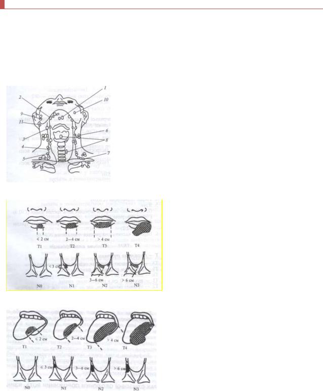
Limthatic apparate of neck
Anatomic scheme of classification of stages of lip‘s cancer
Anatomic scheme of classification of stages of tongue‘s cancer
B.Test tasks for self checking:
1.What histologic forms of a cancer of a lip meet more often? (Answer: ploskokletochny)
2.What stages of a cancer of tongue consider started? (Answer: III and IV stage)
3.Specify an optimum method of a cancer therapy of the bay T1 of NOMO.

(Answer: similarfocus X-rey)
C.Materials for test control. Test tasks with single right answer (α = 2):
1.Cancer cases of a mucous membrane of an oral cavity:
A.Decreases.
B.Stable.
C.Raises.
(Right answer: С)
2.The most frequent morphological form of malignant tumors of a mucous membrane of an oral cavity
is:
A. Planocellular cancer. B. Sarcoma.
C. Mucoepidermoid tumor. D. Cylindroma.
E. Not differentiated cancer.
(Right answer: А)
3.The most frequent localization of a cancer of a mucous membrane of an oral cavity is:
A.Tongue.
B.Mouth floor.
C.Mucous cheeks.
D.Mucous soft palate.
E.Mucous alveolar shoot of a jaw.
(Right answer: В)
D.Educational problems of the 3rd level (atypical tasks):
1.At the patient, in 55 years, in the center of a lower lip the ulcer of 2 cm in diameter, with platen-like edges is observed, the infiltration of tissues which surround an ulcer is observed.
Make the previous diagnosis? In what lymph nodes possible metastasizes? Define a method of treatment of the patient.
(Answer: cancer of a lower lip possible metastasizes in submaxillary lymph nodes. Treatment is combined)
2.The patient, 60 years, addressed with complaints to pain during swallowing that arose about 3 weeks ago and gradually amplifies. During the review on a lateral surface of tongue at the left the found ulcer with transition to a mucous membrane of a mouth floor, to 2 cm in diameter, painful at a palpation, with a bleeding bottom, is observed restriction mobile tongue. Surrounding tissues are infiltrating.
Make the previous diagnosis and specify, what methods of research need to be carried out for diagnosis specification.
(Answer: cancer of a lateral surface of tongue at the left. It is necessary to carry out a biopsy (a histological method) and cytological research of ulcer.)
3.The dentist of policlinic found in sick defeat of a lower lip, suspicious on a cancer.
To what clinical group the patient belongs? What further tactics of the dentist? (Answer: clinical group 1. It is necessary to conduct a biopsy or cytological research , to send the patient to an oncological clinic).
7. Literature:
7.1. Basic literature:
1. Skikevich M. G., Aveticov D. S. The basics of stomatology. Poltava. – 2012, 176 p.
2. Neville BW, Damm DD, Allen CM, et al. Oral & maxillofacial pathology. 2nd ed. Phila., PA: Saunders; 2002; 337–369.
7.2. Additional literature
1.Vaughan Ed, Brown JJ et al: The management of the mandible in mouth cancer. Br. J Oral Maxillofac Surg 32(6):345-6 1999
2.McGurk M, Goodger NM et al: Head and Neck cancer and it‘s treatment. Br. J Oral
Maxillofac. Surg; 38(£):209-20 2000
3.Fedotenko SP et al. Surgical treatment of residual cancer of the oral cavity. Vopr. Onkol;44(5):569-72 1998
4.Kasturi J. Perin MN: Epidemiology of oral cancer. Oral Cancer: 1991:1-8