


|
354 |
|
Sense organs |
|
|
|
|
|
|
|||||
|
|
1 |
|
|
|
Organa sensoria (sensuum). |
19 |
Meridians. Meridiani. Semicircles oriented at |
||||||
|
|
|
SENSE ORGANS. |
|||||||||||
1 |
|
|||||||||||||
|
|
|
|
In the narrow sense, the organs of vision, hear- |
|
right angles to the equator between the anterior |
||||||||
|
|
|
|
ing, smell and taste. |
|
|
|
and posterior poles. D. |
|
|||||
2 |
|
|
|
|
|
|
|
|||||||
2 |
|
ORGAN OF VISION. Organum visus (visuale). |
20 |
External axis of eyeball. Axis bulbi externus. |
||||||||||
|
3 |
|
EYE. Oculus. |
|
|
|
|
|
Line connecting anterior and posterior poles. C |
|||||
|
|
|
|
|
|
|
||||||||
3 |
4 |
|
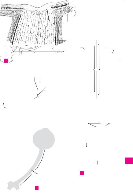
Optic nerve. N. opticus. Fiber bundle beginning |
21 |
Internal axis of eyeball. Axis bulbi internus. |
|||||||||
|
|
|
|
in the retina and extending as far as the optic |
|
Distance from posterior surface of cornea to the |
||||||||
|
|
|
|
|
||||||||||
|
|
|
|
chiasm. |
Histologically |
and |
embryologically |
|
inner surface of retina measured along an im- |
|||||
4 |
|
|
|
|
||||||||||
|
|
|
speaking, it is the tract of the brain that is ac- |
|
aginary line (external axis of eyeball) through |
|||||||||
|
|
|
|
cordingly enclosed by meninges up to the pos- |
|
the anterior and posterior poles. C |
||||||||
5 |
|
|
|
terior aspect of the eyeball. Its axons have no |
22 |
Optic axis. Axis opticus. Line passing through |
||||||||
|
|
|
|
neurilemma (sheath of Schwann) but are myeli- |
|
the midline of the cornea and lens and bisecting |
||||||||
|
|
|
|
|
||||||||||
|
|
|
|
nated. The myelin sheath is formed by the oligo- |
|
the retina between the fovea centralis and optic |
||||||||
6 |
|
|
|
|
||||||||||
|
|
|
dendroglia. A C E |
|
|
|
disc. C |
|
|
|||||
|
5 |
Intracranial part. Pars intracranialis. Segment |
23 |
FIBROUS TUNIC OF EYEBALL. Tunica fibrosa |
||||||||||
7 |
|
|
|
of the optic nerve betweeen the optic canal and |
||||||||||
|
|
|
|
bulbi. External wall of eyeball comprising the |
||||||||||
|
6 |
|
the chiasm. E |
|
|
Pars |
intracanicularis. |
|
cornea and sclera. C |
|
||||
|
|
|
|
|
|
|||||||||
8 |
|
Intracanalicular part. |
24 |
Sclera. The bluish-white outer coat of the eye- |
||||||||||
|
|
|
Segment of the optic nerve located in the optic |
|||||||||||
|
|
|
|
|
ball, which |
consists of |
irregulatory arranged |
|||||||
|
|
|
|
canal. It is partially connected with the canal |
|
|||||||||
9 |
|
|
|
|
collagenous fibers visible through the conjunc- |
|||||||||
|
|
|
wall. E |
|
|
|
|
|
|
|||||
7 |
|
part. Pars orbitalis. Slightly tortuous |
|
tiva. A B C |
|
|
||||||||
|
Orbital |
25 Scleral sulcus. Sulcus sclerae. Shallow groove |
||||||||||||
10 |
|
|
|
segment of the optic nerve measuring about |
||||||||||
|
|
|
|
between the cornea and sclera caused by the |
||||||||||
|
|
|
3 cm in length and occupying the orbit. E |
|
||||||||||
|
|
|
|
|
greater curvature of the cornea. B C D |
|||||||||
|
|
8 Intraocular part. Pars intraocularis. Segment of |
|
|||||||||||
11 |
|
26 |
Corneoscleral junction. Limbus. The concave |
|||||||||||
|
|
|
optic nerve located in the wall of the eyeball. |
|||||||||||
|
9 |
|
Postlaminar |
part. Pars postlaminaris. In- |
|
border of the sclera adjacent to the cornea. B |
||||||||
|
|
|
||||||||||||
12 |
|
27 Trabecular meshwork (pectinate ligament). |
||||||||||||
|
|
|
traocular segment located behind the lamina |
|||||||||||
|
|
|
|
Reticulum |
trabeculare |
(lig. pectinatum) |
||||||||
|
|
|
|
cribrosa and thus at the site where the external |
|
|||||||||
|
|
|
|
|
[[spongium |
iridocorneale]]. Connective tissue |
||||||||
13 |
|
|
|
sheath of the optic nerve (dura) blends into the |
|
|||||||||
|
|
|
|
framework at the iridocorneal (filtration) angle. |
||||||||||
|
|
|
sclera. A |
|
|
|
|
|
|
|||||
|
10 |
|
Intralaminar part. Pars intralaminaris. In- |
28 |
Corneoscleral part. Pars corneoscleralis. Part |
|||||||||
14 |
|
|
of the meshwork attached to the sclera. B |
|||||||||||
|
|
|
traocular segment lying within the lamina cri- |
|
||||||||||
|
|
|
|
brosa. A |
|
|
|
|
|
29 |
Uveal part. Pars uvealis. Part of the trabecular |
|||
|
|
|
|
|
|
|
|
|
||||||
15 |
11 |
|
Prelaminar part. Pars preliminaris. Intraocular |
|
meshwork attached to the iris. B |
|||||||||
|
|
|
|
|
||||||||||
|
|
|
|
segment extending between the lamina cri- |
30 |
Canal of Schlemm. Sinus venosus sclerae. |
||||||||
|
|
|
|
brosa and the nerve fiber layer of the retina. A |
|
Circular vessel occupying the interior aspect of |
||||||||
16 |
|
|
|
|
||||||||||
12 |
|
External sheath. Vagina externa. Dural cover- |
|
the trabecular meshwork. It can be interrupted |
||||||||||
|
|
|
|
ing of the optic nerve extending up to the eye- |
|
or doubled and is involved in the discharge of |
||||||||
17 |
|
|
|
ball. A |
|
|
|
|
|
|
aqueous humor from the anterior chamber. B |
|||
|
13 |
|
Internal sheath. Vagina interna. Pia and |
31 |
Episclera. Lamina episcleralis. Delicate dis- |
|||||||||
|
|
|||||||||||||
18 |
|
|
|
arachnoid coverings acoompanying the optic |
|
placeable connective tissue between the outer |
||||||||
|
|
|
nerve to the eyeball. A |
|
|
|
surface of the sclera and [[Tenon’s capsule]] |
|||||||
|
|
|
|
|
|
|
||||||||
|
14 |
|
Intervaginal |
spaces. |
Spatia intervaginalia. |
|
(bulbar fascia). |
|
||||||
19 |
|
|
|
|||||||||||
|
|
|
Subarachnoid space accompanying the optic |
32 |
Substantia propria sclerae corneal stroma. |
|||||||||
|
|
|
|
nerve and the capillary space between the |
|
The proper substance, i. e., main part of the |
||||||||
20 |
|
|
|
arachnoid and dura. A |
|
|
|
sclera. It consists of irregularly arranged col- |
||||||
|
15 Eyeball. Bulbus oculis. Globe of the eye. It con- |
|
lagenous fibers with sparse elastic fibers. A B |
|||||||||||
|
|
|||||||||||||
21 |
|
|
|
sists of the cornea and sclera together with all of |
33 |
Lamina fusca sclerae. Layer of loose connective |
||||||||
|
|
|
the structures they enclose. D |
|
|
tissue connecting the sclera and the choroid |
||||||||
|
|
|
|
|
|
|||||||||
|
16 Anterior pole. Polus anterior (center of anterior |
|
lying below it. It appears yellowish owing to the |
|||||||||||
22 |
|
|
|
curvature) of the eyeball, which is determined |
|
pigment cells dispersed within it. A |
||||||||
|
|
|
|
by the corneal vertex. D |
|
|
34 |
Lamina cribrosa. Fine, perforated layer of the |
||||||
|
|
|
|
|
|
|||||||||
23 |
17 |
|
Posterior pole. Polus posterior (center of poste- |
|
slcera for the passage of optic nerve fibers from |
|||||||||
|
|
|
|
rior curvature) of the eyeball, which lies lateral |
|
the retina. A |
|
|
||||||
24 |
|
|
|
to the exit of the optic nerve and opposite to the |
|
|
|
|
||||||
|
|
|
anterior pole. D |
|
|
|
|
|
|
|||||
18 Equator. Aequator. Greatest circumference of
25the eyeball located equidistant from the anterior and posterior poles. D
Feneis, Pocket Atlas of Human Anatomy © 2000 Thieme
All rights reserved. Usage subject to terms and conditions of license.

|
356 |
Sense organs |
|
|
|
|
|
|
|
|
|
|
|
|
|||
|
|
1 |
Cornea. The transparent anterior part (1/6) of |
|
16 |
Vascular lamina. Lamina vasculosa. It contains |
|||||||||||
1 |
|
||||||||||||||||
|
|
|
the eyeball with an anterior convex curvature |
|
|
the branchings of the short posterior ciliary ar- |
|||||||||||
|
|
|
|
and a posterior concave curvature. It is 0.9 mm |
|
|
teries. A |
|
|
|
|||||||
2 |
|
|
|
thick in the middle, 1.2 mm thick at its margins. |
|
17 |
Choriocapillaris. |
Lamina |
choroidocapillaris. |
||||||||
|
|
|
|
B D |
|
|
|
|
|
|
|
|
Pigment-free layer of connective tissue with a |
||||
|
|
|
2 Conjunctival ring. Anulus conjunctivae. Junc- |
|
|
||||||||||||
3 |
|
|
|
|
dense network of capillaries extending as far as |
||||||||||||
|
|
|
tion between bulbar conjunctival epithelium |
|
|
the ora serrata. It is often delimited from the |
|||||||||||
|
|
|
|
|
|
||||||||||||
|
|
|
|
and the anterior epithelium of the cornea. D |
|
|
vascular lamina by a special connective tissue |
||||||||||
4 |
|
|
|
|
|
||||||||||||
3 |
Corneoscleral junction. Limbus corneae. D |
|
|
layer. A |
|
|
|
|
|||||||||
|
4 |
Vertex corneae. The most prominent point on |
|
18 |
Basal lamina [[Bruch’s membrane]]. Com- |
||||||||||||
5 |
|
||||||||||||||||
|
|
|
the anterior surface of the cornea. |
|
|
|
|
plexus (lamina) basalis. Homogeneous zone |
|||||||||
|
|
|
5 Anterior surface. Facies anterior. Corneal sur- |
|
|
about 2−4 mm thick between the choriocapil- |
|||||||||||
|
|
|
|
|
laris and the pigment epithelium of the retina. A |
||||||||||||
6 |
|
|
|
|
|||||||||||||
|
|
|
face facing the outside air. D |
|
|
|
|
19 Ciliary body. Corpus ciliare. Enlarged uveal seg- |
|||||||||
|
|
|
6 Posterior surface. Facies posterior. Corneal sur- |
|
|||||||||||||
|
|
|
|
|
ment situated between the ora serrata and root |
||||||||||||
7 |
|
|
|
face facing the anterior chamber. D |
|
|
|
|
|||||||||
|
|
|
|
|
|
|
of the iris. It contains ciliary muscles and |
||||||||||
|
|
|
7 Anterior |
epithelium. Epithelium |
anterius. |
|
|
processes. C |
|
|
|
||||||
|
|
|
|
|
|
|
|
||||||||||
8 |
|
|
|
Stratified |
(about |
5 layers) |
squamous |
|
20 |
Pars plicata (Corona ciliaris). Circular zone oc- |
|||||||
|
|
|
epithelium covering the anterior surface of the |
|
|||||||||||||
|
|
|
|
|
|
cupied by ciliary processes. C |
|
||||||||||
|
|
|
|
cornea with a very smooth surface. B D |
|
|
|
||||||||||
9 |
8 |
|
21 |
Ciliary |
processes. |
Processus ciliares. 70−80 |
|||||||||||
Anterior |
limiting |
(Bowman’s) |
membrane. |
|
|||||||||||||
|
|
|
radially |
oriented, |
capillary-rich folds, |
0.1− |
|||||||||||
|
|
|
|
Lamina limitans |
anterior [[Bowman]]. Basal |
|
|
0.2 mm wide, 1 mm high and 2−3 mm long. |
|||||||||
10 |
|
|
|
membrane of the anterior epithelium, about |
|
|
|||||||||||
|
|
|
|
|
Their epithelium produces aqueous humor. C |
||||||||||||
|
|
|
|
10−20 mm thick. It is continuous posteriorly |
|
22 |
Ciliary folds. Plicate ciliares. Low folds in the re- |
||||||||||
|
|
|
|
|
|||||||||||||
|
|
|
|
with the substantia propria. B |
|
|
|
|
|||||||||
11 |
|
|
|
|
|
|
|
||||||||||
9 |
Substantia propria. Predominant part of the |
|
|
gion of the corona ciliaris and between the cili- |
|||||||||||||
|
|
|
ary processes. C |
|
|
|
|||||||||||
|
|
|
|
avascular cornea consisting of highly organized |
|
|
|
|
|
||||||||
12 |
|
|
|
|
23 |
Pars plana. Orbiculus ciliaris. Circular zone |
|||||||||||
|
|
|
lamellar connective tissue embedded within a |
|
|||||||||||||
|
|
|
|
mucopolysaccharide substance. The state of |
|
|
lying between the corona and ora serrata. It is |
||||||||||
|
|
|
|
|
|
||||||||||||
|
|
|
|
turgescence of its fibers and the distribution of |
|
|
occupied by ciliary folds. C |
|
|
||||||||
13 |
|
|
|
|
|
|
|
||||||||||
|
|
|
its colloidal matrix affect the transparency of |
|
24 Ciliary muscle. M. ciliaris. Smooth muscle oc- |
||||||||||||
|
|
|
|
|
|||||||||||||
|
|
|
|
the cornea. B |
|
|
|
|
|
|
|
cupying the ciliary body. It pulls the choroid for- |
|||||
14 |
|
|
|
|
|
|
|
|
|
|
|||||||
10 |
Posterior |
limiting |
(Descemet’s) |
membrane. |
|
|
ward and, in so doing, relaxes the zonule fibers |
||||||||||
|
|
|
|
Lamina limitans posterior [[Descemet]]. Basal |
|
|
so that the lens can become more strongly |
||||||||||
|
|
|
|
|
|
||||||||||||
15 |
|
|
|
membrane of the |
corneal |
(posterior) en- |
|
|
curved for accomodation of near objects. D |
||||||||
|
|
|
dothelium. At its lateral margin it divides into |
|
25 |
Meridional (longitudinal) fibers. Fibrae mer- |
|||||||||||
|
|
|
|
|
|||||||||||||
|
|
|
|
fibers which radiate into the trabecular mesh- |
|
||||||||||||
16 |
|
|
|
|
|
idionales [fibrae longitudinales]. Larger muscle |
|||||||||||
|
|
|
work of the sclera and iris. Aqueous humor |
|
|
||||||||||||
|
|
|
|
|
fibers oriented meridionally (longitudinally). |
||||||||||||
|
|
|
|
passes through its interstices to drain into the |
|
|
Anteriorly they are attached to the posterior |
||||||||||
17 |
|
|
|
sinus venosus sclerae. B D |
|
|
|
|
|
limiting lamina above the trabecular mesh- |
|||||||
|
|
|
|
|
|
|
|
|
|
|
|
||||||
|
11 |
Posterior epithelium (endothelium). Epithe- |
|
|
work; posteriorly, they insert into the choroid. |
||||||||||||
|
|
|
|||||||||||||||
|
|
|
|
lium posterius. Simple squamous epithelium |
|
|
D |
|
|
|
|
||||||
18 |
|
|
|
|
|
|
|
|
|
||||||||
|
|
|
lining the posterior surface of the cornea. B D |
|
26 |
Circular fibers. |
Fibrae |
circulares. Circular |
|||||||||
|
|
12 VASCULAR TUNIC OF EYEBALL (UVEAL TRACT). |
|
|
muscle lying internal to the meridional fibers. D |
||||||||||||
19 |
|
|
|||||||||||||||
|
|
|
Tunica vasculosa bulbi (tractus uvealis). It rep- |
|
27 |
Radial |
fibers. Fibrae radiales. Muscle |
fibers |
|||||||||
|
|
|
|
resents the middle layer of the wall of the eye- |
|
||||||||||||
|
|
|
|
|
|
crossing perpendicular to the two other muscle |
|||||||||||
20 |
|
|
|
ball and consists of the choroid, ciliary body and |
|
|
|||||||||||
|
|
|
|
|
systems and coursing outwardly. |
|
|||||||||||
|
|
|
iris. |
|
|
|
|
|
|
|
|
|
|||||
|
|
|
|
|
|
|
|
|
|
|
28 Basal lamina. Lamina basalis. Continuation of |
||||||
|
13 |
Choroid. Choroidea. The vascular coat lying be- |
|
||||||||||||||
21 |
|
|
the basal membrane of the choroid. It supports |
||||||||||||||
|
|
|
tween the retina and sclera. A |
|
|
|
|
|
|||||||||
|
|
|
|
|
|
|
|
the epithelium. D |
|
|
|
||||||
|
|
14 Suprachoroid lamina (lamina fusca). Lamina |
|
|
|
|
|
|
|
||||||||
22 |
|
|
|
|
|
|
|
|
|||||||||
|
|
|
suprachoroidea. |
Displaceable |
layer |
directly |
|
|
|
|
|
|
|
||||
|
|
|
|
beneath the sclera. It contains only a few vessels |
|
|
|
|
|
|
|
||||||
23 |
|
|
|
and pigment; its fibers are partly covered by en- |
|
|
|
|
|
|
|
||||||
|
|
|
dothelium. A |
|
|
|
|
|
|
|
|
|
|
|
|
||
|
|
|
|
|
|
|
|
|
|
|
|
|
|
|
|
||
24 |
|
15 Perichoroidal space. Spatium perichoroideale. |
|
|
|
|
|
|
|
||||||||
|
|
|
Spatial system in the suprachoroid lamina, part |
|
|
|
|
|
|
|
|||||||
|
|
|
|
of which forms lymph pathways. It houses the |
|
|
|
|
|
|
|
||||||
25 |
|
|
|
ciliary nerves, long and short posterior ciliary |
|
|
|
|
|
|
|
||||||
|
|
|
|
arteries and the vorticose veins. A |
|
|
|
|
|
|
|
|
|
||||
|
|
|
Feneis, Pocket Atlas of Human Anatomy © 2000 Thieme |
|
|
|
|||||||||||
|
|
|
|
|
|
||||||||||||
|
|
|
All rights reserved. Usage subject to terms and conditions of license. |
|
|||||||||||||


|
358 |
Sense organs |
|
|
||
|
|
1 |
Iris. Frontally-located, round, variably colored |
16 |
Lesser arterial circle of iris. Circulus arteriosus |
|
1 |
||||||
|
|
disk about 10−12 mm in diameter, with a cen- |
|
iridis minor. Ringlike vascular system in the vi- |
||
|
|
|
tral aperture (pupil). The iris forms the poste- |
|
cinity of the pupillary margin formed by anas- |
|
2 |
|
|
rior border of the anterior chamber of the eye. |
|
tomoses between the radial branches of the |
|
|
|
|
Its lateral margins become continuous with the |
|
greater arterial circle. A |
|
|
|
|
ciliary body. A |
17 |
Pupillary membrane. [Membrana pupillaris]. |
|
3 |
|
|
||||
2 |
Pupillary margin. Margo pupillaris. Medial (in- |
|
Anterior part of embryonical vascular mem- |
|||
|
|
|
ternal) margin of the iris bordering the pupil. A |
|
brane around the lens that is situated behind |
|
4 |
|
|
B |
|
the pupil. It is fused to the pupillary margin and |
|
|
3 |
Ciliary margin. Margo ciliaris. Lateral (external) |
18 |
receives blood vessels from there. |
||
|
||||||
|
|
|
margin of iris attached to ciliary body at the ir- |
INTERNAL (SENSORY) TUNIC OF EYEBALL. |
||
5 |
|
|
||||
|
|
idocorneal angle. B |
|
Tunica interna bulbi. It comprises the retina |
||
|
4 |
Anterior surface. Facies anterior. It faces the |
|
with its pigment epithelium. |
||
6 |
|
|
anterior chamber. B |
19 |
Retina. Inner lining of eyeball developed from |
|
|
5 |
Posterior surface. Facies posterior. Surface |
|
the two layers of the optic cup. Most of it is |
||
|
|
|||||
7 |
|
|
facing the posterior chamber. A B |
|
light-sensitive (pars optica). B |
|
|
|
20 Pars optica retinae. Retinal segment capable of |
||||
6 |
Greater ring (circle) of iris. Anulus iridis major. |
|||||
|
||||||
|
|
|
Ciliary segment of the iris, and outer cirucular |
|
transforming light stimuli into nerve impulses. |
|
|
|
|
|
|||
8 |
|
|
|
It lines the posterior aspect of the eyeball and |
||
|
|
zone on the anterior surface of the iris. It is |
|
|||
|
|
|
coarser and broader than the lesser ring. A |
|
extends as far anteriorly as the ora serrata. B |
|
|
|
|
|
|||
9 |
7 |
Lesser ring (circle) of iris. Anulus iridis minor. |
21 |
Pigmented part. Pars pigmentosa. Pigment |
||
|
epithelium arising from the external layer of the |
|||||
|
|
|
Pupillary segment of iris. Narrow, circular inner |
|
||
|
|
|
zone on the anterior surface of iris. Its structure |
|
optic cup. B |
|
10 |
|
|
|
|||
|
|
22 |
Nervous part. Pars nervosa. Retina proper con- |
|||
|
|
is finer than that of the greater ring. A |
||||
|
8 |
Iridial folds. Plicae iridis. Folds passing around |
|
sisting essentially of three nuclear layers lying |
||
|
|
|||||
11 |
|
|
the pupillary margin on the anterior side of the |
|
internal to the pigment epithelium. B |
|
|
|
23 |
Neuroepithelial (photosensitive) layer. Stratum |
|||
|
|
|
iris. They make the pupillary margin appear |
|||
|
|
|
slightly serrated. A |
|
neuroepitheliale (photosensorium). Outer layer |
|
12 |
|
|
|
|||
9 |
Pupil. Pupilla. Aperture in the iris surrounded |
|
of the cerebral stratum. It consists of rods and |
|||
|
|
|
by the pupillary margin of the iris. Its diameter |
|
cones, the outer segments of which affect the |
|
|
|
|
|
|||
13 |
|
|
varies depending upon the intensity of light and |
|
transformation of light stimuli into nerve im- |
|
|
|
|
pulses. Cell bodies of rods and cones form the |
|||
|
|
the focal distance of the observed object. A |
|
|||
|
10 |
M. sphincter pupillae. Network of spirally |
|
outermost layer of the retinal nuclei (external |
||
14 |
|
nuclear layer). D |
||||
|
|
coursing muscle fibers the longitudinal axes of |
|
|||
|
|
24 |
Internal nuclear layer. [[Stratum ganglionare reti- |
|||
|
|
|
which run approximately parallel to the pupil- |
|||
15 |
|
|
lary margin when the pupil is dilated. It is in- |
|
nae]]. Middle layer of cell nuclei mainly con- |
|
|
|
|
sisting of the cell bodies of bipolar and amacrine |
|||
|
|
nervated by parasympathetic fibers from the |
|
|||
|
|
|
oculomotor nerve. B |
|
cells. D |
|
16 |
|
|
|
|||
|
|
25 |
Ganglion cell layer. [[Stratum ganglionare n. op- |
|||
11 |
M. dilator pupillae. Thin layer of smooth |
|||||
|
|
|
muscle mainly comprised of radially oriented |
|
tici]]. Internal layer of nuclei consisting of multi- |
|
|
|
|
|
|||
17 |
|
|
fibers. It is innervated by sympathetic fibers |
|
polar cell bodies of initially non-myelinated |
|
|
|
|
ganglion cells the axons of which form the optic |
|||
|
|
from the carotid plexus. |
|
|||
|
12 |
Stroma iridis. Vascular framework of the iris in- |
|
nerve. D |
||
18 |
|
|||||
26 |
Ora serrata. Serrated margin between the |
|||||
|
|
filtrated by pigmented connective tissue cells. |
||||
|
|
|
Its anterior and posterior portions are thicker |
|
light-sensitive and light-insensitive parts of the |
|
|
|
|
|
|||
19 |
|
|
than the rest and are divided by a fine fibrous |
|
neural retina. B C |
|
|
|
27 |
Pars ciliaris retinae. Light-insensitive retinal |
|||
|
|
|
network. A B |
|||
|
13 |
Pigmented (posterior) epithelium. Epithelium |
|
segment consisting of a bilayered cuboidal |
||
20 |
|
|||||
|
epithelium (ciliary epithelium) forming the |
|||||
|
|
pigmentosum. Bilayered epithelium on the |
|
|||
|
|
|
posterior surface of the iris. It is so heavily pig- |
|
posterior surface of the ciliary body. Its outer |
|
|
|
|
|
|||
21 |
|
|
mented that no nuclei are visible on the surface |
|
layer of epithelium is continuous with the pig- |
|
|
|
|
ment epithelium of the retina and is pigmented, |
|||
|
|
facing the posterior chamber. A |
|
|||
|
14 |
Spaces of iridocorneal angle [spaces of Fon- |
|
whereas the innermost epithelium is continu- |
||
22 |
|
ous with the pars nervosa of the retina and is |
||||
|
|
tana]. Spatia anguli iridocornealis. Interstices |
|
|||
|
|
|
devoid of pigment. B |
|||
|
|
|
between the fibers of the trabecular meshwork. |
28 Pars iridica retinae. Light-insensitive retinal |
||
23 |
|
|
They form passageways that convey aqueous |
|||
|
|
|
segment on the posterior surface of the iris. It is |
|||
|
|
fluid to the sinus venosus sclerae. A |
|
|||
|
15 |
Greater arterial circle of iris. Circulus arterio- |
|
continuous with the pars ciliaris retinae and |
||
24 |
|
forms the bilayered posterior epithelium of the |
||||
|
|
sus iridis major. Ringlike vascular system with |
|
|||
|
|
|
iris. Both layers are heavily pigmented. B |
|||
|
|
|
radiating branches. It is formed by anastomoses |
|
|
|
25between the long and short posterior ciliary arteries. A
Feneis, Pocket Atlas of Human Anatomy © 2000 Thieme
All rights reserved. Usage subject to terms and conditions of license.


 2
2 28
28 

360 |
Sense organs |
|
|
|||
|
|
1 |
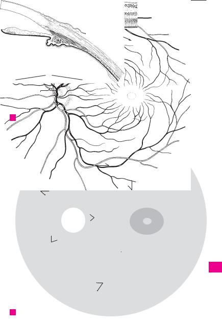
Optic disc (papilla). Discus nervi optici [papilla |
16 |
Iridocorneal angle. Angulus iridocornealis. |
|
1 |
||||||
|
|
nervi optici]. Beginning of the optic nerve as |
|
Angle between the iris and cornea. It houses the |
||
|
|
|
visualized in the fundus about 3−4 mm medial |
|
trabecular meshwork, the interstices of which |
|
2 |
|
|
to the macula. It is about 1.6 mm in diameter. C |
|
serve as passageways that drain aqueous humor |
|
|
|
2 Physiological cup. Excavatio disci. Depression |
|
into the sinus venosus sclerae. A |
||
|
|
17 |
Aqueous humor. Humor aquosus. Fluid pro- |
|||
3 |
|
|
in the middle of the optic disc with the stems of |
|||
|
|
the central retinal artery and vein. C |
|
duced by the epithelium of the ciliary processes |
||
|
|
|
|
|||
|
|
3 Macula [[lutea]]. Transversely oval, yellowish |
|
(total quantity: 0.2−0.3 cm3). The clear fluid |
||
4 |
|
|
consists of 98% water, 1.4% NaCl and traces of |
|||
|
|
area, 2−4 mm in diameter, at the posterior pole |
|
|||
|
|
|
of the retina. C |
|
protein and sugar. It has a refractive index of |
|
|
|
|
|
|||
|
|
|
|
1.336. |
||
|
|
|
|
|
||
54 Fovea centralis. Central fovea, a small depres-
sion in the macula caused by thinning of the upper retinal layers. Its diameter, measured
6from the beginning of the decrease in retinal thickness from one side to the other, is approxi-
7mately 1−2 mm. B C
5Foveola. Thinnest area of fovea centralis with a
8diameter of about 0.2−0.4 mm. Here, the retina is comprised entirely of approx. 2500 closely packed cones. B
96 Retinal blood vessels. Vasa sanguinae retinae. Branches of the central retinal artery and vein
10 |
located on the internal aspect of the retina. |
7Circle of arteries around the optic nerve. Cir-
11culus vasculosus nervi optici. Small vascular ring penetrating the sclera around the optic nerve.
128 Superior temporal arteriole/venule or retina.
Arteriola/venula temporalis retinae superior.
13Lateral upper branch of the central retinal artery and vein. C
149 Inferior temporal arteriole/venule of retina.
Arteriola/venula temporalis retinae inferior. Lateral lower branch of the central retinal artery
15and vein. C
10 Superior nasal arteriole/venule of retina.
16Arteriola/venula nasalis retinae superior. Upper medial branch of the central retinal artery and
vein. C
17
11 Inferior nasal arteriole/venule of retina. Arteriola/venula nasalis retinae inferior. Lower me-
18dial branch of the central retinal artery and vein. C
1912 Superior macular arteriole/venule. Arteriola/ venula macularis superior. They supply and drain the upper part of the macula. C
2013 Inferior macular arteriole/venule. Arteriola/ venula macularis inferior. They supply and
21drain the lower part of the macula. C
14 Medial arteriole/venule of retina. Arteriola/
22venula medialis retinae. Small branches that supply and drain the medial part of retina proximal to the optic disc. C
2314 a CHAMBERS OF THE EYE. Camerae bulbi.
15 Anterior chamber. Camera anterior. Space that
24extends from the anterior surface of the iris to the posterior surface of the cornea and com-
25municates with the posterior chamber via the pupil. A
Feneis, Pocket Atlas of Human Anatomy © 2000 Thieme
All rights reserved. Usage subject to terms and conditions of license.

|
362 |
Sense organs |
|
|
|
|||
|
|
1 |
Posterior chamber. Camera posterior. It ex- |
14 |
Lens fibers. Fibrae lentis. Fibers corresponding |
|||
1 |
||||||||
|
|
tends from the iris and ciliary body to the ante- |
|
to the lens epithelium from which they develop. |
||||
|
|
|
rior surface of the vitreous. A |
|
They form the lens substance measuring 2.5− |
|||
2 |
2 |
Aqueous humor. Humor aquosus. Produced by |
|
12 µm thick and up to 10 mm long. C |
||||
|
|
|
the ciliary processes. It flows between the in- |
15 |
Epithelium of lens. Epithelium lentis. Part of |
|||
|
|
|
||||||
3 |
|
|
terstices of the suspensory ligaments of the lens |
|
the lens confined to the anterior surface and ex- |
|||
|
|
to the anterior surface of the lens and then be- |
|
tending as far as the equator. It is derived |
||||
|
|
|
tween the iris and lens to the pupil, through |
|
embryologically from the anterior epithelium |
|||
4 |
|
|
which it enters the anterior chamber. |
|
of the lens vesicle. C |
|||
|
3 |
Vitreous chamber. Camera vitrea. Space filled |
16 |
Lens capsule. Capsula lentis. Transparent mem- |
||||
|
||||||||
5 |
|
|
up by the vitreous body. B |
|
brane, up to 15 µm thick, covering the lens in- |
|||
4 |
Vitreous body. Corpus vitreum. It consists of |
|
cluding its epithelium. Its anterior pole is |
|||||
|
|
|||||||
|
|
|
about 98% water and primarily contains traces |
|
thicker than the posterior pole. It gives attach- |
|||
6 |
|
|
|
|||||
|
|
of protein and NaCl and a mixture of fine fibrils |
|
ment to the suspensory ligaments. C |
||||
|
|
|
which thicken near the surface to form a lim- |
17 |
Anterior pole. Polus anterior. D |
|||
|
|
|
||||||
7 |
|
|
iting membrane. It has a gelatinous consistency |
18 |
Posterior pole. Polus posterior. D |
|||
|
|
due to its high content of hyaluronic acid. A |
||||||
|
|
|
19 |
Anterior surface. Facies anterior. Less curved |
||||
|
5 |
Hyaloid |
artery. [A. hyaloidea]. Branch of the |
|||||
|
||||||||
8 |
|
|
ophthalmic artery supplying the vascular mem- |
|
lens surface with a radius of 8.3−10 mm. C |
|||
|
|
20 |
Posterior surface. Facies posterior. More |
|||||
|
|
|
brane of the lens. Present only during embry- |
|||||
9 |
|
|
onic development. The proximal portion per- |
|
curved lens surface with a radius of about |
|||
|
|
|
6.5 mm. C |
|||||
|
|
|
sists in the optic nerve as the central retinal |
|
||||
|
|
|
artery. B |
|
|
21 |
Axis. Line connecting anterior and posterior |
|
10 |
|
|
|
|
||||
6 |
Hyaloid canal. Canalis hyaloideus. Canal |
|
poles. D |
|||||
|
|
|
within the vitreous body formerly occupied by |
22 |
Equator. Margin of lens. D |
|||
11 |
|
|
the embryonic hyaloid artery which degener- |
23 |
Radii of lens. Suture line of the individual lens |
|||
|
|
|
ates in |
this region. |
The canal assumes a |
|
fibers. In the young it resembles a triradiate |
|
|
|
|
downward sagging corkscrew shape; it extends |
|
||||
12 |
|
|
|
seam. D |
||||
|
|
from the optic disc to the posterior surface of |
|
|||||
|
|
24 Ciliary zonule. Zonula ciliaris. Suspensory ap- |
||||||
|
|
|
the lens. Its wall is formed by condensed fibers. |
|||||
|
|
|
||||||
|
|
|
|
paratus together with its interstices. It encircles |
||||
13 |
|
|
A |
|
|
|
||
|
|
|
|
|
the lens equator and consists of a radially |
|||
7 |
Hyaloid (lenticular, patellar) fossa. Fossa hy- |
|
||||||
|
|
oriented system of fibers of variable length and |
||||||
|
|
|
aloidea. Fossa on the anterior surface of the vit- |
|
||||
14 |
|
|
|
the folds situated between them. C |
||||
|
|
reous body adjacent to the lens. A |
|
|||||
|
|
25 |
Zonular fibers (suspensory ligaments). Fi- |
|||||
|
8 |
Vitreous |
(hyaloid) |
membrane. Membrana |
||||
15 |
|
brae zonulares. Suspensory fibers attached to |
||||||
|
|
vitrea. Condensation of fibers on the surface of |
|
|||||
|
|
|
the equator and the adjacent anterior and post- |
|||||
|
|
|
the vitreous body. See (4), vitreous body. A |
|
||||
|
|
|
|
erior surfaces of the lens. They arise distally |
||||
16 |
9 |
Stroma of vitreous body. Stroma vitreum. |
|
from the basal lamina of the ciliary body and the |
||||
|
|
|
Fine network of fibers in the virtreous body. Its |
|
pars ciliaris retinae. C |
|||
|
|
|
surface thickens to form the vitreous mem- |
26 |
Zonular spaces. Spatia zonularia. Spaces be- |
|||
17 |
|
|
||||||
|
|
brane. |
|
|
||||
|
|
|
|
|
tween the zonule fibers filled with percolating |
|||
|
|
9 a Vitreous humor. Humor vitreus. Fluid part of |
|
|||||
|
|
|
aqueous humor. C |
|||||
18vitreous body. Primarily consists of mucupolysaccharides and is situated between the fibers of the stroma.
1910 LENS. Structure of the eye situated between the pupil and vitreous body. It is suspended by the
20ciliary zonule (suspensory ligaments), has a diameter of 9−10 mm and is about 4 mm thick. B C D
2111 Substantia lentis. Lens substance situated beneath the lens epithelium and comprising
22the lens nucleus and lens cortex with a refractive index of 1.44−1.55. C
2312 Lens cortex. Cortex lentis. External zone of the lens. It is softer owing to its high water content and blends into the lens nucleus without a
24sharp boundary. C
13 Nucleus of lens. Nucleus lentis. Harder core of
25the lens with a low water content, as is especially evident in the elderly. C
Feneis, Pocket Atlas of Human Anatomy © 2000 Thieme
All rights reserved. Usage subject to terms and conditions of license.


|
364 |
Sense organs |
|
|
|
|
|
||
|
|
1 |
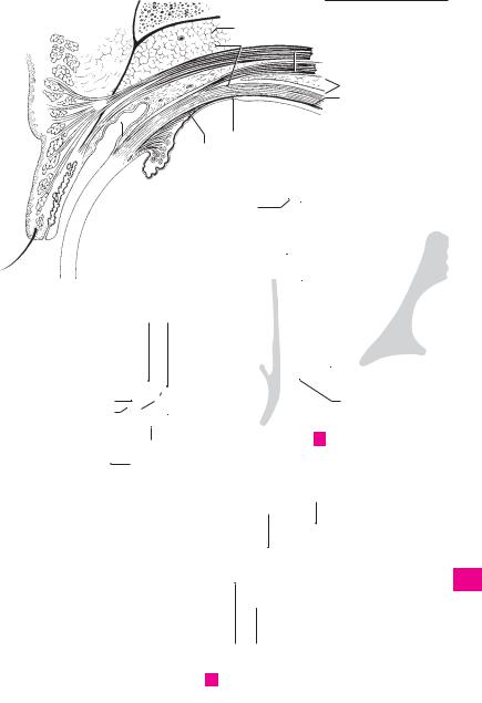
ACCESSORY ORGANS OF EYE. Organa oculi ac- |
15 |
Superficial lamina of levator tendon. |
||||
1 |
|||||||||
|
|
cessoria. |
|
|
|
|
Lamina superficialis. It passes between the tar- |
||
|
2 |
Muscles of eye. Musculi bulbi. Extrinsic ocular |
|
sus and orbicularis oculi to insert into the sub- |
|||||
2 |
|
cutaneous connective tissue of the upper eyelid. |
|||||||
|
|
muscles. |
|
|
|
|
|||
|
3 |
Orbital muscle. M. orbitalis. Thin layer of |
|
It is so broad that it extends mainly laterally to |
|||||
|
|
the wall of the orbit. A |
|||||||
3 |
|
||||||||
|
|
smooth muscle which bridges the inferior orbi- |
16 |
Deep lamina of levator tendon. Lamina pro- |
|||||
|
|
|
tal fissure. C |
|
|
|
|||
|
|
|
|
|
|
|
funda. It inserts into the upper margin and the |
||
|
4 |
Superior rectus. M. rectus superior. o: Common |
|
||||||
4 |
|
anterior surface of the tarsus. A |
|||||||
|
|
|
tendinous ring. i: Along an oblique line in front |
17 |
Orbital fasciae. Fasciae orbitales. |
||||
|
|
|
|||||||
5 |
|
|
of the equator, 7−8 mm posterior to the corneal |
||||||
|
|
18 |
Periosteum of orbit. Periorbita. It is delicate |
||||||
|
|
margin. A: Elevation and medial rotation of su- |
|||||||
|
|
|
perior pole of eyeball. I: Oculomotor nerve. B C |
|
and fused solidly to the bone at the inlet and |
||||
|
|
|
|
||||||
6 |
|
|
D |
|
|
|
|
outlet of the orbit. Anteriorly, it is continuous |
|
5 |
Inferior rectus. M. rectus inferior. o: Common |
|
with the adjacent periosteum, posteriorly with |
||||||
|
|
||||||||
|
|
the dura. A |
|||||||
7 |
|
|
tendinous ring. i: Along an oblique line about |
|
|||||
|
|
19 |
Orbital septum. Septum orbitale. Connective |
||||||
|
|
6 mm behind the corneal margin. A: Depression |
|||||||
|
|
|
and lateral rotation of superior pole of eyeball. I: |
|
tissue septum partly reinforced by tendon. It |
||||
8 |
|
|
Oculomotor nerve. B C D |
|
|
|
passes from the orbital margin below the orbic- |
||
|
6 |
Medial rectus. M. rectus medialis. o: Common |
|
ularis oculi to the external margins of the tarsi |
|||||
|
|
and forms the anterior end of the orbit. A |
|||||||
9 |
|
|
tendinous ring. i: About 5.5 mm from the cor- |
|
|||||
|
|
|
Muscular fasciae. Fasciae musculares. Sheaths |
||||||
|
|
|
neal margin. A: Adduction of corneal pole. I: |
20 |
|||||
|
|
|
Oculomotor nerve. B C |
|
|
|
of Tenon’s capsule enveloping the tendons and |
||
10 |
|
|
|
|
|
||||
7 |
Lateral rectus. M. rectus lateralis. o: Common |
|
muscular bellies of the 6 extrinsic ocular |
||||||
|
|
muscles. A |
|||||||
|
|
|
tendinous ring and lesser wing. i: 5.5 mm be- |
|
|||||
11 |
|
|
21 |
Tenon’s capsule (fascia bulbi). Vagina bulbi. |
|||||
|
|
hind corneal margin. A: Abduction of corneal |
|||||||
|
|
|
pole. I: Abducent nerve. B C D |
|
|
|
Connective tissue gliding membrane between |
||
|
|
|
|
|
|
||||
|
8 |
Tendon of lateral rectus at greater wing. |
|
the eyeball and orbital fat. It is fused to the |
|||||
12 |
|
||||||||
|
sclera posteriorly at the optic nerve. Anteriorly |
||||||||
|
|
|
Lacertus musculi recti lateralis. C |
|
|
|
it ends beneath the conjunctiva. It is separated |
||
|
|
|
|
|
|
||||
13 |
9 |
Common tendinous ring (common annular |
|
from the sclera primarily by the episcleral |
|||||
|
|
tendon). Anulus tendineus communis. Ten- |
|
space. A |
|||||
|
|
|
|
||||||
|
|
|
dinous ring for attachment of the recti ocular |
22 Episcleral space. Spatium episclerale [[inter- |
|||||
14 |
|
|
|||||||
|
|
muscles. It surrounds the optic canal and me- |
|||||||
|
|
|
vaginale]]. Gliding space between the eyeball |
||||||
|
|
|
dial part of the superior orbital fissure. C |
|
|
||||
|
|
|
|
|
and Tenon’s capsule. It is traversed by long, deli- |
||||
15 |
10 |
Superior oblique. M. obliquus superior. o: Body |
|
cate connective tissue fibers. A |
|||||
|
|
|
of sphenoid medial to common tendinous ring. |
23 Orbital fat body. Corpus adiposum orbitae. |
|||||
|
|
|
|||||||
16 |
|
|
i: Posterolateral |
aspect of sclera |
behind |
the |
|
Adipose tissue fills the spaces around the ocular |
|
|
|
equator after its |
tendon passes |
through |
the |
|
|||
|
|
|
muscles, the eyeball and the optic nerve and is |
||||||
|
|
|
trochlea and approaches sclera obliquely from |
|
bordered anteriorly by the orbital septum. A D |
||||
17the medial margin of orbit. A: Abduction, medial rotation and depression. I: Trochlear nerve. B
1811 Trochlea. Cartilaginous sling attached to the medial wall of the orbit [[trochlear spine]] and
19serving as a pulley for the tendon of the superior oblique muscle. B
2012 Tendon sheath of superior oblique muscle
(synovial bursa of trochlea). Vagina tendinis m. obliqui superioris [[bursa synovialis
21trochlearis]]. Synovial sheath (bursa) for the tendon of the superior oblique muscle separat-
22ing the tendon from the trochlea. B
13 Inferior oblique. M. obliquus inferior. o: Lateral to the nasolacrimal canal. i: Posterior to equa-
23tor. A: Elevation, abduction and lateral rotation. I: Oculomotor nerve. D
2414 M. levator palpebrae superioris. o: Bone above optic canal and dura of optic nerve. Its tendon
25broadens anteriorly and splits to form an upper and lower layer. I: Oculomotor nerve. A C D
Feneis, Pocket Atlas of Human Anatomy © 2000 Thieme
All rights reserved. Usage subject to terms and conditions of license.
 7
7
|
366 |
Sense organs |
|
|
|
|||
|
|
1 |
Eyebrow. Supercilium. The transverse eleva- |
19 |
Lateral palpebral raphe. [[Raphe palpebralis |
|||
1 |
||||||||
|
|
tion above the eyes, covered by thick, bristle- |
|
lateralis]]. Delicate band on the lateral palpebral |
||||
|
|
|
like hairs. A |
|
|
|
ligament. It is reinforced by the orbicularis oculi |
|
2 |
2 |
Eyelids. Palpebrae. |
|
|
muscle. D |
|||
|
20 |
Lateral palpebral ligament. Lig. palpebrale |
||||||
|
3 |
Upper eyelid. Palpebra superior. A |
||||||
3 |
|
laterale. Fibrous band that attaches the lateral |
||||||
|
|
|
|
|
|
|||
|
4 Lower eyelid. Palpebra inferior. A |
|
palpebral commissure to the lateral wall of the |
|||||
|
|
|
||||||
|
|
5 Anterior palpebral surface. Facies anterior pal- |
|
orbit in front of the orbital septum. B |
||||
4 |
|
|
||||||
|
21 |
Tarsal [[Meibomian]] glands. Glandulae tar- |
||||||
|
|
pebralis. The anterior external (skin-covered) |
||||||
|
|
|
||||||
|
|
|
surface of the eyelid. E |
|
|
sales. Elongated holocrine glands located in the |
||
5 |
|
|
|
|
||||
6 |
Epicanthus |
(mongolian fold). [Plica palpe- |
|
superior and inferior tarsal plates with openings |
||||
|
|
near the posterior edge of the free margin of the |
||||||
|
|
|
bronasalis] [[epicanthus]]. Vertical fold covering |
|
||||
|
|
|
|
eyelids. They produce a sebaceous secretion for |
||||
6 |
|
|
the medial angle of the eye. It is a continuation |
|
||||
|
|
|
lubrication of the lid margins. E |
|||||
|
|
|
of the upper eyelid at the lateral nasal wall. C |
22 Superior tarsal muscle. M. tarsalis superior. |
||||
|
|
|
||||||
7 |
7 |
Posterior palpebral surface. Facies posterior |
||||||
|
Smooth muscle fibers between the muscle-ten- |
|||||||
|
|
|
palpebralis. |
Surface lined |
by conjunctival |
|
don border of the levator palpebrae muscle and |
|
|
|
|
|
|||||
|
|
|
epithelium |
and containing |
dispersed goblet |
|
||
8 |
|
|
|
the superior tarsal plate. E |
||||
|
|
cells. E |
|
|
|
|||
|
|
|
|
23 |
Inferior tarsal muscle. M. tarsalis inferior. |
|||
|
8 |
Palpebral fissure. Rima palpebrarum. Space |
||||||
9 |
|
Smooth muscle fibers between the inferior for- |
||||||
|
|
between the margins of the upper and lower |
|
nix of the conjunctiva and the inferior tarsal |
||||
|
|
|
eyelids. A E |
|
|
|
plate. E |
|
10 |
|
|
|
|
|
|||
9 |
Lateral palpebral commissure. Commissura |
24 |
Tunica conjunctiva. The lining of the inner sur- |
|||||
|
|
|
palpebralis lateralis. Lateral junction of the |
|
face of the eyelids, which consists of two or more |
|||
|
|
|
|
|||||
11 |
|
|
upper and lower eyelids. A |
|
|
layers of columnar epithelium with goblet cells |
||
10 |
Medial palpebral commissure. Commissura |
|
and a loose, cell-rich lamina propria containing |
|||||
|
|
|||||||
|
|
multiple blood vessels. The tunica extends |
||||||
12 |
|
|
palpebralis medialis. Medial junction of the |
|
||||
|
|
|
around the fornix of the conjunctiva to the eye- |
|||||
|
|
upper and lower eyelids. A |
|
|
||||
|
11 Lateral angle (canthus) of eye. Angulus oculi |
|
ball, which it covers with a layer of stratified |
|||||
13 |
|
squamous epithelium that extends up to the cor- |
||||||
|
|
lateralis. Acute lateral angle of the eye; it is also |
|
neal margin. E |
||||
|
|
|
the lateral end of the palpebral fissure. A |
25 |
Semilunar fold of conjunctive. Plica semi- |
|||
14 |
|
|
||||||
12 |
Medial angle (canthus) of eye. Angulus oculi |
|||||||
|
lunaris conjunctivae. It lies in the medial angle of |
|||||||
|
|
|
medialis. More rounded medial end of the |
|
the eye between the fornix of the upper and |
|||
15 |
|
|
palpebral fissure which delimits a triangular |
|
lower eyelid. F |
|||
|
|
|
space, the lacrimal lake. A |
|
26 Lacrimal caruncle. Caruncula lacrimalis. Mu- |
|||
|
13 |
Limbi palpebrales anteriores. Anterior edges |
||||||
16 |
|
cosal mass in the medial angle of the eye covered |
||||||
|
|
|
of the free margins of the eyelids adjacent to the |
|
by stratified squamous or columnar epithelium. |
|||
|
|
|
external skin. E |
|
|
F |
||
1714 Limbi palpebrales posteriores. Posterior edges of the free margins of the eyelids adjacent to the
18conjunctiva. E
15 Eyelashes. Cilia. The 3−4 rows of hair growing
19near the anterior edge of the free margin of the eyelids. E F
2016 Superior tarsal plate. Tarsus superior. Curved plate about 10 mm high occupying the upper eyelid and consisting of compact, interwoven
21collagenous connective tissue with tarsal glands. B E
2217 Inferior tarsal plate. Tarsus inferior. Plate about
5 mm high within the lower eyelid. It likewise consists of firm, interwoven collagenous con-
23nective tissue with tarsal glands. B E
18 Medial palpebral ligament. [[Lig. palpebrale
24mediale]]. Band of connective tissue between the medial palpebral commissure and the medial
25wall of the orbit. It lies in front of the lacrimal sac. B D
Feneis, Pocket Atlas of Human Anatomy © 2000 Thieme
All rights reserved. Usage subject to terms and conditions of license.


|
368 |
Sense organs |
|
|
|
|
|
|
|
|||
|
|
1 |
|
Bulbar conjunctiva. Tunica conjunctiva bul- |
|
15 |
Rivus lacrimalis. Pathway that conducts tears |
|||||
1 |
|
|
||||||||||
|
|
|
|
baris. Part of conjunctiva covering the eyeball. It |
|
|
from the excretory ducts to the lacrimal lake. It |
|||||
|
|
|
|
|
consists of stratified, nonkeratinized squamous |
|
|
lies within the conjunctival sac between the |
||||
2 |
|
|
|
|
epithelium with only a few goblet cells and a |
|
|
closed eyelids and the eyeball. |
||||
|
|
|
|
|
loose, cell-poor lamina propria permeated with |
|
16 |
Lacrimal lake. Lacus lacrimalis. Space in the me- |
||||
|
|
|
|
|
|
|||||||
3 |
|
|
|
|
elastic fibers. A |
|
|
|
dial angle of |
the eye |
around the lacrimal |
|
|
|
|
|
|
|
|
|
|||||
2 |
|
Palpebral conjunctiva. Tunica conjunctiva pal- |
|
|
caruncle. B C |
|
|
|||||
|
|
|
|
|
|
|||||||
|
|
|
|
|
pebralis. The portion of the conjunctiva covering |
|
17 Papilla lacrimalis. Small cone-shaped elevation |
|||||
4 |
|
|
|
|
|
|||||||
|
|
|
|
the posterior surface of the eyelid. It consists of |
|
|||||||
|
|
|
|
|
|
medial to the inner edge of both the upper and |
||||||
|
|
|
|
|
two or more layers of columnar epithelium with |
|
|
lower eyelids. Each apex houses an opening or |
||||
|
|
|
|
|
|
|
||||||
5 |
|
|
|
|
goblet cells and a loose, vascularized lamina |
|
|
lacrimal punctum. C |
|
|||
|
|
|
|
propria. A |
|
|
|
|
||||
|
|
|
|
|
|
|
18 Lacrimal punctum. Punctum lacrimale. Small |
|||||
|
3 |
|
Superior fornix of |
cunjunctiva. Fornix con- |
|
|||||||
6 |
|
|
|
opening marking the beginning of the lacrimal |
||||||||
|
|
|
|
junctivae superior. Reflected fold of conjunctiva |
|
|
fluid drainage system. C |
|
||||
|
|
|
|
|
|
|
|
|||||
|
|
|
|
|
extending from the eyeball (bulbar) to the upper |
|
19 |
Lacrimal |
canaliculus. Canaliculus lacrimalis. |
|||
7 |
|
|
|
|
eyelid (palpebral). A |
|
|
|||||
|
|
|
|
|
|
|
Small canal, up to 1 cm long, from each lacrimal |
|||||
4 |
|
Inferior fornix of conjunctiva. Fornix conjunc- |
|
|
||||||||
|
|
|
|
punctum to the lacrimal sac. C |
||||||||
8 |
|
|
|
|
tivae inferior. Reflected fold of conjunctiva from |
|
20 |
Ampulla |
of |
lacrimal |
canaliculus. Ampulla |
|
|
|
|
|
|
the eyeball (bulbar) |
on to the lower eyelid |
|
|
canaliculi lacrimalis. Slight enlargement at the |
|||
|
|
|
|
|
(palpebral). A |
|
|
|
||||
9 |
|
|
|
|
|
|
|
bend of the lacrimal canaliculus. C |
||||
5 |
Conjunctival sac. Saccus conjunctivalis. Space |
|
|
|||||||||
|
21 Lacrimal sac. Saccus lacrimalis. It is located in |
|||||||||||
|
|
|||||||||||
|
|
|
|
|
between palpebral and bulbar conjunctivae. Its |
|
||||||
10 |
|
|
|
|
|
|
the lacrimal fossa and is about 1.5 cm long and |
|||||
|
|
|
|
upper and lower ends form the superior and in- |
|
|
||||||
|
|
|
|
|
|
about 0.5 cm wide. It descends directly into the |
||||||
|
|
|
|
|
ferior fornices of the conjunctiva. A |
|
|
nasolacrimal duct. C |
|
|||
11 |
6 |
|
Ciliary glands (of Moll). Glandulae ciliares |
|
|
|
||||||
|
|
22 |
Fornix of lacrimal sac. Fornix sacci lacrimalis. |
|||||||||
|
|
|
|
|
[[Molli]]. Apocrine glands on the lid margin. |
|
|
Dome-shaped upper margin of the lacrimal sac. |
||||
|
|
|
|
|
|
|
||||||
12 |
|
|
|
|
They open either into the hair follicles of the |
|
|
C |
|
|
|
|
|
|
|
|
eyelashes or at the lid margin. A |
|
23 Nasolacrimal duct. Ductus nasolacrimalis. Duct |
||||||
|
7 |
|
Sebaceous glands (of Zeiss). Glandulae se- |
|
||||||||
13 |
|
|
|
that is directly continuous with the larcrimal sac |
||||||||
|
|
|
|
baceae [[Zeiss]]. Small sebaceous glands with |
|
|
and about 1.2−2.4 cm in length. It passes |
|||||
|
|
|
|
|
openings into the hair follicles of the eyelashes. |
|
|
through the nasolacrimal canal and opens into |
||||
14 |
|
|
|
|
A |
|
|
|
the inferior nasal meatus. Its flattened lumen is |
|||
|
8 |
|
Conjunctival glands. Glanduale conjunctivales. |
|
|
lined by a mucosa containing two or more layers |
||||||
|
|
|
|
|||||||||
|
|
|
|
of columnar epithelium bearing cilia at some |
||||||||
15 |
|
|
|
|
Follicular aggregations of lymphocytes at the |
|
|
|||||
|
|
|
|
|
|
sites. C |
|
|
|
|||
|
|
|
|
medial angle of the eye. |
|
|
|
|
|
|||
|
9 |
|
Lacrimal apparatus. Apparatus lacrimalis. The |
|
24 |
Lacrimal fold. Plica lacrimalis. Mucosal fold at |
||||||
16 |
|
|
||||||||||
|
|
|
the opening of the nasolacrimal duct. It is lo- |
|||||||||
|
|
|
|
system of structures that lubricate the cornea |
|
|
||||||
|
|
|
|
|
|
|
cated in the inferior nasal meatus about 3− |
|||||
|
|
|
|
|
and conjunctiva. B |
|
|
|
||||
17 |
|
|
|
|
|
|
|
3.5 cm posterior to the external naris. C |
||||
10 |
Lacrimal gland. Glandula lacrimalis. Gland lo- |
|
|
|||||||||
|
|
|
|
|
|
|||||||
|
|
|
|
|
|
|
||||||
|
|
|
|
|
cated above the lateral angle of the eyelids; it is |
|
|
|
|
|
|
|
18 |
|
|
|
|
separated into an upper and lower portion by |
|
|
|
|
|
|
|
|
|
|
|
|
the tendon of levator palpebrae muscle. Its ex- |
|
|
|
|
|
|
|
|
|
|
|
|
|
|
|
|
|
|
||
|
|
|
|
|
cretory ducts open laterally into the superior |
|
|
|
|
|
|
|
19 |
|
|
|
|
|
|
|
|
|
|
||
|
|
|
|
fornix of the conjunctiva. B |
|
|
|
|
|
|
||
|
11 |
|
Orbital part. Pars orbitalis. Larger portion of |
|
|
|
|
|
|
|||
20 |
|
|
|
|
|
|
|
|||||
|
|
|
|
lacrimal gland located above the tendon of the |
|
|
|
|
|
|
||
|
|
|
|
|
levator palpebrae muscle. B |
|
|
|
|
|
|
|
21 |
12 |
|
Palpebral part. Pars palpebralis. Smaller por- |
|
|
|
|
|
|
|||
|
|
|
|
|
tion of lacrimal gland located below the tendon |
|
|
|
|
|
|
|
|
|
|
|
|
|
|
|
|
|
|
||
|
|
|
|
|
|
|
|
|
|
|
||
22 |
|
|
|
|
of the levator palpebrae muscle. B |
|
|
|
|
|
|
|
|
13 |
Excretory ducts of |
lacrimal gland. Ductuli |
|
|
|
|
|
|
|||
|
|
|
|
|
|
|
|
|||||
23 |
|
|
|
|
exretorii [[glandulae |
lacrimalis]]. 6−14 ducts |
|
|
|
|
|
|
|
|
|
|
opening into the superior fornix of the conjunc- |
|
|
|
|
|
|
||
|
|
|
|
|
|
|
|
|
|
|
||
24 |
|
|
|
|
tiva. B |
|
|
|
|
|
|
|
|
14 |
|
Accessory lacrimal glands. [Gll. lacrimales ac- |
|
|
|
|
|
|
|||
|
|
|
|
|
cessoriae]. Additional smaller lacrimal glands |
|
|
|
|
|
|
|
25 |
|
|
|
|
found scattered especially in the vicinity of the |
|
|
|
|
|
|
|
|
|
|
|
|
superior conjunctival fornix. A |
|
|
|
|
|
|
|
|
|
|
|
Feneis, Pocket Atlas of Human Anatomy © 2000 Thieme |
|
|||||||
|
|
|
|
|
||||||||
|
|
|
|
All rights reserved. Usage subject to terms and conditions of license. |
||||||||

|
|
|
|
|
|
|
|
|
|
|
|
|
|
|
|
|
|
|
|
|
|
|
Sense organs |
369 |
|
|
|
|
|
|
|
|
|
|
|
|
|
|
|
|
|
|
|
|
|
|
|
|
|
|
|
|
|
5 |
|
|
|
3 |
|
|
|
|
|
|
|
|
|
|
|
1 |
||||||||||
|
|
1 |
|
|
|
|
|
|
|
|
|
|
||||||||||||||
|
|
|
|
|
|
|
|
|
|
|
|
|
|
|
|
|||||||||||
|
|
|
|
|
|
|
|
|
|
|
|
|
|
|||||||||||||
|
|
|
|
|
|
|
|
|
|
|
|
|
|
|
|
|
|
|
|
|
|
|
|
|
2 |
|
|
|
|
|
|
|
|
|
|
|
2 |
|
|
|
|
|
|
|
|
|
|
||||||
|
|
|
|
|
|
|
|
|
|
|
|
|
|
|
|
|
|
|
|
|
|
|
||||
|
|
|
|
|
|
|
|
|
|
11 |
12 |
|
|
|
|
|
|
|
|
|||||||
|
|
|
|
|
|
|
|
|
|
|
||||||||||||||||
|
|
|
|
|
|
|
|
|
|
|
|
|
|
|
|
|
|
|
|
|||||||
|
|
|
|
|
|
|
|
|
|
|
|
|
|
|
|
|
|
|
3 |
|||||||
|
|
|
|
|
|
|
|
14 |
|
|
|
|
|
|
|
|
|
|
|
|
||||||
|
|
|
|
|
|
|
|
|
|
|
|
|
|
|
|
|
|
|
|
|
|
|
|
|||
|
|
|
|
|
|
|
|
|
|
|
|
|
|
|
|
|
|
|
|
|
|
|
|
|
||
|
|
|
|
|
|
|
|
|
|
|
|
|
|
|
|
|
|
|
|
|
|
|
|
|
4 |
|
|
|
|
|
|
|
|
|
|
|
|
|
|
|
|
|
|
|
|
|
|
|
|
|
|
|
|
|
|
|
|
|
|
|
|
|
|
|
|
|
|
|
|
|
|
|
|
|
|
|
|
|
||
|
|
|
|
|
|
|
|
|
|
|
|
|
|
|
|
|
|
|
|
|
|
|
|
|
5 |
|
|
|
|
|
|
|
|
|
|
|
|
|
|
|
|
|
|
|
|
|
|
|
|
|
|
|
|
|
|
|
|
|
|
|
|
|
|
|
|
|
|
|
|
|
|
|
|
|
|
|
|
|
||
|
|
|
|
|
|
|
|
|
|
|
|
13 |
|
|
|
|
|
|
|
|
|
6 |
||||
7 |
|
|
|
|
|
|
|
|
|
|
|
|
|
|
|
|
|
|
|
|
|
|
16 |
|
|
|
|
|
|
|
|
|
|
|
|
|
|
|
|
|
|||||||||||||
|
|
|
|
|
|
|
|
|
|
|
|
|
|
Lacrimal gland |
|
|
7 |
|||||||||
|
|
|
|
|
|
|
|
|
|
|
|
|
|
B |
|
|
|
|||||||||
6 |
|
|
|
|
|
|
|
|
|
|
|
|
|
|
|
|
|
|
|
|
|
|
||||
|
|
|
|
|
|
|
|
|
|
|
|
|
|
8 |
||||||||||||
|
|
|
|
|
|
|
|
|
|
|
|
|
|
|
|
|
|
|
|
|
|
|
|
|
||
|
|
|
|
|
|
|
|
|
|
|
|
|
|
|
|
|
|
|
|
|
|
|
|
|
|
|
|
|
|
|
|
|
|
|
|
|
|
|
|
|
|
|
|
|
|
|
|
|
|
|
|
||
|
|
|
|
|
|
|
|
|
|
|
|
|
|
|
|
|
|
|
|
|
|
|
|
|
9 |
|
|
|
|
|
|
|
|
|
|
|
|
|
|
|
|
|
|
|
|
|
|
|
|
|
|
|
|
|
|
|
|
|
|
|
|
|
|
|
|
|
|
|
|
|
|
|
|
|
|
|
|
|
||
|
|
|
|
|
|
|
|
|
|
|
|
|
|
|
|
|
|
|
|
|
|
|
|
|
10 |
|
|
|
|
|
|
|
|
|
|
|
|
|
|
|
|
|
|
|
|
|
|
|
|
|
|
|
|
|
|
|
|
|
|
|
|
|
|
|
|
|
|
|
|
|
|
|
|
|
|
|
|
|
||
|
|
|
|
|
|
|
|
|
|
|
|
|
|
|
|
|
|
|
|
|
|
|
|
|
11 |
|
2 |
|
|
|
|
|
|
|
|
|
|
|
|
|
|
|
|
|
|
|
|||||||
|
|
|
|
|
|
|
|
|
|
|
|
|
|
|
|
|
|
|
|
|||||||
5 |
|
|
|
|
|
|
|
|
|
|
|
|
|
|
|
|
|
|||||||||
|
|
|
|
20 |
|
|
|
|
|
|
12 |
|||||||||||||||
|
|
|
|
|
|
|
|
|
|
|
|
|
|
|
|
|
|
|
|
|
||||||
|
|
|
|
|
|
|
|
|
|
|
|
17 |
19 |
|
|
|
|
|||||||||
|
|
|
|
4 |
|
|
|
13 |
||||||||||||||||||
|
|
A |
Eyelids, sagittal section |
|
|
|
|
18 |
|
|
|
|
|
|
22 |
|
||||||||||
|
|
|
|
|
|
|
|
|
|
|
|
|
||||||||||||||
|
|
|
|
|
|
|
|
|
|
|
|
|
|
|
|
16 |
|
|
|
|
|
|
|
|
|
|
|
|
|
|
|
|
|
|
|
|
|
|
|
|
|
|
|
|
|
14 |
|||||||
|
|
|
|
|
|
|
|
|
|
|
|
17 |
18 |
|
|
|
|
|
|
|
|
|||||
|
|
|
|
|
|
|
|
|
|
|
|
|
|
|
|
|
|
|
19 |
21 |
|
|
15 |
|||
|
|
|
|
|
|
|
|
|
|
|
|
|
|
|
|
20 |
|
|
|
|
|
|
||||
|
|
|
|
|
|
|
|
|
|
|
|
|
|
|
|
|
|
|
|
|
|
|
|
|
|
|
16
17
18
23
19
20
24
21
22
C Lacrimal system
23
24
25
Feneis, Pocket Atlas of Human Anatomy © 2000 Thieme
All rights reserved. Usage subject to terms and conditions of license.

|
370 |
Sense organs |
|
|
|
|
|
|
|
|
|
|
|
||
|
|
1 |
VESTIBULOCOCHLEAR ORGAN. Organum vesti- |
16 |
Basal membrane of semicircular duct. Mem- |
||||||||||
1 |
|||||||||||||||
|
|
bulocochleare. Sensory apparatus housed in the |
|
brana basalis ductus semicircularis. Appears |
|||||||||||
|
|
|
temporal bone for the perception of sound, |
|
upon light microscopy as a homogeneous basal |
||||||||||
2 |
|
|
equilibrium and positional changes. |
|
membrane situated directly below the |
||||||||||
|
2 |
INTERNAL EAR. Auris interna. Part of the vesti- |
|
epithelium. C |
|
|
|
|
|
|
|||||
|
17 |
|
|
|
|
|
|
|
|
||||||
3 |
|
|
bulocochlear organ residing in the petrous tem- |
[[Epithelium |
of |
semicircular |
duct]]. |
||||||||
|
|
poral bone. |
|
|
|
[[Epithelium |
ductus |
semicircularis]]. Simple |
|||||||
|
|
|
|
|
|
||||||||||
|
3 |
MEMBRANOUS LABYRINTH. Labyrinthus mem- |
|
epithelium lining the inner aspect of the mem- |
|||||||||||
4 |
|
branous semicircular duct. The cells are flat and |
|||||||||||||
|
|
branaceus. Complicated system of ducts and di- |
|
||||||||||||
|
|
|
latations within the bony labyrinth, which con- |
|
become cuboidal on their concave side. C |
|
|||||||||
|
|
|
|
|
|||||||||||
5 |
|
|
tains sensory epithelium and is suspended by |
18 |
Membranous ampullae. Ampullae mem- |
||||||||||
|
|
|
connective tissue. A |
|
|
|
branaceae. Dilatations of the semicircular ducts |
||||||||
|
4 |
Endolymph. Fluid contained within the mem- |
|
in the vicinity of the utricle. |
|
|
|
||||||||
6 |
|
|
|
|
|||||||||||
19 |
Anterior |
membranous |
ampulla. |
Ampulla |
|||||||||||
|
|
branous labyrinth. |
|
|
|||||||||||
|
|
|
|
|
|||||||||||
|
5 |
Perilymph. Fluid occupying the osseous laby- |
|
membranacea anterior. Dilatation at the end of |
|||||||||||
7 |
|
anterior (superior) semicircular duct located |
|||||||||||||
|
|
rinth and surrounding the membranous laby- |
|
||||||||||||
|
|
|
rinth. |
|
|
|
near the lateral membranous ampulla. A |
|
|||||||
|
|
|
|
|
|
|
|||||||||
8 |
|
|
|
|
20 |
Posterior |
membranous |
ampulla. Ampulla |
|||||||
6 |
Vestibular labyrinth. Labyrinthus vestibularis. |
||||||||||||||
|
|
|
Portion of membranous labyrinth constituting |
|
membranacea posterior. Dilatation at the end of |
||||||||||
|
|
|
|
the posterior semicircular duct located distal to |
|||||||||||
9 |
|
|
the organ of equilibrium. It includes the semi- |
|
|||||||||||
|
|
|
the other two membranous ampullae. A |
|
|||||||||||
|
|
circular ducts. |
|
|
|
|
|||||||||
|
|
|
|
|
21 |
Lateral |
membranous |
ampulla. |
Ampulla |
||||||
|
7 |
Endolymphatic duct. Ductus endolymphaticus |
|||||||||||||
|
|||||||||||||||
10 |
|
membranacea lateralis. Ampulla of the lateral |
|||||||||||||
|
|
[[aquaeductus vestibuli]]. Slender duct arising |
|
||||||||||||
|
|
|
|
semicircular duct located proximal to the ante- |
|||||||||||
|
|
|
from the utriculosaccular duct and passing |
|
|||||||||||
|
|
|
|
rior membranous ampulla. A |
|
|
|
||||||||
11 |
|
|
through the osseous aqueduct of the vestibule to |
|
|
|
|
||||||||
|
|
22 |
Sulcus ampullaris. Indentation below the am- |
||||||||||||
|
|
|
terminate as the endolymphatic sac. A |
||||||||||||
|
8 |
Endolymphatic sac. Saccus endolymphaticus. |
|
pullary crest bearing branches from the ampul- |
|||||||||||
12 |
|
||||||||||||||
|
lar nerve for innervation of the ampullary crest. |
||||||||||||||
|
|
Blind sac of endolymphatic duct located be- |
|
||||||||||||
|
|
|
|
B |
|
|
|
|
|
|
|
||||
|
|
|
tween two dural layers at the posterior wall of |
|
|
|
|
|
|
|
|
||||
13 |
|
|
23 |
Ampullary crest. Crista ampullaris. Crescent- |
|||||||||||
|
|
the petrous temporal. A |
|
||||||||||||
|
9 |
Utriculosaccular duct. Ductus |
utriculosaccu- |
|
shaped ridge |
projecting |
into |
the |
ampullary |
||||||
|
|
||||||||||||||
14 |
|
|
laris. Slender duct between the saccule and |
|
space. It is covered by sensory epithelium and |
||||||||||
|
|
|
has a base of nerve fibers and connective tissue. |
||||||||||||
|
|
|
utricle. It gives rise to the endolymphatic duct. A |
|
B |
|
|
|
|
|
|
|
|||
|
|
|
|
|
|
|
|
|
|
|
|||||
15 |
10 |
Utricle. Utriculus. Sac 2.5−3.5 mm in diameter, |
|
|
|
|
|
|
|
|
|||||
24 |
[[Neuroepithelium]]. |
Sensory |
epithelium |
of |
|||||||||||
|
|
|
serving as the base for the three semicircular |
|
ampullae |
consisting of supporting |
cells |
and |
|||||||
|
|
|
ducts. A |
|
|
|
|||||||||
16 |
11 |
Ductus |
semicirculares. |
|
sensory cells with hairs (microvilli) projecting |
||||||||||
Semicircular duct. |
|
||||||||||||||
|
from the surface into an overlying cupula. B |
||||||||||||||
|
|
|
Three membranous ducts that resemble two- |
25 |
Cupula. Gelatinous body suspended above the |
||||||||||
17 |
|
|
thirds of a circular arch, each occupying its own |
||||||||||||
|
|
|
ampullary crest as far as the roof of the ampulla |
||||||||||||
|
|
|
osseous semicircular canal oriented perpendic- |
|
and penetrated by hairs of the sensory cells. B |
||||||||||
|
|
|
|
||||||||||||
|
|
|
ular to the others. |
|
|
|
|||||||||
18 |
|
|
|
|
|
||||||||||
|
|
|
|
26 |
Membranous |
crura. |
Crura |
membranacea. |
|||||||
12 |
Anterior (superior) |
semicircular duct. Duc- |
|||||||||||||
|
|
|
tus semicircularis anterior. It is oriented verti- |
|
Limbs of semicircular ducts opening into the |
||||||||||
19 |
|
|
|
utricle. |
|
|
|
|
|
|
|
||||
|
|
cally and somewhat perpendicular to the |
|
|
|
|
|
|
|
|
|||||
|
|
27 |
Simple membranous crus. Crus mem- |
||||||||||||
|
|
|
petrous part of the temporal bone. A |
||||||||||||
|
|
|
|||||||||||||
20 |
13 |
Posterior semicircular duct. Ductus semi- |
|
branaceum simplex. Posterior limb of lateral |
|||||||||||
|
semicircular duct opening independently into |
||||||||||||||
|
|
|
circularis posterior. It is oriented somewhat ver- |
|
the utricle. A |
|
|
|
|
|
|
||||
|
|
|
|
|
|
|
|
|
|
||||||
21 |
|
|
tically in a plane which runs parallel to the longi- |
|
|
|
|
|
|
|
|||||
|
|
28 |
Ampullary membranous crura. Crura mem- |
||||||||||||
|
|
tudinal axis of the petrous part of the temporal |
|||||||||||||
|
|
|
bone. A |
|
|
|
branacea ampullaria. Semicircular duct seg- |
||||||||
22 |
14 |
Lateral semicircular duct. Ductus semicircu- |
|
ments situated between the ampullae and the |
|||||||||||
|
utricle. A |
|
|
|
|
|
|
|
|||||||
|
|
|
laris lateralis. The most lateral, horizontally |
|
|
|
|
|
|
|
|
||||
|
|
|
29 |
Common |
membranous |
crus. Crus mem- |
|||||||||
23 |
|
|
oriented semicircular duct. It may create a bulge |
||||||||||||
|
|
in the medial wall of the tympanic cavity. A |
|
branaceum commune. Common limb formed by |
|||||||||||
|
|
|
|
||||||||||||
|
15 |
Proper membrane of semicircular duct. Mem- |
|
the anterior and posterior semicircular ducts |
|||||||||||
24 |
|
and opening into the utricle. A |
|
|
|
||||||||||
|
|
brana propria ductus semicircularis. Layer |
|
|
|
|
|||||||||
|
|
|
|
|
|
|
|
|
|
|
|||||
|
|
|
below the basal membrane consisting primarily |
|
|
|
|
|
|
|
|
|
|||
25of densely packed fibers which extends into the looser network of the perilymphatic space. C
Feneis, Pocket Atlas of Human Anatomy © 2000 Thieme
All rights reserved. Usage subject to terms and conditions of license.

|
372 |
Sense organs |
|
|
|
|
|
|
|
|
|
|
|||
|
|
1 |
Ductus reuniens. Fine tube connecting the sac- |
19 |
Spiral organ (of Corti). Organum spirale |
||||||||||
1 |
|||||||||||||||
|
|
cule with the cochlear duct. B |
|
|
|
[[Corti]]. Sensory field on the basilar membrane |
|||||||||
|
2 |
Saccule. Sacculus. Round vesicle, 2−3 mm in |
|
that transforms sound waves into nerve im- |
|||||||||||
2 |
|
|
size, equipped with a sensory field. B |
|
|
pulses. D |
|
|
|
|
|
||||
|
3 |
Maculae [[staticae]]. Sensory fields for the per- |
20 |
Basilar membrane. Lamina basilaris. Plate of |
|||||||||||
|
|||||||||||||||
3 |
|
|
ception of the position of the head in space. A B |
|
connective tissue between the cochlear duct |
||||||||||
4 |
Utricular |
macula. Macula utriculi. Horizon- |
|
and scala tympani. It extends between the tym- |
|||||||||||
|
|
|
tally oriented sensory field, 2.3−3 mm in size, |
|
panic lip of the osseous spiral lamina and the |
||||||||||
4 |
|
|
|
spiral crest. E |
|
|
|
|
|||||||
|
|
occupying the floor of the utricle. B |
|
|
|
|
|
|
|
||||||
|
5 |
Saccular |
macula. Macula sacculi. Vertically |
21 Spiral crest (ligament). Crista spiralis (lig. spi- |
|||||||||||
|
|||||||||||||||
5 |
|
|
oriented, arched sensory field, about 1.5 mm |
|
rale). Spirally arranged system of fibers arising |
||||||||||
|
|
|
from the periosteum of the cochlear canal and |
||||||||||||
|
|
wide; in the medial wall of the saccule. B |
|
|
|||||||||||
|
6 |
Statoconia. Calcium concretions, up to 15 µm |
|
radiating into the basilar lamina. E |
|
||||||||||
6 |
|
|
|||||||||||||
22 |
Nerve foramina. Foramina nervosa. Holes in the |
||||||||||||||
|
|
in size, embedded in a gelatinous substance to- |
|||||||||||||
|
|
|
|
basilar lamina |
for transmission of |
cochlear |
|||||||||
|
|
|
gether with the sensory hairs. A |
|
|
|
|||||||||
7 |
|
|
|
|
|
nerve fibers from the hair cells to the spiral gan- |
|||||||||
7 |
Statoconial membrane. Membrana stato- |
|
|||||||||||||
|
glion. D |
|
|
|
|
|
|||||||||
|
|
|
coniorum. Membrane covering the maculae and |
23 |
Limbus |
of osseous spiral |
lamina. Limbus |
||||||||
|
|
|
|||||||||||||
8 |
|
|
consisting |
of a gelatinous ground |
substance |
||||||||||
|
|
|
laminae spiralis osseae. Thickening and trans- |
||||||||||||
|
|
with statoconia on its surface. It is penetrated by |
|
||||||||||||
|
|
|
|
formation of the endosteum on the upper layer |
|||||||||||
|
|
|
bristle-like processes from underlying macular |
|
|||||||||||
|
|
|
|
of the osseous spiral lamina indented externally |
|||||||||||
9 |
|
|
sensory cells. A |
|
|
|
|
||||||||
|
|
|
|
|
|
by the internal spiral sulcus. E |
|
|
|||||||
|
8 |
[[Neuroepithelium]]. |
Pseudostratified, |
pris- |
24 Vestibular lip of limb of osseous spiral lamina. |
||||||||||
10 |
|
|
matic, sensory epithelium of the macula con- |
||||||||||||
|
|
|
Labium |
limbi |
vestibulare. |
Upper, |
shorter |
||||||||
|
|
sisting of |
supporting |
and sensory |
cells. The |
|
|||||||||
|
|
|
|
process of the limbus. Site of attachment of the |
|||||||||||
|
|
|
sensory cells bear 20−25 µm long bristle-like |
|
|||||||||||
|
|
|
|
tectorial membrane. E |
|
|
|
||||||||
11 |
|
|
processes which project into the |
statoconial |
|
|
|
|
|||||||
|
|
25 |
Typmpanic lip |
of limb of |
osseous spiral |
||||||||||
|
|
|
membrane. A |
|
|
|
|||||||||
|
9 |
Labyrinthus |
cochlearis. |
|
lamina. |
Labium limbi |
tympanicum. Lower, |
||||||||
|
Cochlear |
labyrinth. |
|
||||||||||||
|
|
||||||||||||||
12 |
|
longer process of the limbus lying on the basilar |
|||||||||||||
|
|
Complex contents of the osseous cochlea. C |
|
||||||||||||
|
|
|
|
membrane. D E |
|
|
|
|
|||||||
|
10 Perilymphatic space. Spatium perilymphati- |
26 |
|
Membrana |
tectoria. |
||||||||||
13 |
Tectorial |
membrane. |
|||||||||||||
|
|
cum. Space occupied by perilymph and partially |
|||||||||||||
|
|
|
Fibrous membrane resting on the organ of Corti. |
||||||||||||
|
|
|
permeated by connective tissue fibers. It in- |
|
|||||||||||
|
|
|
|
It is narrow at the site where it attaches to the |
|||||||||||
14 |
|
|
cludes the scala vestibuli and tympani. A B |
|
|||||||||||
|
|
|
vestibular lip and ends freely beyond the row of |
||||||||||||
11 |
Scala vestibuli. Perilymphatic canal located |
|
|||||||||||||
|
|
outer hair cells. D E |
|
|
|
||||||||||
|
|
|
|
|
|||||||||||
|
|
|
above the osseous spiral lamina and cochlear |
27 Auditory teeth. Dentes acustici. The ridge-like |
|||||||||||
15 |
|
|
|||||||||||||
|
|
duct. It ascends as far as the apex of the cochlea |
|||||||||||||
|
|
|
rows of cells on the surface of the vestibular lip. |
||||||||||||
|
|
|
(helicotrema). C |
|
|
|
|
||||||||
|
|
|
|
|
|
|
The tectorial membrane attaches near here. D |
||||||||
16 |
12 |
Scala tympani. Perilymphatic canal below the |
|
||||||||||||
28 |
Internal spiral sulcus. Sulcus spiralis internus. |
||||||||||||||
|
13 |
osseous spiral lamina and basilar membrane. C |
|
Groove between the vestibular and tympanic |
|||||||||||
|
|
||||||||||||||
17 |
Cochlear |
aqueduct |
(perilymphatic |
duct). |
|
lips. D E |
|
|
|
|
|
||||
|
|
Aquaeductus cochleae. Pathway connecting the |
|
|
|
|
|
|
|||||||
|
|
|
29 External spiral sulcus. Sulcus spiralis externus. |
||||||||||||
|
|
|
perilymphatic space |
with the subarachoid |
|||||||||||
|
|
|
|
Groove on the outer wall of the cochlear duct be- |
|||||||||||
18 |
|
|
space. B |
|
|
|
|
|
|||||||
|
|
|
|
|
|
|
tween the spiral prominence and the spiral |
||||||||
|
14 |
External aperture of perilymphatic duct. |
|
organ. E |
|
|
|
|
|
||||||
|
|
|
|
|
|
|
|||||||||
19 |
|
|
Apertura externa aquaeductus cochleae. Open- |
|
|
|
|
|
|
|
|||||
|
|
ing for the tympanic nerve in the vicinity of the |
|
|
|
|
|
|
|
||||||
|
|
|
|
|
|
|
|
|
|
||||||
|
|
|
canaliculus. See p. 14.22 |
|
|
|
|
|
|
|
|
|
|||
20 |
|
|
|
|
|
|
|
|
|
|
|
||||
15 |
Cochlear duct. Ductus cochlearis. A spiral en- |
|
|
|
|
|
|
|
|||||||
|
|
|
dolymphatic tube taking 21/2−23/4 turns around |
|
|
|
|
|
|
|
|||||
|
|
|
|
|
|
|
|
|
|
||||||
21 |
|
|
a bony axis (modiolus) before ending blindly at |
|
|
|
|
|
|
|
|||||
|
|
the apex of the cochlea. It houses the sensory |
|
|
|
|
|
|
|
||||||
|
|
|
|
|
|
|
|
|
|
||||||
|
|
|
epithelium for the perception of sound. B C E |
|
|
|
|
|
|
|
|||||
22 |
|
|
|
|
|
|
|
|
|
||||||
16 |
Cupular cecum. Caecum cupulare. Blind end of |
|
|
|
|
|
|
|
|||||||
|
|
|
cochlear duct located at the apex of the cochlea. |
|
|
|
|
|
|
|
|||||
|
|
|
|
|
|
|
|
|
|
||||||
23 |
|
|
B |
|
|
|
|
|
|
|
|
|
|
|
|
17 |
Vestibular cecum. Caecum vestibulare. Blind |
|
|
|
|
|
|
|
|||||||
|
|
|
end of the cochlear duct facing the vestibule. B |
|
|
|
|
|
|
|
|||||
2418 Tympanic wall of cochlear duct (spiral membrane). Paries tympanicus ductus cochlearis
25(membrana spiralis). Inferior wall of cochlear duct situated above the scala tympani. E
Feneis, Pocket Atlas of Human Anatomy © 2000 Thieme
All rights reserved. Usage subject to terms and conditions of license.


374 Sense organs
1 |
1 |
Reticular membrane. Membrana reticularis. |
17 |
Vestibular veins. Vv. vestibulares. They arise |
|
Membrane covering the organ of Corti, formed |
|
from the semicircular ducts in the region of |
|
|
|
by the heads of pillar/Deiters‘ cells. The micro- |
|
the utricle and saccule and drain partly into a |
2 |
|
villi of hair cells emerge through the gaps. B |
|
labyrinthine vein, partly into the vein of the |
|
2 |
Spiral vessel. Vas spirale. Small blood vessel |
|
vestibular aqueduct. C |
|
|
|||
3 |
|
below the tunnel, in the tympanic layer of the |
18 |
Vein of vestibular aqueduct. V. aqueductus |
|
basilar membrane. A |
|
vestibuli. Companion vein of endolymphatic |
|
|
3 |
Vestibular wall of cochlear duct (vestibular |
|
duct. It opens into the inferior petrosal sinus. C |
4[[Reissner’s]] membrane). Paries vestibularis 19 Vein of cochlear aqueduct. V. aqueductus
|
|
ductus cochlearis (membrana vestibularis) [[Re- |
|
cochleae. Companion vein of perilymphatic |
||||
5 |
|
issner]]. Upper wall of the cochlear duct. It is |
|
duct. It carries blood from the basal turn of the |
||||
|
about 3 µm thick. A |
|
|
|
cochlea through the cochlear canaliculus. C |
|||
|
|
|
|
|
||||
|
4 External wall of cochlear duct. Paries externus |
20 OSSEOUS LABYRINTH. Labyrinthus osseus. |
||||||
6 |
||||||||
|
ductus cochlearis. Lateral wall. A |
|
|
Bony capsule enclosing the membranous laby- |
||||
|
5 |
Basilar crest. Crista basilaris. Pointed edge of |
|
rinth. D |
||||
|
|
|||||||
7 |
|
the spiral crest that joins the basilar membrane. |
21 Vestibule. Vestibulum. Part of osseous laby- |
|||||
|
6 |
A |
|
Prominentia |
spiralis. |
|
rinth enclosing the utricle and saccule. D |
|
|
|
|
||||||
8 |
Spiral |
prominence. |
22 |
Spherical recess of vestibule. Recessus spheri- |
||||
|
Marginal ridge projecting above the external |
|
cus. Rounded recess in the medial wall of the |
|||||
|
|
|
||||||
|
|
spiral sulcus. It consists of connective tissue and |
|
vestibule. It is occupied by the saccule. D |
||||
9 |
|
contains a blood vessel. A |
|
23 |
Elliptical recess of vestibule. Recessus ellipti- |
|||
|
7 Vas prominens. The blood vessel in the spiral |
|
cus. Oval depression in the medial wall of the |
|||||
|
|
|||||||
10 |
|
prominence. A |
|
|
|
vestibule. It is occupied by the portion of the |
||
8 |
Stria vascularis. Broad, specialized band of |
|
||||||
|
|
utricle between the posterior ampulla and |
||||||
|
|
highly |
vascularized |
stratified squamous |
|
common crus. D |
||
11 |
|
|
||||||
|
epithelium above the spiral prominence. It is |
24 |
Crest of vestibule. Crista vestibuli. Ridge be- |
|||||
|
|
thought to secrete endolymph. A |
|
|||||
|
|
|
|
tween the spherical and elliptical recesses. D |
||||
12 |
9 |
Spiral ganglion of cochlea. Ganglion spirale |
|
|||||
25 Pyramid of vestibule. Pyramis vestibuli. Upper |
||||||||
|
|
cochlearis. Accumulation of bipolar |
ganglion |
|
broadened part of the crest of the vestibule. D |
|||
|
|
cells in the spiral canal of the modiolus. The |
|
|||||
|
|
|
|
|||||
13afferent, peripheral fibers of these cells arise from the hair cells; the central, efferent fibers
14form the cochlear branch of the vestibulocochlear nerve. C
10 Vessels of inner ear. Vasa auris internae. C
1511 Labyrinthine artery. A. labyrinthina. It arises from the basilar artery in front of the anterior
16inferior cerebellar artery, passes through the internal acoustic meatus with the vestibulocochlear nerve and enters the petrous part of
17the temporal bone, where it ramifies and supplies the inner ear. C
1812 Vestibular branches. Rami vestibulares. Branches supplying the ampullae, maculae semicircular ducts and the lower third of the
19basal turn of the cochlea. C
13 Cochlear branch. Ramus cochlearis. It passes
20into the modiolus where it supplies the spiral ganglion and the cochlear duct except for the lower third of the basal turn of the cochlea. C
2114 Arterial glomeruli of cochlea. Glomeruli arteriosi cochleae. Spirally oriented arterial
22network accompanying the spiral vein. C
15 Labyrinthine veins. Vv. labyrinthinae. Com-
23panion veins of the labyrinthine artery. They pass through the internal acoustic meatus and open either into the inferior petrosal sinus or
24directly into the internal jugular vein. C
16 Spiral vein of modiolus. V. spiralis modioli. It
25takes a spiral course in the modiolus and empties into the labyrinthine vein. C
Feneis, Pocket Atlas of Human Anatomy © 2000 Thieme
All rights reserved. Usage subject to terms and conditions of license.
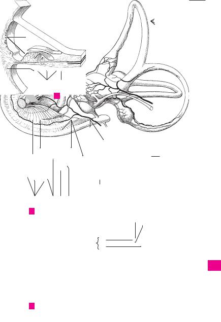

376 |
Sense organs |
|
|||
|
|
1 |
Cochlear recess. Recessus cochlearis. Depres17 |
Ampullary osseous crura. Crura ossea ampul- |
|
1 |
|||||
|
|
sion lying below and in front of the spherical |
laria. Crura of the semicircular canals dilated for |
||
|
|
|
recess. It is occupied by the lower end of the |
accomodation of the ampullae of the mem- |
|
2 |
|
|
cochlear duct. C |
branous labyrinth. B |
|
2Maculae cribrosae. Perforated bony areas 18 Cochlea. It makes 21/2−23/4 turns, measures
3 |
|
transmitting fibers of |
the vestibulocochlear |
|
8−9 mm at its base and is altogether 4−5 mm |
||
|
nerve. |
|
|
|
high. B |
||
|
|
|
|
|
|||
|
3 |
Macula cribrosa superior. Perforated bony |
19 |
Apex of cochlea. Cupula cochlea. Anteroinferi- |
|||
4 |
|||||||
|
area transmitting fibers of the utriculoampul- |
|
orly and laterally directed structure within the |
||||
|
|
lar nerve. C |
|
|
|
cranium. B |
|
|
|
|
|
|
|||
5 |
4 |
Macula cribrosa media. Perforated bony area |
20 |
Base of cochlea. Basis cochlea. Structure with a |
|||
|
|
near the base of the cochlea for passage of |
|
surface pointing approx. in the direction of the |
|||
|
|
|
|||||
|
|
fibers of the saccular nerve. C |
|
|
internal acoustic meatus. A |
||
6 |
|
|
|
||||
5 |
Macula cribrosa inferior. Perforated bony |
21 |
Spiral canal of cochlea. Canalis spiralis |
||||
|
|||||||
|
|
area in the wall of the posterior osseous am- |
|
cochleae. It is partitioned into three canals by |
|||
7 |
|
|
|||||
|
pulla for passage of fibers of the posterior am- |
|
the osseous spiral lamina and the basilar mem- |
||||
|
|
pullar nerve. C |
|
|
|
brane on one side and the vestibular wall of the |
|
|
|
|
|
|
|||
8 |
6 |
Osseous semicircular canals. Canales semi- |
|
cochlear duct on the other. A |
|||
22 |
Modiolus. Conical axis of the cochlea. It is hol- |
||||||
|
|
circulares ossei. Bony canals containing per- |
|||||
|
|
ilymph and enclosing the membranous semi- |
|
lowed out for accomodation of the cochlear |
|||
9 |
|
|
|||||
|
circular ducts (filled with endolymph). C |
|
nerve and forms the medial wall of the spiral |
||||
|
7 |
Anterior (superior) |
semicircular |
canal. |
|
canal. A |
|
|
|
||||||
10 |
|
Canalis semicircularis anterior. Canal sharing a |
23 |
Base of modiolus. Basis modioli. Beginning of |
|||
|
|
commn crus with the posterior semicircular |
|
the cochlear axis. A |
|||
|
|
|
|||||
|
|
canal and oriented vertically, somewhat per- |
24 |
Lamina of modiolus. Lamina modioli. Bony |
|||
11 |
|
||||||
|
pendicular to the axis of the petrous part of |
||||||
|
|
plate extending upward as a continuation of the |
|||||
|
|
the temporal bone. B |
|
|
|
bony spiral lamina. A |
|
12 |
|
|
|
|
|||
8 |
Posterior semicircular canal. Canalis semicir- |
|
|||||
25 |
Spiral canal of modiolus. Canalis spiralis |
||||||
|
|
cularis posterior. Posterior and most |
inferior |
|
modioli. Fine channel in the axial wall near the |
||
|
|
|
|||||
13 |
|
semicircular canal. It lies somewhat parallel to |
|
base of the bony spiral lamina. It contains the |
|||
|
the axis of the petrous part of the temporal |
|
spiral ganglion. A |
||||
|
|
bone. B |
|
|
26 |
Longitudinal canals of modiolus. Canales |
|
14 |
|
|
|
||||
9 |
Lateral semicircular canal. Canalis semicircu- |
||||||
|
longitudinales modioli. Centrally located bony |
||||||
|
|
laris lateralis. Horizontally oriented canal that |
|
||||
|
|
|
channels containing fibers of the cochlear nerve |
||||
15 |
|
sometimes creates a bulge in the medial wall of |
|
leaving the spiral ganglion. A |
|||
|
the tympanic cavity. B |
|
|
|
|||
|
|
|
|
|
|
||
10 Osseous ampullae. Ampullae osseae. Dilata-
16tions close to the base of the semicircular crura. They lodge the membranous ampullae. C
1711 Anterior osseous ampulla. Ampulla ossea anterior. Ampulla of the anterior semicircular canal located proximal to the ampulla of the
18lateral semicircular canal. B
12 Posterior osseous ampulla. Ampulla ossea
19posterior. Ampulla of the posterior semicircular canal. It lies below the plane of the lateral semi-
20circular canal. B
13 |
Lateral osseous ampulla. Ampulla ossea |
|
|
|
lateralis. Ampulla of lateral semicircular canal. |
21 |
|
|
|
It lies near the anterior semicircular ampulla. B |
|
22 |
14 |
Crura ossea. Osseous crura of the semicircular |
|
canals. |
|
15 Common osseous crus. Crus osseum com-
23mune. Posteriorly situated common limb formed by the union of the crura of the superior and posterior semicircular canals. B
2416 Simple osseous crus. Crus osseum simplex. Posteriorly situated crus of the lateral semi-
25circular canal opening independently into the wall of the vestibule. B
Feneis, Pocket Atlas of Human Anatomy © 2000 Thieme
All rights reserved. Usage subject to terms and conditions of license.

Sense organs 377
1
|
|
|
|
|
|
|
|
|
|
|
24 |
2 |
|
|
|
|
|
|
|
|
|
|
|
|
|
|
|
|
|
|
|
|
|
|
|
|
|
|
|
|
|
|
|
|
|
|
|
|
|
|
|
|
|
|
|
|
|
|
|
|
|
|
25 |
|
|
3 |
|||
|
|
|
7 |
|
|
|
|
|
|
||||
|
|
|
|
|
|
|
|
|
|||||
|
|
|
|
|
|
22 |
|
|
4 |
||||
|
|
|
|
|
|
|
|
|
|||||
|
|
|
|
|
|
|
|
|
|
|
|
26 |
|
|
|
|
|
|
|
|
|
|
|
|
|
|
|
|
|
|
|
|
|
|
|
|
|
|
5 |
||
|
|
|
|
|
|
|
|
|
|
|
|
||
|
|
|
|
|
|
|
|
|
|
|
|
|
|
|
|
|
17 |
|
23 |
|
|
|
|||||
|
|
|
|
|
|
|
|||||||
|
|
|
|
21 |
6 |
||||||||
|
|
|
|
|
|
|
|||||||
|
|
|
|
|
|
|
|
|
|
|
|||
|
|
|
|
|
|
|
|
|
Cochlea, |
|
|
|
|
|
|
|
|
|
|
|
|
A |
20 |
|
|||
|
|
|
|
|
|
|
7 |
||||||
|
|
|
|
|
|
|
|
|
longitudinal section |
||||
|
|
|
|
|
|
|
|
|
|
|
|||
15 |
|
|
|
11 |
|
|
|
|
|
|
|
||
|
|
|
|
|
|
|
|
|
8 |
||||
|
|
|
|
|
|
|
|
|
|
|
|
|
|
17 |
13 |
|
|
|
|
|
|
|
|
||||
|
|
|
|
|
|
|
|
||||||
|
|
|
|
|
|
|
9 |
||||||
9 |
|
|
|
|
|
|
|
|
|
|
|||
8 |
|
|
|
|
|
|
|
|
|
|
|
|
|
16 |
|
|
|
|
|
|
|
|
|
|
|
||
|
|
|
|
18 |
|
|
10 |
||||||
|
|
|
|
|
|
|
|
|
|||||
|
|
|
12 |
|
|
|
|
|
|
|
|
||
|
|
|
|
19 |
|
|
11 |
||||||
|
|
|
|
|
|
|
|
|
|
||||
|
|
|
|
|
|
|
|
|
|
||||
|
|
|
|
|
|
|
|
|
|
|
|
|
|
17 |
|
|
|
|
|
|
|
|
|
|
12 |
||
|
|
|
|
|
|
|
|
|
|
|
|
|
|
|
|
|
|
|
|
|
|
|
|
|
|
|
13 |
|
B |
Cast of osseous labyrinth |
|
|
|
|
|
|
|
||||
|
|
|
|
|
|
|
|
|
|||||
|
|
|
|
|
|
|
|
|
|
|
|
|
|
|
|
|
|
|
|
|
|
|
|
|
|
|
14 |
|
|
|
|
|
|
|
|
|
|
|
|
|
|
|
|
|
|
|
|
|
|
|
|
|
|
|
|
|
|
|
6 |
|
|
|
|
|
|
|
|
15 |
|
|
|
|
|
|
|
|
|
|
|
|
|
||
|
|
|
|
10 |
|
|
|
|
|
|
|
|
|
|
|
|
|
|
|
|
|
|
|
16 |
|||
|
|
|
|
|
|
|
|
|
|
|
|
|
|
|
|
|
|
|
|
|
|
|
|
|
|
|
|
|
|
|
|
|
|
|
|
|
|
|
|
|
|
|
|
|
|
|
|
|
3 |
|
|
|
|
|
17 |
|
|
|
|
|
|
5 |
4 |
|
|
|
|
|
|
|
|
|
|
|
|
|
|
|
|
|
|
||
|
|
|
|
|
|
|
|
|
|
|
18 |
||
|
|
|
|
|
|
|
|
|
|
|
|
||
|
|
|
|
|
|
|
|
|
|
|
|
|
|
1
19
20
21
22
23
C Bony labyrinth, posterior wall
24
25
Feneis, Pocket Atlas of Human Anatomy © 2000 Thieme
All rights reserved. Usage subject to terms and conditions of license.

|
378 |
Sense organs |
|
|
|
|
|
||
|
|
1 |
Bony spiral lamina. Lamina spiralis ossea. |
15 |
MIDDLE EAR. Auris media. Part of the ear com- |
||||
1 |
|||||||||
|
|
Bilayered bony plate projecting from the modi- |
|
prising the tympanic (middle ear) cavity, audi- |
|||||
|
|
|
olus into the spiral canal of the cochlea in a spi- |
|
tory tube and mastoid cells. |
|
|||
2 |
|
|
ral fashion. Together with the cochlear duct to |
16 |
Tympanic cavity. Cavitas tympanica (cavum |
||||
|
|
|
which it is attached, it forms a complete parti- |
|
tympani). Obliquely oriented space medial to |
||||
|
|
|
tion between the scala vestibuli and scala tym- |
|
|||||
3 |
|
|
|
the tympanic membrane (eardrum). It contains |
|||||
|
|
pani. B |
|
|
|||||
|
|
|
|
the auditory ossicles and communicates post- |
|||||
|
2 |
Hook of spiral lamina. Hamulus laminae spi- |
|
||||||
|
|
erosuperiorly with the mastoid air cells and an- |
|||||||
4 |
|
|
ralis. Free hook-shaped upper end of the os- |
|
teroinferiorly with the nasopharyngeal cavity |
||||
|
|
|
seous spiral lamina at the apex of the cochlea. B |
|
via the auditory tube. |
|
|||
|
|
|
|
|
|||||
5 |
3 |
Helicotrema. Confluence between scala vesti- |
17 |
Tegmental wall (tegmen tympani). Paries |
|||||
|
|
|
buli and scala tympani at the apex of the |
|
tegmentalis. Thin roof of the tympanic cavity. It |
||||
|
|
|
cochlea. It exists because the bony spiral lamina |
|
lies lateral to the arcuate eminence of the |
||||
6 |
|
|
|
||||||
|
|
and the cochlear duct end before reaching the |
|
petrous part of the temporal bone. C |
|||||
|
|
|
apex of the cochlea. B |
|
18 |
Epitympanic recess (attic). Recessus epitym- |
|||
|
|
|
|
||||||
7 |
4 |
Secondary spiral lamina. Lamina |
spiralis |
||||||
|
|
secundaria. Bony ridge situated in the lower |
|
panicus. Dome |
of tympanic |
cavity located |
|||
|
|
|
|
above the upper margin of the tympanic mem- |
|||||
|
|
|
half of the basal turn. It projects from the outer |
|
|||||
8 |
|
|
|
brane and arching superiorly and laterally. C |
|||||
|
|
wall of the spiral canal across from the bony spi- |
|
||||||
|
|
19 |
Cupular part. Pars cupularis. Upper portion of |
||||||
|
|
|
ral lamina. The inferior part of the basilar mem- |
||||||
|
|
|
|||||||
|
|
|
brane extends between these two |
spiral |
|
epitympanic recess. C |
|
||
9 |
|
|
|
|
|||||
|
|
laminae. B |
|
20 |
Jugular wall. Paries jugularis. Floor of the tym- |
||||
|
|
|
|
||||||
|
|
5 Internal acoustic (auditory) meatus. Meatus |
|
panic cavity facing the jugular fossa. C |
|||||
10 |
|
|
|||||||
|
|
acusticus internus. Arises near the posterior |
21 |
Styloid prominence. Prominentia styloidea. |
|||||
|
|
|
wall of the petrous part of the temporal bone. It |
|
Elevation on the floor of the tympanic cavity |
||||
11 |
|
|
is about 1 cm long and transmits the vestibulo- |
|
produced by the styloid process. C |
||||
|
|
cochlear and facial nerves and the labyrinthine |
|
||||||
|
|
|
22 Labyrinthine wall. Paries labyrinthicus. Medial |
||||||
|
|
|
artery and vein. A |
|
|||||
12 |
|
|
|
|
wall of tympanic cavity. C |
|
|||
6 |
Porus acusticus. Outer opening of internal |
|
|
||||||
23 |
Oval window. Fenestra vestibuli [[ovalis]]. It is |
||||||||
|
|
|
acoustic meatus into the posterior wall of the |
||||||
|
|
|
|||||||
13 |
|
|
petrous part of the temporal bone above the |
|
closed by the base of the stapes. C |
||||
|
|
jugular foramen. A |
|
24 |
Fossula fenestrae vestibuli. Small depression |
||||
|
|
|
|
||||||
|
|
|
|
||||||
|
|
7 Fundus of internal auditory meatus. Fundus |
|
in medial wall of tympanic cavity between the |
|||||
14 |
|
|
|||||||
|
|
meatus acustici interni. Floor of meatus, which |
|
malleus and incus. C |
|
||||
|
|
|
is subdivided into several fields. A |
|
25 |
Promontory. |
Promontorium. |
Prominence |
|
15 |
|
|
|
||||||
8 |
Transverse crest. Crista transversa. Trans- |
||||||||
|
caused by the basal turn of the cochlea. C |
||||||||
|
|
|
versely oriented ridge dividing the fundus of |
26 |
Sulcus promontorii. Groove on the promontory |
||||
|
|
|
|||||||
16 |
|
|
the internal acoustic meatus into an upper and |
|
produced by the tympanic nerve from the tym- |
||||
|
|
lower part. A |
|
|
|||||
|
|
|
|
|
panic plexus. C |
|
|
||
|
|
9 Facial nerve area. Area nervi facialis. Region |
|
|
|
||||
|
|
27 |
Subiculum promontorii. Small bony ridge behind |
||||||
17 |
|
||||||||
|
|
containing the beginning of the facial nerve |
|||||||
|
|
|
canal. A |
|
|
the promontory and round window. C |
|||
|
|
|
|
|
|
|
|
||
1810 Cochlear area. Area cochleae. Large region below the transverse crest. It contains the foraminous spiral tract. A
1911 Foraminous spiral tract. Tractus spiralis foraminosus. Area perforated by fibers of the spiral
20ganglion forming the cochlear part of the vestibulocochlear nerve. It corresponds to the spiral canal of the cochlea. A
2112 Superior vestibular area. Area vestibularis superior. Region lateral to the facial canal and per-
22forated by fibers of the utriculo-ampullar nerve. A
2313 Inferior vestibular area. Area vestibularis inferior. Region located lateral to foraminous spiral tract and perforated by fibers of the sacular
24nerve. A
14 Small opening for the posterior ampullar nerve.
25Foramen singulare. It lies behind the inferior vestibular area. A
Feneis, Pocket Atlas of Human Anatomy © 2000 Thieme
All rights reserved. Usage subject to terms and conditions of license.

|
|
|
|
|
|
|
|
|
|
|
|
|
|
|
Sense organs 379 |
|
|||||
|
|
|
|
|
|
|
|
|
|
|
|
|
|
|
|
|
|
|
|
|
|
|
|
|
|
|
|
|
|
|
|
|
|
|
|
|
|
|
|
|
|
|
1 |
|
|
|
|
|
|
|
|
|
|
|
|
|
|
|
|
|
|
|
|
|
|
|
|
|
|
|
|
|
|
|
|
|
|
|
|
|
|
|
|
|
|
|
|
|
|
|
|
|
|
|
|
|
|
|
|
|
|
|
|
|
|
|
|
|
2 |
|
|
|
|
|
|
|
|
|
|
|
|
|
9 |
12 |
|
|
|
|
|||
|
|
|
|
|
|
|
|
|
|
|
|
|
|
|
|||||||
|
|
|
|
|
|
|
|
|
|
|
|
|
|
3 |
|||||||
|
|
|
|
|
|
|
|
|
|
|
|
|
|
|
|
|
|
|
|
|
|
|
|
|
|
|
|
|
|
|
|
|
|
|
|
|
|
|
|
|
|
|
|
|
|
|
|
|
|
|
|
|
|
|
|
|
|
|
|
|
|
|
|
|
|
|
|
|
|
|
|
|
|
|
|
|
|
|
|
|
|
|
|
|
|
|
|
3 |
|
|
|
|
|
|
|
|
8 |
|
|
|
|
|
4 |
||||||
|
|
|
|
|
|
|
|
|
|
|
|
|
|||||||||
|
|
|
|
|
|
|
|
|
|
|
|
|
|
||||||||
|
|
|
|
|
|
||||||||||||||||
|
|
|
|
|
|
|
|
|
|
|
|
|
|
|
|
14 |
5 |
||||
|
|
|
|
|
|
|
|
|
|
|
|
|
|
|
|
||||||
|
|
|
|
|
|
|
|
|
|
|
|
|
|
|
|
|
|
|
|||
|
|
|
|
6 |
|
|
|
|
|
|
10 |
|
|
|
|||||||
|
|
|
|
|
|
|
|
|
|
|
|
|
|
|
6 |
||||||
|
|
|
|
|
|
|
|
|
|
|
|
|
|
|
|
|
|
|
|
||
|
|
|
|
|
|
2 |
|
|
|
|
|
|
|
|
|
|
|
|
|
|
|
|
|
|
|
|
|
|
|
|
|
||||||||||||
|
|
|
|
|
|
|
|
7 |
|||||||||||||
|
|
|
|
|
|
|
|
|
|
|
|
|
|
|
|
|
|
|
|
|
|
|
|
|
|
|
|
|
|
|
|
|
|
|
|
|
|
|
|
|
|
|
|
|
|
|
|
|
|
|
|
|
|
|
|
|
11 |
13 |
|
|
|
8 |
|||
1 |
|
|
|
|
|
|
|
|
|
|
|
|
|
|
|
|
|
||||
|
|
|
|
|
|
|
|
|
|
|
|
|
|
|
|
|
|
||||
|
|
|
|
|
|
|
|
|
|
|
|
Internal acoustic meatus |
|
|
|
|
|
|
|||
|
|
|
|
|
|
|
|
|
|
|
A |
|
|
|
|
|
9 |
||||
|
|
|
|
|
|
|
|
|
|
|
|
|
|
|
|
|
|
|
|
|
|
|
|
|
|
|
|
|
|
|
|
|
|
|
|
|
|
|
|
|
|
|
|
|
|
|
|
|
|
|
|
|
|
|
|
|
|
|
|
|
|
|
|
|
|
|
|
|
|
|
|
|
|
|
|
|
|
|
|
|
|
|
|
|
|
|
10 |
1 |
|
|
|
|
|
|
|
|
|
|
|
|
|
|
|
|
|||||
|
|
|
|
|
|
|
|
|
|||||||||||||
4 |
|
|
|
|
|
|
|
|
11 |
||||||||||||
|
|
|
|
|
|
|
|
|
|
|
|
|
|
|
|
||||||
|
|
|
|
|
|
|
|
|
|
||||||||||||
|
|
|
|
|
|
|
|
|
|
|
|
|
|
|
|
|
|
|
|
|
|
|
|
Section of cochlea |
|
|
|
|
|
|
|
|
12 |
||||||||||
|
B |
|
|
|
|
|
|
|
|
||||||||||||
|
|
|
|
|
|
|
|
|
|
|
|
|
|
|
|
|
|
|
|
|
|
|
|
|
|
|
|
|
|
|
|
|
|
|
|
|
|
|
|
|
|
|
|
|
|
|
|
|
|
|
|
|
|
|
|
|
|
|
|
|
|
|
|
|
13 |
|
|
|
|
|
|
|
|
|
|
|
|
|
|
|
|
|
|
|
|
|
|
|
|
|
|
|
|
|
|
|
|
|
|
|
|
|
|
|
|
|
|
|
|
19 |
|
|
|
|
|
|
|
|
|
|
14 |
||||||||||
|
|
|
|
|
|
|
|
|
|
|
|||||||||||
|
|
|
|
|
|
|
|
|
|
|
|
|
|
|
|
|
|
|
|
|
|
17 |
|
|
|
|
|
|
|
|
|
|
|
|
|
15 |
|||||||
|
|
|
|
|
|
|
|
|
|
|
|
|
|
|
|
|
|
|
|
|
|
|
|
|
|
|
|
|
|
|
|
|
|
|
|
|
|
|
|
|
|
|
|
18 |
|
|
|
|
|
|
|
|
|
|
|
|
|
16 |
|||||||
|
|
|
|
|
|
|
|
|
|
|
|
|
|||||||||
23 |
|
|
|
|
|
|
|
|
|
|
|||||||||||
|
|
|
|
|
|
|
|
|
|||||||||||||
|
|
|
|
|
|
|
|
17 |
|||||||||||||
24 |
|
|
|
|
|
|
|
|
|
|
|
||||||||||
|
|
|
|
|
|
|
|
|
|
|
|
||||||||||
|
|
|
|
|
|
|
|
|
|||||||||||||
25 |
|
|
|
|
|
|
|
|
|
||||||||||||
|
26 |
|
|
|
|
|
|
18 |
|||||||||||||
|
|
|
|
|
|
|
|||||||||||||||
|
|
|
|
|
|
|
|
|
|
|
|
|
|
|
|
|
|
|
|
|
|
|
|
|
|
|
|
|
|
|
|
|
|
|
|
|
|
|
|
|
|
|
|
27 |
|
|
|
|
|
|
|
|
|
|
|
|
|
|
19 |
||||||
21 |
|
|
|
|
|
|
|
|
|
|
|
|
|
||||||||
20 |
|
|
|
|
|
|
|
|
|
|
|
|
|
|
|
||||||
20
21
22
C Medial wall
of tympanic cavity 23
24
25
Feneis, Pocket Atlas of Human Anatomy © 2000 Thieme
All rights reserved. Usage subject to terms and conditions of license.


Sense organs 381
|
|
|
|
|
|
|
|
|
|
|
|
|
|
|
|
|
1 |
|
|
|
|
|
|
|
|
|
|
|
|
|
|
25 |
|
|
|
|
|
|
|
|
|
|
|
|
|
|
|
|
|
|
|
|
|
|
|
|
|
|
|
|
|
|
|
|
22 |
|
|
|
2 |
||
|
|
|
|
|
|
|
|
|
|
|
|
|
|
|
|||
|
|
|
|
|
|
|
|
|
|
|
|
|
|
|
|
|
|
|
|
|
|
|
|
|
|
|
|
|
|
|
|
|
|
|
|
23 |
|
|
|
22 |
|
|
|
|
|
|
|
|
|
|
|
|
3 |
26 |
|
|
|
|
|
|
|
|
24 |
|
|
|
|
|
|
||
|
|
|
|
|
|
|
|
|
|
|
|
|
|
4 |
|||
|
27 |
|
|
|
|
|
|
|
|
|
|
|
|
|
20 |
||
|
|
|
|
|
|
|
|
|
|
|
|
|
23 |
|
|||
|
|
|
|
|
|
|
|
|
|
|
|
|
|
||||
|
28 |
|
|
|
|
|
|
|
|
|
|
|
|
|
5 |
||
|
|
|
|
|
|
|
|
|
|
|
|
|
|
|
|||
|
|
|
|
|
|
|
|
|
|
|
|
|
|
|
|
||
|
|
|
|
|
|
|
|
|
|
|
|
|
|
|
|
||
|
|
|
|
|
|
|
|
|
|
|
|
|
|
|
|
|
6 |
|
|
|
|
|
|
|
|
|
|
|
|
|
|
|
|
|
|
|
|
|
|
|
|
|
|
|
|
|
|
|
|
|
|
|
|
|
|
|
|
|
|
|
|
|
|
31 |
|
|
|
|
|
|
7 |
|
|
Right eardrum, |
32 |
|
|
|
|
|
Lateral wall of |
|
|
|
|
||||
|
|
|
|
|
|
|
|
|
|
|
|
|
|
||||
|
|
|
|
|
|
|
|
|
|
|
|
|
|
||||
|
A |
|
|
|
|
29 |
|
B |
|
|
|
8 |
|||||
|
|
|
|
|
|
|
|||||||||||
|
|
external view |
33 |
|
|
|
|
|
|
|
|
tympanic cavity |
|
|
|
|
|
|
|
|
|
|
|
|
|
|
|
|
|||||||
|
|
|
|
|
|
|
|
|
|
|
|
|
|
|
|
||
9
10
11
30
12
C Attachment of eardrum
|
|
|
|
|
13 |
9 |
|
|
|
|
14 |
|
|
|
5 |
|
|
|
|
|
|
|
15 |
11 |
|
|
|
|
16 |
12 |
|
|
|
|
|
10 |
|
|
|
|
|
14 |
|
|
|
|
17 |
15 |
|
|
|
|
|
13 |
|
|
4 |
19 |
18 |
|
|
|
|||
|
1 |
3 |
2 |
18 |
|
8 |
|
19 |
|||
|
|
|
|
16 |
|
|
|
20 |
17 |
|
|
|
|
|
|
|
|
|
|
|
|
21 |
|
|
|
Medial wall of tympanic cavity |
|
|
|
|
|
|
|
|
|
|
|
|
|
D |
|
22 |
|
|
|
|
|
|
|
|
|
|
23 |
|
|
|
|
24 |
|
|
|
|
25 |
|
|
|
|
|
Feneis, Pocket Atlas of Human Anatomy © 2000 Thieme
All rights reserved. Usage subject to terms and conditions of license.
|
382 |
Sense organs |
|
|
|||
|
|
1 |
Auditory ossicles (malleus, incus and stapes). |
19 |
Incudomallear joint. Articulatio incudomal- |
||
1 |
|||||||
|
|
Ossicula auditoria (auditus). They operate col- |
|
learis. Joint between the incus and malleus. It |
|||
|
|
|
lectively as a bent lever system transferring |
|
occasionally exhibits an articular cavity. A |
||
2 |
|
|
sound waves from the tympanic membrane to |
20 |
Incudostapedial articulation. Articulatio in- |
||
|
|
|
the inner ear. |
|
cudostapedialis. Union between the lenticular |
||
|
|
|
|
|
|
||
3 |
|
2 Stapes (stirrup). A stirrup-shaped auditory os- |
|
process of the long crus of the incus and the |
|||
|
|
sicle. Its base is inserted into the fenestra vesti- |
|
stapes. A |
|||
|
|
|
|
||||
|
|
|
buli. A B |
|
21 |
Tympanostapedial syndesmosis. Syndesmo- |
|
4 |
|
|
|
||||
|
|
|
|
||||
3 |
Head of stapes. Caput stapedis. It lies opposite |
|
sis tympanostapedialis. Connective tissue at- |
||||
|
|
|
the base of the stapes and articulates with the |
|
taching the base of the stapes to the oval win- |
||
5 |
|
|
lenticular process of the incus. A B |
|
dow. It is broader anteriorly than posteriorly. B |
||
|
4 |
Anterior |
crus (limb.). Crus anterius. The vir- |
22 |
Ligaments of auditory ossicles. Ligg. os- |
||
|
|||||||
6 |
|
|
tually straight anterior limb of the stapes. A B |
|
siculorum auditoriorum. |
||
|
|
23 |
Anterior ligament of malleus. Lig. mallei an- |
||||
|
5 |
Posterior crus (limb.). Crus posterius. The |
|||||
|
|||||||
|
|
terius. Ligament that arises from the anterior |
|||||
7 |
|
|
more curved posterior limb of the stapes. A B |
|
|||
|
|
|
process of the malleus, lies in the anterior mal- |
||||
6 |
Base (footplate) of stapes. Basis stapedis. The |
|
|||||
|
|
lear fold and extends as far as the petrotym- |
|||||
8 |
|
|
plate of bone inserts into the fenestra vestibuli |
|
panic fissure. D |
||
|
|
(ovalis). A B |
|
||||
|
|
|
24 |
Superior ligament of malleus. Lig. mallei su- |
|||
|
|
7 Incus (anvil). The central ossicle situated be- |
|||||
9 |
|
|
perius. It passes from the head of the malleus to |
||||
|
|
tween the head of the malleus and the head of |
|
the roof of the epitympanic recess. C D |
|||
|
|
|
the stapes. A D |
25 |
Lateral ligament of malleus. Lig. mallei |
||
10 |
|
|
|||||
8 |
Body of incus. Corpus incudis. It articulates |
||||||
|
laterale. It unites the neck of the malleus with |
||||||
|
|
|
with the malleus by means of a saddle-shaped |
|
the upper margin of the tympanic notch. C |
||
|
|
|
|
||||
11 |
|
|
joint. A |
|
26 |
Superior ligament of incus. Lig. incudis su- |
|
9 |
Long crus (limb.). Crus longum. Long process |
||||||
|
|
perius. Courses approximately parallel to the |
|||||
|
|
||||||
12 |
|
|
which projects almost vertically downward be- |
|
superior ligament of the malleus and connects |
||
|
|
hind the manubrium of the malleus with the |
|
the body of the incus with the roof of the |
|||
|
|
|
|
||||
|
|
|
lenticular process at its tip. A |
|
epitympanic recess. C D |
||
13 |
|
|
|
||||
10 |
Lenticular process. Processus lenticularis. Small |
27 |
Posterior ligament of incus. Lig. incudis post- |
||||
|
|
|
bony projection on the tip of the long crus. It ar- |
|
erius. It passes from the short limb of the incus |
||
14 |
|
|
ticulates with the stapes. A |
|
to the lateral wall of the tympanic cavity. C D |
||
|
11 |
Short crus (limb). Crus breve. Small posteri- |
28 |
Stapedial membrane. Membrana stapedialis. |
|||
|
|||||||
15 |
|
|
orly directed process attached to the incudal |
|
Thin membrane between the limbs and base of |
||
|
|
fossa by a ligament. A |
|
the stapes. B |
|||
|
|
|
|
||||
|
12 |
Malleus [[hammer]]. It lies between the tym- |
29 |
Anular ligament of stapes. Lig. anulare stape- |
|||
16 |
|||||||
|
|
panic membrane and the incus. A C |
|
diale. Ligament situated between the base of |
|||
|
13 |
Handle |
(manubrium) of malleus. Manu- |
|
the stapes and the margin of the oval window. It |
||
17 |
|
is broader anteriorly than posteriorly. B |
|||||
|
|
brium mallei. Its outer surface is fused to the |
|
||||
|
|
|
|
||||
|
|
|
tympanic membrane as far as the lateral |
|
|
||
18 |
|
|
process. A |
|
|
||
|
14 Head of malleus. Caput mallei. It has a convex |
|
|
||||
|
|
|
|||||
19 |
|
|
articular surface for accomodation of the body |
|
|
||
|
|
of the incus. A |
|
|
|||
|
|
|
|
|
|||
|
15 Neck of malleus. Collum mallei. Structure con- |
|
|
||||
20 |
|
|
|||||
|
|
necting the head and manubrium of the mal- |
|
|
|||
|
|
|
leus. A |
|
|
|
|
21 |
|
|
|
|
|
||
16 |
Lateral process of malleus. Processus later- |
|
|
||||
|
|
|
alis. Short process that projects from the end of |
|
|
||
22 |
|
|
the manubrium and produces the mallear |
|
|
||
|
|
|
prominence. A |
|
|
||
|
17 |
Anterior process of malleus. Processus ante- |
|
|
|||
23 |
|
|
|||||
|
|
|
rior. Long, very thin process. It extends into the |
|
|
||
24 |
|
|
petrotympanic fissure in the newborn, but |
|
|
||
|
|
diminishes in the adult. A |
|
|
|||
18 Articulations of auditory ossicles. Articula-
25tiones ossiculorum auditoriorum. They are not true joints but syndesmoses.
Feneis, Pocket Atlas of Human Anatomy © 2000 Thieme
All rights reserved. Usage subject to terms and conditions of license.

|
384 |
Sense organs |
|
|
|
|
|||
|
|
1 |
Muscles of auditory ossicles. Musculi os- |
13 |
AUDITORY |
(PHARYNGOTYMPANIC, EUSTA- |
|||
1 |
|||||||||
|
|
siculorum auditoriorum. The following two |
|
CHIAN) TUBE. Tuba auditoria (auditiva). Narrow |
|||||
|
|
|
muscles are attached to the ossicles. |
|
4 cm long, partly cartilaginous, partly bony tube |
||||
2 |
2 |
M. tensor tympani. Muscle located in the sem- |
|
between the middle ear and the nasopharynx |
|||||
|
|
|
icanal for the tensor tympani muscle above the |
|
for aeration of the tympanic cavity. A C |
||||
|
|
|
|
||||||
|
|
|
auditory tube. Its tendon bends laterally around |
14 |
Tympanic ostium of auditory tube. Ostium |
||||
3 |
|
|
|||||||
|
|
the cochleariform process almost at a right |
|
tympanicum tubae auditoriae. Opening of the |
|||||
|
|
|
angle and inserts at the base of the manubrium |
|
auditory tube on the anterior wall of the tym- |
||||
4 |
|
|
of the malleus. I: Mandibular nerve. A |
|
panic cavity. It usually lies slightly above the |
||||
|
3 |
M. stapedius. Muscle that originates in a bony |
|
floor of the tympanic cavity. A |
|||||
|
|
||||||||
5 |
|
|
canal on the posterior wall of the tympanic cav- |
15 |
Osseous part of auditory tube. Pars ossea |
||||
|
|
ity. Its tendon passes through an opening at the |
|
tubae auditoriae. Laterally and posterosuperi- |
|||||
|
|
|
apex of the pyramid before inserting at the head |
|
orly situated portion that involves about 1/3 of |
||||
6 |
|
|
of the stapes. By tilting the stapes, it exerts a |
|
the tube’s length. It lies below the semicanal for |
||||
|
|
|
dampening effect on sound vibrations reaching |
|
the tensor tympani muscle and emerges be- |
||||
|
|
|
the inner ear. I: Nerve to stapedius from the fa- |
|
tween the carotid canal and the foramen spino- |
||||
7 |
|
|
|
||||||
|
|
cial nerve. B |
|
|
|
sum. A |
|
||
|
4 |
Mucous membrane of tympanic cavity. Tunica |
16 |
Isthmus [tubae auditoriae]. Narrow portion of |
|||||
8 |
|
|
mucosa cavitatis tympanicae. It consists of |
|
the tube between the cartilaginous and bony |
||||
|
|
|
simple squamous and/or cuboidal epithelium |
|
parts of the auditory tube. A |
||||
|
|
|
and a delicate vascular-rich lamina propria. |
17 |
Air cells. Cellulae pneumaticae. Small fossae in |
||||
9 |
|
|
|||||||
5 |
Posterior mallear fold. Plica mallearis poste- |
|
the wall of the osseous part of the auditory tube. |
||||||
|
|
|
rior. Fold extending from the base of the handle |
|
A |
|
|||
10 |
|
|
of the malleus to the posterior, upper part of the |
18 |
Cartilaginous part of auditory tube. Pars car- |
||||
|
|
|
tympanic ring. It contains the posterior portion |
|
tilaginea [tubae auditoriae]. Located anterome- |
||||
|
|
|
|
||||||
|
|
|
of the chorda tympani. D |
|
dially and has a length of about 2.5 cm. A |
||||
11 |
|
|
|
||||||
6 |
Anterior mallear fold. Plica mallearis anterior. |
19 |
Cartilage of auditory tube. Cartilago tubae |
||||||
|
|||||||||
|
|
|
Fold extending from the base of the manubrium |
|
auditoriae. Hook-shaped in cross section. It be- |
||||
|
|
|
|
||||||
12 |
|
|
to the anterior, upper part of the tympanic ring. |
|
comes lower lateroposteriorly and contains |
||||
|
|
|
It contains the anterior segment of the chorda |
|
elastic cartilage only in the angle between the |
||||
|
|
|
|
||||||
|
|
|
tympani, the anterior process of the malleus |
|
two cartilaginous laminae. A |
||||
13 |
|
|
|
||||||
|
|
and the anterior ligament of the malleus. D |
20 |
Medial cartilaginous lamina. Lamina (car- |
|||||
|
|
7 Fold of chorda tympani. Plica chordae tym- |
|
tilaginis) medialis. Broad plate of cartilage. C |
|||||
14 |
|
|
|||||||
|
|
pani. Fold created by the chorda tympani on the |
21 |
Lateral cartilaginous lamina. Lamina (car- |
|||||
|
|
|
neck of the malleus between the two above- |
|
tilaginis) |
lateralis. Narrow anterolaterally |
|||
|
|
|
|
||||||
15 |
|
|
mentioned folds. D |
|
|
directed plate of cartilage. C |
|||
|
7 a Recess of |
tympanic |
membrane. Recessus |
|
|||||
|
22 |
Membranous lamina. Lamina membranacea. |
|||||||
|
|
||||||||
|
|
|
membranae tympanicae. Mucosal pouch in the |
||||||
|
|
|
|
Membranous portion of the wall of the car- |
|||||
16 |
|
|
|
||||||
|
|
tympanic cavity. |
|
|
|||||
|
|
|
|
tilaginous part of the auditory tube. A C |
|||||
|
8 |
Anterior recess of tympanic membrane. Re- |
23 |
Tunica mucosa. Mucous membrane of auditory |
|||||
17 |
|
|
cessus [membranae tympani] anterior. Mucosal |
|
tube lined by a simple ciliated epithelium. C |
||||
|
|
|
pouch between the anterior mallear fold and |
24 |
Glands of auditory tube. Glandulae tubariae. |
||||
|
|
|
the tympanic membrane. D |
||||||
18 |
|
|
|
Mucous glands especially in the cartilaginous |
|||||
9 |
Superior recess of tympanic membrane |
|
|||||||
|
part of the tube. C |
||||||||
|
|
|
(Prussak’s |
pouch). |
Recessus [membranae |
25 Pharyngeal opening of auditory tube. Ostium |
|||
19 |
|
|
tympani] superior. Recess bounded laterally by |
||||||
|
|
|
pharyngeum tubae auditoriae. Funnel-shaped |
||||||
|
|
|
the flaccid part of the tympanic membrane, me- |
|
to slit-like opening above the levator eminence |
||||
|
|
|
dially by the head and neck of the malleus and |
|
|||||
20 |
|
|
|
at the level of the inferior nasal meatus 1 cm |
|||||
|
|
the body of the incus. D |
|
||||||
|
|
|
lateral and anterior to the posterior wall of the |
||||||
|
10 |
Posterior recess of tympanic membrane. Re- |
|
||||||
|
|
pharynx. A |
|
||||||
21cessus [membranae tympani] posterior. Mucosal pouch between the posterior mallear fold and tympanic membrane. D
2211 Incudal fold. Plica incudialis. Mucosal fold extending from the roof of the epitympanic recess
23to the head of the incus or from the short limb of the incus to the posterior wall of the tympanic cavity. D
2412 Stapedial fold. Plica stpedialis. Mucosal fold extending from the posterior wall of the tympanic
25cavity to the stapes. It covers the stapedius muscle and the stapes. B
Feneis, Pocket Atlas of Human Anatomy © 2000 Thieme
All rights reserved. Usage subject to terms and conditions of license.


|
386 |
Sense organs |
|
|
||
|
|
1 |
EXTERNAL EAR. Auris externa. The part of the |
20 |
Cymba conchalis. Upper, slit-like part of the |
|
1 |
||||||
|
|
ear consisting of the auricle (pinna) and exter- |
|
concha between the crura of helix and anthelix. |
||
|
|
|
nal acoustic meatus. |
|
A |
|
2 |
|
|
|
|||
2 |
External acoustic (auditory) meatus (canal). |
21 |
Cavity of concha. Cavitas (cavum) conchalis. |
|||
|
|
|
Meatus acusticus externus. Flat, partly car- |
|
Main part of the concha located below the crus |
|
|
|
|
|
|||
3 |
|
|
tilaginous, partly bony, S-shaped canal about |
|
of the helix and behind the tragus. A |
|
|
|
2.4 cm long with a diameter of about 6 mm. C |
22 |
Antitragus. Small tubercle present on the infe- |
||
|
|
|
||||
|
3 |
External opening of acoustic canal. Porus |
|
rior continuation of the anthelix and separated |
||
4 |
|
|||||
|
|
acusticus extrnus. C |
|
from the tragus by the intertragic incisure. A D |
||
|
|
3 a Tympanic notch. Incisura tympanica. Defect |
23 |
Tragus. Flat projection in front of the external |
||
|
|
|||||
5 |
|
|
between the greater and lesser tympanic |
|
opening of the acoustic canal. A |
|
|
|
|
spines. In the newborn, it is the superior gap be- |
24 |
Anterior notch. Incisura anterior (auris). It lies |
|
|
|
|
||||
|
|
|
tween the still free ends of the tympanic ring. |
|
between the tragus (supratragic tubercle) and |
|
6 |
|
|
|
|||
|
|
See p. 16.8 |
|
the crus of the helix. A |
||
|
4 |
Cartilaginous part of external acoustic mea- |
25 |
Intertragic incisure. Incisura intertragica. |
||
7 |
||||||
|
|
tus. Meatus acusticus externus cartilagineus. |
|
Notch between tragus and antitragus. A D |
||
|
|
|
Lateral, cartilaginous third of the external |
26 |
Auricular [[Darwin’s]] tubercle. [Tuberculum |
|
|
|
|
||||
8 |
|
|
acoustic meatus. C |
|
auriculare]. It is occasionally present on the |
|
5 |
Cartilage of external acoustic meatus. Car- |
|
||||
|
|
anterior margin of the helix from which it ex- |
||||
|
|
|
tilago meatus acustici. Together with the car- |
|
tends posteroinferiorly. A |
|
9 |
|
|
|
|||
|
|
tilage of the pinna, it forms a groove that opens |
27 Apex of auricle. [Apex auricularis]. Outer mar- |
|||
|
|
|
superiorly and posteriorly. D |
|||
|
|
|
|
gin of the auricular cartilage (helix) which, |
||
10 |
6 |
Incisurae cartilaginis meatus acustici. Two |
|
when present, projects backward, upward and |
||
|
|
|
fissures in the cartilage of the external acoustic |
|
outward. B |
|
|
|
|
|
|||
|
|
|
meatus. They are bridged by connective tissue |
28 Posterior auricular sulcus. Sulcus auricularis |
||
11 |
|
|
||||
|
|
and are usually directed anteriorly. D |
||||
|
|
|
posterior. Shallow indentation between the an- |
|||
|
|
7 Lamina tragi. Lateral part of the meatal car- |
|
|||
|
|
|
titragus and anthelix. A |
|||
12 |
|
|
tilage. It lies in front of the external opening of |
29 |
Supratragic tubercle. [Tuberculum supratragi- |
|
|
|
|
the acoustic meatus. D |
|
cum]. Small tubercle occasionally present at the |
|
|
|
|
|
|||
13 |
8 |
Auricle (Pinna). Auricula. A B |
|
upper end of the tragus. A |
||
|
|
|
|
|
||
9Ear lobe. Lobulus auricularis. Lower end of the pinna devoid of cartilage. A B C
14 |
10 |
Auricular cartilage. Cartilago auricularis. |
|
|
|||
|
|
Structural framwork of the pinna consisting of |
|
15 |
|
||
|
elastic cartilage. D |
||
|
11 Helix. External, curved margin of the auricle. A |
||
|
|||
16 |
|
B C D |
|
|
12 |
Crus of helix. Crus helicis. Site of origin of the |
|
|
|||
17 |
|
helix in the cavity (concha) of the auricle. A B D |
|
13 |
Spine of helix. Spina helicis. Small, forward- |
||
|
|||
|
|
projecting cartilaginous prominence on the |
|
18 |
|
||
|
crus helices. D |
||
|
14 Tail of helix. Cauda helicis. Posterior, inferior |
||
|
|||
19 |
|
end of the helix separated from the antitragus |
|
|
|
by an indentation. D |
|
|
|
||
20 |
15 |
Antihelix [[anthelix]]. Arched projection located |
|
|
|
in front of the posterior part of the helix. A B C D |
|
|
16 |
Tringular fossa. Fossa triangularis. Antero- |
|
21 |
|||
|
superiorly located fossa enclosed by the two |
||
|
|
crura of the anthelix. A D |
|
22 |
|
||
17 |
Crura of anthelix (antihelix). Crura antheli- |
||
|
|
cis. Formed by the bifurcation of the anthelix |
|
|
|
||
23 |
|
superiorly, the two crura form the boundary of |
|
|
the triangular fossa. A D |
||
24 |
18 Scaphoid fossa. Scapha. Narrow fossa situated |
||
|
posteriorly between the helix and anthelix. A D |
||
19 Concha (cavity) of auricle. Concha auricularis.
25It is embraced by the anthelix, antitragus and tragus. A
Feneis, Pocket Atlas of Human Anatomy © 2000 Thieme
All rights reserved. Usage subject to terms and conditions of license.

 16
16
|
388 |
Sense organs |
|
|
|
|
||
|
|
1 Isthmus of auricular cartilage. Isthmus car- |
19 M. antitragicus. Muscle fibers |
on the anti- |
||||
1 |
|
|||||||
|
|
tilaginis auricularis. Narrow bridge of cartilage |
|
tragus. They extend partly to the tail of the |
||||
|
|
|
connecting the cartilage of the external acoustic |
|
helix. B |
|
|
|
2 |
|
|
meatus and tragic lamina with the auricular |
20 M. transversus auricularis. Muscle located on |
||||
|
|
|
cartilage. A |
|
the posterior surface of the auricular cartilage. |
|||
|
|
2 Terminal notch. Incisura terminalis auricularis. |
|
|||||
3 |
|
|
It extends between the eminences of the sca- |
|||||
|
|
Deep notch separating the lamina tragi from the |
|
phoid fossa and concha. C |
|
|||
|
|
|
|
|
||||
|
|
|
auricular cartilage. A |
21 M. obliquus auricularis. It lies between the |
||||
4 |
|
|
||||||
3 |
Fissura antitragohelicina. Deep fissure sepa- |
|||||||
|
eminences of the concha and triangular fossa. C |
|||||||
|
|
|
rating the antitragus and helix inferiorly as well |
22 [M. incisurae helicis]. Muscle fibers occasion- |
||||
5 |
|
|
as the anthelix and helix further superiorly. A |
|||||
|
|
|
ally present in the caudal continuation of trans- |
|||||
|
|
4 Transverse sulcus of anthelix. [[Sulcus antheli- |
|
versus auricularis muscle. C |
|
|||
|
|
|
|
|||||
6 |
|
|
cis transversus]]. Posteromedial depression vis- |
23 |
OLFACTORY ORGAN. Organum olfactorium (ol- |
|||
|
|
ible between the eminences of the triangular |
||||||
|
|
|
|
factus). Organ of smell. |
|
|||
|
|
|
fossa and concha. A |
|
|
|||
7 |
|
|
24 Nasal mucosa of the olfactory region. Regio |
|||||
5 |
Groove of crus of helix. Sulcus cruris helicis. |
|||||||
|
|
|
Shallow furrow on the posterior surface of the |
|
olfactoria tunicae mucosae nasi. This region, |
|||
|
|
|
|
which is covered by olfactory epithelium, is |
||||
8 |
|
|
auricular cartilage. It corresponds to the crus of |
|
||||
|
|
|
about the size of a |
nickel and |
occupies the |
|||
|
|
|
the helix on the anterior surface. A |
|
lateral and medial walls and roof (below the cri- |
|||
|
|
6 Fossa of anthelix. Fossa anthelicis. Furrow on |
|
|||||
9 |
|
|
briform plate) of the nasal cavity at the level of |
|||||
|
|
posterior surface of auricular cartilage corre- |
|
the superior nasal concha. E F |
|
|||
|
|
|
|
|
||||
|
|
|
sponding to the anthelix on the anterior sur- |
25 Olfactory glands (of Bowman). Glandulae ol- |
||||
10 |
|
|
||||||
|
|
face. A |
||||||
|
|
|
factoriae. Serous glands found below the ol- |
|||||
|
7 |
Eminence of concha. Eminentia conchae. |
|
|||||
|
|
factory epithelium. Their predominantly serous |
||||||
11 |
|
|
Elevation on the posterior surface of the auricu- |
|
secretions can enhance or wash away odorous |
|||
|
|
|
lar cartilage corresponding to the cavity of the |
|
substances. F |
|
|
|
|
|
|
concha. A |
26 |
GUSTATORY ORGAN. |
Organum |
gustatorium |
|
12 |
|
|
||||||
|
8 Eminence of scaphoid fossa. Eminentia |
|||||||
|
|
|
(Gustus). Organ of taste, comprised of all the |
|||||
|
|
|
scaphae. Curved elevation on the posterior sur- |
|
taste buds. |
|
|
|
13 |
|
|
|
|
|
|||
|
|
face of the auricular cartilage corresponding to |
27 Taste bud. Caliculus gustatorius (gemma gusta- |
|||||
|
|
|
the scaphoid fossa on the anterior surface. A |
|||||
|
|
|
|
toria). Occupying the full thickness of the |
||||
|
9 |
Eminence of triangular fossa. Eminentia fossae |
|
|||||
14 |
|
epithelium, it consists of supporting cells and |
||||||
|
|
|
triangularis. Elevation on the posterior surface |
|
taste cells. Each of the taste cells has microvilli |
|||
|
|
|
of the auricular cartilage corresponding to the |
|
on its surface which serve as chemoreceptors. |
|||
15 |
|
|
|
|||||
|
|
triangular fossa. A |
|
Distribution of taste buds: aggregated in the |
||||
|
10 |
Auricular ligaments. Ligg. auricularia. Liga- |
|
epithelium of vallate (circumvallate) and foliate |
||||
16 |
|
|
ments of the pinna. They attach the auricular |
|
papillae; solitary on the outside of the tongue. D |
|||
|
|
|
cartilage to the temporal bone. |
28 Taste pore. Porus gustatorius. Space left by the |
||||
|
|
|
||||||
17 |
11 |
Anterior auricular ligament. Lig. auriculare an- |
|
epithelium above the apex of the taste bud. Mi- |
||||
|
crovilli project into it. D |
|
||||||
|
|
|
terius. It extends from the root of the zygomatic |
|
|
|||
|
|
|
arch to the spine of the helix. B |
|
|
|
|
|
1812 Superior auricular ligament. Lig. auriculare superius. It passes from the upper margin of the
19osseous external auditory meatus to the spine of the helix. B
2013 Posterior auricular ligament. Lig. auriculare posterius. It extends from the eminence of the concha to the mastoid process. C
2114 Mm. auriculares. Muscles of the pinna.
15 M. helicis major. It passes upward from the
22spine of the helix to the helix. B
16 M. helicis minor. It lies on the crus of the helix.
23 B
17 M. tragicus. It lies vertically on the lamina tragi.
24 B
18 M. pyramidalis auricularis. Muscle fibers that
25occasionally split off from the tragicus muscle and pass to the spine of the helix. B
Feneis, Pocket Atlas of Human Anatomy © 2000 Thieme
All rights reserved. Usage subject to terms and conditions of license.

 1
1
 24
24