Добавил:
Upload Опубликованный материал нарушает ваши авторские права? Сообщите нам.
Вуз: Предмет: Файл:
ATLAS Feneis, Daubner - Pocket Atlas of Human Anatomy.pdf
Скачиваний:
323
Добавлен:
15.02.2016
Размер:
12.18 Mб
Скачать


320


Cranial nerves


Pars peripherica

14

Decussation of trochlear nerve. Decussatio


1


1


Peripheral nervous system.


(systema nervosum periphericum). The periph-


nervorum trochlearium. The crossing of


eralpartofthenervoussystemwhichincludesall


trochlear nerve fibers in the superior medul-

2


peripheral conducting tracts (nerves). The


lary velum. B


border between it and the central nervous sys-

15

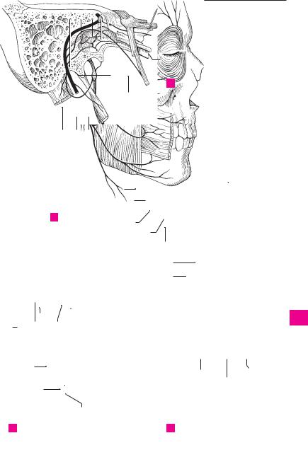
TRIGEMINAL NERVE (V). N. trigeminus [V]. Fifth

3


temliesatthesurfaceofthebrainandspinalcord.


cranial nerve (nerve of the 1st pharyngeal arch).


2

CRANIAL NERVES. Nervi craniales (en-


Nerve that exits laterally from the pons with


cephalici). The 12 pairs of nerves connected


two groups of fibers, supplies the masticatory

4


with the brain. With the exception of the


muscles and provides sensory innervation to


trochlear (IV), all of them emerge from the base


the face. B C


5


of the brain and exit through the base of the

16

Sensory root of trigeminal nerve. Radix sen-


skull (in contrast to the spinal nerves). Area of


soria [portio major]. Sensory part which exits


distribution: head, neck, as well as the thorax


from the pons caudally and enters the trigemi-

6


and abdomen (via vagus nerve).


nal ganglion. C


3

OLFACTORY NERVE (I). Nn. olfactorii (I). First

17


Trigeminal (semilunar, gasserian)

ganglion.

7


cranial nerve, which is formed by about 20


Ganglion trigeminale [[semilunare;

Gasseri]].


small bundles of nonmyelinated axons from the


Semilunar ganglion that is equivalent to a spi-


olfactory cells. It passes through the cribriform


nal ganglion. It is located in an outpocketing of

8


plate of the ethmoid into the olfactory bulb


the subarachnoid space (cavum trigeminale)


(synaptic site). A


above the foramen lacerum at the medial, ante-

9


4 OPTIC NERVE (II). N. opticus [II]. Second cranial


rior border of the petrous part of the temporal


nerve which leaves the eyeball medial to the


bone. C


posterior optic pole and extends up to the optic

18

Motor root. Radix motoria [portio minor].

10


chiasm. B C


Motor portion of trigeminal nerve for innerva-


5

OCULOMOTOR NERVE (III). N. oculomotorius


tion of the masticatory muscles. It is situated

11


[III]. Third cranial nerve, which exits from the


cranially at the exit of the trigeminal nerve and


sulcus on the medial side of the cerebral


below the trigeminal ganglion. C


peduncle. This motor nerve (somatic and

19

Ophthalmic nerve. N. ophthalmicus. First divi-

12


visceral) passes into the orbit through the su-


sion (branch) of trigeminal nerve. It passes


perior orbital fissure. B C


through the superior orbital fissure. C

13


6

Superior ramus (division). Ramus superior.

20

Tentorial (meningeal) branch. Ramus ten-


Superior branch for the superior rectus and le-


torii (meningeus). Recurrent nerve for the ten-

14


vator palpebrae superioris muscles. B


torium cerebelli and falx cerebri. C


7

Inferior ramus (division). Ramus inferior. In-

21

Lacrimal nerve. N. lacrimalis. Passes laterally


ferior branch for the medial and inferior recti


through the superior orbital fissure and sup-

15


and inferior oblique muscles. B


plies the lacrimal gland, conjunctiva and lateral


8

Ciliary ganglion. Ganglion ciliare. Located


portion of upper eyelid. C


16


about 2 cm behind the eyeball and lateral to the

22

Communicating

ramus

with zygomatic


optic nerve. This parasympathetic ganglion


nerve. Ramus communicans [cum. n. zygomat-


serves as a relay station for fibers innervating


ico]. Connection to the zygomatic nerve with

17


the ciliary and sphincter pupillae muscles. B


autonomic fibers extending from the pterygo-


9

Parasympathetic (motor) root. Radix para-


palatine ganglion to the lacrimal gland. C


sympathetica (oculomotoria). Branch of the

23

Frontal nerve. N. frontalis. Nerve that enters


18


oculomotor nerve with preganglionic, para-


the orbit through the superior orbital fissure. It


sympathetic fibers projecting to the ciliary gan-


lies on the levator palpebrae superioris and


19


glion. B


continues toward the forehead. C; see also

10

Short ciliary nerves. Nn. ciliares breves.


p. 323 A


Several (up to 20) nerves penetrating the sclera

24

Supraorbital nerve. N. supraorbitalis. Thickest

20


above and below the optic nerve and carrying


branch of the frontal nerve. It supplies the con-


postganglionic, parasympathetic and sympa-


junctiva, upper eyelid, frontal sinus and the


21


thetic fibers. B


skin of the forehead. C


11

Sympathetic root. Radix sympathetica. Fine,

25

Lateral branch. Ramus lateralis. It passes


postganglionic fiber tract from the internal


through the supra-orbital notch. C


22


carotid plexus with no synapses in the ciliary

26

Medial branch. Ramus medialis. It passes medi-


ganglion. B


ally through the frontal notch. C


23

12

Sensory root. Radix sensoria (nasociliaris).

27

Supratrochlear

nerve.

N. supratrochlearis.


Fine, long connection with afferent fibers to the


Thin, medial branch of frontal nerve. It divides


nasociliary nerve. B


at the medial angle of the eye to form an as-

24


13

TROCHLEAR NERVE (IV). N. trochlearis [IV].


cending and descending branch. C


Fourth cranial nerve. Thin nerve exiting dorsal


25and caudal to the tectal lamina and supplying the superior oblique muscle. B

Feneis, Pocket Atlas of Human Anatomy © 2000 Thieme

All rights reserved. Usage subject to terms and conditions of license.

Cranial nerves 321

1

2

3


3


3


4


5


6


Olfactory nerve


7


A


8


6


9


12

11

4


13

5


8


10


14


11


10


9


12


15


13


7


14


15


Oculomotor and trochlear nerves


B


24


26

16


25


23

21


17


27


18


20


5


4

22


19


19


18


322.1


20


16

21

22

23

15 17

24

C Ophthalmic nerve

25

Feneis, Pocket Atlas of Human Anatomy © 2000 Thieme

All rights reserved. Usage subject to terms and conditions of license.

322 Cranial nerves

1Nasociliary nerve. N. nasociliaris. Most medial 1 branch of the ophthalmic nerve. It courses below the superior rectus and between the superior ob-

2lique and medial rectus. A

2Communicating branch of nasociliary nerve

with ciliary ganglion. Ramus communicans

3[cum ganglio ciliari]. Any branch carrying


sensory fibers from the eye through the ciliary

4

ganglion to the nasociliary nerve. A

Long ciliary nerves. Nn. ciliares longi. Two long,

3

fine twigs with sympathetic fibers supplying the

5dilatator pupillae muscle and afferent fibers from the iris, ciliary body and cornea. A

64 Posterior ethmoidal nerve. N. ethmoidalis posterior. Thin nerve at the posterior end of the orbit supplying the sphenoidal sinus and poste-

7rior ethmoidal cells. A

5Anterior ethmoidal nerve. N. ethmoidalis ante-

8rior. Nerve that enters the cranial cavity (extradural part) through the anterior ethmoidal foramen. It then courses through the cribriform

9plate of the ethmoid into the nasal cavity. A B C

6Nasal branches of anterior ethmoidal nerve. Rami

10nasales [n. ethmoidalis anterior]. Collective term for the following four branches of the anterior ethmoidal nerve.

117 Internal nasal branches. Rami nasales interni.


Rami that innervate the nasal mucosa in front of

12

8

the conchae and for the anterior nasal septum. B


Lateral nasal branches. Rami nasales laterales. In-


13

9

nervate the anterior part of lateral nasal wall. B

Medial nasal branches. Rami nasales mediales. In-


nervate the anterior part of the nasal septum. C

1410 External nasal branch. Ramus nasalis externus. Innervates the skin on the tip of the nose and the

15nasal ala; it passes through the ethmoidal sulcus of the nasal bone. B

16

11

Infratrochlear nerve. N. infratrochlearis. It


passes below the trochlea of the superior oblique


at the inner angle of the eye and supplies the lacri-

17

mal sac, lacrimal caruncle and surrounding skin. A


12Palpebral branches. Rami palpebrales. Rami for part of the upper and lower eyelids. A

1813 Maxillary nerve. N. maxillaris. Second division (branch) of trigeminal nerve. It passes through the

19foramen rotundum to the pterygopalatine fossa and subsequently through the inferior orbital fissure into the orbit. A C

2014 Meningeal nerve. Ramus meningeus [medius]. Branch given off in front of the foramen rotundum.

21It supplies the dura in the region of the frontal branch of the middle meningeal artery. A

2215 Ganglionic branches. Rami ganglionici (ganglionares). Usually two rami from the pterygopalatine ganglion. They contain autonomic fibers for the

23lacrimal gland and sensory fibers from the periosteum of the orbit. A

24

16

Pterygopalatine ganglion. Ganglion pterygo-


palatinum. Parasympathetic ganglion located in


the corresponding fossa close to the spheno-

25


palatine foramen. Their postganglionic fibers in-


nervate the lacrimal and nasal glands. A B C

16 a

Parasympathetic root. Radix parasympathetica.


Communicates with the greater petrosal nerve.

16 b

Sympathetic root. Radix sympathetica. Com-


municates with the deep petrosal nerve.

16 c

Sensory root. Radix sensoria. Communicates


with the maxillary nerve.

17Orbital branches. Rami orbitales. Two to three fine rami which pass into the orbit through the inferior orbital fissure, then through the bone to the posterior ethmoidal cells and to the sphenoidal sinus. B C

18Lateral posterior superior nasal branches.

Rami nasales posteriores superiores laterales. Up to 10 fine rami which pass through the sphenopalatine foramen to the superior and middle nasal conchae and to the posterior ethmoidal cells. B

19Medial posterior superior nasal branches.

Rami nasales posteriores superiores mediales. Two to three branches which pass through the sphenopalatine foramen to the upper part of the nasal septum. C

20Nasopalatine nerve. N. nasopalatinus [[incisivus]]. It passes between the periosteum and mucosa of the nasal septum, then through the incisive canal to the anterior part of the palatine mucosa and the gingiva of the upper incisor teeth. C

20 a Long nasopalatine nerve. Nervus nasopalatinus longus.

20 b Branches to nasal septum. Rami septales nasales.

20 c Short nasopalatine nerves. Nervi nasopalatini breves.

20 d Lateral nasal branches. Rami nasales laterales.

20 e Branches to maxillary sinus. Rami sinus maxillaris.

21Pharyngeal nerve. Nervus pharyngeus. Fine nerve for the pharyngeal mucosa. B

22Greater palatine nerve. N. palatinus major. After passing through the greater palatine canal, it courses through the corresponding foramen and supplies the mucosa of the hard palate and its glands. B

23Posterior inferior nasal branches. Rami nasales posteriores inferiores. Rami for the middle and inferior nasal meatuses as well as the inferior nasal concha. B

24Lesser palatine nerves. Nn. palatini minores. They travel in their respective, slender canals, exit through the lesser palatine foramina and supply the soft palate. B

24 a Tonsillar branches. Rami tonsillares. Branches to the palatine tonsil.

25Zygomatic nerve. N. zygomaticus. It branches into the pterygopalatine fossa, passes through the inferior orbital fissure to the lateral wall of the orbit and provides an anastomotic branch to the lacrimal gland. A

26Zygomaticotemporal branch. Ramus zygomaticotemporalis. It passes through the respective foramen to the lateral wall of the orbit. A

27Zygomaticofacial branch. Ramus zygomticofacialis. It passes through the corresponding foramen to the skin on the zygomatic bone. A

Feneis, Pocket Atlas of Human Anatomy © 2000 Thieme

All rights reserved. Usage subject to terms and conditions of license.

Cranial nerves 323

5

11

14

320.23


2

3


12


25


13

15


16


26

14


27


5


A Nasociliary

and maxillary nerves


17

7

16


10

18

8


21


23


22


24


B Pterygopalatine ganglion and anterior ethmoidal nerve

5

13


17


19

9


16


20

C Nerves of nasal septum

Feneis, Pocket Atlas of Human Anatomy © 2000 Thieme

All rights reserved. Usage subject to terms and conditions of license.

1

2

3

4

5

6

7

8

9

10

11

12

13

14

15

16

17

18

19

20

21

22

23

24

25


324

Cranial nerves


1 Infraorbital nerve. N. infraorbitalis. Terminal

17 Nerve to lateral pterygoid. N. pterygoideus

1


branch of maxillary nerve. It passes through the

lateralis. Motor nerve for the corresponding


inferior orbital fissure and corresponding sul-

muscle. It frequently arises together with the

2


cus and foramen to the skin of the upper eyelid,

buccal nerve. A


nose, upper lip and cheek. C

18 Nerve to medial pterygoid. N. pterygoideus


3


2 Superior alveolar nerves. Nn. alveolares super-

medialis. Motor nerve for the corresponding


iores. Branches to the maxillary teeth.

muscle. It also sends small twigs to the tensor


3 Posterior superior alveolar branches. Rami

veli palatini and tensor tympani muscles. A

4


19 Otic ganglion. Ganglion oticum. Parasympa-


alveolares superiores posteriores. Two to three


branches passing through the alveolar

thetic ganglion located medial to the mandibu-


5


foramina to the inner surface of the maxilla.

lar nerve below the foramen ovale. It receives


They supply the maxillary sinus and the molars

tributaries from the glossopharyngeal nerve via


including their buccal gingiva. C

the lesser petrosal nerve and sends secretory

6


4

Middle superior alveolar branch. R. alve-

fibers to the parotid gland. B


olaris superior medius. It courses through the

20 Ramus communicans [cum nervo ptery-

7


infraorbital sulcus to the maxilla and passes

goideo mediali]. Branch which communicates


along the lateral wall of the maxillary sinus up

with the nerve to the medial pterygoid muscle.


to the superior dental plexus. C

B

85 Anterior superior alveolar branches. Rami 21 Nerve to tensor veli palatini muscle. N.


alveolares superiores anteriores. They run in


musculi tensoris veli palatini. It sometimes

9


their respective canals and via the superior


arises from the nerve to the medial pterygoid


dental plexus to the incisors, canines, pre-


muscle. B


molars and first molar tooth. C

22

Nerve to tensor tympani muscle. N. musculi

10


6

Superior dental plexus. Plexus dentalis superior.


tensoris tympani. It also sometimes arises from


Nerve plexus in the bone above the roots of the


the nerve to the medial pterygoid muscle. B

11


teeth formed by the superior alveolar rami. C

23

Buccal nerve. N. buccalis. Sensory nerve for the


7 Superior dental branches. Rami dentales super-


skin and mucosa of the cheek and the buccal

12


iores. Branches to the individual roots of the


gingiva in the region of the first molar. A


teeth. C

24

Auriculotemporal nerve. N. auriculotem-


13

8

Superior gingival branches. Rami gingivales su-


poralis. It usually encircles the middle mening-


periores. Rami to the gingiva. C


eal artery, sends a small branch to the temporo-


9

Inferior palpebral branches. Rami palpe-


mandibular joint and then passes upward be-

14


tween the ear and superficial temporal artery


brales inferiores. Rami given off to the lower


eyelid outside of the infraorbital foramen. C


to the skin of the temporal region. A


25

Nerve to external acoustic meatus. N. mea-

15

10

External nasal branches. Rami nasales ex-


tus acustici externi. Usually two small branches


terni. Branches to the outside of the nasal ala. C


for the skin of the external acoustic meatus. A


11

Internal nasal branches. Rami nasales interni.


16


26

Fine branches to the tympanic membrane.


Branches to the skin of the nasal vestibule. C


Rami membranae tympani. A


12 Superior labial branches. Rami labiales su-


17

27

Parotid branches. Rami parotidei. Small


periores. Rami to the skin and mucosa of the


upper lip. C


branches supplying the parotid gland. A


18


28

Branches communicating with the facial

13

Mandibular nerve. N. mandibularis. Third divi-


nerve. Rami communicantes [cum n. faciali].


sion (branch) of the trigeminal nerve. It passes


They carry parasympathetic fibers from the otic

19


through the foramen ovale and into the in-


ganglion to the parotid gland via the facial


fratemporal fossa. Besides sensory fibers, it


nerve. A


contains motor fibers for the masticatory


20


muscles. A

29

Anterior auricular nerves. Nn. auriculares


14 Meningeal branch (nervus spinosus). Ramus


anteriores. They supply the anterior surface of


the pinna. A

21


meningeus (n. spinosus). It passes through the


Superficial temporal rami. Rami temporales


foramen spinosum accompanied by both 30


branches of the middle meningeal artery and


superficialis. Branches supplying the skin of the

22


supplies the dura, a part of the sphenoidal sinus


temporal region in front of and above the ear. A


and the mastoid air cells. A


2315 Masseteric nerve. N. massetericus. Motor nerve for the masseter muscle passing above the lateral pterygoid muscle and through the

24mandibular notch. A

16 Deep temporal nerves. Nn. temporales pro-

25fundi. Motor nerves passing to the temporalis muscle from below. A

Feneis, Pocket Atlas of Human Anatomy © 2000 Thieme

All rights reserved. Usage subject to terms and conditions of license.

Cranial nerves 325

30


13


26 14


16

29


24


25


15


17


28

18


27

23


20

21

22

19

A Mandibular nerve

B Otic ganglion

10

1

3


4


9


11


5


66

8


7


12

7


8


C Maxillary nerve

Feneis, Pocket Atlas of Human Anatomy © 2000 Thieme

All rights reserved. Usage subject to terms and conditions of license.

1

2

3

4

5

6

7

8

9

10

11

12

13

14

15

16

17

18

19

20

21

22

23

24

25


326

Cranial nerves


1

Lingual nerve. N. lingualis. A branch of the 17

Otic ganglion. Ganglion oticum. Parasympa-

1


mandibular nerve that arches anteriorly be-

thetic ganglion located medial to the mandibu-


tween the lateral and medial pterygoid muscles

lar nerve below the foramen ovale. It communi-

2


to the floor of the mouth where it lies near a

cates with the glossopharyngeal nerve via the


wisdom tooth directly below the mucosa. A B C

lesser petrosal nerve and sends secretory fibers


2

Branches to isthmus

of

the fauces. Rami

into the parotid gland. C

3


isthmi faucium (rami fauciales). It also 18

ABDUCENT NERVE (VI). N. abducens [VI]. Sixth


branches to the tonsils. A


cranial nerve. It exits the brain in the angle be-

4


3

Communicating rami

to

the hypoglosal

tween the pons and pyramid, penetrates the


dura at the level of the middle of the clivus,


nerve. Rami communicantes [cum n. hypo-

5


glosso]. It lies on the hyoglossus muscle. A

passes laterally into the cavernous sinus and


then through the inferior orbital fissure into


4

Communicating ramus to the chorda tym-


the orbit where it supplies the rectus lateralis

6


pani nerve. Ramus communicans [cum chorda

muscle. D


tympani]. A


75 Sublingual nerve. N. sublingualis. It passes lateral to the sublingual gland into the mucosa of the floor of the mouth and into the gingiva of

8the anterior mandibular teeth. A

6Lingual branches. Rami linguales. Numerous

9rami containing sensory and taste fibers from the anterior two-thirds of the lingual mucosa. A

107 Ganglionic branches. Rami ganglionares. Rami communicating with the submandibular

11


ganglion. A

8 Submandibular ganglion. Ganglion subman-


dibulare. Parasympathetic ganglion above or in

12


front of the submandibular gland. Synaptic sta-


tion for preganglionic fibers of the chorda tym-


13


pani with postganglionic fibers for the sublin-


gual and submandibular glands. A


9 Inferior alveolar nerve. N. alveolaris inferior.

14


Largest branch of mandibular nerve with


sensory and motor components. It passes 1 cm

15


behind the lingual nerve and through the man-


dibular foramen into the mandibular canal. A B


C

16


10

Mylohyoid nerve. N. mylohyoideus. Coursing


in the mylohyoid groove and then below the

17


mylohyoid muscle, this motor nerve supplies


the mylohyloid muscle and the anterior belly of


18


the digastric. A B C

11 Inferior dental plexus. Plexus dentalis infe-


19


rior. Plexus of nerves within the mandibular


canal. B


12

Inferior dental branches. Rami dentales inferi-

20


ores. Rami for the mandibular teeth. B


13 Inferior gingival branches. Rami gingivales inferi-

21ores. Rami for the buccal gingiva of the mandibular teeth (except for the first molar). B

2214 Mental nerve. N. mentalis. Sensory nerve exiting from the mental foramen below the 2nd pre-

23molar tooth. B

15 Mental branches. Rami mentales. Branches

supplying the chin. B

24

16 Labial branches. Rami labiales. Branches sup-

25

plying the lower lip. B

16 a Gingival branches. Rami gingivales. B

Feneis, Pocket Atlas of Human Anatomy © 2000 Thieme

All rights reserved. Usage subject to terms and conditions of license.

Cranial nerves 327


1


2


3


9

4


4


1

5


10


6


2


7

8

8


6


1

7

5

9

9


10


10


3

11


A

Lingual nerve

12

12


16a


11


16


13


15


14

B Inferior alveolar nerve

13 14


15

16

17

18

19

17

1


20


9


10


21


18

22


23


24

C Otic ganglion and branches

D Abducent nerve


25

Feneis, Pocket Atlas of Human Anatomy © 2000 Thieme

All rights reserved. Usage subject to terms and conditions of license.

328 Cranial nerves

1 FACIAL NERVE (VII). N. facialis (n. intermedio- 1 facialis) [VII]. Seventh cranial nerve (nerve of the 2nd pharyngeal arch). It exits between the pons

2and olive, passes with the vestibulocochlear

nerve into the petrous temporal bone and leaves it through the stylomastoid foramen. It supplies

3the muscles of facial expression. A B C D

2Genu of facial nerve. Geniculum [n. facialis].

4

Bend in the facial nerve just below the anterior

wall of the petrous temporal bone. A


3 Nerve to the stapedius. N. stapedius. Slender

5branch supplying the stapedius muscle. A

4Communicating branch of facial never with

6tympanic plexus. Ramus communicans [cum plexus tympanico]. A

7

5 Communicating branch of facial nerve with

vagus nerve. Ramus communicans [cum nervo

vago]. Located directly below the stylomastoid

8foramen.

6Posterior auricular nerve. N. auricularis posterior. Ramifies beneath the stylomastoid foramen,

9passes upward between the mastoid process and

the external acoustic meatus and supplies the

10posterior ear muscles and the occipital belly of the occipitofrontalis muscle. B

11

7

Occipital branch. Ramus occipitalis. Ramus sup-


plying the occipital belly of the occipitofrontalis


8

muscle. B


12

Auricular branch. Ramus auricularis. Branch to


the muscles of the pinna. B

9Digastric branch. Ramus digastricus. It supplies

13


the posterior belly of the digastric muscle. A B


10

Stylohyoid branch. Ramus stylohyoideus.

14


Branch that often arises together with the lingual


branch. It supplies the stylohyoid muscle. A


15 11 Communicating branch of facial nerve with glossopharyngeal nerve. Ramus communicans [cum n. glossopharyngeo]. A

1612 Intraparotid plexus. Plexus intraparotideus. Facial nerve plexus situated in the space accessible

17anteriorly between the two parotid lobes. B

13Temporal branches. Rami temporales. Rami ascending over the zygomatic arch to the muscles

18of facial expression above the palpebral fissure and along the ear. B

1914 Zygomatic branches. Rami zygomatici. Rami

supplying the lateral part of the orbicularis oculi and the muscles of facial expression between the

20 palpebral and oral fissures. B

15 Buccal branches. Rami buccales. Rami supplying

21the buccinator muscle and the muscles of facial expression around the mouth. B

22

16

Lingual branch. Ramus lingualis. Inconstant


ramus to the tongue. It sometimes arises in to-


gether with the stylohyoid ramus.

23


17

Marginal mandibular branch. Ramus marginalis


mandibularis. It passes to the chin and supplies


the muscles of facial expression below the oral

24


fissure. B


18 Cervical branch. Ramus colli (cervicalis). Motor

25branch for the platysma. It anastomoses with the transverse cervical nerve. B

19Sensory root of facial nerve. N. intermedius. It arises from the brainstem independently between the facial and vestibular nerves and transports autonomic and taste fibers. After anastomosing with various vessels, it ultimately unites with the facial nerve in the petrous part of the temporal bone. D

20Geniculate (facial) ganglion. Ganglion geniculi (geniculatum). Equivalent to a spinal ganglion with pseudo-unipolar ganglion cells. It is located in the petrous part of the temporal bone at the bend of the facial nerve. It receives taste fibers from the chorda tympani. A

21Chorda tympani. Nerve bundle with parasympathetic fibers for the submandibular gland and sensory fibers from the taste buds occupying the anterior two-thirds of the tongue. It returns to the tympanic cavity where it passes between the malleus and incus, then goes through the petrotympanic fissure [[Glaser] or sphenopetrosal fissure to subsequently join the lingual nerve. A

22Pterygopalatine ganglion. Ganglion pterygopalatinum. Parasympathetic ganglion located in its respective fossa near the sphenopalatine foramen. It receives preganglionic fibers from the facial nerve via the greater petrosal nerve and sends postganglionic secretory fibers to the lacrimal and nasal glands. C

23Nerve of pterygoid canal. N. canalis pterygoidei [Radix facialis]. Located in the pterygoid canal at the root of the pterygoid process. It contains parasympathetic (facial nerve), sympathetic and sensory fibers destined for the pterygopalatine ganglion. C

24Greater petrosal nerve. N. petrosus major. Branch of facial nerve emerging from the anterior wall of the petrous part of the temporal bone. It incorporates parasympathetic and sensory fibers, and penetrates the covering plate of the foramen lacerum lateral to the internal carotid artery where it is joined by the deep petrosal nerve. A C

25Deep petrosal nerve. N. petrosus profundus. Nerve carrying sympathetic fibers from the internal carotid plexus; it joins the greater petrosal nerve to form the nerve of the pterygoid canal. C

26Submandibular ganglion. Ganglion submandibulare. Located above or in front of the submandibular gland. This parasympathetic ganglion is the synaptic site between preganglionic fibers from the chorda tympani and postganglionic fibers to the sublingual and submandibular glands. C

27Sympathetic branch (to the submandibular ganglion). Ramus sympatheticus (ad ganglion submandibulare). Branch from the internal carotid plexus. Its fibers arrive at the submandibular ganglion above the facial artery and pass through the ganglion without synapsing. C

28Glandular branches. Rami glandulares. Small rami at the inferior margin of the submandibular ganglion that supply the submandibular gland. C

29Sublingual ganglion. [Ganglion sublinguale]. Small group of cells occasionally present on the glandular rami. C

Feneis, Pocket Atlas of Human Anatomy © 2000 Thieme

All rights reserved. Usage subject to terms and conditions of license.

Cranial nerves 329

3 1 2 20

4

21

9 11 10

7

8

B Facial nerve

6

22

25

1 24 23

27

26

28

24

AFacial nerve

in temporal bone

13

14

12

12

1

15

9

17

18

1 19 330.1

29


C Pterygopalatine and submandibular

D Facial and

ganglia

vestibulocochlear nerves

Feneis, Pocket Atlas of Human Anatomy © 2000 Thieme

All rights reserved. Usage subject to terms and conditions of license.

1

2

3

4

5

6

7

8

9

10

11

12

13

14

15

16

17

18

19

20

21

22

23

24

25

330 Cranial nerves

1VESTIBULOCOCHLEAR NERVE (VIII). N. vestibulo-

1 cochlearis [VIII]. Eighth cranial nerve. Nerve that exits at the lower margin of the pons and passes

2through the internal acoustic meatus to the vestibular (equilibrium) and auditory organs. A

2Vestibular nerve.N.vestibularis.Superior,ventral,

3

vestibular portion of vestibulocochlear nerve. A

3

Cochlear nerve.Nervuscochlearis.Inferior,dorsal,

4


cochlear part of vestibulocochlear nerve. A

4

Vestibular nerve. Nervus vestibularis. Portion of


VIII nerve passing from the vestibular nucleus to

5the maculae and ampullary crest. A

5Vestibular ganglion. Ganglion vestibulare. Gan-

6glion located in the floor of the internal acoustic meatus. It contains bipolar neurons associated with the vestibular nerve. A

76 Communicating branch with cochlear nerve.

Ramus communicans cochlearis.

7 Upper part of vestibular ganglion. Pars super-

8ior. It supplies the anterior and lateral semicircular canals, utricle and anterior part of saccule. A

98 Utriculoampullar nerve. N. utriculoampullaris. Superior branch of vestibular nerve with fibers

10


from the macuala and ampullary crest of the su-


perior (anterior) and lateral semicircular canals. A


9

Utricular nerve. N. utricularis. Branch from the

11


macula utriculi. A


10 Anterior ampullar nerve. N. ampullaris anterior.


12


Branch from the ampullary crest of the anterior


semicircular canal. A

11 Lateral ampullar nerve. N. ampullaris lateralis.

13Branch from the ampullary crest of the lateral semicircular canal. A

12 Inferior part of the vestibular ganglion. Pars

14inferior. It supplies the posterior semicircular canal and a part of the sacculus. A

1513 Posterior ampullar nerve. N. ampullaris poste-

rior. Branch from the ampullary crest of the posterior semicircular canal. A

1614 Saccular nerve. N. saccularis. Branch from the macula sacculi. A

1715 Cochlear nerve. Nervus cochlearis. Part of the vestibulocochlear nerve for the auditory organ in the cochlea. A

1816 Cochlear (spiral) ganglion. Ganglion cochleare (spiral cochleae). Helical band of ganglion cells

19directed towards the axis of the cochlea and situated along the base of the osseous spiral lamina. A

17 GLOSSOPHARYNGEAL NERVE (IX). N. glossopha-

20ryngeus [IX]. Ninth cranial nerve (nerve of the 3rd pharyngeal arch). It leaves the brain through the posterolateral sulcus behind the olive, courses

21through the jugular foramen and passes obliquely downward posterior to the stylopharyngeus

22muscle. It contains motor fibers for the pharyngeal constrictors and stylopharyngeus muscle and sensory fibers for the pharyngeal mucosa, tonsils

23and posterior third of the tongue (taste fibers) and parasympathetic fibers to the otic ganglion via the tympanic and lesser petrosal nerves. B

2418 Superior (jugular) ganglion. Ganglion superius [[intracraniale]]. Smaller ganglion in the jugular

25foramen. It contains cell bodies of afferent fibers. B C

19Inferior (petrous) ganglion. Ganglion inferius [[extracraniale]]. Larger ganglion situated directly below the jugular foramen. It contains cell bodies of afferent fibers. B C

20Tympanic nerve. N. tympanicus. First branch of the IX nerve. It branches off from the inferior ganglion and passes between the jugular foramen and carotid canal, then through the tympanic canaliculus to the tympanic cavity. C

21Tympanic englargement (ganglion). Intumescentia tympanica (ganglion tympanicum). Irregularly scattered ganglion cells forming a dilatation in the course of the tympanic nerve. C

22Tympanic plexus. Plexus tympanicus. N. plexus in the mucosa over the promontory. It is formed by the tympanic n., internal carotid plexus and communicating ramus of the facial n. with the tympanic plexus. C

23Tubal branch of tympanic plexus. Ramus tubarius (tubalis). Ramus for the auditory tube. C

24Caroticotympanic nerves. Nn. caroticotympanici. Sympathetic fibers of the tympanic plexus derived from the internal carotid plexus. C

25Communicating branch of glossopharyngeal nerve with auricular branch of the vagus nerve. Ramus communicans [cum ramo auriculari nervi vagi]. Fine branch from the inferior ganglion to the auricular ramus of the vagus. B

26Pharyngeal branches of IX nerve. Rami pharyngeales (pharyngei). Three to four rami passing into the pharyngeal plexus. B

27Ramus to stylopharyngeus muscle. Ramus m. stylopharyngei. B

28Branch to carotid sinus. Ramus sinus carotici. It also passes to the carotid body and communicates with the sympathetic trunk and vagus nerve. B

29Tonsillar branches. Rami tonsillares. Branches supplying the mucosa of the palatine tonsil and its surroundings. B

30Lingual branches. Rami linguales. Taste fibers from the posterior third of the tongue including the valate papillae which are also supplied by the lingual nerve via the chorda tympani. B

31Otic ganglion. Ganglion oticum. Parasympathetic ganglion located medial to the mandibular nerve below the foramen ovale. It receives preganglionic fibers from the glossopharyngeal nerve via the lesser petrosal nerve and sends postganglionic secretory fibers to the parotid gland. D

32Lesser petrosal nerve. N. petrosus minor. Nerve containing parasympathetic fibers from the glossopharyngeal nerve. It emerges from the tympanic plexus, penetrates the anterior wall of the petrous temporal bone and leaves the middle cranial fossa through the sphenopetrosal fissure. Its fibers synapse in the otic ganglion. C D

33Communicating branch with the meningeal branch. Ramus communicans [cum ramo meningeo]. D

34Communicating branch with auriculo-tem- poral nerve. Ramus communicans [cum n. auriculotemporali]. It includes postganglionic parasympathetic fibers that supply the parotid gland. D

35Communicating branch with chorda tympani.

Ramus communicans [cum chorda tympani]. It contains sensory nerve fibers. D

Feneis, Pocket Atlas of Human Anatomy © 2000 Thieme

All rights reserved. Usage subject to terms and conditions of license.

Cranial nerves 331


1


2


3


3


11

10

9

8


4


1


4


2


7

5


15


5


12


6

13

AVestibulocochlear nerve, schematic

17

18

25

19

27 29

28

26

30

28

14


7


8


16


32


9


10


22


11


12


21


13


20


24

23


18


14


19


15

16

C Tympanic nerve with branches


32

17


18


19


33


20

34


31


21

35


22


23


24

B

Glossopharyngeal and vagus nerves

D

Otic ganglion


25

Feneis, Pocket Atlas of Human Anatomy © 2000 Thieme

All rights reserved. Usage subject to terms and conditions of license.

332 Cranial nerves

1 VAGUS NERVE (X). N. vagus [X]. Tenth cranial nerve 1 (nerve of 4th and 5th pharyngeal arches). Together with the IX nerve, it exits the brain in the

2poserolateral sulcus and passes through the jugular foramen. Its supply region extends into the thoracic and abdominal cavities. A

32 Superior (jugular) ganglion. Ganglion superius. Small superior sensory ganglion of vagus situated

4in the jugular foramen. A

3Inferior (nodose) ganglion. Ganglion inferius. Large inferior, spindle-shaped ganglion of the

5vagus nerve. A

4Meningeal branch. Ramus meningeus. Recurrent

6branch from the superior ganglion supplying the dura of the posterior cranial fossa in the region of the transverse and occipital sinuses. A

75 Auricular branch. Ramus auricularis. Branch that arises from the superior ganglion of the vagus

8nerve, passes through the mastoid canal, exits through the tympanomastoid fissure and supplies

the posterior surface of the pinna and the posterio-

9inferior wall of the external acoustic meatus. A

6Communicating branch with IX nerve. Ramus

10communicans [cum. n. glossopharyngeo]. Anastomotic branch from the auricular ramus to the glossopharyngeal nerve. A

117 Pharyngeal branch. Ramus pharyngealis


(pharyngei). Branch radiating into the pharyngeal

12

plexus. A


8Pharyngeal plexus. Plexus pharyngealis. Nerve plexus below the middle pharyngeal constrictor

13formed by the glossopharyngeal and vagus nerves and the cervical sympathetic trunk. A

149 Superior cervical cardiac branches. Rami cardi-


aci cervicales superiores. Branches given off at

15

variably high cervical levels; they travel to the

deep part of the cardiac plexus. A

10 Superior laryngeal nerve. N. laryngealis superior.

16Nerve that arises from the inferior ganglion and passes downward medial to the internal carotid artery to supply the larynx. A

1711 External branch of superior laryngeal nerve

(external laryngeal nerve). Ramus externus.

18Nerve that branches to the inferior pharyngeal constrictor and then, covered by the infrahyoid musculature, passes to the cricothyroid muscle.

19A

12 Internal branch (internal laryngeal nerve).

20Ramus internus. Together with the superior laryngeal artery, it penetrates the thyrohyoid membrane and emerges below the mucosa of the

21piriform recess. It supplies the mucosae of the epiglottic valleculae, the epiglottis and the larynx as far down as the level of the vocal folds.

22A

13 Branch communicating with the recurrent laryngeal

23nerve. Ramus communicans [cum. n. laryngeali recurrenti]. A

24

14

Inferior cervical cardiac branches. Rami cardiaci


cervicales inferiores. On the right, they pass to


the deep part of the cardiac plexus; on the left,

25


they pass to the superficial part of the cardiac


plexus accompanied by the vagus nerve. A

15Recurrent laryngeal nerve. N. laryngealis recurrens. On the right, it loops around the subclavian artery whereas, on the left, it curves around the arch of the aorta before ascending in the groove between the trachea and esophagus. Its terminal branch penetrates the inferior pharyngeal constrictor and enters the larynx, where it supplies the mucosa up to the vocal folds and all intrinsic laryngeal muscles except the cricothyroid. It also provides a communicating branch to the internal laryngeal nerve. A

16Tracheal branches. Rami tracheales. A

17Esophageal branches. Rami oesophageales. A

17 a Rami pharyngeales. Pharyngeal branches to the inferior pharyngeal constrictor.

18Inferior laryngeal nerve. [[N. laryngeus inferior]]. Term sometimes used to denote the terminal branch of the recurrent laryngeal nerve, which supplies the intrinsic muscles of the larynx except the cricothyoid and projects a communicating branch to the internal laryngeal nerve. A

19[[R. communicans [cum ramo laryngeo interno]].

Communicating branch of the inferior (recurrent) laryngeal nerve to the internal laryngeal nerve. A

20Thoracic cardiac branches. Rami cardiaci thoracici. Rami to the thoracic inlet. A

21Bronchial branches. Rami bronchiales. Rami projected to the hilum of the lung below the recurrent laryngeal nerve. A

22Pulmonary plexus. Plexus pulmonalis. Nerve plexus located anterior and posterior to the hilum of the lung for innervation of bronchi, vessels and visceral pleura. A

23Esophageal plexus. Plexus oesophagealis. Nerve plexus around the esophagus formed directly by the two vagus nerves and superiorly also by the left recurrent laryngeal nerve. A

24Anterior vagal trunk. Truncus vagalis anterior. Weak anterior plexus emerging from the esophageal plexus and containing fibers from the both vagi. A

25Posterior vagal trunk. Truncus vagalis posterior. Better developed posterior nerve plexus arising from the esophageal plexus and containing fibers from both vagi. A

26Anterior gastric branches. Rami gastrici anteriores. Rami extending from the anterior vagal trunk to the anterior surface of the stomach. A

27Posterior gastric branches. Rami gastrici posteriores. Rami extending from the posterior vagal trunk to the posterior surface of the stomach. A

28Hepatic branches. Rami hepatici. Rami to the hilum of the liver. A

29Celiac branches. Rami coeliaci. Rami to the celiac plexus. A

30Renal branches. Rami renales. Rami to the renal plexus. A

31ACCESSORY NERVE (XI). N. accessorius [XI]. Eleventh cranial nerve. Its two roots unite in the skull and pass through the jugular foramen together with the IX and X nerves. B

Feneis, Pocket Atlas of Human Anatomy © 2000 Thieme

All rights reserved. Usage subject to terms and conditions of license.


Cranial nerves

333


32

Cranial roots (vagal part). Radices craniales

1


4


1


(pars vagalis). Fibers from the nucleus ambiguus


2


which leave the accessory nerve in the jugular


5


foramen and join the vagus nerve. B

3


6


2


33 Spinal roots (spinal part). Radices spinales (pars


spinalis). They arise from the base of the anterior


7


3


horn of the cervical spinal cord (C1−6) and form


a trunk which ascends into the subarachnoid

10


space of the skull where it unites temporarily


8


4


with fibers from the cranial roots. B


34 Accessory nerve trunk. Truncus nervi accessorii.

11


5


It is formed by the union of both roots. B


35

Internal branch. Ramus internus. Fiber tract as-

12


sociated with the vagus nerve and formed by the


13


6


united cranial roots of the accessory nerve. B

18


19


36

External branch. Ramus externus. United spinal

9


14


7


root fibers of the accessory nerve. They supply


16


the sternocleidomastoid and trapezius muscles.


20


B

15


8


37 Muscular branches. Rami musculares. Branches


17


supplying the sternocleidomastoid and trapezius


muscles. B


15


9


21


10


11


22

34

35


23


25


24


27


32


29


33


36


26


33

28


28


33


30


30

A Vagus nerves with branches

37

B Accessory nerve

Feneis, Pocket Atlas of Human Anatomy © 2000 Thieme

All rights reserved. Usage subject to terms and conditions of license.

12

13

14

15

16

17

18

19

20

21

22

23

24

25

Spinal nerves

334

1


1

HYPOGLOSSAL NERVE (XII). N. hypoglossus

15 Posterior (dorsal) branches. Rami posteriores.


[XII]. Twelfth cranial nerve. Formed by numer-


Posterior branches of the spinal nerve that


ous roots emerging from the brain between the


supply the nuchal muscles and the skin lateral

2


pyramid and olive. It passes through the hypo-


to the nuchal region and near the occiput. A


glossal canal and descends between the inter-

16

Medial branch of posterior ramus. Ramus

3


nal jugular vein and internal carotid artery. At


medialis. Branch with motor and sensory fibers


the level of the angle of the mandible it then


supplying the muscles and skin. A


proceeds anteriorly above the posterior margin

17

Lateral branch of posterior ramus. Ramus

4


of the floor of the mouth to enter the tongue. B


lateralis. Purely motor branch passing obliquely


2 Lingual branches. Rami linguales. Rami begin-


laterad into the muscles. A

5


ning lateral to the hyoglossus muscle and sup-

18

Suboccipital nerve. N. suboccipitalis. Poste-


plying the styloglossus, hyoglossus and genio-


rior branch of the first cervical spinal nerve. It


glossus muscles as well as the intrinsic muscles


exits between the vertebral artery and poste-


of the tongue. B


6


rior arch of the atlas and supplies the short


3

SPINAL NERVES. Nervi spinales. They are


muscles of the neck. D

7


formed by two roots and, in contrast to the

19

Greater occipital nerve. N. occipitalis major.


cranial nerves, they exit through the interverte-


Posterior branch of the second cervical spinal


bral foramina. A C


nerve. It emerges between the axis and ob-


4

Root filaments. Fila radicularia. Fine root fibers


8


liquus capitis inferior muscle, pierces the


emerging from the spinal cord within the ante-


trapezius and supplies the nuchal muscles and

9


rior and posterior roots of the individual spinal


skin of the occipital region. D


nerves. A


20

Third occipital nerve. N. occipitalis tertius.


5 Anterior (ventral) root. Radix anterior (mo-


Posterior branch of the third cervical spinal

10


toria). Motor root. A


nerve. It supplies the skin of the nuchal region


6 Posterior (dorsal) root. Radix posterior (sen-


close to the midline. D

11


soria). Sensory root. A


21 Anterior (ventral) branches. Rami anteriores.


7

Spinal (dorsal root) ganglion. Ganglion spinale


Anterior rami of cervical spinal nerves. They


(sensorium). Ganglion situated in the inter-


form the cervical and brachial plexuses. A

12


vertebral foramen, composed of pseudo-uni-

22 Cervical plexus. Plexus cervicalis. Nerve plexus


polar cells. It lies in the posterior root just in


formed by the anterior rami of spinal nerves

13


front of the site where it joins the anterior root. A


C1−4. They supply the skin and muscles of the


8

Spinal nerve trunk. Truncus nervi spinalis.


neck.


Segment between the union of the two roots

23 Nerve loop from C1−3. Ansa cervicalis [hypo-

14


and the first branch of the spinal nerve. A C


glossi]. A nerve loop in the neck (C1-C3) that


9

Anterior (ventral) branch. Ramus anterior.


supplies the infrahyoid muscles. B

15


Larger anterior branch of a spinal nerve. It com-

24

Anterior (ventral) root. Radix anterior. The


municates with adjacent anterior rami to form


anterior root, part of which supplies the genio-


large plexuses. In the thoracic region it be-


hyoid and thyrohyoid muscles via the hypo-

16


comes continuous with an intercostal nerve. A


glossal nerve. B


10

Posterior (dorsal) branch. Ramus posterior.

25

Posterior (dorsal) root. Radix posterior. Pos-

17


Weaker branch supplying the skin of the back


terior root. B


and autochthonous back muscles. A

26

Thyrohyoid branch of the ansa cervicalis.


18


11

Rami

communicantes.

Communicating


Ramus thyrohyoideus. Branch supplying the


branches connecting the spinal nerve and the


thyrohyoid muscle. B


sympathetic trunk. A


27

Lesser occipital nerve. N. occipitalis minor.


19


11 a

Gray communicating ramus. Ramus griseus.


Uppermost cutaneous branch of the cervical


Postganglionic part. A


plexus. It passes upward at the posterior mar-


11 b

White communicating ramus. Ramus albus.


gin of the sternocleidomastoid and, at the oc-

20


Preganglionic part. A


ciput, ramifies as a lateral communicating


12 Meningeal branch. Ramus meningeus. Deli-


nerve of the greater occipital nerve. D


21


cate, recurrent ramus. It passes in front of the

28 Great auricular nerve. N. auricularis magnus. It


spinal nerve to re-enter the vertebral canal


courses to the ear, thereby crossing the sterno-


through the intervertebral foramen and supply


cleidomastoid vertically somewhat above its

22


the meninges of the spinal cord, where it unites


middle. D


with other meningeal rami to form a plexus. It

29

Posterior (dorsal) branch. Ramus posterior. It

23


contains sensory and sympathetic fibers. A


supplies the skin of the posterior surface of the


13 Cauda equina. Collection of all spinal nerve


pinna and the adjacent area. D


roots extending from L1−2 caudally in addition

30

Anterior (ventral) ramus. Ramus anterior. It

24


to the filum terminale. C


supplies the skin of the anterior surface of the


14

CERVICAL NERVES. Nervi cervicales. Eight spi-


ear up to the angle of the mandible. D

25nal nerves emerging from the cervical spinal cord. B

Feneis, Pocket Atlas of Human Anatomy © 2000 Thieme

All rights reserved. Usage subject to terms and conditions of license.

Spinal nerves 335

3;8

1

14

26

25

24

23

3;8

13

4

16

6

17

5

4

7

10;15


12

11a 11b 9;21

ASpinal nerve with roots and branches

B Hypoglossal nerve

2and ansa cervicalis


18


19


29


30


27


20


28


C

Cauda equina

D

Nerves of the nape of the neck

Feneis, Pocket Atlas of Human Anatomy © 2000 Thieme

All rights reserved. Usage subject to terms and conditions of license.

1

2

3

4

5

6

7

8

9

10

11

12

13

14

15

16

17

18

19

20

21

22

23

24

25

336 Spinal nerves

1Transverse cervical (colli) nerve. N. transversus

1 colli. Arises from C3 and is the third nerve occupying the “nerve point” at the posterior margin

2of the middle third of the sternocleidomastoid

muscle where it turns anteriorly and passes beneath the platysma to supply the skin. It re-

3ceives motor fibers for the platysma from the cervical branch of the facial nerve. B

4

2

Superior branches. Rami superiores. They as-


cend to the suprahyoid region. B


3 Inferior branches. Rami inferiores. They de-

5scend to the infrahyoid region. B

4Supraclavicular nerves. Nn. supraclaviculares.

6Cutaneous branches from C3−4. They spread out fanlike to the shoulder and clavicular regions. B

5 Medial supraclavicular nerves. Nn. supra-

7claviculares mediales. They pass over the middle third of the clavicle and supply the skin of the

8neck in this region and the thorax as far as the sternal angle, as well as the sternoclavicular joint. B

9 6 Intermediate supraclavicular nerves. Nn. supraclaviculares intermedii. They descend beneath the platysma and over the middle third

10 of the clavicle to supply the skin up to the 4th rib. B

11 7 Lateral (posterior) supraclavicular nerves.

Nn. supraclaviculares laterales (posteriores). Posterior group of nerves supplying the skin over

12the acromion, deltoid muscle and the acromioclavicular joint. B

138 Phrenic nerve. N. phrenicus. It arises from C4

with additional rami from C3 and C5, extends on the scalenus anterior muscle, then passes

14through the middle mediastinum to the diaphragm. Some of its fibers extend further into the

15peritoneum. A C


9

Pericardial branch. Ramus pericardiacus.

16


Slender branch to the anterior surface of the peri-


cardium. A


10

Phrenicoabdominal branches. Rami phreni-

17


coabdominales. Fibers supplying the peritoneum


up to the gallbladder and pancreas. On the right


side they pass through the foramen for the vena

18


cava, on the left side further anteriorly through


the diaphragm near the left margin of the heart. A


19

11

Accessory phrenic nerves. Nn. phrenici acces-


sorii. Frequent additional roots of the phrenic

20


nerve from C5 and C6 via the nerve to the sub-


clavius. A C

12 BRACHIAL PLEXUS. Plexus brachialis. Nerve

21plexus formed by the anterior rami of spinal nerves C5−T1. Supplying the arm and part of the shoulder girdle, it passes between the scalenus

22anterior and medius extending as far as the head of the humerus. C

2312 a Nerve roots. Radices.

13 Trunks. Trunci. Three primary trunks make up the brachial plexus and each is usually formed

24from one or two anterior rami of spinal nerves.

14 Upper trunk. Truncus superior. Formed by the

25union of C5 and C6 spinal nerves, it generally arises lateral to the scalenus gap. C

15Middle trunk. Truncus medius. A continuation of C7 spinal nerve itself. C

16Lower trunk. Truncus inferior. Formed by the union of C8 and T1 spinal nerves; it lies within the scalenus gap posterior to the subclavian artery. C

17Anterior divisions. Divisiones anteriores. Portions of brachial plexus formed by the anterior branches of the three trunks. They supply the flexor muscles.

18Posterior divisions. Divisiones posteriores. Portions of brachial plexus formed by the posterior branchesofthethreetrunks.Theyunitetoformthe posterior cord and supply the extensor muscles.

18 a Cords. Fasciculi. Three nerve bundles formed by the union of branches (anterior and/or posterior) from the three trunks.

19SUPRACLAVICULAR PART. Pars supraclavicularis. Part of brachial plexus extending up to the superior margin of the clavicle. C

20Dorsal scapular nerve. N. dorsalis scapulae. Nerve that arises from C5 directly lateral to the intervertebral foramen, penetrates the scalenus medius and then courses below the levator scapulae and the two rhomboid muscles, which it innervates. C

21Long thoracic nerve. N. thoracicus longus. Nerve that arises from C5−7, penetrates the scalenus medius and travels on the serratus anterior, which it supplies. C

22Nerve to subclavius. N. subclavius. Slender nerve from the upper trunk with fibers from C4−6 for the subclavius muscle. It frequently sends a branch (11) to the phrenic nerve. C

23Suprascapular nerve. N. suprascapularis. Nerve that arises from C5−6, passes over the brachial plexus to the scapular notch and then goes under the superior transverse ligament of the scapula to innervate the supraand infraspinatus muscles. C

24INFRACLAVICULAR PART. Pars infraclaviculares. Portion of brachial plexus below the clavicle. It extends from the upper margin of the clavicle to the level where the cords divide into the individual nerves. C

25Lateral cord. Fasciculus lateralis. Located lateral to the axillary artery, it is formed by the union of the anterior divisions of the upper and middle trunks, thus from C5−7. C

26Medial cord. Fasciculus medialis. Cord located medial to the axillary artery. It is formed solely by the anterior division of the lower trunk, thus from C8−T1. C

27Posterior cord. Fasciculus posterior. Cord located posterior to the axillary artery; it is formed by the union of the posterior divisions of all three trunks, thus from C5−T1. C

28Medial pectoral nerve. N. pectoralis medialis. Nerve formed by fibers from the medial cord, thus from C8−T1. It supplies the pectoralis major and minor muscles. C

29Lateral pectoral nerve. N. pectoralis lateralis. Fibers from C5−7 that supply the two pectoral muscles. C

Feneis, Pocket Atlas of Human Anatomy © 2000 Thieme

All rights reserved. Usage subject to terms and conditions of license.

Spinal nerves 337


1

2

8


3


11


7

4

5


6


9


9


B

Nerves forming the “nerve point”


10


8


10


20


C V


14


A

Phrenic nerve


C VI


19


23

15


C VII


25


16


Th I


22


27

28


8


26

12


11


29


24


21


11


8


C

Brachial plexus


Feneis, Pocket Atlas of Human Anatomy © 2000 Thieme

All rights reserved. Usage subject to terms and conditions of license.

1

2

3

4

5

6

7

8

9

10

11

12

13

14

15

16

17

18

19

20

21

22

23

24

25


338

Spinal nerves


1

Musculocutaneous nerve. N. musculocu15

Common digital nerves. Nn. digitales com-

1


taneus. Nerve that arises from the lateral cord


munes. Nerves that run in the spaces between


(C5−7), penetrates the coracobrachialis and


the first four fingers and then divide. A

2


supplies it as well as the biceps and brachialis

16

Proper palmar digital nerves. Nn. digitales


muscles. It ends as the lateral antebrachial cu-


palmares proprii. Terminal branches of com-


taneous nerve. A


3


mon palmar digital nerves. They supply the


2

Muscular branches. Rami musculares.


palmar aspect of the skin of the radial 31/2 fin-


Branches to the coracobrachialis, biceps and


gers and dorsal aspect of the skin of the radial

4


brachialis muscles. A


21/2 distal phalanges. A


3

Lateral antebrachial cutaneous nerve. N. cu-

17

Ulnar nerve. N. ulnaris. Nerve that arises from


5


taneus antebrachii lateralis. Terminal branch of


the medial cord (C8, T1). It initially lies in the


musculocutaneous nerve. It penetrates the fas-


medial bicipital groove, breaks through the me-


cia at the bend of the elbow and supplies the


dial intermuscular septum and then, after pas-

6


skin of the lateral forearm. A


sage in the groove for the ulnar nerve, pene-


trates the flexor carpi ulnaris. C

4Medial brachial cutaneous nerve. N. cutaneus

7


brachii medialis. Nerve that arises from the me-

18

Muscular branches. Rami musculares. They


dial cord (C8, T1) and supplies the skin of the


supply the flexor carpi ulnaris and the ulnar

8


medial upper arm together with the interco-


part of the flexor digitorum profundus. C


stobrachial nerve. A


19

Dorsal branch of ulnar nerve. Ramus dorsalis


5 Medial antebrachial cutaneous nerve. N. cu-


n. ulnaris. Cutaneous branch passing between

9


the distal

and middle

third of

the forearm


taneus antebrachii medialis. Nerve that arises


from the medial cord (C8, T1) and penetrates


beneath the flexor carpi ulnaris to innervate the

10


dorsum of the hand. B C


the fascia at about the middle of the upper arm


and accompanies the basilic vein. It supplies

20

Dorsal digital nerves. Nn. digitales dorsales.


11


the skin on the medial side of both the distal


Individual branches to the little finger, ring fin-


upper arm and the forearm. A


ger and the ulnar side of the middle finger. This


6 Anterior branch. Ramus anterior. It supplies


area of innervation can be displaced by the


12


radial nerve. B


the medial flexor side of the forearm. A


7

Posterior (ulnar) branch. Ramus posterior [[ul-

21 Palmar branch of ulnar nerver. Ramus pal-

13


maris nervi ulnaris. Nerve that arises in the dis-


naris]]. It supplies the medial upper 2/3 of the


tal third of the forearm, penetrates the deep


posterior aspect of the forearm. A B


fascia and supplies the skin on the palmar sur-

14


8

Median nerve. N. medianus. Nerve formed by


face of the hand. C


the union of medial and lateral roots from the

22

Superficial

branch.

Ramus

superficialis.

15


medial and lateral cords (C6−T1). A


Branch that courses beneath the palmar


9 Medial root. Radix medialis. Part of the median


aponeurosis and divides to form the common


16


nerve coming from the medial cord with fibers


palmar distal nerves and a fine branch to the


from C8 and T1. A


palmaris brevis. C


10

Lateral root. Radix lateralis. Part of the median

23

Common palmar digital nerves. Nn. digitales

17


nerve arising from the lateral cord with fibers


palmares communes. Usually only one branch


from C6−7. A


which runs in the region between the ring and

18

11

Anterior interosseous nerve of forearm. N. in-


little fingers. C


24

Proper palmar digital nerves. Nn. digitales pal-


terosseus

(antebrachii)

anterior.

Nerve that

19


arises from the bend of the elbow from the


mares proprii. Cutaneous nerves to the little


finger and the ulnar side of the ring finger. They


posterior side of the median nerve, runs on the


interosseous membrane and supplies the


also supply the dorsal aspect of the middle and

20


distal phalanges of the 11/2 ulnar fingers. C


radiocarpal joint, intercarpal joints, flexor polli-


cis longus, flexor digitorum profundus (radial

25

Deep branch (of ulnar nerve). Ramus profun-

21


part) and pronator quadratus. A


dus. Branch that curves around the hamulus to

12

Muscular

branches.

Rami

musculares.


supply the

muscles of

the hypothenar emi-


nence, the interossei, the two ulnar lumbricals,

22


Branches that supply the pronator teres, flexor


the adductor pollicis and the deep head of the


carpi radialis, palmaris longus and flexor digi-


flexor pollicis brevis. C


torum superficialis muscles. A


2313 Palmar branch of median nerve. Ramus palmaris n. mediani. Nerve that arises in the distal

24third of the forearm and supplies the skin of the lateral palm. A

2514 Communicating branch to the ulnar nerve.

Ramus communicans [cum nervo ulnari]. A

Feneis, Pocket Atlas of Human Anatomy © 2000 Thieme

All rights reserved. Usage subject to terms and conditions of license.

Spinal nerves 339

1

2


9


1


10


3


4


8


17

4


2


5


7


6


7


5


8


7

9

10

11


6

18

12


19


11


12


19

13


3


14


21

15


20


16


15


14


22

17


13

25


18


23


19

B

Cutaneous nerves

24


of forearm

20

16


21

22


Nerves of upper limb, frontal view


Ulnar nerve


A

C

23


24


25


Feneis, Pocket Atlas of Human Anatomy © 2000 Thieme

All rights reserved. Usage subject to terms and conditions of license.


340

Spinal nerves


1 Radial nerve. N. radialis. Nerve that originates

13 Axillary nerve. N. axillaris. Nerve that arises

1


from the posterior cord (usually with fibers


from the posterior cord (C5−6) and passes to-


from C5−T1), takes a spiral course around the


gether with the posterior circumflex humeral

2


posterior aspect of the humerus while within


artery through the axilla to the teres minor and


the groove for the radial nerve, then proceeds


deltoid muscles. D

3


laterally between the brachialis and bra-

14 Muscular branches. Rami musculares. Fibers to


chioradialis

as

well as

both

extensor carpi


radialis muscles. At the elbow it divides to form


the teres minor and deltoid muscles. D


4


its deep and superficial rami. A B D


15 Superior lateral brachial cutaneous nerve. N.


2

Posterior

brachial cutaneous

nerve. N. cu-


cutaneus brachii lateralis superior. It supplies


the area of the skin located somewhat over the

5


taneus brachii

posterior.

Small

cutaneous


deltoid muscle. D


branch supplying the skin on the extensor side


16

THORACIC NERVES. Nn. thoracici. Twelve

6


of the upper arm. A


3 Lateral brachial cutaneous nerve. N. cutaneus


thoracic spinal nerves emerging below thoracic


vertebrae 1−12, respectively. C


brachii laterialis inferior. Second cutaneous


7


17

Posterior branches. Rami posteriores. Rami


branch for the lateral and dorsal surfaces of the


upper arm below the deltoid muscle. A


that pass dorsally through the autochthonous

8


4

Posterior antebrachial cutaneous nerve. N.


muscles of the back, then divide to form lateral


and medial cutaneous branches. C


cutaneus

antebrachii

posterior.

Cutaneous

18

Lateral/medial

muscular branches. Ramus

9


branch for the field between the lateral and


medial antebrachial cutaneous nerves. B


muscularis lateralis/medialis. C

10


5 Muscular branches. Rami musculares. Motor

19 Posterior cutaneous branch. Ramus cutaneus


rami to the triceps, anconeus, brachioradialis


posterior. C


11


and extensor carpi radialis longus muscles. A

20 Anterior branches (intercostal nerves). Rami


6 Deep branch. Ramus profundus. Branch that


anteriores (nn. intercostales). Rami forming the


intercostal nerves ventrally in the thoracic re-


supplies the extensors of the forearm. It pene-


gion. C


12


trates the supinator, supplying it and all exten-


sors (except the extensor carpi radialis longus)

21

Lateral cutaneous branch (pectoral/abdomi-

13


and the abductor pollicis longus. A B


nal). Ramus

cutaneus lateralis (pectoralis/


7

Posterior interosseous nerve of forearm. N.


abdominalis). Nerve arising from the middle of


the intercostal nerve. It passes obliquely ven-


interosseus

(antebrachii)

posterior. Terminal


14


trad and appears between the slips of the serra-


branch of the deep ramus that lies on the inter-


osseous membrane in the distal third of the


tus anterior muscle and the latissimus dorsi. C


15


forearm beneath the extensors and extends to

22

Lateral mammary branches. Rami mammarii


the wrist joint. A


laterales. Rami of lateral cutaneous branches

16


8

Superficial

branch.

Ramus

superficialis.


arising from T4−6 and passing anteriorly to the


mammary region. C


Branch that runs along the brachioradialis to-


17


gether with the radial artery, crosses under its

23

Intercostobrachial nerves. Nn. intercostobr-


accompanying muscle and then arrives at the


achiales. Lateral cutaneous rami arising usually


dorsum of the hand and fingers as a cutaneous


from T1, but also from T1−3 and passing to the

18


nerve. A B


upper arm. C


9 Communicating branch to the ulnar nerve.

24 Anterior cutaneous branch (pectoral/abdomi-


19


Ramus communicans ulnaris. It joins the dorsal


nal). Ramus

cutaneus anterior (pectoralis/


ramus of the ulnar nerve on the dorsum and the


abdominalis). Branch that emerges medially


hand. A


and anteriorly and divides to form medial and


20

10

Dorsal digital nerves. Nn. digitales dorsales.


lateral branches. C

25

Medial mammary branches. Rami mammarii


Terminal rami of the superficial branch passing

21


on the radial and ulnar sides of the extensor


mediales. Medial branches from the anterior


aspect of the lateral 21/2, sometimes also 31/2


cutaneous rami of intercostal nerves 2−4. C


22


fingers. A


26

Subcostal nerve. N. subcostalis. Anterior


11 Subscapular nerves. Nervi subscapulares. Two


branch of the 12th thoracic nerve located below


to three branches from the brachial plexus (su-


the 12th rib.


23praclavicular part or posterior cord) supplying the subscapularis and teres major muscles. D

2412 Thoracodorsal nerve. N. thoracodorsalis. Longest subscapular nerve with fibers from C6−

258. It courses along the lateral margin of the scapula and supplies the latissimus dorsi. D

Feneis, Pocket Atlas of Human Anatomy © 2000 Thieme

All rights reserved. Usage subject to terms and conditions of license.

Spinal nerves 341


19

18


1


17


16


2


20


3


1


4


23


III


5


IV


6


21


7

2


3


8


24


9


5


25


10


24


6


11


8


22


12


7


13


Intercostal nerves


C


14


1


15


6


16


8


17


4


11


18


9


12


15


13


19


14


20

10


1


21


22


23


Radial nerve


Posterior antebrachial


Axillary nerve


24


A

B

D


cutaneous nerve


25

Feneis, Pocket Atlas of Human Anatomy © 2000 Thieme

All rights reserved. Usage subject to terms and conditions of license.


342

Spinal nerves


1

LUMBAR NERVES. Nn. lumbales (lumbares). 18

Ilioinguinal nerve. N. ilioinguinalis. Nerve aris-

1


Five lumbar spinal nerves each emerging below

ing from L1, possibly also T12. It appears at the


its respective lumbar vertebra.

lateral margin of the psoas and courses between

2

2

Posterior branches. Rami posteriores. They

the kidney and quadratus lumborum, then be-


supply the autochthonous back muscles and

tween the transversus abdominis and internal


abdominal oblique (muscular branches) to enter

3


the skin overlying them. C


the inguinal canal. C


3Medial branch. Ramus medialis. Weak motor


branch in the lumbar region. C

19

Anterior scrotal nerves. Nn. scrotales anteri-

4


4

Lateral branch. Ramus lateralis. Predominantly


ores. Sensory branches to the anterior skin of the


scrotum, mons pubis and adjacent skin of the


sensory. C


5


thigh. C

5

Superior clunial (gluteal) branches. Rami


20

Anterior labial nerves. Nn. labiales anteriores.


clunium (gluteales) superiores. Lateral rami of


Sensory rami to the labium majus, mons pubis

6


L1−3 supplying the skin up to the greater tro-


and adjacent skin of the thigh. C


chanter (buttock region). B

21 Genitofemoral nerve. N. genitofemoralis. Nerve

7

6

Anterior branches. Rami anteriores. Ventral


that arises from L1−2, penetrates the psoas


branches forming the lumbar plexus. C


major and courses on top of it. C


7SACRAL NERVES AND COCCYGEAL NERVE. Nn.

8sacrales et n. coccygeus. Five sacral and one 22 Genital branch. Ramus genitalis. Branch that


coccygeal nerve.


courses through the inguinal canal and supplies

9

8

Posterior branches. Rami posteriores. Sensory


the cremaster muscle, skin of scrotum (labium


majus) and adjacent skin of the thigh. C


and motor rami emerging from the posterior


23

Femoral branch. Ramus femoralis. Branch that

10


sacral foramina. A B


9

Medial branch. Ramus medialis. It supplies the


passes through the vascular lacuna (between


femoral artery and iliopectineal arch), then


multifidus and the skin over the sacrum and


11


through the saphenous hiatus to supply the skin


coccyx. A B


10 Lateral branch. Ramus lateralis. Sensory branch


located there. C

12


24 Lateral femoral cutaneous nerve. N. cutaneus


for the skin over the coccyx formed by the union


femoris lateralis. Nerve that arises from L2−3. It


of sensory fibers from the posterior rami of S1−3.


appears at the lateral margin of the psoas and

13


A B


courses beneath the iliac fascia and through the

11

Medial

clunial

(gluteal)

nerves. Rami


lateral part of the muscular lacuna into the thigh,


clunium (gluteales) mediales. Sensory nerves


14


where it proceeds below or above the sartorius


from S1−3. They penetrate the gluteus maximus


to the lateral skin of the thigh. C


and supply the skin of the medial, upper gluteal

25

Obturator nerve. N. obturatorius. Nerve that

15


region. B


12

Anterior branches. Rami anteriores. They pass


arises from L2−4. It passes beneath the psoas, be-


hind the internal iliac artery and lateral to the

16


through the anterior sacral foramina and form


ureter, then through the obturator canal to the


the sacral plexus. A C


adductor group and to the medial skin of the


13

LUMBOSACRAL PLEXUS. Plexus lumbosacralis.


thigh. C

17


Collective term for the combined lumbar and

26 Anterior branch. Ramus anterior. Nerve that


sacral plexuses joined together by fibers from L4


courses on top of the adductor brevis and obtura-

18


and mutually supplying the lower limb. C


tor externus muscles and beneath the adductor

14

LUMBAR PLEXUS. Plexus lumbalis (lumbaris).


longus and pectineus muscles. It supplies these


Nerve network formed by spinal nerves L1−4


and the gracilis muscles. C

19


which lie at the anterior side of the leg.

27

Cutaneous branch. Ramus cutaneus. Variable


15

Iliohypogastric nerve. N.

iliohypogastricus.

20


terminal branch which appears between the ad-


Nerve that contains sensory and motor fibers


ductor longus and gracilis muscles and supplies


from T12 and L1 for the abdominal muscles. It


the distal 2/3 of the skin of the thigh. C

21


traverses the psoas major, then courses between


28

Posterior branch. Ramus posterior. It pierces


the transversus abdominis and internal abdomi-


nal muscles and pierces the latter medial to the


the obturator externus and supplies it and the

22


anterior superior iliac spine. C


adductor magnus and brevis. Via a sensory


16

Lateral

cutaneous

branch. Ramus cutaneus


branch it extends as far as the posterior wall of


the knee joint. C

23


lateralis. It can reach as far as the lateral gluteal


29

Muscular branches. Rami musculares. Branches


region. C


24


17

Anterior cutaneous branch. Ramus cutaneus


supplying the previously named muscles. C


30

Accessory obturator nerve. N. obturatorius ac-


anterior. It frequently penetrates the aponeuro-


sis of the external oblique just above the superfi-


cessorius. It frequently arises as an additional

25


cial inguinal ring and supplies the skin in this


obturator nerve from L3−4 to supply the pec-


area. C


tineus and hip joint.


Feneis, Pocket Atlas of Human Anatomy © 2000 Thieme

All rights reserved. Usage subject to terms and conditions of license.


Spinal nerves

343


12


1


5


2


L III


3


4


8


5


8


6

9


10


11


9


7


A

Exit of a sacral nerve


3

4


8


10


2


9


B


Middle and superior


6


10

21


clunial nerves


15


11

15


18


18


12


21


23


13


22


14


13


24

15


12

17

16


16

25

22


23


17


17


18

28


18


19

29


20


19;20


26


21


22


27


23


24

C Lumbosacral plexus

25

Feneis, Pocket Atlas of Human Anatomy © 2000 Thieme

All rights reserved. Usage subject to terms and conditions of license.

344 Spinal nerves

1Femoral nerve. N. femoralis. Nerve arising from 1 L2−4. It appears at the lateral margin of the psoas and runs between the iliac and psoas major

2

muscles to pass through the muscular lacuna. A

2 Muscular branches. Rami musculares. Branches


to the sartorius, pectineus and quadriceps

3femoris muscles. A

3Anterior cutaneous branches. Rami cutanei anteriores. Main branches for the distal 3/4 of the

4

anterior surface of the thigh up to the patella. A

4Saphenous nerve. N. saphenus. Longest, purely

5sensory ramus of the femoral nerve. It begins in the femoral triangle, passes beneath the ”vasto-

adductor membrane”, which it pierces, arrives 6 beneath the skin between the sartorius and gracilis muscles and, together with the great

7saphenous vein, proceeds as far as the medial side of the foot. A

5 Infrapatellar branch. Ramus infrapatellaris. It

8penetrates the sartorius and arrives at the skin below the patella. A

6 Medial cutaneous branches of the leg. Rami

9 cutanei cruris mediales. Branches of saphenous nerve to skin of the lower leg and foot. A

107 Lumbosacral trunk. Truncus lumbosacralis. Connection to the lumbar plexus formed by L5 and a part of L4. A

118 SACRAL PLEXUS. Plexus sacralis. Plexus arising from L5−S3 and a part of L4 and S4, lying anterior

12to and beneath the fascia of the piriformis muscle beneath its fascia. Its nerves pass to the posterior side of the lower limb. A

139 Nerve to obturator internus muscle. N. musculi obturatorii interni. Nerve that arises from L5−S2 and passes through the greater sciatic foramen

14into the ischioanal fossa from which it extends to the obturator internus muscle.

1510 Nerve to piriformis muscle. N. musculi piriformis. Nerve that arises from S1−2 and proceeds to the anterior side of the piriformis.

1611 Nerve to quadratus femoris. N. musculi quadrati femoris. Nerve that arises from L4−S1, passes

17through the greater sciatic foramen and proceeds down to the quadratus femoris and the hip joint.

12 Superior gluteal nerve. N. gluteus superior.

18Nerve that arises from L4−S1, passes through the greater sciatic foramen cranial to the piriformis [[“suprapiriform foramen”]] and then between

19the gluteus medius and minimus as far as the tensor fasciae latae. It supplies all of the above-

20mentioned muscles except the piriformis. B

13Inferior gluteal neve. N. gluteus inferior. Nerve that arises from L5−S2, passes through the in-

21frapiriform foramen and supplies the gluteus maximus. B

2214 Posterior femoral cutaneous nerve. N. cutaneus

femoralis posterior. Nerve that arises from S1−3, passes through the greater sciatic foramen below

23the piriformis [[“infrapiriform foramen”]] and supplies the skin on the posterior side of the thigh and proximal portion of lower leg. B

2415 Inferior clunial (gluteal) rami. Rami clunium

(gluteales) inferiores. Cutaneous branches pass-

25ing upward around the lower margin of the gluteus maximus. B

16Perineal branches. Rami perineales. Branches that arise from the lower margin of the gluteus maximus and pass beneath the ischial tuberosity medially to the scrotum (labia). One of the branches ascends as far as the coccyx. B

16 a Perforating cutaneous nerve. N. cutaneus perforans. Ramus of the posterior femoral cutaneous nerve supplying the skin of the anus. B

17Sciatic nerve. N. ischiadicus (sciaticus). Nerve that arises from L4−S3. It is the thickest nerve of the body. It leaves the pelvis through the greater sciatic foramen below the piriformis [[infrapiriform foramen]] and passes downward lateral to the ischial tuberosity, beneath the gluteus maximus and the long head of the biceps. B

18Common peroneal (fibular) nerve. N. fibularis communis. Nerve that arises from L4−S2, branches off from the sciatic nerve at a variable level, passes in company with the biceps tendon as far as the posterior aspect of the head of the fibula and then crosses obliquely forward between the skin and fibula. B

19Lateral sural cutaneous nerve. N. cutaneus surae lateralis. It usually arises in the popliteal fossa and supplies the skin on the lateral aspect of the proximal 2/3 of the posterior side of the lower leg. A B

20Communicating branch of common peroneal (fibular) nerve. Ramus communicans fibularis. It passes beneath the fascia over the lateral head of the gastrocnemius and joins the medial sural cutaneous nerve to form the sural nerve. B

21Superficial peroneal (fibular) nerve. N. fibularis superficialis. One of the terminal branches of the common fibular nerve. It descends between the peroneal muscles and extensor digitorum longus. A B

22Muscular branches. Rami musculares. Branches to the peroneus longus and brevis.

23Medial dorsal cutaneous nerve. N. cutaneus dorsalis medialis. It runs over the extensor retinacula and supplies the skin of the dorsum of the foot, the medial side of the big toe and the halves of the 2nd and 3rd toes facing one another. A

24Intermediate dorsal cutaneous nerve. N. cutaneus dorsalis intermedius. Lateral cutaneous branch of the superficial fibular nerve to the middle and lateral aspect of the dorsum of the foot. A

25Dorsal digital nerves of foot. Nn. digitales dorsales pedis. Branches for all toes except the distal phalanges. A

26Deep peroneal (fibular) nerve. N. fibularis profundus. It proceeds beneath the peroneus longus, then lateral to the tibialis anterior muscle to supply the dorsum of the foot. A B

27Muscular branches. Rami musculares. Rami passing to the tibialis anterior, extensor hallucis longus and brevis, and extensor digitorum longus and brevis muscles. A

28Dorsal digital nerves of lateral surface of great toe and of medial surface of second toe. Nn. digitales dorsales, hallucis lateralis et digiti secundi medialis. Sensory branches. A

Feneis, Pocket Atlas of Human Anatomy © 2000 Thieme

All rights reserved. Usage subject to terms and conditions of license.

Spinal nerves 345

7


8


1


12


13


2

14


16a

15


16


3


14


2


4


17


5


18


19


20


21


26


19


6

19


21


21


27

4

26

23

24

27 B Nerves of lower limb from behind

28

25

A Nerves of lower limb from front

Feneis, Pocket Atlas of Human Anatomy © 2000 Thieme

All rights reserved. Usage subject to terms and conditions of license.

1

2

3

4

5

6

7

8

9

10

11

12

13

14

15

16

17

18

19

20

21

22

23

24

25


346

Spinal nerves


Tibial nerve. N. tibialis. Nerve arising from L4−

14

Common plantar digital nerves. Nn. digitales

1


1


S3. It is the second terminal branch of the sci-


plantares communes. Two branches, one pass-


atic nerve. It passes through the popliteal fossa,


ing to the little toe and giving off a branch to the

2


disappears beneath the tendinous arch of the


flexor digiti minimi brevis, the other proceed-


soleus muscle and proceeds, accompanied by


ing to the interval between the 4th and 5th toes.

3


the posterior tibial artery, around the medial


A


malleolus to the sole of the foot. A

15

Proper plantar digital nerves. Nn. digitales


4


2

Muscular branches. Rami musculares. Rami to


plantares proprii. They pass to the fibular and


the gastrocnemius, plantaris, soleus and the


tibial sides of the little toe as well as to the fibu-


deep flexors at the lower leg. A


lar side of the 4th toe. A

5


3

Interosseous nerve of leg. N. interosseus

16

Deep branch. Ramus profundus. Muscular


cruris. It is accompanied by the anterior tibial


branch passing in company with the plantar

6


artery and contains fibers for the bones and ti-


arch to the interossei, adductor hallucis and the


biofibular joint. A


lateral three lumbrical muscles. A

7


4

Medial sural cutaneous nerve. N. cutaneus

17

Pudendal nerve. N. pudendus. Arising from


surae medialis. Nerve that arises from the tibial


S2−4, it passes through the greater sciatic fora-


nerve in the popliteal fossa, then descends sub-


men below the piriformis [[”infrapiriform fora-

8


fascially lateral to the small saphenous vein and


men”]] to the ischioanal fossa. C


joins the communicating branch of the com-

18

Inferior rectal nerves. Nn. rectales (anales) in-

9


mon fibular nerve to form the sural nerve. A B


feriores. Fibers from S3−4 for the external anal


5

Sural nerve. N. suralis. Continuation of the


sphincter and the anal skin. C


10


medial sural cutaneous nerve after its union

19

Perineal nerves. Nn. perineales. Collective


with the communicating branch of the com-


term for the nerves of the perineum.

11


mon fibular nerve. B

20

Posterior scrotal (labial) nerves. Nn.


6

Lateral dorsal cutaneous nerve. N. cutaneus


scrotales/libiales posteriores. They reach the


dorsalis lateralis. It passes to the lateral aspect


scrotum (labium majus) from behind. C

12


of the dorsum of the foot and anastomoses with

21

Muscular

branches. Rami musculares. They


the intermediate dorsal cutaneous nerve. B


supply the muscles of the perineum.


7 Lateral calcaneal branches. Rami calcanei later-


13


22

Dorsal nerve

of

penis. N. dorsalis penis.


ales. Lateral branches to the calcaneus. B


Paired nerves lying on the dorsum of the penis


14


8

Medial calcaneal branches. Rami calcanei


with branches

also

to the underside of the


mediales. Branches arising directly from the ti-


penis. C


15


bial nerve. They pass to the medial aspect of the

23

Dorsal nerve of clitoris. N. dorsalis clitoridis.


calcaneus. B


Smaller nerve corresponding to the dorsal


9

Medial plantar nerve. N. plantaris medialis.


nerve of the penis. C

16


The larger terminal branch of the tibial nerve. It

24

Coccygeal

nerve. N. coccygeus. Last spinal


proceeds beneath the flexor retinaculum and


nerve. It

emerges

between the coccyx and

17


the abductor hallucis to the sole of the foot


sacrum and anastomoses with S4−5 nerves. C


which it supplies, as well as the skin and the


25

Coccygeal plexus. Plexus coccygeus. Nerve


flexor hallucis brevis and flexor digitorum

18


brevis. A


plexus formed by fibers from a part of S4, all of


S5 and the coccygeal nerve. It supplies the skin


10

Common plantar digital nerves. Nn. digitales


over the coccyx. C


19


plantares communes. They course in the inter-

26

Anococcygeal nerves. Nn. anococcygei. Several


val between toes 1−4 and divide to form the


proper plantar digital nerves. A


fine nerves from the coccygeal plexus. They

20


pierce the anococcygeal ligament and supply

11

Proper plantar digital nerves. Nn. digitales plan-


the skin lying over it. C


tares proprii. Cutaneous nerves passing on the


21tibial and fibular sides of the flexor aspect of the medial 31/2 toes. They supply the distal

phalanges, including their dorsal aspect. A

22

12Lateral plantar nerve. N. plantaris lateralis. Smaller terminal branch of the tibial nerve. It

23passes beneath the flexor digitorum brevis medial to the lateral plantar artery as far as the

24base of the 5th metatarsal bone. A

13 Superficial branch. Ramus superficialis. Perdom-

25inantly sensory branch of lateral plantar nerve. A

Feneis, Pocket Atlas of Human Anatomy © 2000 Thieme

All rights reserved. Usage subject to terms and conditions of license.

Spinal nerves 347


1


2


4


3


4


2


2

4


5


1


6


2


7


2


8


9


10


11


3


5


12


13


Cutaneous nerves


B


14


of leg from behind


15


8


16


7


17


9


12


6


18


16

10

11

13


19


25


20

14


24


22

26


21


23


15


17


22


20

18


23


24

A

Nerves of leg

C

Pudendal nerve


and foot from behind

25

Feneis, Pocket Atlas of Human Anatomy © 2000 Thieme

All rights reserved. Usage subject to terms and conditions of license.

Соседние файлы в предмете Нормальная анатомия