

hemeoncalgorithms
.pdf
|
|
|
Malignant Disorders |
M. Weyl Ben Arush · J.M. Pearce |
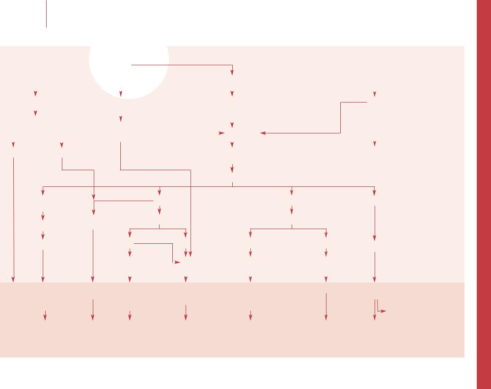
Assessment of a mediastinal mass |
Assessment of a mediastinal mass
76
Pallor fever
Purpura
Abnormal CBC
Blasts on blood smear
Large anterior mass
Bone marrow aspirate ± biopsy
Anteromedial mass
AFPHCG Biopsy
Negative AFP and/or HCG 
Symptoms leading to chest X-ray
Neck, supraclavicular or generalized adenopathy
Chest X-ray (AP and lateral)
Mediastinal mass
Nl CBC
Chest CT |
|
Posterior mass |
|
||
|
|
 MRI spine
Cough, dysphagia, hoarseness, new or refractory wheezing, dyspnea, orthopnea, facial swelling, and/or plethora
Superior vena cava syndrome
(see ‘Recognition and management of superior vena cava syndrome’, p 96)
Positive AFP and/or HCG |
|
|
Urine VMA/HVA |
||||
|
|
Biopsy |
|||||
|
|
|
|
|
|
|
|
|
|
|
|
|
|
|
|
Germ cell tumors |
|
|
|
|
|
||
Positive urine VMA/HVA Negative urine VMA/HVA |
|||||||
|
|
||||||
Leukemia or |
|
Lymphoma |
Benign tumor |
Thymoma |
Mature teratoma |
Immature |
Neuroblastoma |
Neurofibroma |
||||||||||
lymphoma |
|
|
|
|
|
|
|
|
|
|
|
teratoma |
|
|
Neurofibrosarcoma |
|||
with marrow |
|
|
|
|
|
|
|
|
|
|
|
|
|
|
|
Schwannoma |
||
|
|
|
|
|
|
|
|
|
|
|
|
|
|
|
||||
involvement |
NHL |
Hodgkin |
|
|
|
|
|
|
|
|
|
Lymphoma |
||||||
|
|
|
|
|
|
|
|
|
|
|
Ganglioneuroma |
|||||||
|
|
|
|
|
|
|
|
|
|
|
|
|
|
|
|
|
Ewings sarcoma |
|
|
|
|
|
|
|
|
|
|
|
|
|
|
|
|
|
|
||
|
|
|
|
|
|
|
|
|
|
|
|
|
|
|
|
|
|
|
|
|
|
|
|
|
|
|
|
|
|
|
|
|
|
|
|
Rhabdomyosarcoma |
|
|
|
Bone marrow |
|
|
|
|
|
|
|
|
|
|
|
|
Benign masses |
|||
|
|
CT (abdomen/pelvis) |
|
|
|
|
|
|
|
|
|
|
||||||
|
|
|
|
|
|
|
|
|
|
|
|
|
|
|||||
|
|
|
|
|
|
|
|
|
|
|
|
|
|
|||||
(see ‘Assessment of |
Gallium/PET |
|
|
|
|
|
|
|
|
|
|
|
|
|
|
|||
a child with suspected |
Bone scan |
|
|
|
|
|
|
|
|
|
|
|
|
|
|
|||
leukemia’, p 74) |
|
|
|
|
|
|
|
|
|
|
|
|
|
|
|
|
|
|
|
|
|
|
|
|
|
|
|
|
|
|
|
|
|
|
|
||
|
|
|
|
|
|
|
|
|
|
|
|
|
|
|
|
|
|
|
|
|
Chemotherapy |
Chemotherapy |
|
Surgery |
Surgery |
Chemotherapy |
Chemotherapy |
Diagnosis |
|||||||||
|
|
|
|
|
± radiotherapy |
|
± radiotherapy |
|
|
Surgery |
Surgery |
dependent |
||||||
|
|
|
|
|
|
|
|
|
|
|
|
|
|
|
Radiation |
|
|
|
|
|
|
|
|
|
|
|
|
|
|
|
|
|
|
|
|
|
|

–– See algorithms on generalized and localized lymphadenopathy for additional details of clinical presentation. Cancerous nodes are generally very firm and fixed to underlying structures. It is often possible to discern several different nodes matted together. They are not warm, tender or fluctuant and do not respond to antibiotics. Supraclavicular adenopathy is most often associated with malignancy.
–– It is important to assess the airway with any anteromedial mediastinal mass – airway compression can be a life-threatening emergency. On plain chest X-ray the thymus may look like a mediastinal mass, but it has a characteristic shape and does not cause tracheal deviation. Presence of paratracheal and mediastinal adenopathy increases the suspicion of malignancy.
–– Evaluation of chest masses by CT scan is usually sufficient, especially with the high resolution and thin sections of spiral CT when available. MRI is valuable in defining posterior mediastinal masses and potential spinal canal involvement, which is an oncologic emergency to preserve neural function.
–– Diagnosis is confirmed by bone marrow examination. The arbitrary line between leukemia with adenopathy vs. lymphoma with marrow involvement is the percentage of blasts in the marrow. Greater than 25% blasts is defined as leukemia. T cell ALL is the leukemia most commonly associated with mediastinal masses, usually with a high WBC count.
–– Emergency low-dose radiation is indicated when there is life-threatening airway compromise from tracheal compression. Tissue radiation can make the pathologic diagnosis difficult. Ideally, biopsy of the mass occurs before radiation or from a node outside the radiation field. Chemotherapy can also be used before a biopsy, but within 24–
48 h all of the tumor will be affected and it may be difficult to impossible to make a pathologic diagnosis thereafter.
–– Mediastinal non-Hodgkin lymphoma is most commonly of T cell phenotype. It is possible to make the diag-
77nosis from pleural fluid or bone marrow if those tissues are involved. Evaluate the spinal fluid for malignant involvement. Except for emergency radiation for the airway, therapy is based on chemotherapy alone.
–– 20–30% of children present with B symptoms (night sweats, weight loss of >10% body weight, and/or fevers >38°C). Nodes are generally not painful or tender but have a ‘rubbery’ firmness, and often have a variable growth rate. Gallium scan is positive in 40–60% of the cases and can be used to follow the disease response with treatment, but it is also positive in many nonmalignant inflammatory conditions. PET scanning is gaining favor as it is more specific to identification of lymphoma sites and as a marker of disease response is more highly predictive of cure.
–– Benign tumors are usually located in the anteromedial mediastinum: bronchogenic cysts, goiter, lipoma, lymphangioma, and enteric cysts are the most frequent and are diagnosed by open biopsy. In the posterior mediastinum, neurenteric cysts are generally associated with congenital abnormalities of the thoracic spine. The CT findings will often suggest these diagnoses.
–– Nearly one-half of these tumors are asymptomatic, but up to 30% of all patients, with or without symptoms, can have myasthenia gravis. The histologic diagnosis is made through open biopsy or mediastinoscopy. Treatment is surgical excision.
–– These tumors are usually asymptomatic until they reach a considerable size and may produce tracheal and bronchial compression. Tumor markers such as -fetopro- tein (AFP) and human chorionic gonadotrophin ( HCG), both detected in serum, may be the only clues to diagnosis. Interpreting AFP in infants can be difficult as they normally have a high level at birth and a variable half-life. Mature teratomas are negative for these markers and, therefore, the presence of a positive marker indicates a malignant component within the mass. A biopsy, open or by mediastinoscopy, is necessary to differentiate the subtype of germ cell tumor although treatment is currently the same for all groups. Completely mature teratomas respond only to surgical resection.
–– Posterior masses adjacent to the vertebra can invade the spinal cord. If the cord and clinical function are compromised it is possible to preserve neurologic function by immediate initiation of chemotherapy, spot radiation or surgical laminectomy with removal of tumor.
–– Elevation of urinary HVA and/or VMA can be diagnostic of neuroblastoma. Minor elevations can be due to diet intake, as phenylalanine and tyrosine raise these levels. To be significant they should be 3 SD above the mean. VMA and HVA can be measured on spot urine samples by normalizing them against creatinine levels on the same sample.
–– Neuroblastoma in the posterior mediastinum tends to be stage I–III with more histologic differentiation and a decreased rate of metastases. Surgical removal can be difficult because of adjacent vital structures. The posterior mediastinum is also the most common site of a fully mature ganglioneuroma (often VMA/HVA negative) as well as ganglioneuroblastoma.
–– Neurofibroma, neurofibrosarcoma, and malignant schwannomas are all derived from the neural crest and are most commonly seen in patients with neurofibromatosis. Enteric cysts and thoracic meningoceles are very rare. Very rarely, Ewing and rhabdomyosarcoma occur in the posterior mediastinum.
Selected reading
For chapters on non-Hodgkin lymphoma, Hodgkin disease, acute lymphoblastic leukemia and neuroblastoma see Emedicine Pediatric Medicine, E medicine, 2002 (http://emedicine.com).
Hudson MM, Donaldson SS: Hodgkin’s disease. Pediatr Clin North Am 1997;44:891–906.
Shad A, Magrath I: Non-Hodgkin’s lymphoma. Pediatr Clin North Am 1997;44:863–890.
Strollo DC, Rosado de Christenson ML, Jett JR:
Primary mediastinal tumors. I. Tumors of the anterior mediastinum. Chest 1997;112:511–522.
Strollo DC, Rosado de Christenson ML, Jett JR:
Primary mediastinal tumors. II. Tumors of the middle and posterior mediastinum. Chest 1997;112:1344–1357.
Malignant Disorders |
M. Weyl Ben Arush · J.M. Pearce |
Assessment of a mediastinal mass |
|
|
|
|
|
|

|
|
|
Malignant Disorders |
M. Weyl Ben Arush · J.M. Pearce |
Assessment of an abdominal mass |
Assessment of an abdominal mass
78
Hepatosplenomegaly
Adenopathy
CBC, blood smear
Viral titers
Intrahepatic mass
AFP, HCG
MRI
Biopsy
Clinical signs and examination |
|
|
|
|
Cushingoid |
Isolated mass with or without pain |
||
Obstructive symptoms of GI or GU systems |
Feminization |
|
|
|
Virilization |
|
|
|
|
|
|
Abdominal imaging |
|
|
Ultrasound/plain film |
|
|
CT scan |
|
|
 US/CT scan abdomen
If no abdominal mass with feminization/virilization, MRI/CT brain Pediatric endocrine evaluation
Biliary tract and pancreatic |
Renal |
Adenopathy |
Adrenal |
|
Other |
Abdominal |
|||||||
|
Renal disease |
|
|
Tumor lysis labs |
Urine VMA/HVA |
|
AFP |
mass present |
|||||
|
|
|
|
||||||||||
|
|
|
|
|
|
||||||||
|
|
|
|
|
|
||||||||
|
Excision or biopsy |
|
|
|
|||||||||
|
Bone marrow |
|
|
|
|
HCG |
|
|
|||||
|
without tumor |
|
|
CSF |
|
|
|
|
|
|
|
||
|
|
|
|
|
|
|
|
|
|
|
|
|
|
|
|
|
|
|
|
|
+ |
– |
|
|
|||
|
|
|
|
|
|
|
|
|
|||||
|
Hydronephrosis |
|
|
|
|
|
|
|
|
|
|
||
|
Polycystic kidney |
|
|
|
|
|
|
|
|
|
|
||
|
|
|
|
|
|
|
|
|
|
|
|||
|
Dysplastic kidney |
|
|
Biopsy |
Biopsy |
|
Biopsy |
Biopsy |
|||||
Biopsy |
|
|
|
||||||||||
|
|
|
|
|
|
|
|
|
|
|
|
|
|
|
|
|
|
|
|
|
|
|
|
|
|
|
|
|
|
|
|
|
|
|
|
|
|
|
|
|
|
|
|
|
|
|
|
|
|
|
|
|
|
|
|
|
|
|
|
|
|
Leukemia |
Metastatic disease |
Rhabdomyosarcoma |
|
Wilms tumor |
B cell/Burkitt Neuroblastoma |
Adrenal |
Sarcomas |
Adrenal tumor |
|||||||||||
Viral illness |
Hepatoblastoma |
Other sarcomas |
|
Other renal tumors |
lymphoma |
|
Pheochromocytoma incidentaloma |
PNET |
Extragonadal germ |
||||||||||
|
Hepatocellular carcinoma |
Pancreatic tumor |
|
|
|
|
|
Hodgkin disease |
|
Ganglioneuroma Teratoma |
cell tumor |
||||||||
|
Benign liver tumors |
|
|
|
|
|
|
|
|
|
|
|
|
|
Other adrenal |
|
|
|
|
|
|
|
|
|
|
|
|
|
|
|
|
|
|
|
|
tumors |
|
|
|
|
|
|
|
|
|
|
|
|
|
|
|
|
|
|
|
|
|
|
|
|
|
|
|
|
|
|
|
|
|
|
|
|
|
|
|
|
|
|
|
(see ‘Lym- |
CT chest |
|
|
|
CT chest |
|
|
CT chest |
|
|
|
|
|
|
|
|
|||
phadenophathy. |
Metastatic evaluation |
|
|
|
Metastatic evaluation |
Gallium/PET |
|
|
|
|
|
|
|
|
|||||
1. Generalized |
|
|
|
|
|
|
|
|
|
|
|
|
|
|
|
|
|
|
|
|
|
|
|
|
|
|
|
|
|
|
|
|
|
|
|
|
|
|
|
lymphadenopathy’, |
|
|
|
|
|
|
|
|
|
|
|
|
|
|
|
|
|
|
|
p 46) |
|
|
|
|
|
|
|
|
|
|
|
|
|
|
|
|
|
|
|
|
|
|
|
|
|
|
|
|
|
|
|
|
|
|
|
|
|
|
|
|
|
|
|
|
|
|
|
|
|
|
|
|
|
|
|
|
|
|
|
|
Surgery |
Surgery |
Surgery when |
Surgery |
|
|
Chemotherapy |
Surgery |
|
Diagnosis- |
Surgery |
||||||||
|
Chemotherapy ± |
Chemotherapy |
appropriate |
Chemotherapy ± |
± radiation |
Chemotherapy |
dependent |
Chemotherapy |
|||||||||||
|
radiation if malignant |
± radiation if malignant |
|
|
|
radiation if malignant |
|
|
|
± radiation if malignant |
|
if malignant |
|||||||
|
|
|
|
|
|
|
|
|
|
|
|
|
|
|
|
|
|
|
|

–– Plain X-rays are most helpful to evaluate for GI obstruction or to rule out constipation as a cause of the ‘mass’. US is more useful for rapid evaluation of abdominal masses. In neonates, cystic and fluid filled masses are generally benign but solid masses must be considered malignant until proven otherwise. More than 50% of flank masses in the newborn are non-malignant renal anomalies. In Wilms tumor US can often clarify whether tumor extends into the inferior vena cava. CT with oral and i.v. contrast can define the extent of the mass, helping the surgeon decide whether to attempt total resection or biopsy only. High resolution spiral CT or MRI better define the extent
of hepatic lesions.
–– Hepatoblastoma is the most common pediatric liver tumor in most areas, but where chronic active hepatitis is common, hepatocellular carcinoma is more prevalent. Hepatoblastoma, an embryonal tumor, arises in normal liver, is most commonly unifocal and in the right lobe. However, it can be multifocal and involve the entire liver. Hepatocellular carcinoma is more frequent in older children, more likely involves both lobes at diagnosis, generally arises in already damaged liver tissue and is difficult to cure. Both present with hepatomegaly and abdominal pain and can have elevated AFP (in 90% of hepatoblastoma and 60% of hepatocellular carcinoma). Staging is dependent on site, vessel invasion, resectability and metastases. The combination of US, CT and MRI can define many of these issues, including vascular involvement.
–– The tumors which most often metastasize to the liver are Wilms tumor, neuroblastoma and ovarian tumors.
–– One third of primary liver tumors are benign and include hemangioendothelioma, mesenchymal hamartoma, focal nodular hyperplasia and adenoma.
–– Pancreatic tumors are rare but include benign papillary-cystic tumors and hemangiomas. Malignant tumors are similar to those of adults.
–– Most children are asymptomatic and diagnosed be-
79cause a flank mass is felt by a family member or physician.
The tumor is generally ballotable and smooth. Those with symptoms have abdominal pain, fever, anemia (from intratumor hemorrhage), hematuria (20%) and/or hypertension. While most children with Wilms tumor are otherwise normal, there is an increased incidence in children with Beck- with-Wiedemann syndrome, aniridia, hemihypertrophy and
genitourinary anomalies. Anaplastic histology confers a worse prognosis. In Europe it is common to make the diagnosis by biopsy and then chemotherapy is delivered before definitive resection. In the United States total resection is attempted at diagnosis, followed by chemotherapy – survival rates are the same with both approaches.
–– Non-Wilms renal tumors include rhabdoid (2% of renal tumors, probably neurogenic in origin, often presents with metastatic disease in the lung, liver or brain, and has a very poor prognosis), clear cell sarcoma of the kidney
(4% of renal tumors, 40–60% have bone metastases, poor prognosis), renal cell carcinoma (very rare in children and behaves like adult cases) and congenital mesoblastic nephroma (two subtypes, one benign and one malignant, treated like Wilms tumor).
–– CSF and/or bone marrow involvement in B-cell lymphoma permits diagnosis without an abdominal biopsy as may paracentesis of malignant ascites. Tumor lysis syndrome can be present at diagnosis in these rapidly dividing tumors. See ‘Recognition and management of tumor lysis syndrome’, p 94.
–– See ‘Assessment of a pelvic mass’ (p 80) for more information on B cell lymphomas. When Hodgkin disease arises in the abdomen, there may be no signs or symptoms until there is spread to clinically palpable nodes. Eosinophilia occurs in approximately 15% of patients (see ‘Assessment of a mediastinal mass’ (p 76).
–– The urinary catecholamines (VMA and HVA) are measured. Elevation of urinary catecholamines HVA and/or VMA occurs in 95% of neuroblastomas; diagnosis by these catecholamines alone does not assess prognostic markers such as histologic classification or MYCN gene amplification. Involved bone marrow can confirm the diagnosis and MYCN status.
–– Incidentalomas are adrenal masses coincidentally identified by ultrasound, CT or MRI done for other reasons. They occur on about 3% of all abdominal CTs. They are small (usually <3 cm), may have calcifications and are probably residual from old adrenal trauma. 10% of these secrete catecholamines and need further intervention – the rest are ‘incidental’, benign and may be left alone.
–– Embryonal neural tumors from the sympathetic nervous system range from malignant (neuroblastoma) to com-
pletely benign (ganglioneuroma). Prognosis is dependent on age (less than one improves prognosis), stage, tumor histology and resectability. Molecular biologic markers like MYCN gene amplification confer a worse prognosis. Cortical bony metastases confer a worse prognosis. Children less than 1 year of age with metastatic disease not involving cortical bone, and a small primary mass have stage IVs which can spontaneously regress without treatment.
–– Pheochromocytomas are very rare in childhood and less than 10% are malignant. Adrenal carcinomas and benign adenomas are very rare in childhood. 80% are hormone-secreting tumors and in 60% there are symptoms of hormone excess with weight loss and malaise.
–– CT/MRI of brain is necessary to identify pituitary tumors that can cause precocious puberty.
–– Adrenal tumors can cause Cushing syndrome or either feminization or virilization. Extragonadal abdominal germ cell tumors are generally retroperitoneal, usually in children under the age of 2 years, and can cause precocious puberty. They present with pain and/or obstructive symptoms of bowel or urinary tract. Hepatic teratomas are seen soon after birth and are associated with very elevated AFP levels.
Selected reading
Broecker B: Non-Wilms’ renal tumors in children. Urol Clin North Am 2000;7:463–469.
Castleberry RP: Biology and treatment of neuroblastoma. Pediatr Clin North Am1997;44:919–937.
Graf N, Tournade MF, deKraker J: The role of preoperative chemotherapy in the management of Wilms tumor: The SIOP Studies. Urol Clin North Am 2000;27:443–454.
Neville HL, Ritchey ML: Wilms’ tumor, overview of the National Wilms Tumor Study Group results. Urol Clin North Am 2000;27:435–442.
Schteingart DE: Management approaches to adrenal incidentalomas. Endocrinol Metab Clin 2000;29:127–139.
Stringer MD: Liver tumors. Semin Pediatr Surg 2000;9:196–208.
Malignant Disorders |
M. Weyl Ben Arush · J.M. Pearce |
Assessment of an abdominal mass |
|
|
|
|
|
|
|
|
|
Malignant Disorders |
M. Weyl Ben Arush · J.M. Pearce |
Assessment of a pelvic mass |
Assessment of a pelvic mass
80 |
|
|
|
|
|
|
|
|
|
Clinical symptoms |
|
|
|
|
|
|
|
|
|
|
|
|
|
|
|
|
|
|
|
|
|
|
|
|
|
|
|
|
|
|
|
|
|
Amenorrhea |
|
|
Feminization or |
|
Pain, vaginal bleeding |
Bowel obstruction |
|||||
|
|
|
|
|
|
virilization |
|
Fever, palpable mass on examination |
Bladder obstruction |
||||
|
|
|
|
||||||||||
|
|
|
|
|
|
|
|
|
|
(abdomen, rectal, gynecologic) |
Neurologic symptoms from spinal cord |
||
|
|
|
|
|
|
|
|
|
|||||
|
|
HCG |
|
|
|
|
|
|
|
|
and nerve root compression |
||
|
|
|
|
|
|
|
|
|
|
||||
|
|
|
|
|
|
Sex hormone evaluation |
|
|
|
|
|
||
|
|
|
|
|
|
|
|
|
|
||||
|
|
|
|
|
|
Ultrasound pelvis and abdomen |
|
|
Plain films |
|
|
||
|
|
|
|
|
|
|
|
|
|
||||
|
|
|
|
|
|
CT scan/MRI |
|
|
|
|
|
|
|
|
|
|
|
|
|
|
|
|
|||||
+ |
|
– |
|
|
|
Ultrasound pelvis and abdomen |
Emergency surgery? |
||||||
|
|
|
|||||||||||
|
|
|
|
|
|
|
|
|
|
CT scan/MRI |
|
|
|
|
|
|
Origin of mass |
|
Pelvic bone |
|
Ovary, uterus |
Retroperitoneum |
Intestinal, adenopathy |
involvement |
|
|
|
|
|
Bulging hymen |
HCG, AFP |
Urinary VMA/HVA |
|
Bone scan |
Typical US |
|
|
|
Biopsy |
– |
+ |
– |
+ |
|
|
|
Biopsy or paracentesis |
|
|
|
|
|
|
|
|
Drainage of abscess |
|
Biopsy |
Biopsy |
Biopsy |
|
||||
|
|
|
|
||||||||
|
|
Biopsy cyst/mass |
|
(ascitic fluid) |
|
|
|
|
|
||
|
|
|
|
|
|
|
|||||
|
|
|
|
|
|
|
|
|
|
|
|
|
|
|
|
|
|
|
|
|
|
|
|
Pregnancy Ewing |
Hydro- |
Abscess |
|
Gonadal tumor |
Rhabdomyosarcoma |
Neuroblastoma |
Burkitt non-Hodgkin |
||||
sarcoma |
metrocolpos |
Ovarian cyst |
|
Germ cell tumor |
Other sarcomas |
|
|
lymphoma |
|||
Primitive neuro- |
|
Ovarian carcinoma |
|
Non-germ cell tumor |
Primitive neuroectodermal tumor |
|
|
|
|||
ectodermal tumor |
|
Leiomyosarcoma |
|
|
|
Germ cell tumor |
|
|
|
||
(see ‘Recognition and management of tumor lysis syndrome’, p 94)
Bone marrows |
Surgery |
Surgical intervention |
Staging laparotomy |
Excision when possible |
Bone marrows |
Spinal tap |
Chest CT |
|
± antibiotics |
and chemotherapy if |
Chemotherapy ± |
Bone scan |
Bone marrows |
|
|
Chemotherapy for |
malignant |
radiotherapy |
Chemotherapy |
Chemotherapy |
|
|
malignancy |
|
|
|
|
|
|
|
|
|
|
|

–– A bone scan can determine the extent of tumor in the pelvic bones and other distant sites of osseous involvement.
–– There are several options for obtaining tissue for histologic diagnosis. US-guided biopsy (fine-needle aspirate or needle-core biopsy), biopsy through rectoscopy or cystoscopy, laparoscopy or open biopsy through laparotomy.
If the tumor has associated ascites, a paracentesis may yield diagnostic fluids (especially in GCT and lymphomas). The approach is determined by the skill and experience of the surgeon as well as the information needed (such as whether there are peritoneal studs in a GCT which may require direct visualization by laparotomy). Fine-needle aspirate may be performed in very sick children but in most cases does not provide enough material to obtain all of the histologic and molecular genetic information required.
–– Ewing sarcoma and primitive peripheral neuroectodermal tumor (PNET) belong to the same family of neuralderived tumors. PNET tends to be more differentiated and has a higher incidence of positive pathology markers such as neuron-specific enolase (NSE) and S100 which are indicators of more mature neural differentiation within the tumor.
–– -Fetoprotein (AFP) levels are normally very high in neonates and this may mask tumor-induced elevation of AFP. The majority of germ cell tumors (GCT) in neonates are benign, requiring surgical excision only. Sacrococcygeal tumors are more frequent in children under the age of three. Fifty percent of these are in neonates, primarily in girls.
–– Gonadal tumors are ovarian and testicular. The majority of gonadal tumors are germ cell in origin and onethird of these GCT arise in the ovary; peak incidence is at 10 years of age, and 70% are mature teratomas which require surgical excision only. Ovarian tumors may not present until they are very large and may present with torsion or rupture. The pattern of spread of malignant GCT is ascites and peritoneal implants followed by metastases to lung, lymph nodes and liver. GCT occurring in extragonadal sites are most frequently benign mature teratomas, but they may contain some malignant components requiring therapy;
81therefore, careful pathologic study of the entire tumor is essential. The majority of malignant GCT involve extraembryonal differentiation and secrete either AFP and/or HCG (AFP in yolk sac tumor and HCG in choriocarcinoma). Elevation of these markers in the serum is of great value both in diagnosis and management. GCT can arise outside the gonad, most commonly as sacrococcygeal teratoma
identified as an external protuberance. GCT occasionally originate in the retroperitoneum. Non-GCT gonadal tumors include sex cord-stromal tumors (granulosa and Sertoli Leydig cell) or epithelial tumors. Ovarian carcinomas are extremely rare in childhood.
–– Rhabdomyosarcoma (RMS) arises from mesenchymal cells committed to developing into striate muscle. The histologic subtypes are correlated with the prognosis. Botryoid and spindle-cell RMS are the rarest subtypes and have the best prognosis. Alveolar RMS (high incidence of a t2;13 chromosomal translocation) is generally seen in the extremities in adolescents, and has the worst prognosis. Two-thirds of all RMS are embryonal which are more often seen in the trunk of younger children and have an intermediate prognosis. About 25% of all RMS occur in the GU/retroperitoneal area and they can be very large masses before causing physical symptoms such as bladder obstruction. Botryoid RMS typically presents as a grape-like cluster protruding from the cervix or vagina in very young girls. RMS are sensitive to both chemotherapy and radiation with most successful treatment regimens incorporating both
if the resection is not complete (for sarcomas other than rhabdomyosarcoma, see ‘Assessment of a soft tissue mass’, p 82).
–– Neuroblastoma rarely originates in the pelvis but when it does it can extend into the neural foramina and compress the nerve roots (see ‘Assessment of an abdominal mass’, p 78).
–– The pelvis/abdomen is the most frequent primary site (90%) of Burkitt (B cell) lymphoma. The most common presentation is a rapidly growing mass, often producing ascites. This leads to pain, abdominal swelling and intestinal obstruction, and can serve as a lead point for an intussusception. Tumor lysis is very common and often life threatening. Diagnosis can be made from tumor biopsy, ascitic fluid
or involved bone marrow. The cells are clonal B cells with surface immunoglobulin and there are characteristic translocations seen that are pathognomonic (t8;14, t8;22 or t2;8). Chemotherapy is very effective.
–– Precocious puberty or virilization can result from ovarian non-germ cell (stromal) tumors, less often from germ cell tumors, and rarely from extragonadal pelvic germ cell tumors. Keep in mind that adrenal adenomas and carcinomas will cause similar symptoms. Pediatric endocrine assessment is indicated.
–– Plain X-rays and ultrasound can demonstrate calcifications in the case of benign gonadal tumors or neuroblastoma. US is very useful in assessing ureteral obstruction which could require emergency urinary diversion to preserve renal function. Ovarian masses can be determined to be cystic or solid and hydrometrocolpos can be identified.
–– Contrast-enhanced CT gives information on the primary tumor and intra-abdominal metastases. Spiral CT and MRI have the advantage of imaging the tumor in the coronal and sagittal planes. MRI better defines spinal cord invasion.
–– Vaginal bleeding can be due to a botryoid rhabdomyosarcoma which presents as a grape-like cluster of clear tissue protruding from the vagina or cervix, which should be excised or biopsied. Vaginal bleeding is also caused by feminization due to sex-hormone-secreting ovarian or adrenal tumors.
–– The predominant symptoms of malignant pelvic tumors are pain, palpable swelling/masses, fever, and signs of obstruction of either GI or GU tract. Compression of nerve roots can lead to leg weakness and paresthesias. These symptoms may increase rapidly over a short period or remain constant for several months.
Selected reading
Cheville JC: Classification and pathology of testicular germ cell and sex cord-stromal tumors. Urol Clin North Am 1999;26:595–609.
Groff DB: Pelvic neoplasms in children. J Surg Oncol 2000;77:65–71.
Pappo AS, Shapiro DN, Crist W: Rhabdomyosarcoma: Biology and treatment. Pediatr Clin North Am 1997;44: 953–972.
Pfeifer SM, Gosman GG: Evaluation of adnexal masses in adolescents. Pediatr Clin North Am 1999;46:573–592.
Malignant Disorders |
M. Weyl Ben Arush · J.M. Pearce |
Assessment of a pelvic mass |
|
|
|
|
|
|

|
|
|
Malignant Disorders |
M. Weyl Ben Arush · J.M. Pearce |
Assessment of a soft tissue mass |
Assessment of a soft tissue mass
82 |
|
|
History |
|
|
|
Physical examination |
||
|
|
|
||
|
|
|
|
|
|
|
|
|
|
Painful, indurated |
|
|
|
|
Not painful or erythematous |
||
Local inflammation |
|
|
|
|
Firm, ± fixed to underlying tissue |
||
Local adenopathy |
|
|
|
|
± Persistent growth |
||
Fever |
|
|
|
|
|
|
|
|
|
|
|
|
|
||
|
|
Underlying bone lesion |
|
Plain X-rays |
|||
|
|
|
|||||
|
|
|
|||||
|
|
|
|
|
|
|
|
|
|
|
|
|
|
|
|
|
|
(see ‘Assessment of |
|
No bone disease |
|||
|
|
bone lesions’, p 84) |
|
|
|
||
|
|
|
|
|
|||
|
|
|
|
|
|
Ultrasound |
|
|
|
|
|
|
|
± CT/MRI |
|
|
|
|
|
|
|
|
|
|
|
|
|
|
|
|
|
CBC, ultrasound |
Cystic lesions |
|
Solid lesion |
||||
Blood and local cultures if feasible |
|
|
|
|
|
|
|
|
|
|
|
|
|
||
|
|
Excision if indicated |
|
Biopsy, possible excision |
|||
|
|
|
|||||
|
|
|
|
|
|
|
|
|
|
|
|
|
|
|
|
|
|
|
(see ‘Management of biopsy tissue in |
||||
|
|
|
children with possible malignancies’, p 100) |
||||
|
|
|
|
|
|
|
|
|
|
|
|
|
|
|
|
|
|
Multiple subcutaneous nodules |
||
|
|
|
|
|
|
|
|
|
|
|
|
CBC, blood smear |
||
|
|
|
|
|
|
|
|
|
|
|
|
|
|
|
Normal |
|
Blasts, abnormal CBC |
||
|
|
|
|
|
|
|
|
|
|
|
|
|
Bone marrow aspirate |
|
|
|
|
||
Infant often with hepatomegaly
± palpable abdominal mass
Urine for VMA, HVA Ultrasound of abdomen
± CT/MRI ± CXR to look for primary tumor site
Biopsy of primary tumor
Infection |
Benign |
Malignant |
Neuroblastoma, |
Nonmalignant causes of |
Acute nonlymphoblastic |
||||||
|
|
Lipoma |
Rhabdomyosarcoma |
likely stage IVS |
subcutaneous nodules |
leukemia |
|||||
|
|
||||||||||
|
|
Hemangioma |
Other sarcomas |
|
|
Post-immunization |
Lymphoma |
||||
|
|
|
|
||||||||
|
|
Cysts |
Malignant fibrous histiocytoma |
|
|
Acute rheumatic fever |
|
|
|||
|
|
Neurofibroma |
Chloroma |
|
|
Rheumatoid arthritis |
|
|
|||
|
|
Lymphangioma |
|
|
|
|
Erythema nodosum |
|
|
||
|
|
|
|
|
|
|
|
||||
|
|
Fibromatosis |
|
|
|
|
Panniculitis |
|
|
||
|
|
Schwannoma |
|
|
|
|
Gardner syndrome |
|
|
||
|
|
Desmoid tumor |
|
|
|
|
|
|
|
|
|
|
|
|
|
|
|
|
|
|
|
||
|
|
Leiomyoma |
|
|
|
|
|
|
|
|
|
|
|
|
|
|
|
|
|
|
|
|
|
|
|
|
|
|
|
|
|
|
|
|
|
|
|
|
|
|
|
|
|
|
|
|
|
Incision and drainage |
Dependent on |
Surgery |
Observation vs |
Evaluate based on |
Chemotherapy |
||||||
if fluctuant |
diagnosis and location |
Chemotherapy |
chemotherapy |
nature of nodules and |
|
|
|||||
Antibiotics |
|
|
± radiation therapy |
|
|
overall clinical picture |
|
|
|||
|
|
|
|
|
|
|
|
|
|
|
|

–– Pain, induration and inflammation are hallmarks of infection. Although superficial tumors can get irritated
by trauma and demonstrate similar symptoms, infections are usually easily identified clinically. A trial of antibiotic therapy is reasonable before proceeding to an expensive evaluation. If there is fluid or fluctuance (which may be better defined by ultrasound), needle aspiration and culture or incision and drainage may take priority over other tests. Ultrasound can also help define the extent of a deep soft tissue infection.
–– Most tumors, whether benign or malignant, are painless and firm. Malignant lesions can be fixed to underlying tissue as they infiltrate tissue planes but even some benign lesions can be very infiltrative, aggressive and locally destructive.
–– Ultrasound can be helpful if the soft tissue mass has no underlying bone pathology. It can distinguish cystic from solid lesions and is very helpful in identifying hemangiomas. In defining soft tissue masses, MRI appears to be superior to CT scan. The former can often identify vascular involvement without contrast enhancement.
–– Plain X-rays may identify bone fractures, hematomas, calcifications, cystic components or a primary bone lesion with a soft tissue extension/reaction. In Langerhans cell histiocytosis, there may be soft tissue swelling over bony osteolytic lesions.
–– Fine-needle aspirates and needle core biopsies can establish malignancy but often do not provide adequate material to identify subtypes of sarcoma or to do necessary cytogenetic and molecular studies. Excisional biopsy may be done especially if likely benign or if significant damage can be avoided to normal tissue. Soft tissue cysts are almost always benign and are simply excised if clinically indicated.
–– Benign tumors can be locally invasive and destructive but do not metastasize. Lipomas are common and occur most in subcutaneous fat. Hemangiomas most commonly occur in the skin with 60% in the head and neck region.
83Large hemangiomas can produce serious complications such as airway obstruction or thrombocytopenia from platelet consumption. Most start in the neonatal period and often enlarge for the first few months following birth, followed by spontaneous involution. If they are causing symptomatology they will often regress in response to high dose
corticosteroids and/or interferon- . Neurofibromas and schwannomas primarily occur along peripheral nerves but can also occur in the central trunk. They are seen primarily in individuals with neurofibromatosis and can be very large and locally destructive. Surgical resection is the only effective treatment modality. Rarely, they can become malignant. Desmoid tumors are derived from fibroblasts and are locally very aggressive. They do respond to low-dose chemotherapy which can make resection easier. Fibromatosis arises from neoplastic myoblastic-fibroblastic tissue and is most common in early childhood. Infantile myofibromatosis arises as a solitary mass in the very young. Lymphangiomas are lymphatic malformations that can be localized or generalized, and commonly occur in the cervicofacial region, thorax or axilla. Leiomyoma is a very rare smooth muscle tumor and must be differentiated from leiomyosarcoma.
–– Rhabdomyosarcoma (RMS) is the most common soft tissue tumor (STS) in childhood (see ‘Assessment of a pelvic mass’, p 80, for more details on RMS in general).
–– Non-rhabdomyosarcoma soft tissue tumors (NRSTS) are a heterogeneous group of tumors which range from well-differentiated and nonaggressive to aggressive and metastasizing. They are rare in children but increase in frequency with age. The most common sites are extremities, trunk wall and peritoneum. Involvement of regional nodes is less frequent than in rhabdomyosarcoma. In general, larger tumors have a worse prognosis. Because of their rarity, our therapeutic experience is limited. Complete surgical resection is critical. Most do respond to chemotherapy. Preresection chemotherapy may shrink an inoperable mass and make its complete resection possible. In metastatic disease chemotherapy is the prime treatment. Radiation therapy may be beneficial when the surgical margins are not free of tumor. Some NRSTS include: peripheral primitive neuroectodermal tumor is neural-derived and presents most commonly around the chest. Synovial sarcoma (40% of NRSTS) often has fibrous and glandular components with epithelial differentiation, usually around the knee or thigh. The translocation tX;18 is seen in this tumor. Fibrosarcoma (13% NRSTS) is the most common NRSTS in children less than one year of age. It arises from fibrous tissue and in children under age five it is usually low grade. In older children fibrosarcomas are much more aggressive with a high local recurrence rate and lung metastases. Malignant fibrous histiocytoma can be very aggressive locally and
also has the ability to metastasize. The cell of origin is not known in this tumor. Neurofibrosarcoma (10% NRSTS) and schwannomas arise from peripheral nerves, with almost 50% occurring in patients with neurofibromatosis (NF1). They have a very poor long-term prognosis. Alveolar soft part sarcoma is a slow-growing tumor arising in the head, neck and extremities. However, over 10–20 years it is almost universally fatal so aggressive surgical management is warranted at diagnosis. Hemangiopericytoma, angiosarcoma and hemangioendothelioma arise from vascular tissue with lymphangiosarcoma originating from vascular or lymphatic endothelium. Angiosarcomas most commonly present in the liver. Hemangiopericytomas have an infantile form that has an excellent prognosis. Leiomyosarcomas occur primarily in the uterus or the GI tract. Those in the GI tract are more likely to have metastases and a poorer prognosis. There is an increased incidence of this type of sarcoma in chronically immunosuppressed patients. Chloromas are soft tissue masses due to acute nonlymphoblastic leukemia, which may precede CBC changes.
–– Children with neuroblastoma can have subcutaneous nodules of tumor that can appear and regress without treatment. This is most common in stage IVS which by definition is children under the age of one with a small primary and no bone metastases. Urinary VMA/HVA are elevated in 95% of neuroblastomas (see ‘Assessment of an abdominal mass’, 78).
–– Many benign disorders cause subcutaneous nodules, a number of which are included in the algorithm.
Selected reading
Coffin CM: Soft tissue tumors in the first year of life: A report of 190 cases. Pediatr Pathol 1990;10:509–526.
Coffin CM: Pathologic evaluation of pediatric soft tissue tumors. Am J Clin Pathol 1998;109(suppl 1):S38–52.
Cripe T: Rhabdomyosarcoma. Emedicine Pediatric
Medicine 2002 (http://emedicine.com).
Kimmelman A, Liang B: Familial neurogenic tumor syndromes. Hematol Oncol Clin North Am 2001;15: 1073–1084.
Prieto VG, Shea CR: Selected cutaneous vascular neoplasms. Dermatol Clin 1999;17:507–520.
Malignant Disorders |
M. Weyl Ben Arush · J.M. Pearce |
Assessment of a soft tissue mass |
|
|
|
|
|
|

|
|
|
|
|
|
|
|
|
|
|
|
|
|
|
|
|
|
|
|
|
|
|
|
|
|
|
|
|
|
|
Malignant Disorders |
|
|
M. Weyl Ben Arush · J.M. Pearce |
|
Assessment of bone lesions |
|
|
|
|
|||||||||||||
|
|
|
|
|
|
|
|
|
|
|
|
|
|
|
|
|
|
|
|
|
|
|
|
|
|
|
|
|
|
|
|
|
Assessment of bone lesions |
|
|
|
|
|
|
|
|
|
|
||||||||||
|
|
|
|
|
|
|
|
|
|
|
|
|
|
|
|
|
|
|
|
|
||||||
|
|
|
|
|
|
|
|
|
|
|
|
|
|
|
|
|
|
|
|
|
|
|
|
|
|
|
|
|
|
|
|
|
|
|
|
|
|
|
|
|
|
|
|
|
|
|
|
|
|
|
|
|
|
84 |
|
|
|
|
|
|
|
|
|
|
Symptoms: bone pain (often chronic, localized versus diffuse), limp (with/without pain), |
|
|
|||||||||||||
|
|
|
|
|
|
|
|
|
|
|
refusal to use limb, palpable, hard mass or pathologic fracture |
|
|
|
|
|
|
|||||||||
|
|
|
|
|
|
|
|
|
|
|
|
|
|
|
|
|||||||||||
|
|
|
|
|
|
|
|
|
|
|
|
|
|
|
|
|
|
|
|
|
|
|
|
|
|
|
|
|
|
|
|
|
|
|
|
|
|
|
|
|
|
|
|
|
|
|
|
|
|
|
|
|
|
|
|
|
|
|
|
|
|
|
|
|
Plain X-ray |
|
|
|
|
|
|
|
|
|
|
|
|
|
||
|
|
|
|
|
|
|
|
|
|
|
|
|
|
|
|
|
|
|
|
|
|
|
||||
|
|
|
|
|
|
|
|
|
|
|
|
|
|
|
|
|
|
|
|
|
|
|
|
|
|
|
|
|
Characteristic |
Leukemic lines |
|
|
|
|
Lytic lesion with |
|
|
|
|
|
Lytic lesion(s) without periosteal reaction |
|
|
||||||||||
|
|
single lesions |
|
|
|
|
|
|
periosteal reaction |
|
|
|
|
|
|
|
|
|
|
|||||||
|
|
|
|
|
|
|
|
|
|
|
|
|
|
|
|
|
|
|||||||||
|
|
|
|
|
± Fever, adenopathy |
|
|
|
|
|
|
|
|
|
|
|
|
|
|
|
|
|
|
|
||
|
|
|
|
|
|
|
|
|
|
|
|
|
|
|
|
Single or multiple lesions |
Fever, often ill appearing |
|
|
|||||||
|
|
|
|
|
|
|
|
|
|
|
|
|
|
|
|
|
|
|||||||||
|
|
|
|
|
Diffuse bone pain |
|
|
|
|
Bone scan, CT/MRI of site |
|
Vertebral collapse |
Usually abdominal mass |
|
|
|||||||||||
|
|
|
|
|
|
|
|
|
|
|
|
|
|
|
|
|
|
± multisystem disease |
|
|
|
|
||||
|
|
|
|
|
|
|
|
|
|
|
|
|
|
|
|
|
|
|
|
|
|
|||||
|
|
|
|
|
CBC + blood smear |
|
|
|
|
|
|
|
|
|
|
|
|
|
|
|
|
Abdominal ultrasound |
|
|
||
|
|
|
|
|
|
|
|
|
Biopsy |
|
|
|
|
|
|
|
|
|
|
|
||||||
|
|
|
|
|
|
|
|
|
|
|
|
|
|
|
|
|
|
|
|
|
|
|
Urine VMA/HVA |
|
|
|
|
|
|
|
|
|
|
|
|
|
|
|
|
|
|
|
|
|
|
|
|
|
|
|
|
|
|
|
|
|
|
|
Bone marrow |
|
|
|
|
|
|
|
|
|
|
|
|
|
|
|
|
|
|
|
|
|
|
|
|
|
|
|
|
|
|
|
|
|
|
|
|
|
Biopsy |
|
|
Biopsy primary lesion |
|
|
|||||
|
|
|
|
|
|
|
|
|
|
|
|
|
|
|
|
|
|
|
|
|
|
|||||
|
|
|
|
|
|
|
|
|
|
|
|
|
|
|
|
|
|
|
|
|
|
|
|
|
||
|
|
|
|
|
|
|
|
|
|
|
|
|
|
|
|
|
|
|
|
|
|
|
|
|
|
|
|
|
|
|
|
|
|
|
|
|
|
|
|
|
|
|
|
|
|
|
|
|
|
|
|
|
|
|
|
Benign lesions |
Leukemia |
Osteosarcoma |
Ewing |
Lymphoma |
Osteomyelitis |
Langerhans cell |
Neuroblastoma |
|
|
|||||||||||||||
|
|
Osteoid osteoma |
Neuroblastoma |
Chondrosarcoma |
sarcoma |
|
|
|
|
|
|
histiocytosis |
|
|
Other metastatic |
|
|
|||||||||
|
|
|
|
|
|
|
|
|
|
|
|
|||||||||||||||
|
|
Benign fibrous |
|
|
Fibrosarcoma |
|
|
|
|
|
|
|
|
|
|
|
|
|
disease |
|
|
|||||
|
|
|
|
|
|
|
|
|
|
|
|
|
|
|
|
|
|
|
||||||||
|
|
cortical defect |
|
|
|
|
|
|
|
|
|
|
|
|
|
|
|
|
|
|
|
|
|
|
||
|
|
|
|
|
|
|
|
|
|
|
|
|
|
|
|
|
|
|
|
|
|
|
|
|||
|
|
Fibrous dysplasia |
|
|
|
|
|
|
|
|
|
|
|
|
|
|
|
|
|
|
|
|
|
|
||
|
|
Osteochondroma |
|
|
|
|
|
|
|
|
|
|
|
|
|
|
|
|
|
|
|
|
|
|
||
|
|
|
|
|
|
|
|
|
|
|
|
|
|
|
|
|
|
|
|
|
|
|
|
|||
|
|
Endochondroma |
|
|
CT of lungs |
CT of lungs |
CT of lungs |
Culture |
|
Bone scan/skeletal survey |
|
|
|
|
||||||||||||
|
|
Aneurysmal |
|
|
|
|
|
|
|
|||||||||||||||||
|
|
|
|
|
|
|
Bone |
Bone marrow |
|
|
|
|
Chest X-ray |
|
|
|
|
|
|
|||||||
|
|
bone cyst |
|
|
|
|
|
|
|
|
|
|
|
|
|
|
|
|||||||||
|
|
|
|
|
|
|
marrow |
Gallium/PET scan |
|
|
|
|
CBC, chemistries |
|
|
|
|
|
|
|||||||
|
|
|
|
|
|
|
|
|
|
|
|
|
|
|
|
|
|
|
|
|||||||
|
|
|
|
|
|
|
|
|
|
|
|
|
|
|
|
|
|
|
|
|
|
|
|
|
|
|
|
|
|
|
|
(see ‘Assessment of |
|
|
|
|
|
|
|
|
|
|
|
|
|
|
|
|
|
|
|
|
|
|
|
|
|
|
|
|
|
|
|
(see ‘Assessment of |
|
|
|
|
|
|
|
|
|
|
|
|
|
|||
|
|
|
|
|
a child with suspected |
|
|
|
|
|
|
|
|
Focal disease |
Multifocal |
(see ‘Assessment of |
|
|
||||||||
|
|
|
|
|
leukemia’, |
|
|
|
|
|
a mediastinal mass’, |
|
|
|
|
|
||||||||||
|
|
|
|
|
|
|
|
|
|
|
|
|
|
|
|
|
|
|
an abdominal mass’, p 78) |
|
|
|||||
|
|
|
|
|
p 74) |
|
|
|
|
|
p 76) |
|
|
|
|
|
|
|
|
|
|
|
||||
|
|
|
|
|
|
|
|
|
|
|
|
|
|
|
|
|
|
|
|
|
|
|
||||
|
|
|
|
|
|
|
|
|
|
|
|
|
|
|
|
|
|
|
|
|
|
|
|
|
|
|
|
|
(see individual notes for |
|
|
Chemotherapy |
Chemotherapy |
|
|
Antibiotics |
No treatment |
Chemotherapy |
|
|
|||||||||||||
|
|
description and therapy) |
|
|
Surgical resection |
Surgical resection |
|
|
|
Intralesional steroids |
± radiation |
|
|
|||||||||||||
|
|
|
|
|
|
|
|
|
|
± radiotherapy |
|
|
|
|
|
Surgery/curretage |
|
|
|
|
|
|
||||
|
|
|
|
|
|
|
|
|
|
|
|
|
|
|
|
|
Low-dose radiation |
|
|
|
|
|
|
|||
|
|
|
|
|
|
|
|
|
|
|
|
|
|
|
|
|
|
|
|
|
|
|
|
|
|
|
|
|
|
|
|
|
|
|
|
|
|
|
|
|
|
|
|
|
|
|
|
|
|
|
|
|
|

–– The radiologic appearance of most benign bone tumors establishes the diagnosis without CT, MRI or biopsy.
–– Occurs primarily in second decade, usually in lower extremities. Severe pain at rest is common. The characteristic X-ray pattern is a radiolucent nidus of osteoid tissue surrounded by sclerotic bone.
–– Peak occurrence is at 4–10 years of age with 90% located in the distal femur. 50% of cases are bilateral. X-ray shows a loculated lesion with a sclerotic medullary border. They usually regress spontaneously.
–– The most common developmental osseous anomaly. It occurs primarily in adolescence, is more common in males, and can present with a pathologic fracture. Generally, resection is delayed until growth is complete.
–– The most common benign tumor of bone in adolescence generally occurs near the knee. If it causes pain it can be removed surgically. Classically, it is sessile and pediculated, arising from the cortex of a long tubular bone, adjacent to the epiphyses.
–– Single or multiple lesions of mature hyaline cartilage that can be locally aggressive and painful, and can undergo malignant degeneration. 30% arise in either metacarpals or metatarsals. Plain films show areas of rarefied bone and stippled calcifications.
–– These cysts affect any bone and appear as a lytic expansile lesion that is well demarcated. Treatment is surgical.
–– 25% of children with ALL present with symptoms of bone pain that can be multifocal. X-rays can be normal or show leukemic lines (faint lines that look like growth arrest).
–– Osteosarcoma is the most common malignant bone tumor with a peak incidence during adolescence and early adult life. It arises from mesenchyme and produces abnormal osteoid tissue. It can arise in any bone but mostly in the long bones with distal femur being the most common
85(followed by proximal humerus and proximal tibia). 15% are metastatic at diagnosis with the lungs most commonly involved. Osteosarcoma is relatively radiation resistant so therapy is based on chemotherapy and definitive resection at the time of limb salvage after several cycles of chemotherapy, when surgically possible. Tumors with >95% necrosis at the time of limb salvage have a better prognosis.
–– Chondrosarcoma: most common in older adults and primarily in the pelvis.
–– Fibrosarcoma: primarily in distal extremities, behaves similarly to osteosarcoma but does not produce osteoid.
–– Ewing sarcoma originates in neural tissue of the intramedullary cavity but often invades cortex into surrounding soft tissue. Pathologically, it is a small round blue cell tumor similar to lymphoma and neuroblastoma. However, neural markers can be present and there is a characteristic chromosomal translocation t11:22 which forms a chimer between the EWS gene and a known oncogene, Fli1. Some tumors have a t21:22 translocation instead. Half of Ewing sarcomas occur in the axial skeleton and half in the extremities. 25–30% of the tumors are metastatic at diagnosis. It is both chemotherapy and radiation sensitive. Radiation
is reserved for tumors that are unresectable after initial chemotherapy, or after partial excision. As with osteosarcoma, limb salvage procedures are used when possible and the degree of necrosis at the time of limb salvage is prognostically significant.
–– Destructive bony lesions can have soft tissue extension (most common in Ewing sarcoma). In osteogenic sarcoma there may be calcifications from aberrant new bone formation. Any destructive lesion can show periosteal reactions – slower-growing lesions typically show onion-skin- ning (thin arches of concentric periosteum over the area of destroyed bone) while rapid growth causes Codmans triangle (rapidly elevated periosteum appears angulated) or a sunburst pattern. Only biopsy can definitively distinguish these lesions.
–– CT and MRI can delineate the extent of the lesion,
for both soft tissue and intraosseous extension. It is helpful to do these scans before the biopsy to help the surgeon analyze the best approach. Bone scans are necessary to look for evidence of distant bony metastases.
–– When a malignant bone tumor is anticipated, the biopsy is best done by an oncologic orthopedist experienced with limb salvage procedures. After biopsy and chemotherapy, the mass is removed while avoiding amputation when surgically possible. For local tumor control, the biopsy site must be excised so it is best that one sur-
geon does both procedures. Needle biopsies may provide enough tissue for a diagnosis, but not enough for important biologic studies, and the procedure may allow tumor spread into soft tissue sites.
–– Primary bone lymphoma is rare in children, occurring mainly in adolescents. Radiographically, it most resembles Ewing sarcoma. It is generally a large B cell lymphoma histologically. A bone lymphoma is considered disseminated at diagnosis and treated with aggressive chemotherapy with radiation reserved for resistant lesions.
–– A well-circumscribed osteolytic lesion is typical of Langerhans cell histiocytosis. Isolated lesions often present as a lump, with or without pain. Any bone can be affected, but skull is the most common. Diagnosis is confirmed by biopsy. This nonmalignant disorder can be localized to one or more osseous sites or it can have multiorgan involvement (including pituitary with diabetes insipidus, skin rashes resembling seborrheic dermatitis, liver, lung and bone marrow). Treatment modalities include simple curettage or intralesional corticosteroids for isolated bony lesions, chemotherapy (depending on location and extent of the disease) and, occasionally, low-dose radiation. Smaller lesions in less critical, non-weight-bearing bones often resolve spontaneously without therapy.
–– Bone metastases present as a lump or as a persistent area of pain. Most often due to neuroblastoma with positive urinary VMA and HVA, but rarely due to other malignancies.
Selected reading
Himelstein BP, Dormans JP: Malignant bone tumors of childhood. Pediatr Clin North Am 1996;43:967–984.
Hornicek FJ, Gebhardt MC, Sorger JI, Mankin HJ: Tumor reconstruction. Orthop Clin North Am 1999;30:673–684.
Leet AI: Evaluation of the acutely limping child. Am Fam Physician 2000;6:1011–1018.
Miller SL, Hoffer FA: Malignant and benign bone tumors. Radiol Clin North Am 2001;39:673–699.
Toretsky JA: Ewing sarcoma and primitive neuroectodermal tumors, see Emedicine – Pediatric Medicine, 2002 (http://emedicine.com)
Malignant Disorders |
M. Weyl Ben Arush · J.M. Pearce |
Assessment of bone lesions |
|
|
|
|
|
|