

hemeoncalgorithms
.pdf
|
|
|
Red Cell Disorders |
R.H. Sills · A. Deters |
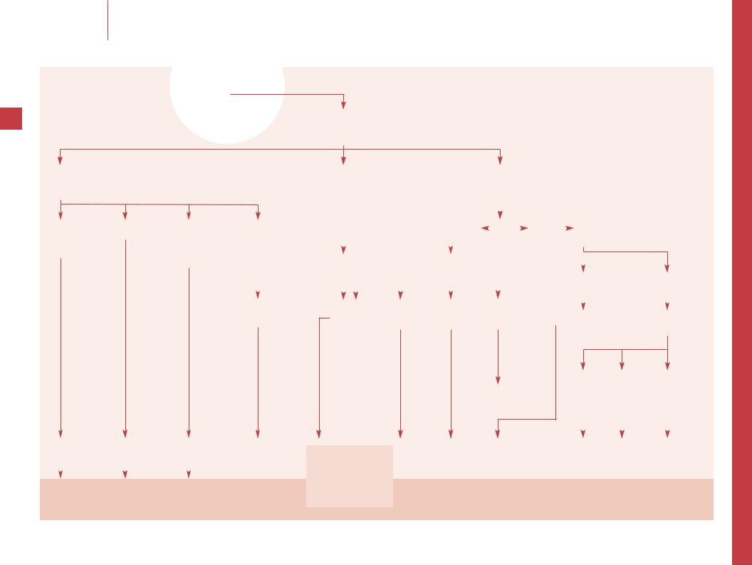
Microcytic anemia |
Microcytic anemia
6 |
RBC indices |
|
Blood smear |
|
7 Fe intake or blood loss |
± Family Hx of anemia or |
Chronic illness or |
+ Target cells |
& Pb level |
Rare disorders |
||||
7 RBC, & RDW ± 7 MCHC |
thalassemia |
infection |
+ Family Hx |
|
|
||||
Anisocytosis |
Mediterranean/Asian/African |
|
|
African/Asian |
|
|
|||
Mild ovalocytosis |
ancestry |
|
|
ancestry |
|
|
|||
± dimorphic population |
|
|
|
|
|
|
|
|
|
|
|
|
|
|
|
|
|
||
|
|
|
|
|
|
|
|
|
|
|
|
|
|
|
|
|
|
|
|
Trial oral Fe |
& RBC, Nl or |
||
|
|
|
minimal & RDW |
|
|
|
Hb >9 g/dl (1.4 mmol/l) |
|
|
|
No hepatosplenomegaly |
|
|
|
Target cells |
|
|
|
|
& Hb Hb fails to & |
± basophilic stippling |
||
|
|||
(see ‘Presumed iron deficiency anemia which fails to respond to oral iron’, p 22)
Hepatosplenomegaly |
Fe, TIBC, TS, ferritin Hb electrophoresis |
||
Hb <9 g/dl (1.4 mmol/l) |
|
|
|
|
|
||
Severe hypochromia |
|
|
|
Anisopoikilocytosis |
|
|
|
Target cells |
|
|
|
Normoblastemia |
|
|
|
|
|
|
|
|
|
|
|
Thalassemia intermedia |
7 Fe, 7 TIBC, |
|
or major |
± 7 TS, & ferritin |
|
Hb H disease |
|
|
|
|
|
|
|
|
|
|
|
|
|
|
|
|
|
Iron deficiency anemia |
Thalassemia trait |
Anemia of |
Hb C or E |
Pb poisoning |
Sideroblastic anemia |
|||
|
|
|
|
|
infection or |
Sickle thalassemia |
|
Protein calorie |
|
|
|
|
|
|
|||
|
|
|
|
|
chronic disease |
Unstable |
|
malnutrition |
|
|
|
|
|
|
|||
|
|
(see ‘Thalassemia’, p 24) |
|
|
hemoglobinopathy |
Metabolic defects of |
||
|
|
|
|
|
|
Fe absorption and |
||
|
|
|
|
|
|
|
|
|
|
|
|
|
|
|
|
|
metabolism |
|
|
|
|
|
|
|
|
|
Continue Fe therapy |
|
|
|
|
|
|
|
|
|
|
|
|
|
|
|
|
|

|
–– Iron deficiency anemia (IDA) is most |
|
|
common in infants and young children with |
|
|
poor Fe intake (often cow’s milk intake of a |
|
|
liter or more daily), but with malnutrition it is |
|
|
seen at any age. Blood loss should always be |
|
|
considered, but is more likely in older children |
|
|
and particularly in adolescent girls. An under- |
|
|
lying bleeding disorder, such as von Wille- |
|
|
brand disease, can cause excessive blood loss. |
|
|
Typical findings of IDA are 7 RBC and & RDW. |
|
|
Smear reveals hypochromia, microcytosis, and |
|
|
often ovalocytes. A dimorphic population |
|
|
(microcytic hypochromic cells mixed with |
|
|
normocytic, normochromic cells) is commonly |
|
|
present early in the disease or following the |
|
|
onset of Fe therapy. |
|
|
–– The diagnosis of IDA is usually estab- |
|
|
lished by a successful trial of oral Fe therapy. |
|
|
Use ferrous sulfate in a dose of 2–3 mg/kg/day |
|
|
of elemental Fe (10–15 mg/kg/day of ferrous |
|
|
sulfate) divided t.i.d.; doses of up to 6 mg/ |
|
|
kg/day of elemental Fe are used for severe |
|
|
Fe deficiency to increase the Hb to safer levels |
|
|
more quickly. If there is no response in |
|
|
2–3 weeks, see the algorithm for failure of |
|
|
IDA to respond to Fe. The etiology of the Fe |
|
|
deficiency is usually apparent and measure- |
|
|
ments of serum Fe (7), TIBC (&), TS (7), and |
|
|
ferritin (7) are not usually necessary. |
|
|
–– Fe treatment is usually continued for at |
|
|
least 3–4 months to correct the anemia and |
|
|
rebuild Fe stores. Changes in diet (particularly |
|
|
a decrease in cow’s milk intake) and manage- |
|
|
ment of blood loss, when appropriate, are |
|
|
necessary to prevent recurrence. |
|
|
–– Mediterranean, Asian or African ancestry |
|
|
is not universal, but is usually present. The |
|
|
Mentzer index (MCV/RBC) may be helpful to |
|
|
differentiate thalassemia minor. In IDA the |
|
7 |
index is generally >13, whereas in thalassemia |
|
trait it is usually <13. |
||
|
–– Hepatosplenomegaly, more severe anemia (typically Hb <9.0 g/dl, 1.4 mmol/l) and more severe hypochromia are consistent
with -thalassemia major or -thalassemia intermedia, but are not expected in either - or -thalassemia trait. Hb levels can be as high as 10.0 g/dl (1.51 mmol/l) in Hb H disease. Normoblastemia, the presence of nucleated erythrocytes in peripheral blood, is prominent in severe forms of -thalassemia. However, small numbers of normoblasts may be seen in severe anemia of any etiology.
–– The anemia of chronic disease can be associated with any severe infection or inflammatory disorder. It is usually normocytic, but microcytosis occurs in 20–30% of patients. Anemia is usually fairly mild with Hb levels 2–3 g/dl (0.31–0.47 mmol/l) below expected normals for age. The combinations of a low serum iron and TIBC, with an elevated ferritin, are typical and help to differentiate it from IDA.
–– Homozygous hemoglobin C or E disease,
E-thalassemia and sickle thalassemia are associated with 7 MCV and prominent numbers of target erythrocytes. These are also seen in E trait, but without anemia. Rare unstable hemoglobins may be associated with microcytic anemia and the Hb electrophoresis may be normal; they are suspected with hemolysis of unidentified etiology. Hb stability studies can establish this diagnosis.
–– Microcytic anemia in Pb poisoning is more often due to concomitant IDA and not the Pb poisoning itself; anemia is a late sign of
Pb poisoning. Basophilic stippling is often but not consistently present.
–– There are several rare etiologies of microcytosis. The hereditary sideroblastic anemias are a rare heterogenous group of disorders characterized by anemia, reticulocytopenia and abnormal patterns of iron deposition in marrow erythroblasts. Protein calorie malnutrition usually causes normocytic/normochromic anemia but it can cause microcytosis without IDA. There are a number of metabolic defects of Fe absorption and metabolism, but they are very rare.
Selected reading
Chui DH, Wage JS: Hydrops fetalis caused by alpha thalassemia: An emerging health care problem. Blood 1998;91:2213–2222.
Clark M, Royal J, Seeler R: Interaction of iron deficiency and lead and the hematologic findings in children with severe lead poisoning. Pediatrics 1988;81:247–254.
Glader BE, Look KA: Hematologic disorders in children from Southeast Asia. Pediatr Clin N Am 1996;43:665–681.
Kwiatkowski JL, West TB, Heidary N, Smith-Whitely K: Severe iron deficiency in young children. J Pediatr 1999;135:514–516.
Schwartz J, Landrigan PJ, Baker EL, et al:
Lead-induced anemia: Dose-response relationships and evidence for a threshold. Am J Publ Hlth 1990;80;165–168.
Red Cell Disorders |
R.H. Sills · A. Deters |
Microcytic anemia |
|
|
|
|
|
|

|
|
|
Red Cell Disorders |
R.H. Sills · A. Deters |
Normocytic anemia |
Normocytic anemia
8 |
|
|
|
|
Evaluate clinical and laboratory evidence of blood loss |
|
Nl reticulocytes |
|
|
|
|
|
|
Indirect bilirubin |
|
Nl indirect bilirubin |
|
|
|
|
|
|
Reticulocyte count |
|
No blood loss |
|
|
|
|
|
|
Blood smear |
|
|
|
|
|
|
|
|
|
|
|
|
|
|
|
|
|
|
|
|
|
|
|
|
|
|
|
|
|
|
|
|
|
|
|
|
|
|
|
+ Blood loss |
& reticulocytes |
|
|
|
|
Clinical evaluation |
|
|
|
|
|
|
|
|
|
|
|
|
Macro-ovalocytosis |
||||||||
± & reticulocytes |
& indirect bilirubin |
|
|
Smear: no PMN hypersegmentation |
|
|
|
|
|
|
|
|
|
|
|
|
and/or PMN |
||||||||||
|
|
|
|
|
|
|
|
|
|
|
|
|
|
||||||||||||||
Nl indirect bilirubin |
No blood loss |
|
|
|
|
or macro-ovalocytosis |
|
|
|
|
|
|
|
|
|
|
|
|
hypersegmentation |
||||||||
Nl SMEAR or |
|
|
|
|
|
|
|
|
|
|
|
|
|
|
|
|
|
|
|
|
|
|
|
|
|
|
|
polychromasia |
|
|
|
|
|
|
|
|
|
|
|
|
|
|
|
|
|
|
|
|
|
|
|
|
|
|
|
and/or dimorphic |
|
|
|
|
|
|
|
|
|
|
|
|
|
|
|
|
|
|
|
|
|
|
|
|
|
|
|
|
|
|
|
|
|
|
|
|
|
|
|
|
|
|
|
|
|
|
|
|
|
|
|
|
|
||
|
population |
|
|
|
|
Pregnant |
Viral or bacterial infection |
Chronically ill |
|
|
No evidence of recent infection |
Dimorphic |
Serum B12 + |
||||||||||||||
|
|
|
|
|
|
|
|
||||||||||||||||||||
|
|
|
|
|
|
|
|
|
|
|
|
|
|
|
|
|
|
|
or anemia of chronic disease |
population RBC folate |
|||||||
|
|
|
|
|
|
|
|
|
|
|
|
|
|
|
|
|
|
|
|||||||||
|
|
|
|
|
|
|
|
|
|
|
|
|
|
|
|
|
|
|
Hb <9 or persistent anemia |
|
|
|
|
|
|||
|
|
|
|
|
|
|
|
|
|
|
|
|
|
|
|
|
|
|
|
|
|
|
|
||||
|
|
Hemolysis |
Starvation |
|
|
Hb >9 g/dl, |
Hb <9 g/dl |
BUN or creatinine |
|
|
|
|
|
|
|
|
|
||||||||||
|
|
|
|
|
|
|
|
Trial of Fe |
|
|
|||||||||||||||||
|
|
|
|
|
|
|
|
|
|
||||||||||||||||||
|
|
|
|
Anorexia nervosa |
|
not ill |
and/or ill |
|
|
|
|
|
Bone marrow aspirate/biopsy |
|
|
||||||||||||
|
|
|
|
|
|
|
|
appearing |
appearing |
|
|
|
|
|
|
|
|
|
|
||||||||
|
|
|
|
|
|
|
|
|
|
|
|
|
|
|
|
|
|
|
|
|
|
|
|
|
|
|
|
|
|
(see ‘Hemolytic |
|
|
|
|
|
|
|
|
|
|
Abnormal |
Nl |
|
|
|
|
|
|
|
|
|
||||
|
|
|
|
|
|
|
|
|
|
|
|
|
|
|
|
|
|
|
|
|
|||||||
|
|
|
|
|
|
|
|
|
|
|
|
|
|
|
|
|
|
|
|
|
|||||||
|
|
anemia’, p 18) |
|
|
|
|
|
|
|
|
|
|
|
|
|
|
|
|
|
|
|
& Hb |
|
|
|
||
|
|
|
|
|
|
|
|
Observe and |
|
|
|
|
|
|
|
Nl Erythroid |
Other |
|
|
|
|||||||
|
|
|
|
|
|
|
|
|
|
|
|
|
|
|
|
|
|
|
|
||||||||
|
|
|
|
|
|
|
|
repeat Hb in |
|
|
|
|
|
|
|
|
hypoplasia |
findings |
|
|
|
|
|
||||
|
|
|
|
|
|
|
|
|
|
|
|
|
|
|
|
|
|
|
|
|
|||||||
|
|
|
|
|
|
|
|
|
|
|
|
|
|
|
|
|
|
|
|
|
|
|
|
||||
|
|
|
|
|
|
|
|
3–4 weeks |
|
|
|
|
|
Fe/TIBC, ferritin |
|
|
|
|
|
|
|
||||||
|
|
|
|
|
|
|
|
|
|
|
|
|
|
|
|
|
|
|
|
||||||||
|
|
|
|
|
|
|
|
|
|
|
|
|
|
|
|
|
|
|
|
|
|
|
|
||||
|
|
|
|
|
|
|
|
|
|
|
|
|
|
|
|
|
|
|
|
|
|
|
|
|
|
|
|
|
|
|
|
|
|
|
|
|
|
|
|
|
|
|
|
|
|
|
|
|
|
|
|
|
|
|
|
|
|
|
|
|
|
|
|
Nl |
Abnormal |
|
|
|
|
|
|
|
|
|
|
|
|
|
|
|
|
||
|
|
|
|
|
|
|
|
|
|
|
|
|
|
|
|
|
|
|
|
|
|
|
|
||||
|
|
|
|
|
|
|
|
|
|
|
|
|
|
|
|
|
7 Fe and TIBC |
|
|
|
|
|
|
|
|
|
|
|
|
|
|
|
|
|
|
|
|
|
|
|
|
|
|
|
|
|
|
|
|
|
|
|
|
||
|
|
|
|
|
|
|
|
|
|
|
|
|
|
|
|
|
& ferritin |
|
|
|
|
|
|
|
|
|
|
|
|
|
|
|
|
|
|
|
|
Reassess |
|
|
|
|
|
|
|
|
|
|
|
|
|
|
|||
|
|
|
|
|
|
|
|
|
|
|
|
|
|
|
|
|
|
|
|
|
|
|
|
|
|
||
|
|
|
|
|
|
|
|
|
|
|
|
|
|
|
|
|
|
|
|
|
|
|
|
|
|
||
|
|
|
|
|
|
|
|
|
|
|
|
|
|
|
|
|
|
|
|
|
|
|
|
|
|
|
|
|
|
|
|
|
|
|
|
|
|
|
|
|
|
|
|
|
|
|
|
|
|
|
|
|
|
|
|
Recent/ongoing |
|
|
Protein-calorie |
Anemia of |
|
|
Anemia of |
|
|
Anemia of |
Anemia of |
|
TEC |
|
IDA |
Early megaloblastic |
|||||||||||
hemorrhage |
|
|
malnutrition |
pregnancy |
|
infection |
|
|
renal disease |
chronic |
|
DBA |
|
|
|
anemia or combined |
|||||||||||
|
|
|
|
|
|
|
|
|
|
|
|
|
|
|
|
|
disease |
|
Acquired pure |
|
|
IDA + megaloblastic |
|||||
|
|
|
|
|
|
|
|
|
|
|
|
|
|
|
|
|
|
|
|
||||||||
|
|
|
|
|
|
|
|
|
|
|
|
|
|
|
|
|
|
|
|
RBC aplasia |
|
|
|
anemia |
|||
|
|
|
|
|
|
|
|
|
|
|
|
|
|
|
|
|
|
|
|
|
|
|
|
|
|
|
|
|
|
|
|
|
|
|
|
|
|
|
|
|
|
|
|
|
|
|
|
|
|
|
|
|
|
|
|
|
|
|
|
|
|
|
|
|
|
|
|
|
|
|
|
|
|
|
|
|
|
|
|
|
|
|
|
Ensure patient |
|
|
|
|
|
|
No further |
Further Dx and Rx |
|
|
|
|
|
|
|
|
|
|
|
R/O IDA |
|||||||
is stable |
|
|
|
|
|
|
evaluation |
|
|
|
|
|
|
|
|
|
|
|
|
|
(see ‘Macrocytic anemia’, p 10) |
||||||
|
|
|
|
|
|
|
|
|
|
|
|
|
|
|
|
|
|
|
|
|
|
|
|
|
|
|
|

–– Acute blood loss is usually obvious and most often due to epistaxis, hematemesis, hematochezia, hematuria, menorrhagia and trauma. Chronic GI blood loss may be occult and melena may not be recognized as significant; stools should be examined for occult blood. Consider blood sampling and postoperative losses. Typically, pallor is noted without icterus.
–– The reticulocytosis in response to hemorrhage is usually delayed 3–7 days after the onset of bleeding. Intercurrent infection or illness may also inhibit the reticulocyte response. Bilirubin should be normal (except
in neonates with enclosed hemorrhage in whom it may be &). Smear usually reveals polychromasia from the reticulocytosis, and a dimorphic population in early or partially treated iron deficiency anemia.
–– Severe protein-calorie malnutrition is associated with normocytic anemia; 1/3 of patients with anorexia nervosa have normocytic anemia.
–– Hb levels fall to as low as 10 g/dl
(1.55 mmol/l) normally in pregnancy because of hemodilution. Lower values require investigation. Fluid overload in the absence of pregnancy can cause dilutional anemia, but this is usually clinically evident.
–– The most common cause of anemia in children is viral and, less often, bacterial infection. If the anemia is mild (e.g. Hb >9.0 g/dl, 1.4 mmol/l) and the child is not very ill, observe the child and repeat the Hb in 2–4 weeks. It will then usually be normal and no further investigation will be necessary; if the anemia persists then reassessment should be undertaken to ensure a more serious diagnosis if not
9 being missed. During active inflammation, the fall in Hb has been estimated at 13% per week.
–– Infections such as bacterial septicemia (staphylococcal, streptococcal, pneumococcal),
Bartonella, Clostridia, malaria, and HIV are often associated with severe normocytic anemia
–– The anemia of chronic disease is common. Hemoglobin is usually 7–11 g/dl (1.09–1.7 mmol/l) and the MCV is normal in most patients. Endocrinologic disorders such as hypothyroidism can also cause anemia.
–– If the anemia is mild (Hb 9–11, 1.4–
1.7 mmol/l) it may be reasonable to observe the child since it might be due to an unrecognized viral illness. If the anemia is more severe or there are concerning clinical signs, a bone marrow aspirate and biopsy should be done. A concerning laboratory finding is myelophthisic anemia (leukoerythroblastosis) which usually results from bone marrow invasion; it presents with erythroblasts and immature WBCs, teardrop-shaped RBCs and giant platelets in peripheral blood.
–– TEC is the most common cause of pure RBC aplasia in children. Other acquired etiologies are rare and usually occur in young adults and most have no recognized underlying disorder. Drug-induced RBC aplasia occurs with carbamazepine, phenytoin and valproate and usually resolves following discontinuation of the drug. Diamond-Blackfan anemia (DBA) is congenital pure RBC aplasia; although often macrocytic, it may be normocytic.
–– Other rare diagnoses based on marrow findings include myelodysplastic syndromes, congenital dyserthyropoietic anemias and myelofibrosis.
–– Particularly if the MCV is near the lower limit of normal, developing iron deficiency anemia should be considered. A dimorphic population of normal and hypochromic RBCs and an elevated RDW are usually evident and a trial of oral Fe is often diagnostic.
–– Macro-ovalocytosis and PMN hypersegmentation (see ‘Macrocytic anemia’, p 10) may be evident before the development of macrocytosis in megaloblastic anemia. In addition, the combination of IDA with B12 and/or folate deficiency may remain normocytic; macro-ovalocytosis and hypersegmentation are still expected as is a high RDW reflecting marked anisocytosis (variation in cell size). The potential irreversible neurologic damage associated with B12 deficiency makes it critical to consider this diagnosis.
Selected reading
Abshire TC, Reeves JD: Anemia of inflammation in children.
J Pediatr 1983;103:868.
Abshire TC: Anemia of inflammation. Pediatr Clin N Am 1996;43:623–637.
Beutler E: G6PD deficiency.
Blood 1994;84:3613–3636.
Lee M, Trumann JT: Anemia acute; in Johnston JM, Windle ML, Bergstrom SK, Gross S, Arceci RJ (eds): Pediatric Medicine, Emedicine.com, 2002 (http://www.emedicine.com).
Sieff CA, Nisbet-Brown E, Nathan DG:
Congenital bone marrow failure syndromes. Br J Haematol 2000;111:30–42.
S, Arceci RJ (eds): Pediatric Medicine, Emedicine.com, 2002 (http://www.emedince.com).
Vlachos A, Lipton JM: Bone marrow failure in children. Curr Opin Pediatr 1996;8:33–41.
Red Cell Disorders |
R.H. Sills · A. Deters |
Normocytic anemia |
|
|
|
|
|
|

|
|
|
Red Cell Disorders |
R.H. Sills · A. Deters |
Macrocytic anemia |
Macrocytic anemia
10 |
|
|
|
|
|
|
|
|
|
|
|
|
|
|
RBC indices |
|
|
|
|
|
|
|
|
|||
|
|
|
|
|
|
|
|
|
|
|
|
|
|
|
Blood smear |
|
|
|
|
|
|
|
|
|||
|
|
|
|
|
|
|
|
|
|
|
|
|
|
|
|
|
|
|
|
|
|
|
||||
|
|
|
|
|
|
|
|
|
|
|
|
|
|
|
|
|
|
|
|
|
|
|
|
|
||
|
|
|
|
|
|
|
|
|
|
|
|
|
|
|
|
|
|
|
|
|
||||||
|
|
|
|
|
|
Macro-ovalocytosis |
|
|
|
|
|
|
|
|
No macro-ovalocytosis |
|
|
|||||||||
|
|
|
|
|
|
PMN hypersegmentation |
|
|
|
|
|
|
|
|
No PMN hypersegmentation |
|
|
|||||||||
|
|
|
|
|
|
|
|
|
|
|
|
|
|
|
|
|
|
|
|
|
|
|
|
|
|
|
|
|
|
|
|
|
|
|
|
|
|
|
|
|
|
|
|
|
|
|
|
|
|
|
|
|
|
|
|
|
|
|
|
Megaloblastic anemia |
|
|
|
|
|
|
|
|
Reticulocytes |
|
|
|
|
|||||||
|
|
|
|
|
|
|
|
|
|
|
|
|
|
|
|
|
|
|
|
|
|
|
|
|
|
|
|
|
|
|
|
|
|
|
|
|
|
|
|
|
|
|
|
|
|
|
|
|
|
|
|
|
|
|
|
|
|
|
|
|
|
|
|
|
|
|
|
|
|
|
|
|
|
|
|
|
|
|
|
|
|
|
|
|
|
|
|
|
|
|
|
|
|
|
|
|
|
|
|
|
|
|
|
|
|
|
|
|
|
|
|
|
|
Serum B12 |
|
|
|
|
|
|
|
|
|
|
|
|
|
|
Normal or 7 reticulocytes |
|||||
|
|
|
|
|
|
RBC folate |
|
|
|
|
|
|
|
|
|
|
|
|
|
|
||||||
|
|
|
|
|
|
|
|
|
|
|
|
|
|
|
|
|
|
|
|
|
|
|
|
|||
|
|
|
|
|
|
|
|
|
|
|
|
|
|
|
|
|
|
|
|
|
|
|
|
|
|
|
|
|
|
|
|
|
|
|
|
|
|
|
|
|
|
|
|
|
|
Anemia |
|
|
|
|
Nl Hgb or |
||
|
|
7 B12 level |
7 folate |
|
|
|
Nl B12 and folate |
|
|
|
|
|
|
|
|
|||||||||||
|
|
|
|
|
|
|
|
|
|
|
|
|
|
|
only mildly 7 |
|||||||||||
|
|
|
|
Nl B12 |
|
|
|
|
|
|
|
|
|
|
|
|
|
|
|
|
|
|
|
|
|
|
|
|
|
|
|
|
|
|
|
|
|
|
|
|
|
|
|
Bone marrow |
|
|
|
|
|
|
|||
|
|
|
|
|
|
|
|
|
Megaloblastic anemia |
|
|
|
|
|
|
|
|
|
|
|||||||
|
|
|
|
|
|
|
|
|
|
|
|
|
aspirate |
|
|
|
|
|
|
|||||||
|
|
|
|
|
|
|
|
|
not due to vitamin |
|
|
|
|
|
|
|
|
|
|
|||||||
|
|
|
|
|
|
|
|
|
|
|
|
|
and biopsy |
|
|
|
|
|
|
|||||||
|
|
|
|
|
|
|
|
|
deficiency |
|
|
|
|
|
|
|
|
|
|
Spurious |
|
|
||||
|
|
|
|
|
|
|
|
|
|
|
|
|
|
|
|
|
|
|
|
|
|
|
|
|
||
|
|
|
|
|
|
|
|
|
|
|
|
|
|
|
|
|
Pure RBC |
Hypocellular |
|
|
|
|
||||
|
|
|
|
|
|
|
|
|
|
|
|
|
|
|
|
|
|
|
|
|
||||||
|
|
|
|
|
|
Appropriate |
|
Drug |
|
|
No implicated |
|
|
|
|
& MCV |
|
|
||||||||
|
|
|
|
|
|
|
|
aplasia |
& Hgb F |
|
|
|
|
|||||||||||||
|
|
|
|
|
|
drug |
Hx |
drugs |
|
|
|
|
|
|
|
|
||||||||||
|
|
|
|
|
|
|
|
± congenital |
|
|
|
|
|
|
|
|
||||||||||
|
|
|
|
|
|
|
|
|
|
|
|
|
|
|
|
|
|
|
|
|
|
|
|
|
||
|
|
|
|
|
|
|
|
|
|
|
|
|
|
|
|
|
anomalies |
|
|
|
|
|
|
|
|
|
|
|
|
|
|
|
|
|
|
|
|
|
|
|
|
|
|
|
|
|
|
|
|
|
|
|
|
|
|
|
|
|
|
|
|
|
|
|
|
|
|
|
|
|
|
|
|
|
|
|
|
|
|
|
|
|
|
|
|
|
|
|
|
|
|
|
|
|
|
|
|
|
|
|
|
|
|
|
|
|
|
|
|
B12 deficiency |
Folate |
Drug-induced |
|
|
|
|
Rare |
Reticulo- |
Diamond- |
Fanconi |
Rare |
Cold agglutinins |
Drugs |
|||||||||||
|
|
|
|
deficiency |
megaloblastic |
|
|
|
|
disorders |
cytosis |
Blackfan |
anemia |
findings |
Hyperglycemia |
Congenital heart |
||||||||||
|
|
|
|
|
|
anemia |
|
|
|
|
|
|
|
|
anemia |
|
|
|
|
Leukocytosis |
disease |
|||||
|
|
|
|
|
|
|
|
|
|
|
|
|
|
|
|
|
|
|||||||||
|
|
|
|
|
|
|
|
|
|
|
|
|
|
|
|
|
|
|
|
|
|
|
|
|
Down syndrome |
|
|
|
|
|
|
|
|
|
|
|
|
|
|
|
|
|
|
|
|
|
|
|
|
|
|
Hypothyroidism |
|
|
|
Identify cause |
Identify |
Consider if |
|
|
|
|
|
|
Evaluate for |
Corticosteroid |
Confirmed with |
|
|
Liver disease |
||||||||||
|
|
|
|
|
|
|
|
|
|
Asplenia |
||||||||||||||||
|
|
R/O pernicious |
cause |
drug can be |
|
|
|
|
|
|
hemolysis, |
Rx |
lymphocyte chromosomal |
|
|
|||||||||||
|
|
|
|
|
|
|
|
|
|
|
|
|||||||||||||||
|
|
anemia |
Rx folate |
discontinued |
|
|
|
|
|
|
blood loss, |
|
|
analysis ± DNA studies |
|
|
|
|
||||||||
|
|
Rx B12 deficiency |
|
|
|
|
|
|
|
|
|
|
|
recovering aplasia |
|
|
|
|
|
|
|
|
|
|
||
|
|
|
|
|
|
|
|
|
|
|
|
|
|
|
|
|
|
|
|
|
|
|
|
|
|
|

–– Megaloblastic anemia, most commonly due to B12 or folate deficiency, is the result of impaired DNA synthesis.
It is critical to differentiate the megaloblastic anemias from the other etiologies of macrocytosis. Hypersegmented neutrophils (>5% of PMNs with 5 nuclear lobes and >1% with 6 lobes) are found in 98% of patients with megaloblastic hematopoiesis.
The combination of macro-ovalocytosis and neutrophil hypersegmentation has a specificity of 96–98% and a positive predictive value of 94% for either folate or B12 deficiency in studies in adults. In the absence of malnutrition, megaloblastic anemia is uncommon in children; no cases of folate or B12 deficiency were observed in 146 American children evaluated because of macrocytosis.
–– Serum folate reflects recent intake while RBC folate reflects longer term intake and is a more reliable indicator of folate deficiency. Serum B12 levels are usually diagnostic.
–– B12 deficiency is most commonly caused by pernicious anemia, but may also be due to ileal disease, malabsorption, vegan diet, and fish tapeworm. The most common etiology in infancy may be selective malabsorption of cobalamin (Imerslund-Gräsbeck disease).
–– It is critical to exclude B12 deficiency in megaloblastic patients because of its potential irreversible neurologic damage. Combined deficiency occurs, so even if folate levels are low B12 levels must be determined as well. Treatment of B12 deficiency with folate may correct the hematologic findings and the patient’s improvement can mask the progression of neurologic disease. If the diagnosis is not certain, normal serum methylmalonic acid and total homocysteine levels effectively exclude B12 deficiency; a normal homocysteine alone suggests that it is not folate deficiency.
–– Folate deficiency can be due to drugs (e.g. phenytoin, barbiturates, valproate, methotrexate, trimethoprim), malnutrition, malabsorption, diet of goat’s milk, alcoholism, and increased cell turnover (hemolysis, pregnancy).
–– Drug-induced macrocytic anemias are the most common etiology of macrocytosis in children in industrialized nations. The pathogenesis is not always clear, but it is likely due
11
to megaloblastic changes and/or marrow injury. The drugs most commonly involved are chemotherapeutic agents (e.g. 6-mercaptopurine and hydroxyurea), but also anticonvulsants (most often carbamazepine, valproic acid, phenytoin, phenobarbital), zidovudine, immunosuppressive agents and sulfa drugs.
–– Megaloblastic anemias unrelated to vitamin deficiencies or drugs are rare and may be congenital (deficiency of transcobalamin II or intrinsic factor, other metabolic defects or congenital dyserythropoietic anemia), or acquired (myelodysplastic syndrome, alcoholism). BMA and BMB may be necessary to exclude myelodysplastic syndrome.
–– Even without macro-ovalocytosis and hypersegmentation, B12 (particularly important given its neurologic complications) and folate levels should be obtained if an obvious etiology for the macrocytosis is not identified.
–– Reticulocytes are approximately 20% larger than mature RBCs so a substantially elevated reticulocyte count increases the overall MCV of an otherwise normocytic RBC population.
–– Anemic children who have true macrocytic, nonmegaloblastic anemia usually have diminished or abnormal erythropoiesis so both BMA and BMB are generally indicated.
–– Diamond-Blackfan anemia is congenital RBC aplasia, which usually presents in the first 3 months of life. Most are macrocytic especially if diagnosed after the first months of life. Associated congenital malformations are common. Most patients require long-term corticosteroid therapy. TEC is not a macrocytic process unless recovery is occurring with a reticulocytosis.
–– Fanconi anemia does not usually present with isolated anemia, but the macrocytosis often precedes other hematologic changes. Congenital anomalies are typical but are absent in approximately 25% of patients. Marrow hypoplasia is usually noted, but hyperplasia with dyserythropoiesis (disordered cell development) can be seen. Hb F is &. The most utilized diagnostic finding is enhanced chromosomal breakages in peripheral blood lymphocytes stimulated in culture. Other rare bone marrow failure syndromes, such as dyskeratosis congenita, should be considered if Fanconi anemia is excluded.
–– Other rare marrow disorders associated with macrocytosis include myelodysplasia, dyserythropoiesis and sideroblastic anemia. MDS is diagnosed on the basis of myelodysplastic changes in the marrow; the finding of a clonal chromosomal abnormality in a majority of patients is useful confirmation. Dyserythropoietic changes suggest the rare diagnosis of congenital dyserythropoietic anemia. Sideroblastic anemias demonstrate a ring of iron staining which surrounds the nucleus; some hereditary forms are associated with macrocytosis.
–– Spurious macrocytosis is most often due to cold agglutinins which cause RBCs clumped together to be counted as a single cell; the same artifact decreases the RBC. Warming the sample normalizes the MCV. This cold agglutinin effect often occurs in the absence of hemolysis or anemia. Extreme hyperglycemia and leukocytosis occasionally cause spurious macrocytosis.
–– A number of disorders, most associated with no or minimal anemia, can alter RBC size. Normal neonates are often mistakenly identified as macrocytic because the normal MCV of a full-term neonate is 98–118 fl. Drugs can cause isolated macrocytosis, including those that usually cause obvious megaloblastic changes. Congenital heart disease (usually cyanotic) and, independently, Down syndrome are also associated with macrocytosis; the mechanism by which this occurs is not well understood. Excessive membrane lipids can cause macrocytosis, usually without anemia, in hypothyroidism, liver disease and after splenectomy. In these instances, the RBCs have a larger surface area causing them to spread more widely on blood smears, but this may not be associated with an actual increase in measured volume (MCV).
Selected reading
Bessman JD, Banks D: Spurious macrocytosis: A common clue to erythrocyte cold agglutinins. Am J Clin Pathol 1980;74:797–800.
Holt JT, DeWandler MJ, Arvan DA: Spurious elevation of the electronically determined mean corpuscular volume and hematocrit caused by hyperglycemia. Am J Clin Pathol 1982;77:561–567.
Lindenbaum J: Status of laboratory testing in the diagnosis of megaloblastic anemia. Blood 1983;61: 624–627.
McPhedran P, et al: Interpretation of electronically determined macrocytosis. Ann Intern Med 1973;78: 677–783.
Pappo AS, Fields BW, Buchanan GR: Etiology of red blood cell macrocytosis during childhood: Impact of new diseases and therapies. Pediatrics 1992;89:1063–1070.
Rasmussen SA, Fernhoff PM, Scanlon KS:
Vitamin B12 deficiency in children and adolescents. J Pediatr 2001;138:10–17.
Red Cell Disorders |
R.H. Sills · A. Deters |
Macrocytic anemia |
|
|
|
|
|
|
|
|
|
Red Cell Disorders |
R.H. Sills · A. Deters |
Pancytopenia |
Pancytopenia
12
Clinical evaluation
Evidence hemolysis
Direct Coombs test
Clinical evidence + DCT |
Splenomegaly |
Chronic hemolysis |
|
of sepsis and/or |
± spherocytes |
± hemoglobinuria |
|
shock |
Liver disease |
|
|
|
Portal hypertension |
|
|
|
|
|
|
Flow cytometry
CD55/CD59
Red cell indices
Blood smear
Reticulocytes – indirect bilirubin
Blasts on peripheral smear or |
|
|
|
|
Nl or 7 reticulocytes |
|
|
|
|
||||||||||||
leukoerythroblastosis |
|
|
|
|
Nl indirect bilirubin |
|
|
|
|
||||||||||||
|
|
|
|
|
|
|
|
|
No other evidence hemolysis |
|
|
||||||||||
|
|
|
|
|
|
|
|
|
|
||||||||||||
|
|
|
|
|
|
|
|
|
|
|
|
& MCV |
|
|
|
|
|
|
|||
|
|
|
|
|
|
|
|
|
|
|
|
|
|
|
|
|
|
|
|||
|
|
|
|
|
Nl MCV |
|
|
MCV |
|
|
|
Macro-ovalocytosis |
|
|
|||||||
|
|
|
|
|
|
|
|
|
|
|
|||||||||||
|
|
|
|
|
|
|
|
|
|
|
|
|
|
|
|
|
|
Hypersegmentation PMN |
|||
|
|
|
|
|
|
|
|
|
|
|
|
|
|
|
|
|
|
||||
|
|
|
|
|
|
|
|
|
|
|
|
|
|
|
|
|
|
|
|
|
|
BMA/BMB |
|
|
BMA/BMB |
|
|
|
|
|
|
|
|
|
|
|
|
|
|
||||
|
|
|
|
|
|
|
|
|
|
|
|
|
|
|
|
||||||
|
|
|
|
|
|
|
|
|
|
|
|
– |
+ |
||||||||
|
|
|
|
|
|
|
|
|
|
|
|
|
|
|
|
|
|
||||
|
|
|
|
|
|
|
|
|
|
|
|
|
|
|
|
|
|
||||
|
|
|
|
|
|
|
|
|
|
|
|
|
|
|
|
|
|
||||
|
|
|
|
|
|
|
|
|
|
|
|
|
|
|
|
|
|
|
|
|
|
|
|
|
|
|
|
|
|
|
|
|
|
|
|
|
|
|
|
|
|
|
|
Bone marrow |
Nl or mild |
BM failure |
Fibrosis or |
|
|
|
|
||||||||||||||
|
|
|
|
||||||||||||||||||
replacement |
hypoplasia |
|
|
|
myelodysplasia |
± congenital |
RBC folate |
||||||||||||||
|
|
|
|
|
|
|
|
|
|
|
|
|
|
|
|
|
|
malformations |
Serum B12 |
||
|
7 B12 |
7 RBC |
Nl B12 |
|||
MDS |
|
|
folate |
and |
||
|
|
|||||
|
|
|
|
folate |
||
Myelofibrosis |
|
|
|
|
||
|
|
|
|
|||
|
|
|
|
|
|
|
|
|
|
|
|
|
|
|
|
|
|
|
|
|
Sepsis |
Autoimmune |
Hypersplenism |
PNH |
Leukemia |
Infection Aplastic |
Fanconi anemia |
B12 |
Folate |
MDS |
|||
|
|
pancytopenia |
|
|
|
Lymphoma |
anemia |
Dyskeratosis |
deficiency |
deficiency |
Other |
|
|
|
|
|
|
||||||||
|
|
|
|
|
|
|
Neuroblastoma |
|
congenita |
|
|
|
|
|
|
|
|
|
|
|
|
|
|
||
|
|
|
|
|
|
|
LCH |
|
|
|
|
|
Antibiotics |
Probable |
Determine underlying etiology |
Osteopetrosis |
|
|
|
|
|
||||
Other solid tumors |
|
|
|
|
|
|||||||
Supportive care |
corticosteroids |
R/O portal hypertension |
|
|
|
|
|
|||||
|
|
|
|
|
|
|||||||
R/O DIC

–– Pancytopenia consists of leukopenia, anemia and thrombocytopenia; if the WBC is normal, but the absolute neutrophil count is decreased (neutropenia), the patient should still be considered pancytopenic. More than 1/3 of the children with acute lymphoblastic anemia have a normal WBC, but most are neutropenic.
–– Evidence for hemolysis: indirect hyperbilirubinemia,
reticulocytosis, abnormal smear (e.g., anisocytosis, RBC fragmentation, spherocytosis, elliptocytosis), & RDW (reflecting the variation in cell size with & numbers of
larger reticulocytes and overall variation in cell size),
7 haptoglobulin, hemoglobinuria (if the hemolysis is intravascular), and & LDH (also & in many malignancies).
–– Sepsis can cause pancytopenia due to both diminished production and/or increased destruction. DIC should be considered in critically ill children.
–– Concomitant AIHA, ITP and autoimmune neutropenia occurs occasionally and the direct Coombs test is almost always positive. It may occur in isolation or in association with collagen vascular disorders, particularly SLE.
–– Hypersplenism can result in mild-to-moderate (but rarely severe) pancytopenia from a combination of splenic sequestration and hemolytic anemia. Splenomegaly is usually prominent and its etiology is evident. Chronic liver disease and/or portal hypertension should be considered.
Disorders such as cavernous transformation of the portal vein may present with only pancytopenia and/or splenomegaly.
–– Paroxysmal nocturnal hemoglobinuria is a rare acquired clonal disorder which typically presents with chronic hemolytic anemia. It can also cause varying degrees of marrow aplasia so it should be considered in the child with pancytopenia and a reticulocytosis. The traditional screening tests (sucrose hemolysis test and Ham test) should be replaced by specific flow cytometric analysis for deficient CD55 or CD59.
13–– Blasts in peripheral blood are consistent with leukemia. Leukoerythroblastosis (myelophthisic anemia) is less common in children and usually results from bone marrow invasion. Anemia may be accompanied by several findings in peripheral blood, including erythroblasts
(nucleated erythrocytes), more immature neutrophils, tear- drop-shaped RBCs, and giant platelets. This is usually due to malignant replacement but is also caused by benign conditions such as osteopetrosis, storage disease, infection, myeloproliferative disorders, severe hemolytic disease, thalassemia major and hypoxia. Bone marrow examination should be performed.
–– When examining the bone marrow, an aspirate is always done; in addition to more routine morphologic studies, including special stains, samples for chromosomal and flow cytometric analysis should be obtained. Unless
a diagnosis of leukemia is strongly suspected, a bone marrow biopsy should also be done to provide information concerning cellularity, myelofibrosis, infiltrative processes and storage diseases.
–– Bone marrow examination should be strongly considered in these children. If the pancytopenia is mild and consistent with a transient infection, marrow examination can be deferred.
–– Many viral and bacterial illnesses cause pancytopenia, but the marrow is rarely aplastic and recovery usually coincides with resolution of the infection. Examples include EBV, CMV, HIV, brucellosis, tuberculosis, Q fever, and Legionaires disease.
–– Aplastic anemia is idiopathic in approximately 3/4 of patients. Most others are drug- (chloramphenicol, gold compounds and non-steroidal anti-inflammatory agents most clearly implicated) or toxin-induced (most often benzene), related to infection (most often hepatitis not usually associated with a specific type), constitutional (Fanconi anemia, Shwachman-Diamond syndrome), and, very rarely, PNH.
–– Rare findings include myelodysplasia or myelofibrosis. Myelodysplasia is an acquired clonal disorder of the bone marrow characterized by abnormal maturation of one or more hematopoietic cell lines. Chromosomal analysis of BMA is abnormal in 50–80%. Myelofibrosis is diagnosed by special staining of bone marrow biopsy specimens.
It is very rare in children but when it occurs it is usually in toddlers with trisomy 21 and represents a form of acute megakaryoblastic leukemia.
–– Pancytopenia is common in children with megaloblastic anemia. The simplest initial screen for megaloblastic anemia in the macrocytic patient is to review the peripheral smear. Hypersegmented neutrophils (>5% of PMNs with 5 lobes and >1% with 6 lobes) are found in 98% of patients with megaloblastic anemias. The combination of macro-ovalocytosis and hypersegmentation has a specificity of 96–98% and the positive predictive value for either folate or B12 deficiency is ~94% in adult studies.
–– Fanconi anemia is an autosomal-recessive disorder in which aplastic anemia develops later in the first decade. The MCV is often >100 fl even before anemia develops. Most patients have dysmorphic features which are present at birth. The hypersensitivity of chromosomes to diepoxybutane (DEB) establishes the diagnosis. Other rarer congenital aplastic anemias may be macrocytic, including dyskeratosis congenita.
–– Serum B12 levels are generally reliable but assays of RBC folate are more accurate than serum levels.
–– Megaloblastic anemias can occur in the absence
of deficiencies of folate or B12 and include myelodysplasia, drug-induced changes (e.g. zidovudine, hydroxyurea), congenital dyserythropoietic anemia, and metabolic defects of folate or B12 metabolism (e.g. methylmalonic acidurias).
Selected reading
Alter BP, Young NS: The bone marrow failure syndromes; in Nathan DG, Orkin SH (eds): Hematology of Infancy and Childhood, ed 5. Philadelphia, Saunders, 1998, p 239.
Altschuler S (ed): Pediatric Medicine, Emedicine, 2002 (available at http://www.emedicine.com) has several chapters on anemia useful as references.
Schwartz CL, Cohen HT: Preleukemic syndromes and other syndromes predisposing to leukemia. Pediatr Clin North Am 1988;35:653–871.
Red Cell Disorders |
R.H. Sills · A. Deters |
Pancytopenia |
|
|
|
|
|
|

|
|
|
Red Cell Disorders |
R.H. Sills · A. Deters |
Anemia in the neonate |
Anemia in the neonate
14
Reticulocytes <2%
Nl indirect bilirubin
(see ‘Neonatal anemia due to impaired RBC production’, p 16)
CBC |
Indirect bilirubin |
RBC indices |
Blood smear |
Reticulocytes |
Direct Coombs |
|
|
|
|
|
|
Reticulocytes usually >5–8% |
|
|
Direct Coombs + |
||||||||
|
|
|
|
|
|
Direct Coombs negative |
|
|
Reticulocytes >5–8% |
||||||||
|
|
|
|
|
|
|
|
|
|
|
|
|
|
& Indirect bilirubin |
|||
|
|
|
|
|
|
|
|
|
|
|
|
|
|
||||
|
|
|
|
|
|
|
|
|
|
|
|
|
|
|
|
|
|
|
|
|
|
|
|
|
|
|
& Indirect bilirubin |
|
|
ABO and Rh type infant and |
|||||
|
|
|
|
|
|
|
|
|
|
|
|||||||
Nl blood smear |
|
|
|
|
|
Abnormal smear |
|
|
mother |
|
|
||||||
Nl bilirubin |
|
|
|
|
|
|
|
|
|
|
Specificity anti-RBC antibody |
||||||
|
|
|
|
|
|
|
|
|
|
|
|
|
|
||||
|
|
|
Sick infant |
|
|
|
|
|
|
|
|
|
|
|
|
||
|
|
|
|
|
|
|
|
|
|
|
|
|
|
|
|||
|
|
|
|
|
|
|
|
|
|
|
|
|
|
|
|||
|
|
|
|
|
|
|
|
|
|
|
|
|
|
|
|
|
|
|
|
Evidence of |
Shock |
Nonspecific |
|
Specific smear |
Mother type O |
Infant Rh+ |
No ABO/Rh |
||||||||
|
|
infection |
Hypoxia |
blood smear |
|
abnormalities |
Baby A or B |
Mother Rh– |
incompatibility |
||||||||
|
|
|
|
Acidosis |
|
|
|
|
|
|
Anti-A or -B Ab |
+ anti-Rh Ab |
Elute RBC |
||||
|
Iatrogenic |
|
|
G6PD testing |
|
|
|
|
|
|
|
antibody |
|||||
|
|
|
|
|
|
|
|
|
|
|
|
||||||
|
|
|
|
|
|
|
|
|
|
|
|
||||||
|
blood loss |
|
|
|
|
|
|
|
|
|
|
|
|
|
|
||
|
|
|
|
– |
+ |
|
|
|
|
|
|
|
|
|
|||
|
|
|
|
|
|
|
|
|
|
|
|
|
|
|
|
|
|
|
|
|
|
|
|
|
|
|
|
|
|
|
|
|
|
|
|
Blood loss |
Viral infections |
DIC |
Viral illness |
G6PD |
ABO disease |
ABO |
Rh |
Minor blood |
||||
|
|
Bacterial infections |
|
Rare |
deficiency |
Hereditary spherocytosis |
disease |
disease |
group incom- |
|||
|
|
|
||||||||||
|
|
Fungal infections |
|
etiologies |
|
|
Hereditary elliptocytosis |
|
|
|
|
patibility |
|
|
|
|
|
|
|
|
|
||||
|
|
Protozoal infections |
|
|
|
|
Hereditary pyropoikilocytosis |
|
|
|
|
|
|
|
|
|
|
|
|
Hereditary stomatocytosis |
|
|
|
|
|
|
|
|
|
|
|
|
MAHA |
|
|
|
|
|
|
|
|
|
|
|
|
Malaria |
|
|
|
|
|
Ensure stable |
|
|
|
Avoid oxidant |
Low risk of |
High risk of |
|
|||||
|
|
|
|
|
||||||||
Identify source |
|
|
|
agents |
|
kernicterus |
kernicterus |
|
||||
R/O coagulopathy |
± late anemia |

–– Neonatal pallor is a sign of asphyxia, shock, hypothermia and hypoglycemia as well as anemia. Pallor is usually apparent when the Hb is <7–8 g/dl (1.09–1.24 mmol/l). Neonatal anemia requires immediate investigation. Anemia at birth is usually due to hemorrhage or severe alloimmunization. Anemia manifesting in the first 2 days of life is often due to internal or external hemorrhage, while anemia after the first 48 h is usually hemolytic and associated
with jaundice. When assessing whether a neonate is anemic, consider that capillary samples can average 3.5 g/dl (0.54 mmol/l) higher than venous samples. Hb also varies with age and with gestational age.
–– Reticulocyte counts vary from 3 to 7% of RBCs in the first 2 days of life, decreasing to 0–1% by 7 days. Most neonates with hemolysis have reticulocyte counts of 5–8% or higher. In the first few days of life, nucleated erythrocytes are normally seen in peripheral blood as are small numbers of spherocytes. As an indicator of hemolysis, indirect hyperbilirubinemia is limited by the frequency of hyperbilirubinemia in infants without hemolysis; however, hemolytic anemia in the neonate is almost always associated with a bilirubin level >10–12 mg/dl (171–180 µmol/l).
–– Anemia unaccompanied by jaundice is usually due to hemorrhage in the first 24–72 hours of life. Ensure that the infant is not dangerously hypovolemic and that blood loss is not continuing. With acute blood loss, the Hb may not fall immediately, the MCV will be normal and a reticulocytosis is usually delayed for 3–7 days. Obstetrical complications cause anemia in approximately 1% of newborns; common etiologies include abruptio placentae, placenta previa, twintwin transfusion, ruptured cord, emergency Caesarian section, cephalohematomas, and feto-maternal hemorrhage. The latter is very common and is most easily diagnosed using a Kleihauer-Betke test to identify fetal cells in maternal blood. Common etiologies of serious internal hemorrhage include intracranial or subgaleal, intra-abdominal (particularly hepatic or splenic rupture or hematomas), and pulmonary hemorrhage. Iatrogenic blood loss (phlebotomy, accidents with catheters) should be considered. The Apt test differentiates neonatal gastrointestinal hemorrhage
15from swallowed maternal blood. Bleeding disorders, such as vitamin K deficiency, DIC, neonatal alloimmune thrombocytopenia and hemophilia may be responsible for hemorrhage.
–– Iatrogenic blood loss is a routine component of anemia in sick neonates.
–– Viral and bacterial infections are often associated with hemolysis as well as impaired erythroid production. The hemolysis is usually a direct result of infection, but in very ill infants may also be a consequence of DIC. Infection is often associated with hepatosplenomegaly. One-half of the newborns with toxoplasmosis have anemia, which may be severe. CMV, rubella and herpes simplex are usually associated with mild anemia. Bacterial infection, whether complicated by DIC or not, is often associated with anemia. Malaria must be considered in endemic areas.
–– Hemolytic anemia often accompanies neonatal asphyxia, regardless of etiology, and is often due to DIC. Shock, regardless of etiology, can trigger DIC.
–– Anisocytosis, poikilocytosis, polychromasia, occasional spherocytes or fragmented erythrocytes, are findings which suggest hemolysis but are not specific.
–– G6PD screening tests are useful, but false-negative results are common in mild variants during a reticulocytosis. Perform the more accurate G6PD assay or alternatively repeat the screen once the reticulocyte is normal. Note
that 3% of the world population is G6PD deficient, with neonates most often affected in Mediterranean and Chinese populations.
–– Rare etiologies of hemolytic anemia include other enzyme deficiencies (pyruvate kinase and glucose phosphate isomerase deficiencies most frequently), vitamin E deficiency, oxidizing agents, and metabolic disorders (e.g. galactosemia, amino acid disorders and lysosomal storage diseases).
–– Specific smear abnormalities can establish a diagnosis. Frequent spherocytes suggest hereditary spherocytosis or ABO incompatibility. Hereditary elliptocytosis and hereditary stomatocytosis are easily recognized by a large number of these cells in peripheral blood. Hereditary pyropoikilocytosis presents with microcytosis and bizarrely shaped, fragmented or budded red cells as well as elliptocytes and spherocytes. Microangiopathic hemolytic anemias are identified by the predominant pattern of red cell fragmentation usually accompanied by thrombocytopenia. Malarial parasites may be seen on routine smears, but thick smears may be necessary when the intensity of parasitemia is low.
–– The direct Coombs test may be negative in ABO incompatibility, but this diagnosis may be confirmed by eluting and identifying anti-A or anti-B antibodies from neonatal erythrocytes.
–– Half of the newborns with hereditary spherocytosis are icteric and some may require exchange transfusion. Anemia is frequent in the neonate but does not predict disease severity later in life. The family history will be negative in 1/4–1/3 of families.
–– There is a strong relationship between hereditary elliptocytosis and hereditary pyropoikilocytosis; 1/3 of pyropoikilocytosis patients have family members with typical hereditary elliptocytosis and many patients with pyropoikilocytosis proceed to develop typical HE.
–– See the ‘Consumptive coagulopathy’ algorithm, p 68.
–– Malaria must be considered in endemic areas since transplacental infection rates are as high as 9%. Most neonates are asymptomatic, developing manifestations at 3–12 weeks of age. Progressive hemolytic anemia is common and severe disease can resemble erythroblastosis fetalis.
–– Hemolysis due to blood group incompatibility is very common in the first day of life. Maternal blood type should be determined if the baby is Rh+ or type A or B. The antigen specificity of anti-RBC antibodies in the neonate’s sera or on RBCs should be determined when incompatibility is present or when the direct Coombs test is positive.
–– Clinically apparent minor blood group incompatibility is usually due to Kell, E or c antigen incompatibility. Elution of the specific antibody from the neonate’s red cells allows identification of the specific antigen involved. Maternal autoimmune hemolytic anemia can cause transient neonatal hemolysis but this is rarely seen.
Selected reading
Blanchette VS, Zipursky A: Assessment of anemia in newborn infants. Clin Perinatol 1984;11:489–510.
Christensen RD: Hematologic Problems of the Neonate.
Philadelphia, Saunders, 2000.
Matsunaga AT, Lubin BH: Hemolytic anemia in the newborn. Clin Perinatol 1995;22:803–828.
Red Cell Disorders |
R.H. Sills · A. Deters |
Anemia in the neonate |
|
|
|
|
|
|