

hemeoncalgorithms
.pdf
|
|
|
Red Cell Disorders |
M.M. Heeney · R.E. Ware |
Newborn screening for hemoglobinopathies |
Newborn screening for hemoglobinopathies
26 |
|
|
|
|
|
|
|
|
|
|
|
|
|
|
Population hemoglobinopathy screening |
|
|
|
|
|
|
|
|
|||||||
|
|
|
|
|
|
|
|
|
|
|
|
|
|
|
|
|
|
|
|
|
|
|
|
|
|
|
|
|
|
|
|
|
|
|
|
|
|
|
|
|
|
|
|
|
|
|
|
|
|
|
|
|
|
|
|
|
|
|
|
|
|
|
|
|
|
|
|
|
|
|
|
|
|
|
|
|
Hemoglobin phenotype |
|
|
|
|
|
|
|
|
|
|
|
||||
|
|
|
|
|
|
|
|
|
|
|
|
|
|
|
|
|
|
|
|
|
|
|
|
|
|
|
|
|
|
|
|
|
|
|
|
|
|
|
|
|
|
|
|
|
|
|
|
|
|
|
|
|
|
|
|
|
|
|
|
|
|
|
|
|
|
|
|
|
|
|
|
|
|
|
|
|
|
|
FSA |
|
|
|
|
|
|
|
|
AF |
||||
|
|
|
FA |
|
|
FAS |
FAC |
FA ‘X’ |
FS |
FSC |
F ‘X’ |
F only |
|
|
||||||||||||||||
|
|
|
|
|
|
|
|
|
|
|
|
|
|
|
|
|
|
|
|
|
|
|
|
|
|
|
|
|
|
|
|
|
|
|
|
|
|
|
|
|
|
|
|
|
|
|
|
|
|
|
|
|
|
|
|
|
|
|
|
|
|
|
|
|
|
|
|
|
|
|
|
|
|
|
|
|
|
|
|
|
|
|
|
|
|
|
|
|
|
|
|
|
|
|
|
|
|
|
|
|
|
|
|
|
|
|
|
|
|
|
|
|
|
|
|
|
|
|
|
|
|
|
|
|
|
|
|
|
|
|
|
|
|
|
|
|
|
|
Sickle cell |
|
|
|
Unusual |
|
|
|
|
|
Transfused |
|||||
|
|
|
|
|
|
|
|
|
|
|
|
|
|
disease phenotype |
|
|
|
|
|
|
|
|
||||||||
|
|
|
|
|
|
|
|
|
|
|
|
|
|
|
|
|
phenotype |
|
|
|
|
|
phenotype |
|||||||
|
|
|
|
|
|
|
|
|
|
|
|
|
|
|
|
|
|
|
|
|
|
|
|
|
||||||
|
|
|
|
|
|
|
|
|
|
|
|
|
|
|
|
|
|
|
|
|
|
|
|
|
|
|
|
|
|
|
|
|
|
|
|
|
|
|
|
|
|
|
|
|
|
|
|
|
|
|
|
|
|
|
|
|
|
|
|
|
|
|
|
|
|
|
|
|
|
|
|
|
|
|
|
|
|
|
|
|
|
Confirmatory |
Family studies |
|
|
|
|
|
||||
|
|
|
|
|
|
|
|
|
|
|
|
|
|
Confirmatory testing |
|
|
|
|
|
|
|
|
||||||||
|
|
|
|
|
|
|
|
|
|
|
|
|
|
|
|
|
|
|
|
|
|
|||||||||
|
|
|
|
|
|
|
|
|
|
|
|
|
|
|
|
|
testing |
|
|
|
|
|
|
|
|
|||||
|
|
|
|
|
|
|
|
|
|
|
|
|
|
Family studies |
|
|
|
|
|
|
|
|
|
|
|
|||||
|
|
|
|
|
|
|
|
|
|
|
|
|
|
|
|
|
|
|
|
|
|
|
|
|
||||||
|
|
|
|
|
|
|
|
|
|
|
|
|
|
|
|
|
Family studies |
|
|
|
|
|
|
|
|
|||||
|
|
|
|
|
|
|
|
|
|
|
|
|
|
|
|
|
|
|
|
|
|
|
Both parents |
|
|
|
|
|
||
|
|
|
|
|
|
|
|
|
|
|
|
|
|
|
|
|
|
|
|
|
|
|
|
|
|
|
|
|||
|
|
|
|
|
|
|
|
|
|
|
|
|
|
|
|
|
|
|
|
|
|
|
|
|
|
|
|
|||
|
|
|
|
|
|
|
|
|
|
|
|
|
|
|
|
|
|
|
|
|
|
|
|
|
|
|
|
|||
|
|
|
|
|
|
|
|
|
|
|
|
|
|
|
|
|
|
|
|
Reference |
thalassemia |
|
|
|
|
|
||||
|
|
|
|
|
|
|
|
|
|
|
|
|
|
|
|
|
|
|
|
|
|
|
|
|
||||||
|
|
|
|
|
|
|
|
|
|
|
|
|
|
|
|
|
|
|
|
minor (& HbA2) |
|
|
|
|
||||||
|
|
|
|
|
|
|
|
|
Both parents |
Parental |
Parental |
Parental |
Parental |
laboratory |
|
|
|
|
||||||||||||
|
|
|
|
|
|
|
|
|
|
|||||||||||||||||||||
|
|
|
|
|
|
|
|
|
|
|
|
Parental |
||||||||||||||||||
|
|
|
|
|
|
|
|
|
|
|
|
|||||||||||||||||||
|
|
|
|
|
|
|
|
|
|
|
|
|
|
|
||||||||||||||||
|
|
|
|
|
|
|
|
|
HbS donors |
HbF donor and |
microcytosis/ |
HbC donor and |
microcytosis/ |
|
|
|
|
|
|
|||||||||||
|
|
|
|
|
|
|
|
|
|
|
|
|
|
|
HgbF donor |
|||||||||||||||
|
|
|
|
|
|
|
|
|
(e.g. HbAS) |
HbS donor |
elevated HbA2 |
HbS donor |
elevated HbA2 |
|
|
|
|
|
|
|||||||||||
|
|
|
|
|
|
|
|
|
|
|
|
|
|
|
|
|
|
Parents |
||||||||||||
|
|
|
|
|
|
|
|
|
|
|
|
|
|
|
|
|
|
|||||||||||||
|
|
|
|
|
|
|
|
|
|
|
|
|
|
|
|
|
|
|
|
|
|
|
|
|
|
|
|
|
||
|
|
|
|
|
|
|
|
|
|
|
|
|
|
|
|
|
|
|
|
|
|
|
|
|
|
|
|
|
||
|
|
|
|
|
|
|
|
|
|
|
|
|
|
|
|
|
|
|
|
|
|
|
|
|
|
|
|
|
normal |
|
|
|
|
|
|
|
|
|
|
|
|
|
|
|
|
|
|
|
|
|
|
|
|
|
|
|
|
|
|
|
|
|
|
|
|
|
|
|
|
|
|
|
|
|
|
|
|
|
|
|
|
|
|
|
|
|
|
|
|
|
|
|
|
|
|
|
|
|
|
|
|
|
|
|
|
|
|
|
|
|
|
|
|
|
|
|
|
|
|
|
|
|
|
|
|
|
|
|
|
|
|
|
|
|
|
|
|
|
|
|
|
|
|
|
|
|
|
|
|
|
|
|
|
|
|
|
|
Normal |
Trait |
HbSS |
HbS-HPFH |
HbS 0 T |
HbSC |
HbS + T |
-Thalassemia |
|
HPFH |
|
|
Extreme |
|||||||||||||||
|
|
|
phenotype |
phenotype |
|
|
|
|
HbS + T |
|
|
|
|
|
major |
|
|
|
|
|
prematurity |
|||||||||
|
|
|
|
|
|
|
|
|
|
|
|
|
|
|
|
|
|
|
|
|
|
|
|
|
|
|
|
|
|
|
|
|
|
|
|
|
|
|
|
|
|
|
|
|
|
|
|
|
|
|
|
|
|
|
|
|
|
|
|
|
|
|
|
|
|
|
|
|
|
|
|
|
|
|
|
|
|
|
|
|
|
|
|
|
|
|
|
|
|
|
|
|
|
|
|
|
|
|
|
|
|
|
|
|
|
|
|
|
|
|
|
|
|
|
|
|
|
|
|
|
|
|
|
|
|
|
|
|
|
Genetic counseling |
|
|
Refer to comprehensive sickle cell center |
|
|
|
|
|
|
|
|
|
|
|
||||||||||
|
|
|
|
|
|
No further testing required |
|
|
|
|
|
|
|
|
|
|
|
|
|
|
|
|
|
|
|
|
||||
|
|
|
|
|
|
|
|
|
|
|
|
|
|
|
|
|
|
|
|
|
|
|
|
|
|
|
|
|
|
|

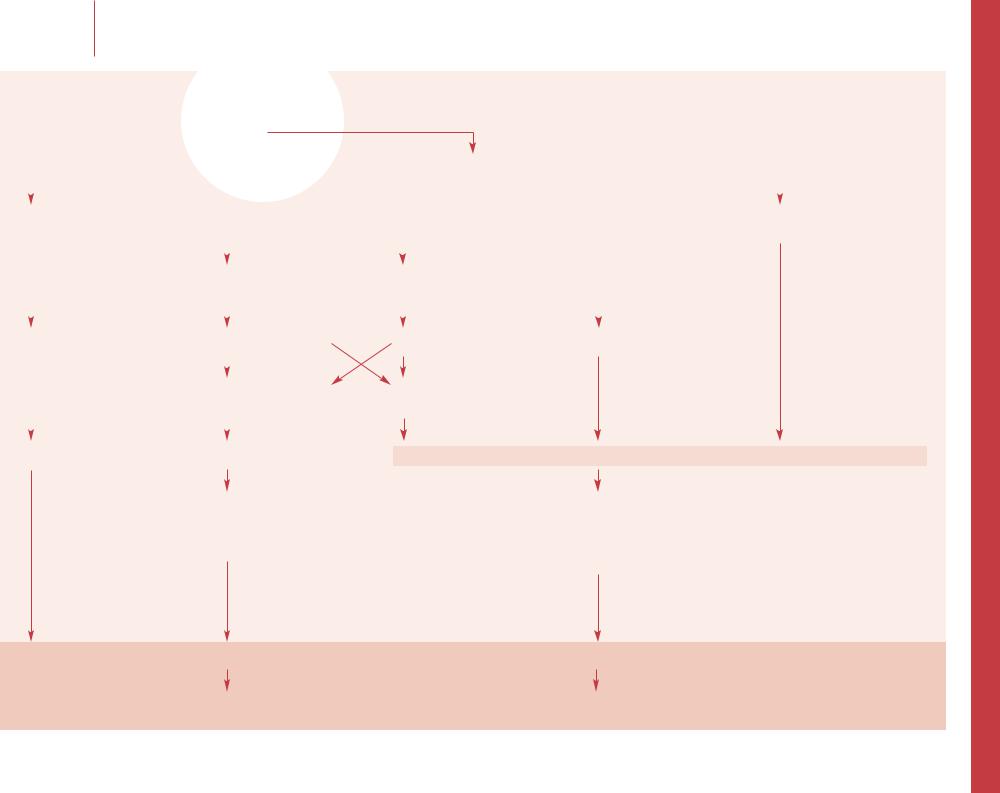
–– This is usually combined with other newborn screening using capillary blood blotted onto filter paper, and identifies a group of autosomally co-dominant inherited disorders of -glo- bin. Identification of homozygous s (HbSS or sickle cell anemia) at birth allows comprehensive clinical care and decreases mortality. Compound heterozygotes where s from one parent is co-inherited with another interacting -globin variant such as C, DPunjab, E or OArab have similar but often less severe clinical problems. Compound heterozygotes of s and -thalassemia (HbS 0T or HbS +T) can be as clinically severe as HbSS depending on the severity of the -thalassemia variant. Targeted screening of highrisk ethnic groups is more cost-effective than no screening, but universal population screening is recommended for most states in USA. Screening programs are also being used in Brazil, parts of France, the UK and a pilot program in Spain. Practitioners should be familiar with the screening programs in their area.
–– Hemoglobin phenotype nomenclature follows a standardized format in which the order of the letters indicates the relative quantity of hemoglobin present (i.e. HbFSA indicates HbF>HbS>HbA in the sample). Therefore, HbFSA (due to HbS +T) and HbFAS (due to sickle cell trait) are not equivalent, and have significantly different implications for treatment and follow-up. HbF is the predominant hemoglobin of gestational life; shortly after birth the level decreases steadily to <2% by 12 months of age in normal infants.
–– At birth, trait phenotypes are dominated by HbF, followed by HbA with either HbS, C or other -globin variant (variously reported as ‘X’ for unknown, ‘V’ for variant, or ‘U’ for unknown). These require no specific hematological follow-up, but family studies with genetic counseling can clarify the risks of disease in future children, especially in populations with high carrier rates. Certain atypical results may require further elucidation with the aid of hemoglobin reference laboratories.
–– Trait phenotypes have no significant medical concerns but have the potential to result in affected offspring. The ‘X’ refers to unknown or variant -globins (e.g. D, E, G). These may need further elucidation by more specialized techniques (high-perform- ance liquid chromatography, or -globin sequencing) by hemoglobin reference laboratories.
–– Identification of a disease phenotype is the primary goal
of newborn hemoglobinopathy screening. These phenotypes can
27be divided into severe and less severe sickle hemoglobinopathies. The severe phenotypes, including HbSS, HbS 0 thalassemia, HbSOArab, require medical and preventive care. The less severe phenotypes may include compound heterozygotes for HbS and non-sickle hemoglobinopathies ( + -thalassemia, HbC, HPFH) and require determination of the precise genotype
and subsequent follow-up dictated by the exact hemoglobinopathy identified. Hemoglobin reference laboratories, in addition to family studies, may be required to elucidate the abnormality.
–– High HbF levels of prematurity ( <34 weeks) and exogenous HbA at the time of newborn screen (transfusion, maternofetal transfusion) can mask disorders such as HbSS. Screening methods vary in sensitivity and accuracy. Sickle solubility tests are inadequately sensitive to the small quantities of HbS present at birth and cannot distinguish sickle cell disease from trait. Cellulose acetate electrophoresis is inexpensive, and widely used despite low sensitivity if HbS <10%; iso-electric focusing (IEF) gives improved resolution and is better suited to mass screening using filter paper blood samples; HPLC has advantages of high automation, speed, accuracy, reproducibility and the ability to accurately quantify many hemoglobin species, but is more expensive and may miss Hb Barts with standard set-up.
–– Testing both biological mother and father is necessary for diagnostic precision and genetic counseling. At a minimum it should include hemoglobin electrophoresis to identify the parents' donation of -globin variants to their infants (i.e. HbF, HbC, HbS, etc.). Complete blood count (with RBC indices and RDW)
and quantitative HbA2 are helpful in identifying the microcytosis and increased HbA2 of -thalassemia carrier parents. Beware of revealing non-paternity.
–– Although typical of HbS 0-thalassemia, this may also represent HbS +-thalassemia when small amounts of HbA are missed.
–– The severe sickle cell phenotypes are best managed, when possible, with comprehensive sickle cell centers which emphasize parental education, symptom recognition, and preventive care measures including complete vaccination (including
Haemophilus influenzae type b, Streptococcus pneumoniae, Neisseria meningitides and Hepatitis B), antibiotic prophylaxis and anticipatory guidance.
–– Homozygous non-sickle -globinopathies have variable clinical severity. -Globin variant E has a high frequency in Southeast Asian populations. Newborns with FE or FAE are usually asymptomatic, but compound heterozygote HbE 0 thalassemia patients have the clinical features of -thalassemia major and are usually transfusion dependent. Newborns with FC have either homozygous HbCC or HbC/ -thalassemia; these infants do not have a sickling hemoglobinopathy but should be followed intermittently with blood counts. The Hb Barts ‘Fast Band’ with an otherwise normal phenotype represents an -tha- lassemia carrier. In high risk groups, this may represent Hb H disease (see ‘Thalassemia’, p 24). Hb Barts disappears quickly after
the neonatal period. Other unusual phenotypes may require the aid of hemoglobin reference laboratories to help identify the - globin mutation.
–– The ’HbF only’ phenotype indicates an absence of detectable -globin production. It may represent -thalassemia major (which will require intensive intervention), the more benign entities of hereditary persistence of fetal hemoglobin (HPFH) (either in its homozygous form or heterozygously with 0-tha- lassemia trait), or extreme prematurity in which a HbA band may not yet be detectable. These usually can be differentiated using family studies and, with the exception of -thalassemia major, there is no need for hematological follow-up. HPFH is a genetically heterogeneous failure to suppress -globin production post-natally, with elevated HbF throughout life without significant clinical consequence.
–– ‘HbAF’ is normally found only outside the neonatal period. In newborns it indicates transfusion of HbA (materno-fetal or therapeutic). Repeat testing at 3–4 months of age will help establish the correct diagnosis.
Selected reading
Loeber G, Webster D, Aznarez A: Quality evaluation of newborn screening programs. Acta Paediat Suppl 1999; 432:3–6.
Panepinto JA, Magid D, Rewers MJ, Lane PA: Universal versus targeted screening of infants for sickle cell disease: A cost effective analysis. J Pediatr 2000;136:201–208.
Reed W, Lane PA, Lorey F, et al: Sickle-cell disease not identified by newborn screening because of prior transfusion. J Pediatr 2000;136:248–250.
Strickland DK, Ware RE, Kinney TR: Pitfalls in newborn hemoglobinopathy screening: Failure to detect + thalassemia.
J Pediatr 1995;127:304–308.
Zimmerman SA, Ware RE, Kinney TR: Gaining ground in the fight against sickle cell disease. Contemp Pediatr 1999; 14:154–177.
Online references
Huisman THJ, Carver MFH, Efremov GD: A Syllabus of Human Hemoglobin Variants (1996). Accessed via www http://globin.cse.psu.edu/globin/html/huisman/variants/
Lane PA: Newborn Screening for Hemoglobin Disorders. Revised January 9, 2001. Accessed via www http://sickle.bwh.harvard.edu/screening.html
Red Cell Disorders |
M.M. Heeney · R.E. Ware |
Newborn screening for hemoglobinopathies |
|
|
|
|
|
|
|
|
|
Red Cell Disorders |
A.S. Al-Seraihy · R.E. Ware |
Sickle cell anemia with fever |
Sickle cell anemia with fever
28 |
|
|
|
|
Assess risk of bacteremia and sepsis |
|||
|
|
|
|
|
|
|
|
|
|
|
|
|
|
|
|
|
|
|
|
|
|
|
|
|
|
|
|
Nontoxic appearance |
|
|
|
Ill, toxic appearance |
|||
|
Examine for source of infection |
|
|
|
Signs and symptoms of sepsis |
|||
|
|
|
|
|
|
|
|
|
|
|
|
|
|
|
|
|
|
|
|
HbSC or HbS + thalassemia |
HbSS or HbS 0 thalassemia, any age |
|||||
|
|
and age >5 years |
Other genotypes age <5 years |
|||||
|
|
|
|
|
|
|
|
|
|
|
|
|
|
|
|
|
|
All sickle cell disease patients |
Temperature >38.5°C |
||
with temperature <38.5°C |
|
|
|
|
|
||
|
|
|
|
|
|
WBC >5 or <30 × 106/l |
|
|
|
|
|
|
|
|
|
Low risk |
|
Intermediate risk |
|
|
|
Blood culture
Urine culture
Chest X-ray
Other tests
Temperature <40°C |
Temperature >40°C |
WBC <5 or >30 × 106/l
Band count >10%
High risk
Blood culture
Urine culture
LP if indicated
Chest X-ray
Other tests
Symptomatic therapy |
i.v. antibiotics |
i.v. antibiotics |
Observation |
Admission |

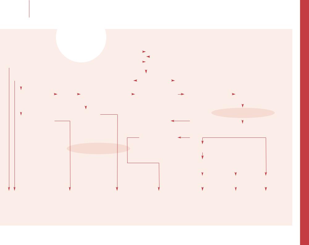
–– Fever is the only reliable indicator of potential infection in patients with SCD. There are no rapidly available laboratory tests on hand that can rule out all bacterial infections such as pneumococcal bacteremia. All febrile children with SCD must be evaluated for serious bacterial infection. For children with HbSS younger than 5 years of age, the risk of acquiring sepsis and meningitis is more than 15% and mortality rate is 30–50%. Work-up and management depend on the clinical evaluation considering age, specific hemoglobinopathy, and physical examination. CBC with differential, reticulocyte count, and blood culture must always be obtained. Chest X-ray should be included in the evaluation of young patients, those with any pulmonary symptoms, and those with leukocytosis or with increased circulating immature neutrophils. Routine U/A and culture is mandatory for all infants, and older children with urinary symptoms, but therapy should not be delayed while awaiting urine collection.
–– SCD patients with fever should be triaged rapidly and evaluated immediately. After taking a brief history, focused physical examination should be done with emphasis on vital signs, O2 saturation, degree of pallor, cardiopulmonary status, evidence of systemic and localized infection, spleen size (compared with baseline) and neurological examination.
–– HbSC is generally milder disease than HbSS. However, children under 5 years of age with HbSC have an increased risk of bacteremia and fatal sepsis. In general, children with HbSC should be managed with the same degree of caution with regard to infection as those with HbSS. Children with HbS 0 thalassemia are considered to be of the same clinical severity as those with HbSS. However, those with HbS + thalassemia have a milder course, and their risk of infection is much less than high-risk patients.
–– The height of initial fever is the most reliable indicator of septicemia. This is particularly true when the temperature is 40°C or greater, especially in children 24 months of age or younger, but sepsis can occur with any degree of fever. Recent antipyretics may reduce the fever but will not
29change the risk of bacteremia.
–– Low-risk patients can be managed symptomatically like patients without sickle cell disease, but the higher risk of acute chest syndrome, osteomyelitis and other complications must be considered.
–– The WBC count tends to be higher among bacteremic children in the first 2 years of life. Most studies show
that the WBC count is not reliable for predicting sepsis in an individual child with SCD. However, among children 24 months of age or younger who have the highest incidence of sepsis, the presence of leukopenia or extreme leukocytosis with high fever are ominous signs.
–– Intermediate risk patients should receive a longacting antibiotic (e.g. ceftriaxone 50 mg/kg) immediately after obtaining the blood culture. The presence of a focus of infection (e.g. otitis) does not alter the urgency of giving parenteral antibiotics. Urine culture and chest X-ray should be done if clinically indicated. Patients should be observed for several hours to ensure they are clinically stable. Only if the family is reliable can the patient be discharged home with specific plan for out-patient follow-up. Minimum fol- low-up includes phone contact the next day. Repeated physical examination and a second dose of ceftriaxone may be advisable in some cases. Physicians should consider admission if patient is less than 1 year, or has a previous history of bacteremia or sepsis, or becomes toxic, or receives clindamycin as a substitute for ceftriaxone, or if there is any concern about the follow-up.
–– Patients with chest X-ray infiltrate should have culture of blood, sputum and stool. Those with hypoxemia should receive supplemental oxygen to keep pulse-oximetry above 92%, and incentive spirometry to help prevent atelectasis. Blood transfusion should be given when oxygen carrying capacity is needed, but not as a routine. Because of the overwhelming incidence of pneumococcal pneumonia, patients should be treated with parenteral antibiotics
(e.g. cefuroxime). Atypical pneumonia with Mycoplasma pneumoniae occurs commonly in SCD, and may lead to acute chest syndrome. Macrolide therapy (erythromycin or azithromycin) should be added to antibiotic coverage in treating SCD patients with pneumonia. A positive stool culture may be the only evidence for Salmonella pneumonia. Patients with clinical findings that are highly suggestive of septic arthritis or osteomyelitis should have needle aspiration and culture of the joint or bone. Antibiotic choice should include agents effective against Salmonella species and Staphylococcus aureus. Abdominal ultrasonography, liver function tests, amylase, and lipase should be considered for patients with RUQ, epigastric or severe abdominal pain to rule out cholelithiasis, cholecystitis,
and pancreatitis.
–– High-risk patients: Parenteral antibiotics should be administered immediately after obtaining the blood count and the blood culture, but before taking the radiograph and waiting for the laboratory results. Lumbar puncture should be performed on toxic children and those with signs of meningitis. Nontoxic children with temperature below 40°C, but with chest X-ray infiltrate, or with WBC >30 or <5 × 10⁶/l should be admitted and treated with parenteral antibiotics. Antibiotic choice should be selected based on the ability to kill both Pneumococcus and H. influenzae and to penetrate into the CSF. Toxic patients or patients suspected of having meningitis should be treated with ceftriaxone (50–75 mg/kg) or cefotaxime (45 mg/kg/dose), and vancomycin (10–15 mg/kg/dose for resistant organisms). If the patient is known or suspected to have allergy to cephalosporin, clindamycin can be substituted. Documented sepsis should be treated parenterally for a minimum of 1 week. Bacterial meningitis should be treated for a minimum of 10 days or 1 week after the CSF has been sterilized. Patients can be discharged from the hospital if afebrile for 24 h with 48 h negative cultures, able to take oral fluids well, with resolution of any respiratory symptoms and adequate oxygenation on room air, and no evidence of worsening of anemia (e.g. aplastic or sequestration crisis).
Selected reading
Cole TB, Smith SJ, Buchanan GR: Hematological alterations during acute infection in children with sickle cell disease. Pediatr Infec Dis J 1987; 6:454–457.
Lane PA, Rogers ZR, Woods GM, et al: Fatal pneumococcal septicemia in hemoglobin SC disease. J Pediatr 1995;127:685–690.
Rogers ZR, Morrison RA, Vedro DA: Outpatient management of febrile illness in infants and young children with sickle cell anemia. J Pediatr 1990;117:736–739.
West TB, West DW, Oheme-Frempong K: The presentation, frequency, and outcome of bacteremia among children with sickle cell disease and fever. Pediatr Emerg Care 1994;10:141–143.
Wilimas JA, Flynn PM, Harris S, et al: A randomized study of outpatient treatment with ceftriaxone
for selected febrile children with sickle cell disease. N Engl J Med 1993;329:472–479.
Red Cell Disorders |
A.S. Al-Seraihy · R.E. Ware |
Sickle cell anemia with fever |
|
|
|
|
|
|

Red Cell Disorders |
M.M. Heeney · R.E. Ware |
Management of painful vaso-occlusive episodes in sickle cell disease
Management of painful vaso-occlusive episodes in sickle cell disease
30
Painful episode
History and physical examination
Chest |
|
|
No |
|
Limb |
|
|
No |
|
|
Abdominal |
|
|
|
|
No |
|
Hand/foot |
|
|
No |
|
Penile |
|
No |
|
|
Uncomplicated painful |
|||||||||||
|
|
|
|
|
|
|
|
|
|
|
|||||||||||||||||||||||||||||
pain |
|
|
pain |
|
|
|
|
pain |
|
|
|
|
|
|
pain |
|
|
pain |
|
|
|
|
vaso-occlusive event |
||||||||||||||||
|
|
|
|
|
|
|
|
|
|
|
|
|
|
|
|
|
|
|
|
|
|
|
|
|
|
|
|
|
|
|
|
|
|||||||
|
|
|
|
|
|
|
|
|
|
|
|
|
|
|
|
|
|
|
|
|
|
|
|
|
|
|
|
|
|
|
|
|
|
|
|
|
|
|
|
Yes |
|
|
Yes |
|
|
|
|
Yes |
|
|
|
|
|
|
Yes |
|
|
Yes |
|
|
|
|
Initial management |
||||||||||||||||
|
|
|
|
|
|
|
|
|
|
|
|
|
|
|
|
|
|
|
|
|
|
|
|
|
|
|
|
|
|
|
|
|
|
|
|
|
Identify and eliminate |
||
|
|
|
|
|
|
|
|
|
|
|
|
|
|
|
|
|
|
|
|
|
|
|
|
|
|
|
|
|
|
|
|
|
|
|
|
|
aggravating factors |
||
|
|
|
|
|
|
|
|
|
|
|
|
|
|
|
|
|
|
|
|
|
|
|
|
|
|
|
|
|
|
|
|
|
|
|
|
|
|||
Fever |
|
|
Weakness |
Right upper |
|
|
|
|
Left upper |
|
Swelling of digits |
|
|
Sustained |
|
|
|
|
Laboratory and |
||||||||||||||||||||
|
|
|
|
|
|
|
|
|
|
|
|
|
radiographic evalutation |
||||||||||||||||||||||||||
CXR infiltrate |
|
|
Swollen joints |
quadrant |
|
|
|
|
quadrant |
|
Tenderness |
|
|
erection |
|
|
|
|
Analgesia, hydration, oxygen |
||||||||||||||||||||
Hypoxia |
|
|
Hemiplegia |
|
|
|
|
|
|
|
|
|
|
|
|
|
|
Erythema |
|
|
|
|
|
|
|
|
|
Frequent reassessment of pain |
|||||||||||
|
|
|
|
|
|
|
|
|
|
|
|
|
|
|
|
|
|
|
|
|
|
|
|
|
|||||||||||||||
Cough |
|
|
|
|
|
|
|
|
|
|
|
|
|
|
|
|
|
|
|
|
|
|
|
|
|
|
|
|
|
|
|
|
|
|
|
|
|||
|
|
|
|
|
|
|
|
|
|
|
|
|
|
|
|
|
|
|
|
|
|
|
|
|
|
|
|
|
|
|
|
|
|
|
|
||||
Tachypnea |
|
|
|
|
|
|
Murphy’s sign |
Splenomegaly |
|
|
|
|
|
|
|
|
|
|
|
|
|
|
|
|
|
||||||||||||||
|
|
|
|
|
|
|
|
|
|
|
|
|
|
|
|
|
|
|
|
|
|
|
|
|
|
|
|||||||||||||
|
|
|
|
|
|
|
|
|
|
Fever |
|
|
|
|
Anemia |
|
|
|
|
|
|
|
|
|
|
Adequate |
|
Inadequate |
|||||||||||
|
|
|
|
|
|
|
|
|
|
|
|
|
|
|
|
|
|
|
|
|
|
|
|
|
|||||||||||||||
|
|
|
|
|
|
|
|
|
|
Elevated bilirubin |
|
|
|
|
|
|
|
|
|
|
|
|
|
|
|
|
|||||||||||||
|
|
|
|
|
|
|
|
|
|
|
|
|
|
|
|
|
|
|
|
|
|
|
|
|
analgesia |
|
analgesia |
||||||||||||
|
|
|
|
|
|
|
|
|
|
(above baseline) |
|
|
|
|
|
|
|
|
|
|
|
|
|
|
|
|
|||||||||||||
|
|
|
|
|
|
|
|
|
|
|
|
|
|
|
|
|
|
|
|
|
|
|
|
|
|
|
|
|
|
|
|
||||||||
|
|
|
|
|
|
|
|
|
|
Elevated alkaline |
|
|
|
|
|
|
|
|
|
|
|
|
|
|
|
|
|
|
|
|
|
|
|||||||
|
|
|
|
|
|
|
|
|
|
|
|
|
|
|
|
|
|
|
|
|
|
|
|
|
|
|
|
|
|
|
|
||||||||
|
|
|
|
|
|
|
|
|
|
phosphatase |
|
|
|
|
|
|
|
|
|
|
|
|
|
|
|
|
|
|
|
|
|
|
|||||||
|
|
|
|
|
|
|
|
|
|
|
|
|
|
|
|
|
|
|
|
|
|
|
|
|
|
|
|
|
|
|
|
|
|
|
|
|
|
Failed outpatient |
|
|
|
|
|
|
|
|
|
|
|
|
|
|
|
|
|
|
|
|
|
|
|
|
|
|
|
|
|
|
|
|
|
|
|
|
|
|
|
||
|
|
|
|
|
|
|
|
|
|
Ultrasound |
|
|
|
|
|
|
|
|
|
|
|
|
|
|
|
|
|
|
|
|
|
|
|
|
management |
||||
|
|
|
|
|
|
|
|
|
|
|
|
|
|
|
|
|
|
|
|
|
|
|
|
|
|
|
|
|
|
|
|
|
|
|
|
||||
|
|
|
|
|
|
|
|
|
|
|
|
|
|
|
|
|
|
|
|
|
|
|
|
|
|
|
|
|
|
|
|
|
|
|
|
||||
|
|
|
|
|
|
|
|
|
|
|
|
|
|
|
|
|
|
|
|
|
|
|
|
|
|
|
|
|
|
|
|
|
|
|
|
|
|
|
|
|
|
|
|
|
|
|
|
|
|
|
|
|
|
|
|
|
|
|
|
|
|
|
|
|
|
|
|
|
|
|
|
|
|
|
|
|
|
|
|
|
|
|
|
|
|
|
|
|
|
|
|
|
|
|
|
|
|
|
|
|
|
|
|
|
|
|
|
|
|
|
|
|
|
|
|
|
|
|
|
Acute chest |
|
|
CVA |
Cholelithiasis |
Splenic |
|
Dactylitis |
|
|
Priapism |
|
|
|
|
|
|
|
||||||||||||||||||||||
syndrome |
|
|
Osteomyelitis |
Cholecystitis |
sequestration |
|
|
|
|
|
|
|
|
|
|
|
|
|
|
|
|
|
|||||||||||||||||
|
|
|
|
|
|
Septic arthritis |
|
|
|
|
|
|
|
|
|
|
|
|
|
|
|
|
|
|
|
|
|
|
|
|
|
|
|
|
|
|
|||
|
|
|
|
|
|
|
|
|
|
|
|
|
|
|
|
|
|
|
|
|
|
|
|
|
|
|
|
|
|
|
|
|
|
|
|
|
|
|
|
Prescribe out-patient |
Admission |
treatment plan |
|

–– Evaluate the onset, location, severity, quality, duration and response to therapy of pain using observation and patient reporting. Obtain history of self-directed treatment and prior experiences with pain and analgesics. Use accepted pain scales (numerical, color, facial expression) for serial evaluation of treatment efficacy. Recognize signs of other disease complications (e.g. fever, cough, dyspnea, vomiting, swelling, neurologic deficit) as well as disorders affecting normal children (e.g. viral gastroenteritis, appendicitis). Evaluate vital signs, O2 saturation, spleen size, pallor, icterus, hydration status, joints, extremities, penis and neurologic function, and compare to the patient’s baseline.
–– Focus on the location of the pain (chest wall, pleural or cardiac) and symptoms suggestive of a complicating diagnosis. With rib or sternal pain, secondary splinting and decreased diaphragmatic excursion may lead to atelectasis, V/Q mismatch and acute chest syndrome (ACS) if pain is not properly treated with analgesics and supplemented with incentive spirometry. Cough, tachypnea, fever or hypoxia requires CXR (PA and lateral). ACS, the most common cause of mortality in SCD, is defined as a new infiltrate, hypoxia, leukocytosis ± fever, and necessitates prompt intervention. Its etiology is often multifactorial. Therapy of ACS is determined by its severity and may include incentive spirometry, supplemental O2 if hypoxic, broad-spectrum antibiotics, transfusion/erythrocytapheresis, serial bronchoscopy and mechanical ventilation.
–– Limb and joint pain usually are due to a vaso-occlusive event, but one must differentiate limited mobility due to pain from neurologic deficit. Suspicion of neurologic deficit (e.g. hemiparesis, asymmetry in muscular tone, slurred speech/cranial nerve abnormality), should prompt immediate intervention and investigation of a possible cerebral vascular accident. Osteomyelitis (often with Salmonella), should be considered when focal tenderness, swelling or fluctuance and fever are present. Joint effusion and severely limited range of motion with fever can be caused by septic arthritis. Consider avascular necrosis of the femoral or humeral heads with chronic or recurrent pain localized to the hip or shoulder.
–– Common diagnoses for normal children with abdominal pain should be considered, especially if surgical intervention is needed. Splenic sequestration is associated with increasing splenomegaly and a >2 g/dl decrease in Hb; thrombocytopenia, shock and an adequate reticulocyte count are frequent. Serial ultrasound studies estimating splenic volume may be helpful. Constipation secondary to opioid analgesics may cause left sided or generalized abdominal pain.
–– Dactylitis (‘hand-foot syndrome’) is painful swelling of the hands and feet secondary to infarction of the small bones. It can be limited to a single digit or be generalized to all four extremities. This vaso-occlusive event can be recurrent but is seen almost solely in infancy.
–– Priapism often starts at night and can be continuous or intermittent/stuttering. Timely detumescence is necessary to prevent future impotency secondary to fibrosis. Improvement should be observed within 24–36 h but complete resolution may require 5–10 days. Initially provide analgesia and hydration with progression to exchange transfusion/erythrocytapheresis if prolonged. Direct corporeal aspiration or irrigation with -adrenergic agonists can be attempted by experienced personnel, while surgical corpus cavernosal shunting should be considered only if rigid tumescence persists despite exchange transfusion or erythrocytapheresis.
–– Factors that aggravate or heighten sensitivity to pain include anxiety, cold exposure/hypothermia, and dehydration. Laboratory evaluation: CBC, differential, reticulocytes with comparison to baseline values. Presenting signs and symptoms dictate additional studies. Transfusion is not indicated in an uncomplicated vaso-occlusive painful episode but obtain a type and screen and determine if the patient’s RBC phenotype is known; this ensures availability of blood product and a reduced risk of allo-immunization (which can be a serious problem for these children) should complications require transfusion. No empiric radiographic investigations are required. CXR to R/O ACS is indicated with hypoxia or pulmonary symptoms. If CVA is suspected, delay MRI/MRA until therapeutic exchange transfusion/erythrocytopheresis is performed. Dehydration should be corrected with bolus isotonic fluid administration. Maintenance fluids should be 150% of normal maintenance, using 0.25–0.5 normal saline to prevent intracellular dehydration associated with hypernatremia (goal serum Na 130–135). When concerned about excessive hydration (e.g. suspicion of ACS, CVA or aplastic crisis), use moderate fluid administration to 75–100% maintenance to prevent complications of overhydration (e.g. pleural effusions, pulmonary edema, cerebral edema). Intravenous is the preferred route of fluid administration, but total fluid intake should include i.v. plus p.o. Empiric supplemental O2 is not indicated in an uncomplicated painful episode unless evidence of hypoxia, tachypnea or dyspnea.
–– Vaso-occlusive pain is episodic, severe and should be considered a medical emergency. Analgesia is instituted quickly in a stepwise approach. Nonpharmacological treatment includes relaxation techniques, diversion and heating pads. Acetaminophen or NSAIDs may control mild pain and then weak opiods (e.g. codeine, oxycodone) are added. Stronger opioids (e.g. morphine, fentanyl, hydromorphone) are the mainstay of treatment of more severe pain, in combination with acetaminophen, NSAIDs and other adjuvant agents. Ambulatory treatment commenced early in an episode may abort debilitating pain and reduce hospitalization and school absence. Failure to achieve relief at home with oral hydration and analgesia necessitates parenteral opioid therapy. Patients may become tolerant to opioids so dosage should be titrated to effect. Physical dependence on opioid medication should never be mistaken for psychological dependence/addiction. As pain recedes, the opioid dosage can be tapered without withdrawal. Opioid toxicity includes hypoventilation and atelectasis (incentive spirometry required), constipation (stool softeners and cathartics required), nausea (antinauseants), urinary retention and pruritus (antihistamines and, if necessary, low-dose naloxone infusion).
Selected reading
Benjamin LJ, Dampier CD, Jacox AK, Odesina V, Phoenix D, Shapiro B, Strafford M, Treadwell M: Guideline for the Management of Acute and Chronic Pain in Sickle-Cell Disease. Glenview, APS Clinical Practice Guidelines Series, No 1, 1999.
Bruno D, Wigfall DR, Zimmerman SA, Rosoff PM, Wiener JS:
Genitourinary complications of sickle cell disease. J Urol 2001; 166:803–811.
Frush K, Ware RE, Kinney TR: Emergency department visits by children with sickle hemoglobinopathies: Factors associated with hospital admission. Pediatr Emergency Care 1995;11:9–12.
Yaster M, Kost-Byerly S, Maxwell LG: The management of pain in sickle cell disease. Pediatr Clin N Am 2000;47:699–710.
Zimmerman SA, Ware RE, Kinney TR: Gaining ground in the fight against sickle cell disease. Contemp Pediatr 1999;14: 154–177.
National Institutes of Health, National Heart, Lung, and Blood Institute: Management and Therapy of Sickle Cell Disease, ed 3. NIH Publ 96-2117. Available at: http://www.nhlbi.nih.gov/health/prof/blood/sickle/sickmt.pdf
31X
Red Cell Disorders |
M.M. Heeney · R.E. Ware |
|
|
Management of painful vaso-occlusive episodes in sickle cell disease

|
|
|
Red Cell Disorders |
A.S. Al-Seraihy · R.E. Ware |
Evaluation and management of anemia in sickle cell disease |
Evaluation and management of anemia in sickle cell disease
32
|
|
|
|
|
|
Anemia |
|
|
|
|
|
|
|
|
|
|
|
|
|
|
|
|
|
|
|
|
|
|
|
|
|
|
|
|
|
|
|
|
|
|
|
Measurement of Hb concentration |
|
|
|||
Ill-appearing patient |
|
|
|
|
Well-appearing patient |
|
|
|
|
||
Hb >2 g/dl below baseline |
|
|
Hb 1–2 g/dl below baseline |
|
|
|
|
||||
|
|
|
|
|
|
|
|
|
|
|
|
|
|
|
|
|
|
|
|
|
|
|
|
Enlarged spleen |
Enlarged liver |
No hepatosplenomegaly |
Normal spleen size |
Enlarged spleen size |
Normal spleen size |
||||||
Low or normal BP |
Low or normal BP |
Low or normal BP |
Low reticulocytes <5% |
Reticulocytes present >5% |
High reticulocytes >15% |
||||||
Reticulocytes >5% |
Reticulocytes >5% |
Very low reticulocytes <1% |
|
|
|
|
|
|
|||
|
|
|
|
Parvovirus serology (IgG, IgM) |
|
|
|
|
|
|
|
|
|
|
|
|
|
|
|
|
|
||
|
|
|
|
|
|
|
|
|
|
|
|
|
|
|
|
|
|
|
|
|
|
|
|
Acute splenic |
Acute hepatic |
Transient aplastic crisis |
|
|
Sub-acute splenic |
Hemolytic crisis |
|||||
sequestration crisis |
sequestration crisis |
|
|
|
|
sequestration crisis |
Rule out concomitant |
||||
|
|
|
|
|
|
|
|
|
|
hemolytic conditions |
|
|
|
|
|
|
|
|
|
|
|
G6PD deficiency |
|
|
|
|
|
|
|
|
|
|
|
Spherocytosis |
|
|
|
|
|
|
|
|
|
|
|
Drug exposure |
|
|
|
|
|
|
|
|
|
|
|
Infection |
|
|
|
|
Folate |
Iron |
Chronic renal |
Bone marrow |
|||
|
|
|
deficiency |
deficiency |
disease |
necrosis |
|||
|
|
|
|
|
|
|
|
|
|
|
|
|
|
|
|
|
|
|
|
|
|
|
|
|
|
|
|
|
|
Transfusion |
Transfusion |
Observation |
Folic acid |
Iron |
Erythropoietin |
|
|||
|
|
versus transfusion |
supplementation |
supplementation |
|
|
|
||
|
|
|
|
|
|
|
|
|
|

–– Although the degree of anemia in sickle cell disease is extremely variable among affected individuals, in any given patient the steady state Hb concentration after the age of
1 year, reticulocyte count, and degree of hemolysis are relatively constant. Measuring Hb concentration and comparing it with the baseline level is the first step to identifying an exacerbation of anemia.
–– Exacerbation of the usual degree of anemia is defined as a drop of more than 2 g/dl, or an absolute Hb concentration of less or equal to 5 g/dl. Patients with a slow and steady Hb drop are more stable than those with an acute drop who might present with shock.
–– Acute splenic sequestration crisis (ASSC) is a sudden pooling of a large amount of blood into the spleen leading to acute splenomegaly, profound anemia, hypotension, and in severe cases, hypovolemic shock. Death may occur in a few hours. ASSC usually occurs prior to autoinfarction of the spleen, with the vast majority occurring before 2 years and almost all before 6 years of age. ASSC is less common in HbSC disease, but may occur in older patients, since splenomegaly persists into adulthood. ASSC often occurs in association with non-specific viral or bacterial infection. Parents should be taught to palpate the spleen regularly, and seek immediate medical care if the spleen enlarges. Laboratory findings may include severe anemia with increased reticulocytes and nucleated red cells, an increased WBC with shift to the left, and a decreased platelet count due to platelet trapping. The immediate treatment of ASSC is directed toward correction of hypovolemia and anemia. Isotonic volume support can be used acutely while awaiting blood for transfusion. The goal of transfusion is primarily to prevent shock, not to restore Hb to normal or to the steady state level. After transfusion the spleen shrinks and Hb often increases more than predicted due to the release of trapped RBC from the spleen. ASSC recurs in approximately 50% of cases. Long-term management for recurrent ASSC may include chronic transfusion therapy in the very young patient in order to avoid splenectomy.
–– Although uncommon hepatic sequestration crisis
33does occur and is characterized by rapid enlargement of the liver accompanied by drop in the Hb. It should be approached in the same manner as ASSC.
–– Transient aplastic crisis (TAC) is an exacerbation of anemia due to transient cessation of erythropoiesis, and can occur in all chronic hemolytic anemias including SCD. An acute aplastic crisis is often associated with infection, and parvovirus B19 is the causative agent in most severe cases. Patients present with signs of severe anemia, although the condition is often discovered incidently during an evaluation for febrile illness. If anemia is severe enough the patient should be admitted for observation. The decision to transfuse RBC should be based first on clinical presentation, the degree of anemia, and reticulocyte count. The goal of transfusion is to prevent congestive heart failure and shock, which occurs when the Hb is below 4–5 g/dl. An increased risk of stroke has been associated with severe anemia.
–– Splenic sequestration is not always acute, but it can be subacute and even chronic. Splenomegaly associated with anemia and thrombocytopenia may be evidence of a subacute or chronic splenic sequestration. Close clinical observation and monitoring of the Hb concentration are mandatory. Many of these children eventually require splenectomy.
–– Hyperhemolytic crisis is very unusual, but may ensue in association with certain drugs, acute infection or G6PD deficiency. Patients show increased scleral icterus and may have abdominal pain, fall in Hb, increased reticulocyte count and bilirubin. After several days hemolysis subsides.
–– Folate deficiency as a cause of exaggerated anemia is a very rare event in the USA, but common worldwide. Nevertheless, it is a common practice to prescribe folic acid 1 mg to patients with SCD unless they have adequate dietary intake of folate. Macro-ovalocytosis from folate deficiency is difficult to recognize in a patient with recticulocytosis. Hypersegmentation of the neutrophil may provide a clue to the correct diagnosis.
–– Iron deficiency is not a major problem in SCD. The diagnosis can be established by measuring serum ferritin, and an increase in Hb and MCV in response to iron therapy. Iron supplementation should never be prolonged since iron overload is a common long-term problem in many patients with SCD. The diagnosis should be considered when the MCV falls below the patient’s baseline.
–– SCD in older patients is associated with chronic renal disease. As a consequence of chronic renal failure, erythropoietin (Epo) production is impaired for the degree of the anemia. It is well established that administration of recombinant erythropoietin improves severe anemia in
SCD patients with renal failure.
–– Bone marrow necrosis is a rare event in pediatric patients with SCD, and is typically due to repeated vasoocclusive infarction.
Selected reading
Balkaran B, Char G, Morris JS, Thomas PW, Serjeant BE, Serjeant GR: Stroke in a cohort of patients with homozygous sickle cell disease. J Pediatr 1992;120:360–366.
Goldstein AR, Anderson MJ, Serjeant GR: Parvovirus associated aplastic crisis in homozygous sickle cell disease. Arch Dis Child 1987;62:585–588.
Hemandez P, Dortics E, Espinoza E, Gonzalez X, Svarch E: Clinical features of hepatic sequestration in
sickle cell anemia. Haematologia (Budap) 1989;22:69–74.
Kinney TR, Ware RE, Shultz WH: Long-term management of splenic sequestration in children with sickle cell disease. J Pediatr 1991;117:1941–1946.
Powell RW, Levine GL, Yang YM, Mankad VN: Acute splenic sequestration crisis in sickle cell disease early detection and treatment. J Pediatr Surg 1992;27:215–219.
Steinberg MH: Erythropoietin for anemia of renal failure in sickle cell disease. N Engl J Med 1991;324:1369–1370.
Steinberg MH, West MS, Gallagher D, Mentzer W, Cooperative Study of Sickle Cell Disease: Effect of G6PD deficiency upon sickle cell anemia. Blood 1988;71: 748–752.
Vichinsky E, Kleman K, Emury S, Lubin B: The diagnosis of iron deficiency anemia in sickle cell disease. Blood 1981;58:963–968.
Young N: Hematologic and hematopoietic consequences of B19 parvovirus. Semin Hematol 1988;25:159–172.
Red Cell Disorders |
A.S. Al-Seraihy · R.E. Ware |
Evaluation and management of anemia in sickle cell disease |
|
|
|
|
|
|
|
|
|
Red Cell Disorders |
A.E. Kulozik · A. Deters |
Polycythemia (erythrocytosis) |
Polycythemia (erythrocytosis)
|
|
|
|
|
|
|
|
|
|
|
|
|
|
|
|
34 |
|
|
|
|
|
History |
|
|
|
|
|
Laboratory criteria |
|||
|
|
|
|
|
|||||||||||
|
|
|
|
|
|
|
|
|
|
|
|
|
|||
|
|
|
|
|
|
|
|
|
|
|
|
|
CBC, HCT >upper end of |
||
|
|
|
|
|
|
|
|
||||||||
|
|
|
|
|
|
|
|
|
|
|
|
|
|||
|
|
|
|
|
|
Physical examination |
|
|
|
|
|
reference range on 2 occasions |
|||
|
|
|
|
|
|
||||||||||
|
|
|
|
|
|
|
|
|
|
|
|
|
|
|
RCM <25% of mean predicted value |
|
|
|
|
|
|
|
|
|
|
|
|
|
|
|
|
|
|
|
|
|
RCM >25% of mean predicted value |
|
|
Red cell mass |
|
||||||
|
|
|
|
|
|
|
|
= relative erythrocytosis and no further |
|||||||
|
|
|
|
|
= absolute erythrocytosis |
|
|
|
(RCM) |
|
hematologic evaluation |
||||
|
|
|
|
|
|
|
|
|
|
|
|
|
|
|
|
SpO2 |
|
Normal |
|
Abdominal ultrasound scan |
|
|
Normal ± splenomegaly |
|||||
|
|
|
|
|||||||||
Blood gas analysis |
|
|
|
Liver/renal chemistry |
|
|
|
|||||
(sleep study) |
|
|
|
|
|
|
|
|
|
|
||
|
|
|
|
|
|
|
|
|
|
|||
|
|
|
|
|
|
|
|
|
|
|
|
|
|
|
|
|
|
|
Abnormal |
|
|
& Erythropoietin |
|||
7 SpO2 or nocturnal |
|
|
|
|
||||||||
|
|
|
|
|
|
|
|
|
||||
|
|
|
|
|
|
|
|
|
||||
O2 desaturation |
|
|
|
|
|
|
|
|
|
|
||
Carboxyhemoglobin |
|
|
|
|
|
|
|
|
|
|
||
|
|
|
|
|
|
|
|
|
|
|
|
Methemoglobin & |
|
|
|
|
|
|
|
|
|
|
|
|
Left shift of oxygen |
|
|
|
|
|
|
|
|
|
|
|
|
dissociation curve |
|
|
|
|
|
|
|
Secondary |
|||||
|
|
|
|
|
|
|
|
|
|
|||
|
|
|
|
|
|
erythrocytosis |
|
|
|
|||
|
|
|
|
|
|
|
|
|
|
|
|
|
Erythropoietin |
|
7 Erythropoietin |
||||
|
||||||
Oxygen dissociation study |
|
± Thrombocytosis/leukocytosis |
||||
Met-Hemoglobin |
|
|
|
|
||
|
|
|
|
|||
|
|
|
|
|
|
|
|
|
Primary erythrocytosis |
|
|||
|
|
|
|
|
|
|
|
|
|
|
|
|
|
|
|
|
|
Bone marrow histology |
||
|
|
|
|
Clonal markers |
||
|
Normal |
|
|
|
|
|
|
|
|
|
|
||
|
|
|
|
|
||
|
|
|
|
Erythropoietin receptor-gene analysis |
|
|
|||
|
|
|
|
|
|
|
|
||
|
|
|
|
|
|
|
|
|
|
|
|
|
|
|
|
|
|
|
|
|
|
|
|
Normal |
Abnormal |
Abnormal |
|||
|
|
|
|
|
|
|
|
|
|
|
|
|
|
|
|
|
|
|
|
|
|
|
|
|
|
|
|
|
|
Neonate |
Acquired |
Acquired |
Congenital |
Idiopathic |
Congenital |
Acquired |
|||
Twin-twin transfusion |
Arterial hypoxemia |
Renal tumors/cysts |
Hb variants with |
erythrocytosis |
EPO receptor |
Polycythemia |
|||
Maternal-fetal transfusion |
Chronic lung disease |
Hydronephrosis |
high oxygen affinity |
|
|
truncation |
(rubra) vera |
||
Diabetic mother |
Congenital heart disease |
Renal artery stenosis |
Methemoglobinemia |
|
|
|
|
|
|
Trisomy 21 |
Sleep apnea |
Adrenal tumors |
|
|
|
|
|
|
|
Maternal hyperthyroidism |
& Carbon monoxide |
Hepatoma |
|
|
|
|
|
|
|
Congenital adrenal hyperplasia |
(smoking) |
Liver dysfunction |
|
|
|
|
|
|
|
|
|
|
|
|
|
|
|
|
|

–– Consider patient’s age; in neonates, ask about delayed cord clamping, maternal diabetes or other causes for chronic fetal hypoxia. Consider symptoms and signs of pulmonary or cardiac disease, such as dyspnea and cyanosis.
Note positive family history of erythrocytosis. Dehydration can cause an increased HCT, but since the red cell mass is normal this represents relative and not absolute erythrocytosis.
–– Erythrocytosis should be considered if the HCT is above the upper age related reference range on two separate occasions. Note thrombocytosis and/or leukocytosis as signs of polycythemia vera.
–– Red cell mass is determined to differentiate between absolute and relative (or apparent) erythrocytosis by 51Cror 99mTc-red blood cell dilution method. In relative erythrocytosis, HCT is above normal range while red cell mass is not increased, e.g. due to reduced plasma volume. The erythrocytosis in these patients
is not a clinical concern, although the cause of decreased plasma volume would be. In absolute erythrocytosis there is a true increase in
red blood cell mass which needs investigation. When an underlying etiology of polycythemia is evident (e.g. chronic lung disease, congenital heart disease), evaluation of red cell mass may not be necessary.
–– Secondary erythrocytosis is due to an excess of erythropoietin as a manifestation of decreased tissue oxygenation or inappropriate erythropoietin secretion from a tumor. The former occurs as a consequence of decreased renal oxygen supply either due to arterial hypoxemia in chronic lung disease or congenital heart disease or due to impaired renal perfusion, e.g. in renal tumors/cysts, hydronephro-
35
sis, and renal artery stenosis. Often serum erythropoietin is increased. Impaired liver function or hepatic tumors can also cause increased erythropoietin levels. Erythropoietin producing adrenal tumors (e.g. adrenocortical carcinoma, pheochromocytoma, hemangioblastoma, e.g. in Hippel-Lindau disease) can cause secondary erythrocytosis.
–– Methemoglobinemia or hemoglobin variants with high oxygen affinity lead to decreased tissue oxygenation and therefore to secondary erythrocytosis. Ideally, perform an oxygen-dissociation curve of the patient’s hemoglobin. Hemoglobin electrophoresis alone is insufficient, since many hemoglobins with abnormal oxygen affinity co-migrate with HbA and will be overlooked. Consider family studies and molecular genetic studies.
–– This heterogeneous group of patients with idiopathic erythrocytosis emerges from those patients with an absolute erythrocytosis without a cause of primary or secondary erythrocytosis. In some patients a cause for secondary erythrocytosis becomes apparent during follow-up.
–– In primary erythrocytosis, erythropoiesis is defective, in contrast to the secondary type, where erythrocytosis is increased in response to increased erythropoietin secretion. Primary erythrocytosis can be caused by truncation of the cytoplasmic portion of the erythropoietin receptor that is responsible for switching off the signal following erythropoietin binding. Erythroid precursor cells are then hypersensitive to erythropoietin leading to erythroid hyperplasia. This condition was shown to be dominantly inherited, but spontaneous somatic mutations have also been recognized. Consider family studies and molecular genetic studies.
–– Polycythemia (rubra) vera is a myeloproliferative, monoclonal disease that is very rare in childhood. Red cell mass must be increased and other common findings include normal arterial oxygen saturation, splenomegaly, thrombocytosis, leukocytosis and bone marrow hypercellularity. A useful marker is the ability of peripheral blood cell fractions to form endogenous erythroid colonies in the absence of erythropoietin.
Selected reading
Messinezy M, Pearson TC: The classification and diagnostic criteria of the erythrocytosis (polycythaemias). Clin Lab Haematol 1999; 21:301–316.
Nathan and Oski’s Hematology of Infancy and Childhood, ed 5. Philadelphia, Saunders, 1998.
Pearson TC, et al: A Polycythemia Vera Update: Diagnosis, Pathobiology, and Treatment. Hematology (Am Soc Hematol Educ Program) 2000:51–68.
Red Cell Disorders |
A.E. Kulozik · A. Deters |
Polycythemia (erythrocytosis) |
|
|
|
|
|
|