

hemeoncalgorithms
.pdf
|
|
|
|
|
|
|
|
|
|
|
|
|
|
|
|
|
|
|
|
|
|
|
|
|
|
|
|
|
|
|
|
|
Red Cell Disorders |
|
|
R.H. Sills · A. Deters |
|
|
|
Neonatal anemia due to impaired RBC production |
|
|
|||||||||||||||
|
|
|
|
|
|
|
|
|
|
|
|
|
|
|
|
|
|
|
|
|
|
|
|
|
|
|
|
|
|
|
|
|
|
|
|
Neonatal anemia due to impaired RBC production |
|
|
|||||||||||||||||||
|
|
|
|
|
|
|
|
|
|
|
|
|
|
|
|
|
|
|
|
|
|
|
|
|
|
|
|
|
|
|
|
|
|
|
|
|
|
|
|
|
|
|
|
|
|
|
|
|
|
|
|
|
|
|
|
|
|
|
|
|
|
|
|
|
|
|
|
|
|
|
|
|
|
|
|
|
|
|
|
|
|
|
|
|
|
|
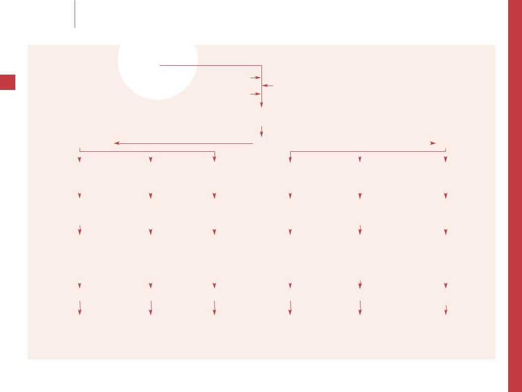
16 |
|
|
|
|
|
|
|
|
|
|
|
Reticulocytes <2% |
|
|
|
|
|
|
|
|
|
|||||||
|
|
|
|
|
|
|
|
|
|
|
|
Nl indirect bilirubin |
|
|
|
|
|
|
|
|
|
|||||||
|
|
|
|
|
|
|
|
|
|
|
|
|
|
|
|
|
|
|
|
|
||||||||
|
|
|
|
|
|
|
|
|
|
|
|
CBC – RBC indices – Blood smear |
|
|
|
|
|
|
|
|
|
|||||||
|
|
|
|
|
|
|
|
|
|
|
|
|
|
|
|
|
|
|
|
|
|
|
|
|
|
|
|
|
|
|
|
|
|
|
|
|
|
|
|
|
|
|
|
|
|
|
|
|
|
|
|
|
|
|
|
|
|
|
|
|
|
|
|
|
|
|
|
|
|
|
|
|
|
|
|
|
|
|
|
|
|
|
|
|
|
|
|
|
|
|
7 MCV |
|
|
|
|
|
|
Nl MCV |
|
|
|
|
|
|
|
|
|
|
± & MCV |
|
|
||||
|
|
|
|
|
|
|
|
|
|
|
|
|
|
|
|
|
|
|
|
|
|
|
|
|
Macro-ovalocytosis |
|
|
|
|
|
|
|
|
|
|
|
|
|
|
|
|
|
|
|
|
|
|
|
|
|
|
|
|
PMN hypersegmentation |
|
|
|
|
|
Mediterranean, |
± Obstetrical |
|
|
|
Sick infant |
Pancytopenia |
|
|
No evidence |
|
|
|
|
|
||||||||||||
|
|
|
|
|
|
|
|
|
|
|
|
|||||||||||||||||
|
|
Asian or African |
complications |
|
|
|
|
|
|
|
|
|
|
|
|
|
of underlying |
|
|
|
|
|
||||||
|
|
|
|
|
|
|
|
|
|
|
|
|
|
|
|
|
|
|
|
|||||||||
|
|
ancestry |
Twin gestation |
|
|
|
|
|
|
|
|
|
|
|
|
|
disease |
|
|
|
|
|
||||||
|
|
|
|
|
|
|
|
|
|
|
|
|
|
|
|
|
|
|
|
|
|
|
|
|
|
|
|
|
|
|
|
|
|
|
|
|
|
|
|
|
|
|
|
|
|
|
|
|
|
|
|
|
|
|
|
|
|
|
|
|
|
|
|
|
|
|
|
|
|
|
|
|
Evidence of infection |
BM aspirate |
|
|
|
|
|
|||||||
|
|
|
|
|
|
|
|
|
|
|
|
|
|
|
|
|
|
|
|
|
|
|
|
|
|
|
|
|
|
|
|
|
|
|
|
|
|
|
|
|
|
|
|
|
|
|
|
|
|
|
|
|
|
|
|
|
|
|
|
|
|
|
|
|
Iatrogenic blood loss |
|
Yes |
|
|
No |
|
|
|
|
|
|
|
|
|
|||||||
|
|
|
|
|
|
|
as contributing factor |
|
|
|
|
|
|
|
|
|
|
|
|
|
|
|
|
|
||||
|
|
|
|
|
|
|
|
|
|
|
|
|
|
|
|
|
|
|
|
|
|
|
|
|||||
|
|
|
|
|
|
|
|
|
|
|
|
|
|
|
|
|
|
|
|
|
|
|
|
|
|
|
|
|
|
|
|
|
|
|
|
|
|
|
|
|
|
|
|
|
|
BM aspirate |
Pure RBC |
Normal |
|
|
|
||||||
|
|
|
|
|
|
|
|
|
|
|
|
|
|
|
|
|
and biopsy |
aplasia |
|
|
|
|
|
|||||
|
|
|
|
|
|
|
|
|
|
|
|
|
|
|
|
|
|
|
|
|
|
|
|
|
|
|
|
|
|
|
|
|
|
|
|
|
|
|
|
|
|
|
|
|
|
|
|
|
|
|
|
|
|
|
|
|
|
|
|
|
|
|
|
|
|
|
|
|
|
|
|
|
+ |
|
|
|
|
|
|
|
|
|
|
|
||
|
|
|
|
|
|
|
|
|
|
|
|
|
|
|
|
|
|
|
|
|
|
|
|
|
|
|||
|
|
|
|
|
|
|
|
|
|
|
|
|
|
|
|
|
|
|
|
|
|
|
|
|
|
|
||
|
|
|
|
|
|
|
|
|
|
|
|
|
|
|
|
|
|
|
|
|
|
|
|
|
|
|
|
|
|
|
|
|
|
|
|
|
|
|
|
|
|
|
|
|
|
|
|
|
|
|
|
|
|
|
|
|
|
|
|
|
|
|
|
|
|
|
|
|
|
|
|
|
|
Megaloblastic |
|
|
|
|
|
|
||||||
|
|
|
|
|
|
|
|
|
|
|
|
|
|
|
Bone marrow |
|
|
|
|
|
|
|
||||||
|
|
|
|
|
|
|
|
|
|
|
|
|
|
|
replacement |
|
changes |
|
|
|
|
|
|
|
||||
|
|
|
|
|
|
|
|
|
|
|
|
|
|
|
|
|
|
|
|
|
|
|
|
|
|
|
|
|
|
|
|
|
|
|
|
|
|
|
|
|
|
|
|
|
|
|
|
|
|
|
|
|
|
|
|
|
|
|
|
|
|
|
|
|
|
|
|
|
|
|
|
|
|
|
|
|
|
|
|
|
|
|
|
|
|
|
|
|
-Thalassemia trait |
Blood loss and |
Acute and |
Infection |
Neuroblastoma |
|
|
DBA |
|
Megaloblastic |
|
|
|||||||||||||||
|
|
Hb H disease |
resulting iron |
chronic disease |
|
|
|
Congenital leukemia |
|
|
Rare diagnoses |
anemia |
|
|
||||||||||||||
|
|
|
|
|
|
deficiency |
|
|
|
|
|
|
LCH |
|
|
|
|
|
|
|
|
|
||||||
|
|
|
|
|
|
|
|
|
|
|
|
|
|
|
|
|
|
|
|
|
||||||||
|
|
(see ‘Thalassemia’, p 24) |
|
|
|
|
|
|
|
|
|
Osteopetrosis |
|
|
|
|
|
|
|
|
|
|||||||
|
|
|
|
|
|
|
|
|
|
|
|
|
|
|
|
|
|
|
|
|
|
|
|
|
||||
|
|
|
|
|
|
|
|
|
|
|
|
|
|
|
|
|
|
|
|
|
|
|
|
|
||||
|
|
|
|
|
|
|
|
|
|
|
|
|
|
|
|
|
|
|
|
|
|
|
|
|
|
|
|
|
|
|
|
|
|
|
|
|
|
|
|
|
|
|
|
|
|
|
|
|
|
|
|
|
|
|
|
|
|

–– MCV normally varies with postnatal age as well as gestational age. The lower limit of normal in cord blood at term is 98, 95 in the first 3 days from capillary samples and 88 at 1 week of age.
–– -Thalassemias manifest in the neonate because both Hb A and F contain chains. Note that hydrops fetalis (which will be clinically obvious) and Hb H disease are almost never seen in people of African descent.
–– Chronic blood loss, usually prenatal, causes iron deficiency and associated microcytosis. It is most often due to feto-maternal hemorrhage, twin-twin transfusions and placentae previa. The reticulocyte count is often decreased because iron deficiency inhibits reticulocyte production.
–– Although iatrogenic and other types of blood loss are not disorders of impaired production, they often exacerbate anemia primarily due to impaired erythrocyte production. Blood loss can also cause Fe deficiency anemia.
–– Critically ill neonates, usually with multiple medical problems, often develop anemia and reticulopenia. This is particularly common in infants with bronchopulmonary dysplasia.
–– Viral, bacterial and other infections can impair erythroid production in the neonate; specific viral agents include rubella, cytomegalovirus, adenovirus and parvovirus.
17
–– Bone marrow replacement is uncommon in the neonate. It is most often caused by neuroblastoma and congenital leukemia, but is also seen with Langerhans cell histiocytosis (which is particularly severe in neonates) and osteopetrosis. Aplastic anemia in the neonate is very rare; most forms of hereditary aplastic anemia, such as Fancon’s anemia, present later in life.
–– With no evident etiology for more severe or persistent anemia, Diamond-Blackfan anemia (congenital hypoplastic anemia) should be considered. Bone marrow aspirate reveals virtual absence of erythroid precursors. Other forms of hypoplastic anemia in neonates are rare and include drug-induced RBC aplasia, Aase syndrome (associated with skeletal anomalies), Pearson’s syndrome (associated with hypoplastic sideroblastic anemia), and congenital dyserythropoietic anemia.
–– Ill neonates may have persistent anemia without a recognized etiology and a normal bone marrow. Most often these are ill neonates with multiple and persistent medical problems.
–– Megaloblastic anemia is rare in the newborn. Initially, macrocytosis, then anemia, and finally pancytopenia develops. B12 deficiency can be seen in breast-fed infants of vegan B12- deficient mothers or infants with GI abnormalities such as necrotizing enterocolitis or short gut syndrome. Folate deficiency is seen in infants receiving goat’s milk or milk that has been boiled, and in those with malabsorption. A number of rare metabolic defects, including transcobalamin II deficiency and orotic aciduria, can cause megaloblastic anemia at birth or soon after. Macrocytosis is often not evident because of the relatively high normal MCV in newborns.
Selected reading
Blanchette VS, Zipurskey A: Assessement of anemia in newborn infants. Clin Perinatol
1984;11:489–510.
Christensen RD: Hematologic Problems of the Neonate. Philadelphia, Saunders, 2000.
Gallagher PG, Ehrenkrenz RA: Nutritional anemias in infancy. Clin Perinatol 1995;22: 671–692.
Ohls RK, Hunter DD, Christensen RD:
A randomized, placebo-controlled trial of recombinant erythropoietin as treatment for the anemia of bronchopulmonary dysplasia. J Pediatr 1993;123:996.
Quirolo K, Foote D, Vichinsky EP: Changing outcome of homozygous alpha thalassemia: Cautious optimism. J Pediatr Hematol/Oncol 2000;22:539–542.
Red Cell Disorders |
R.H. Sills · A. Deters |
Neonatal anemia due to impaired RBC production |
|
|
|
|
|
|

|
|
|
Red Cell Disorders |
A. Deters · A.E. Kulozik |
Hemolytic anemia |
Hemolytic anemia
18 |
History |
|
|
|
Laboratory criteria |
|
|
|
|||
|
|
|
|
||
|
|
|
|
CBC, & reticulocyte count, & indirect bilirubin, |
|
|
|
|
|
|
|
|
Physical examination |
|
|
|
abnormal peripheral blood smear, ± & LDH, 7 haptoglobin |
|
|
|
|
Neonate |
|
Positive |
|
|
|
Coombs test |
|
|
|
Negative |
|
|
|
|
|
|||
|
|
|
|
|
|
|
|
|
|
|
|
|||||||
|
|
|
|
|
|
|
|
|
|
|
|
|
|
|
|
|
||
|
|
|
|
|
|
|
|
|
|
|
|
|
|
|
|
|
|
|
|
Immune hemolysis |
|
|
|
|
|
|
|
Peripheral blood smear |
|
|
|
|
|
||||
|
|
|
|
|
|
|
|
|
|
|
|
|
||||||
Rh/ABO incompatibility |
|
|
|
|
|
|
|
|
|
|
|
|
|
|
|
|
|
|
|
|
|
|
|
|
|
|
|
|
|
|
|
|
|
|
|
||
|
|
|
|
|
|
|
|
|
|
|
|
|
|
|
|
|
|
|
|
|
|
|
|
Spherocytes or elliptocytes |
Target/sickle cells |
Red cell fragmentation |
Other |
Normal |
|||||||||
|
|
|
|
|
|
|
|
|
|
|
|
|
|
abnormalities |
|
|
||
|
|
|
|
|
|
|
|
|
|
|
|
|
|
|
|
|||
|
|
|
|
|
|
|
|
|
|
|
|
|
|
|
|
|
|
|
|
|
Coombs specificity |
Osmotic fragility test |
Hb analysis |
(see ‘Consumptive coagulopathy’, p 68) |
|
|
|
G6PD assay |
|||||||||
|
|
Type and cross-match |
Family studies |
|
|
|
|
|
|
|
|
|
|
|
|
|||
|
|
|
|
|
|
|
|
|
|
|
|
|
|
|||||
|
|
Platelet count |
RIA/ELISA |
|
|
|
|
|
|
|
|
|
|
|
|
|||
|
|
|
|
|
|
|
|
|
|
|
|
|
|
|||||
|
|
ANA |
|
|
|
|
|
|
|
|
|
|
Positive |
Negative |
||||
|
|
PTT |
|
|
|
|
|
|
|
|
|
|
||||||
|
|
|
|
|
|
|
|
|
|
|
|
|
|
|
|
|||
|
|
|
Family study |
|
Autoimmune |
||
|
Red cell enzyme panel |
||
|
hemolytic anemia |
Flow cytometry for GPI-linked |
|
|
|
|
surface proteins (PNH) |
|
|
|
|
|
|
|
|
Primary |
Secondary |
Hereditary |
HbSS |
Malaria |
G6PD deficiency Other enzyme deficiencies |
Warm reactive (IgG) |
Autoimmune disease |
spherocytosis |
HbSC |
Sepsis/infection |
Paroxysmal nocturnal |
Cold reactive (IgM) |
Malignancy |
Hereditary |
HbS T |
Hereditary |
hemoglobinuria |
Paroxysmal |
Immunodeficiency |
elliptocytosis |
Other unstable |
pyropoikilocytosis |
|
cold reactive (IgG) |
Infection |
Hypersplenism |
Hb variants |
Hereditary |
|
|
(i.e. mycoplasmal, viral) |
Coombs negative AIHA |
|
stomatocytosis |
|
|
Drugs |
|
|
|
|
|
|
|
|
|
|

–– Symptoms that suggest severe hemolysis include headache, dizziness, syncope, fever, chills, dark urine (see on ‘Hemoglobinuria’,
p 20) and abdominal/back pain. Possible precipitating factors include infection, medications, and foods (e.g. fava beans in G6PD deficiency). Past medical history should inquire about jaundice as a neonate or later. A history of recurrent infections, arthritis, rash, mouth ulcers or thyroid disease suggests autoimmune hemolysis. Family history should include ancestry (African, Mediterranean, or Arab ancestry suggests G6PD deficiency [mainly but not exclusively in males] or sickle cell disease) and address anemia, jaundice, splenectomy or unexplained gallstones (the latter especially in the young).
–– Clinical signs of anemia are dependent on Hb and cardiovascular dynamics: pallor, tachycardia, tachypnea, hypotension, or shock. Note fever as a sign for intravascular hemolysis, acute infection or autoimmune disease. Growth retardation suggests a longstanding anemia or autoimmune disease. Splenomegaly can be a cause or consequence of hemolytic anemia and petechiae or bruising are signs of coagulopathy or thrombocytopenia.
–– CBC: A normal Hb does not exclude hemolysis. An increased reticulocyte count (ideally corrected for variation in RBC count) suggests hemolysis, but a low or normal reticulocyte count occurs when a hypoplastic crisis complicates hemolytic anemia. Microcytosis can be a sign of hemoglobinopathy or coexistent iron deficiency. Other criteria for hemolysis include elevated indirect bilirubin, decreased haptoglobin, free hemoglobin (acute/severe hemolytic anemia), and increased LDH (which is not very specific).
19–– Perform both direct and indirect Coombs tests. Direct Coombs test detects antibodies on the red cell surface, whereas indirect Coombs test identifies anti-erythrocyte antibodies in serum. Coombs negative autoimmune hemolytic anemia occurs, but is rare in children.
–– Determine thermal amplitude (warm [23°C] vs. cold [4/10°C]), antigen specificity of the antibody and whether IgG, C3 or both are present on red cells. These tests differentiate warm-reactive (mostly IgG) from cold-reactive autoantibodies (mostly IgM, the exception being paroxysmal cold hemoglobinuria). Knowing the antigen specificity will help choosing a safe blood product. Attempt to find compatible units of packed RBCs, but avoid transfusion if possible. Most cases of autoimmune hemolytic anemia in childhood are idiopathic or related to infection, and are transient. Concomitant thrombocytopenia, neutropenia, prolonged PTT, or positive ANA suggest underlying autoimmune or other systemic disease, and in these patients the autoimmune hemolytic anemia is much more likely to be chronic.
–– Spherocytes or elliptocytes occur in many clinical settings. Hereditary spherocytosis and elliptocytosis are usually autosomaldominant and are common. Therefore, obtain blood smears from family members and look for splenomegaly. In hereditary spherocytosis, parents are normal in 5–10% of cases so that these patients are considered to have new mutations. In about 20% of cases both parents are clinically normal but have slight laboratory abnormalities that may suggest a carrier state as in autosomal-recessive diseases. Enhanced osmotic fragility usually confirms the diagnosis of hereditary spherocytosis. Membrane protein studies are helpful in selected cases. Coombs negative autoimmune hemolytic anemia can also cause spherocytosis and a pathological osmotic fragility; use RIA/ELISA to look for antierythrocyte antibodies if there is reason to suspect the diagnosis of AIHA. With hereditary elliptocytosis, the diagnosis is usually made simply by the presence of large numbers of elliptocytes on smear. The majority of patients are asymptomatic.
–– Obtain a quantitative Hb analysis (electrophoresis) and, if necessary, DNA analysis. This should identify a hemoglobinopathy such as sickle cell anemia and its related diseases or other unstable Hb variants such as Hb Köln causing inclusion body anemia.
–– Red cell fragmentation suggests a microangiopathic hemolytic process. Consider DIC or HUS in the acutely ill child, among other diagnoses. (see ‘Consumptive coagulopathy’). History/physical examination may identify a cardiac prosthesis as a cause of hemolysis.
–– A variety of abnormalities on peripheral blood smear may lead to a diagnosis: malarial parasites, the classic fish mouth stomatocytes of hereditary stomatocytosis, the irregular fragments of pyropoikilocytosis, and findings of infection (toxic granulation, Döhle bodies, vacuolization, visible bacteria).
–– Obtain G6PD testing, noting that screening tests can produce false-negative results in milder variants during a reticulocytosis. If the G6PD testing is negative, consider red cell enzyme panel to look for other enzyme deficiencies. A rare cause for hemolysis in childhood may be paroxysmal nocturnal hemoglobinuria (PNH), a clonal abnormality of a hematopoietic stem cell, characterized by a membrane protein defect that renders red blood cells susceptible to damage by serum complement. Diagnosis can be made by flow cytometry for GPIlinked surface proteins (such as CD 55/59) on erythrocytes and granulocytes.
Selected reading
Berman S: Pediatric Decision Making, ed 2. Philadelphia, Dekker, 1991.
Beutler E, Luzzatto L: Hemolytic anemia. Semin Hematol 1999;36(suppl 7):38–47.
Nathan and Oski’s Hematology of
Infancy and Childhood, ed 5. Philadelphia, Saunders, 1998.
Red Cell Disorders |
A. Deters · A.E. Kulozik |
Hemolytic anemia |
|
|
|
|
|
|

|
|
|
Red Cell Disorders |
A. Deters · A.E. Kulozik |
Hemoglobinuria |
Hemoglobinuria
20
Fever
Recent travel |
History of |
||
|
|
burns or |
|
|
|
||
|
|
severe open |
|
|
|
trauma |
|
|
|
|
|
|
|
|
|
Malaria testing |
Culture |
||
Blackwater fever |
Clostridium |
(rare complication of |
welchii |
malaria) |
septicemia |
|
|
|
|
History |
|
|
|
|
|
Laboratory criteria |
|
|
|
|
|
|
|
|
|
|
|
|
|
|
|
|
|
|
|
|
|
||||||
|
|
|
|
|
|
|
|
|
|
U/A, CBC, blood smear and reticulocyte count |
|
|
|
|
R/O hematuria/ |
|||
|
|
Physical examination |
|
|
|
|
|
Free hemoglobin in plasma, 7 haptoglobin, & LDH |
myoglobinuria |
|||||||||
|
|
|
|
|
|
|
|
|
|
|
|
|
|
|
|
|||
|
|
|
|
|
|
|
|
|
|
|
|
|
|
|
|
|
|
|
|
|
|
|
Acute intravascular hemolysis |
|
|
|
|
|
|
|
|
||||||
|
|
|
|
|
|
|
|
|
|
|
|
|
|
|
|
|||
|
|
|
|
|
|
|
|
|
|
|
|
|
|
|
|
|
|
|
|
|
|
|
|
|
|
|
|
|
|
|
|
|
|
||||
|
|
|
Positive family history |
|
|
|
Blood transfusion |
Recent infection |
||||||||||
|
|
|
± known G6PD deficiency |
|
± pain |
(e.g. gastroenteritis) |
||||||||||||
|
|
|
± potential trigger |
|
|
|
|
|
|
|
|
|
|
|
||||
|
|
|
|
|
|
|
|
|
|
|
|
|
|
|||||
|
|
|
drugs/food |
|
|
|
|
|
|
|
|
|
|
|
||||
|
|
|
|
|
|
|
|
|
|
|
|
|
|
|
|
|||
|
|
|
|
|
|
|
|
|
|
|
|
|
|
|
|
|
|
|
|
|
|
G6PD activity |
|
|
|
Repeat |
BUN |
|
|
||||||||
|
|
|
in erythrocytes |
|
|
|
cross-match |
CBC |
|
|
||||||||
|
|
|
|
|
|
|
|
|
|
|
|
|
Blood smear |
|||||
|
|
|
|
|
|
|
|
|
|
|
|
|
||||||
|
|
|
|
|
|
|
|
|
|
|
|
|
|
|
|
|
Intensive |
|
|
|
|
|
|
|
|
|
|
|
|
|
|
|
|
|
|
||
|
|
|
|
|
|
|
|
|
|
|
|
|
|
|
|
|
exercise |
|
|
|
|
|
|
|
|
|
|
|
|
|
|
|
|
|
|
||
Coombs test positive |
|
|
|
|
|
Acid serum (Ham test) |
|
|
Thrombocytopenia |
|
||||||||
Donath-Landsteiner |
|
|
|
|
|
or sucrose lysis test |
|
|
Fragmented RBC |
|
||||||||
antibodies positive |
|
|
|
|
|
|
|
|
|
|
Uremia |
|
|
|||||
|
|
|
|
|
|
|
|
|
|
|
|
|||||||
|
|
|
|
|
|
|
|
|
|
|
|
|
|
|
|
|
|
|
|
|
|
|
|
|
|
|
Flow cytometry for |
|
|
|
|
|
|
|
|
||
|
|
|
|
|
|
|
|
GPI-linked surface |
|
|
|
|
|
|
|
|
||
|
|
|
|
|
|
|
|
proteins on |
|
|
|
|
|
|
|
|
||
|
|
|
|
|
|
|
|
RBC/granulocytes |
|
|
|
|
|
|
|
|
||
|
|
|
|
|
|
|
|
|
|
|
|
|
|
|
|
|||
|
|
|
|
|
|
|
|
|
|
|
|
|
|
|
|
|
|
|
Paroxysmal cold |
G6PD deficiency |
Paroxysmal |
ABO incompatibility |
Hemolytic |
March |
|||||||||||||
hemoglobinuria |
|
|
|
|
|
nocturnal |
or other transfusion |
uremia |
hemoglobinuria |
|||||||||
|
|
|
|
|
|
|
|
hemoglobinuria |
mismatch |
syndrome |
|
|
||||||

–– Note past medical and family history of hemolytic anemia, especially G6PD deficiency. Ask about potential trigger such as drugs, food, and recent travel (particularly to areas endemic for malaria). In cases with severe open trauma or burns, Clostridium welchii septicemia may cause acute hemolysis. Prior transfusion followed by abdominal pain and hemoglobinuria suggests a transfusion reaction. Consider hemolytic uremic syndrome in an acutely ill child with a history of recent gastroenteritis.
–– Look for clinical signs of anemia such as pallor, tachycardia/tachypnea, hypotension or shock. Note fever as a sign of intravascular hemolysis or acute infection.
–– Hemoglobinuria occurs when the renal threshold for urinary excretion of Hb of approximately 150 mg/dl (0.023 mmol/l) is exceeded. Differentiate hematuria, and myoglobinuria from hemoglobinuria: urine test strips for Hb will be positive for all three. In hematuria, the color of centrifuged urine is normally clear
and microscopic examination of unspun urine shows red blood cells. In myoglobinuria and hemoglobinuria, spun urine remains red. Myoglobinuria is excluded immunochemically. Additionally, in hemoglobinuria free Hb can be measured and even visually observed in plasma or serum.
–– If acute intravascular hemolysis is identified, look for the underlying disease as discussed under note 1 above (and see ‘Hemolytic anemia’, p 18).
21
–– Beware of renal failure as a complication of hemoglobinuria. The mechanism for acute renal failure in hemoglobinuria is not completely understood. The following could be involved: (1) Intranephronal obstruction resulting from precipitation or polymerization of the globin portion of Hb with acidic mucoproteins.
(2) Renal ischemia due to concomitant release of vasoconstrictive substances. (3) Direct nephrotoxicity of breakdown products such as ferrihemate resulting in tubular necrosis. As prophylactic measures use forced diuresis and alkalinization of urine to pH >7.0 using i.v. sodium bicarbonate. In prolonged oliguria/ anuria from acute renal failure, peritoneal or hemodialysis may be needed.
–– Paroxysmal cold hemoglobinuria (PCH) is a form of primary autoimmune hemolytic anemia characterized by cold reactive anti-ery- throcyte autoantibodies of the IgG subtype (Donath-Landsteiner antibodies). This class of antibodies binds the polysaccharide P autoantigen on RBC surfaces and fixes complement at 4°C. On warming to 37°C, the complement is activated and hemolysis induced. PCH should be considered if the patient has hemoglobinuria and C3 alone is present on the RBC. Children may develop PCH after a viral-like illness.
–– A rare cause for hemolysis in childhood is paroxysmal nocturnal hemoglobinuria (PNH), a clonal abnormality of the hematopoietic stem cell. It is characterized by a membrane protein that renders red blood cells susceptible to damage by serum complement. Classically, patients have intermittent episodes of dark urine, most commonly in the morning.
–– Hemoglobinuria can occur following strenuous physical exertion such as running on hard surfaces or after repeated blows to the hands from karate exercises. This phenomenon has been termed march hemoglobinuria and is caused by physical injury sustained by RBC in the affected blood vessels.
Selected reading
Massry and Glassock’s Textbook of Nephrology, ed 4. Baltimore,
Lippincott/Williams & Wilkins, 2001.
Nathan and Oski’s Hematology of Infancy and Childhood, ed 5. Philadelphia, Saunders, 1998.
Packman CH: Pathogenesis and management of paroxysmal nocturnal haemoglobinuria. Blood Rev 1998;12:1–11.
Wynn RF, et al: Paroxysmal cold haemoglobinuria of childhood: A review of the management and unusual presenting features of six cases. Clin Lab Haematol 1998;20:373–375.
Red Cell Disorders |
A. Deters · A.E. Kulozik |
Hemoglobinuria |
|
|
|
|
|
|

Red Cell Disorders |
R.H. Sills · A. Deters |
Presumed iron deficiency anemia which fails to respond to oral iron
Presumed iron deficiency anemia which fails to respond to oral iron
22
CBC, Fe, TIBC, TS, ferritin, Pb, blood smear
7 MCV, 7 Fe, & TIBC |
Nl TS, Fe, TIBC |
7 Fe, 7 TIBC & Pb |
|||||||
7 TS, 7 ferritin, 7 RBC, & RDW, ± 7 MCHC |
Ferritin |
± 7 TS |
|||||||
|
|
|
|
|
|
|
|
|
& ferritin |
|
|
|
|
|
|
|
|
|
|
|
|
|
|
|
|
|
|
|
|
|
|
|
|
|
Hb electrophoresis |
|
|||
Dx of iron deficiency anemia |
|
||||||||
|
|
|
|
|
|||||
|
|
|
Normal |
|
|
Abnormal |
|
||
|
|
|
|
|
|
||||
Ongoing blood loss? |
|
|
|||||||
|
|
|
|||||||
|
|
|
|
|
|
|
|||
|
|
|
|
|
|
|
|||
|
|
|
|
|
|
|
|||
|
|
|
|
|
|
|
|
|
|
|
|
|
|
|
|
|
|
|
|
Yes |
No |
|
Confirm Fe |
|
Rare diagnoses -Thalassemia |
& Hb A2 ± & Hb F Hb C, E or S |
|||||||||||
|
|
|
|
|
|
|
|
|
|
|
|||||||
|
|
|
|
|
|
|
|
|
|
|
|||||||
|
|
|
given properly |
|
|
|
|
|
|
|
|
|
|
||||
|
|
|
|
|
|
|
|
|
|
|
|
||||||
|
|
|
|
|
|
|
|
|
|
|
|
|
|
|
|
|
|
|
|
|
|
|
|
|
|
|
|
|
|
|
|
|
-Thalassemia |
|
|
|
|
|
No |
Yes |
|
Possible malabsorption |
|
No |
|
||||||||
|
|
|
|
|
|
|
|
||||||||||
|
|
|
|
|
|
|
|
||||||||||
|
|
|
|
|
(see ‘Thalassemia’, p 24) |
||||||||||||
|
|
|
|
|
|
|
|
|
|
|
|
|
|
|
|||
|
|
|
|
|
|
|
|
|
|
|
|
|
|
|
|||
|
|
|
|
|
|
|
|
Yes |
|
Failure to |
|||||||
|
|
|
|
|
|
|
|
|
|
|
|
||||||
|
|
|
|
|
|
|
|
|
|
|
|
utilize iron |
|
|
|
||
|
|
|
|
|
|
|
|
Fe absorption test |
|
|
|
|
|
|
|
||
|
|
|
|
|
|
|
|
|
|
|
|
|
|
|
|||
|
|
|
|
|
|
|
|
|
|
|
|
|
|
|
|||
|
|
|
|
|
|
|
|
|
|
|
|
|
|
|
|
|
|
|
|
|
|
|
|
|
|
|
|
|
|
|
|
|
|
|
|
+–
&TS
&Fe
&TIBC
Bone marrow aspirate
Ringed sideroblasts
Iron deficiency |
Poor compliance |
Malabsorptive syndrome Concurrent B12 or |
Concurrent |
S-thalassemia ( or ) |
Anemia of |
Pb |
Sideroblastic |
|||||
anemia due |
Incorrect |
|
folate deficiency |
illness |
CC or EE disease |
infection or |
poisoning |
anemia |
||||
|
||||||||||||
to blood loss |
medication/dose |
|
Defects of |
|
|
Hb E – -thalassemia |
inflammation |
|
|
|
||
|
|
|
|
|
|
|||||||
|
|
|
|
Fe metabolism |
|
|
Hb C – -thalassemia |
|
|
|
|
|
|
|
|
|
|
|
|
|
|
|
|
||
|
|
|
|
|
|
|
E – trait |
|
|
|
|
|
Determine site of bleeding |
Establish etiology of malabsorption |
|
|
|
|
|
Possible chelation |
|||||
|
|
|
Possible parenteral iron |
|
|
|
|
|
|
|
|
|
|
|
|
|
|
|
|
|
|
|
|
|
|

–– Oral Fe therapy is 3 mg/kg/day of elemental Fe (maximum 200 mg) in the form of ferrous sulfate divided t.i.d. Doses up to
6 mg/kg/day of elemental iron (30 mg/kg/day of ferrous sulfate) may maximize the speed of response if the IDA is severe. A reticulocytosis is usually noted within 3–7 days and an increase in hemoglobin should be evident within 2–3 weeks.
–– These are the typical findings of IDA. However, false-negative and -positive results are frequent with these studies particularly in mild IDA. TS is the percentage of saturation
of transferrin (TIBC) with Fe. The serum Fe and TIBC should be measured in the morning because of the diurnal variation in serum Fe. The blood smear demonstrates microcytosis, anisocytosis and mild ovalocytosis.
–– Ongoing loss of blood (and therefore Fe) can result in failure to respond to oral Fe. Losses are most commonly due to gastrointestinal bleeding, menorrhagia, epistaxis, and in tropical areas, hookworm. Consider phlebotomy and blood donation losses when appropriate. Idiopathic pulmonary hemosiderosis is rare, but should be considered with chronic pulmonary symptoms.
–– Review type of Fe given, dosage and interval, and if administered properly or at all. Stools often turn black during Fe replacement, but melena should be excluded by testing for occult blood. Fe therapy does not result in false-positive tests for occult blood. The Afifi test can be used to document the presence of Fe in the stools of patients who are actually taking Fe.
–– Consider clues to an underlying malabsorptive state; diarrhea, bulky or fatty stools,
23 failure to thrive, and increased gastric pH which inhibits Fe absorption (e.g. antacids, blocking agents).
–– This study can document the failure of Fe absorption. Use ferrous sulfate in a dose of 10 mg/kg p.o., measuring serum Fe immediately before and 2 h later. The average increase in serum Fe is 274 µg/dl (49 µmol/l); an increase of <100 µg/dl (18 µmol/l) suggests malabsorption.
–– Failure to absorb Fe is usually due to an underlying malabsorptive syndrome and further evaluation is indicated. IDA itself may impair intestinal absorption of Fe in some patients and may necessitate parenteral Fe for correction.
–– Concurrent acute or chronic illness or vitamin deficiency (e.g. B12 or folate) may impair a response to Fe in IDA. Metabolic
defects in Fe metabolism can impair Fe utilization, but are very rare.
–– The clinical presentation is consistent with an underlying acute or chronic illness. The classic findings of the anemia of chronic disease are 7 Fe as found in IDA, but it is differentiated by 7 TIBC and & ferritin. Microcytosis occurs in 20–30% of patients. Hb rarely falls below 7 g/dl (1.09 mmol/l).
–– Obtain routine Hb electrophoresis and quantitative assays of Hb A2 and F. The quantitative assays are necessary for the diagnosis of-thalassemia as their measurement by routine Hb electrophoresis is inaccurate.
–– Other diagnoses are rare and include protein calorie malnutrition, congenital dyserythropoietic anemias and metabolic defects of Fe metabolism.
–– Thalassemia minor is commonly confused with IDA. In thalassemia minor the RBC count is Nl or &, RDW is Nl or only slightly & and Hb is >9.0 g/dl (1.4 mmol/l). In IDA there is 7 RBC, & RDW, and Hb often <9 g/dl (1.4 mmol/l). This may not be valid for more severe forms of thalassemia.
–– MCV is decreased in hemoglobin CC disease, hemoglobin EE disease, E- -thalassemia, C- -thalassemia and may be decreased in either S- -thalassemia or S- -thalassemia. Individuals with E trait are microcytic but not anemic. Rare unstable hemoglobins may be associated with microcytic anemia and routine Hb electrophoresis may be normal; they are suspected with hemolysis of unidentified etiology. Hb stability studies are diagnostic.
–– Pb poisoning itself does not usually cause a microcytic anemia unless it is severe. However, it is often associated with IDA (in part because of the pica, which is a complication
of IDA).
–– Sideroblastic anemias are a very rare heterogenous group of acquired and congenital disorders characterized by anemia, reticulocytopenia and abnormal patterns of iron deposition in marrow erythroblasts.
Selected reading
Gross SJ, et al: Malabsorption of iron in children with iron deficiency. J Pediatr 1976;88:795.
Macdougall LG: A simple test for the detection of iron in stools.
J Pediatr 1970;76:764–765.
Moore DF Jr, Sears DA: Pica, iron deficiency and the medical history. Am J Med 1994;97:390–393.
Wharton BA: Iron deficiency in children: Detection and prevention.
Br J Haematol 1999;106:270–280.
Red Cell Disorders |
R.H. Sills · A. Deters |
|
|
Presumed iron deficiency anemia which fails to respond to oral iron
|
|
|
Red Cell Disorders |
A.E. Kulozik · A. Deters |
Thalassemia |
Thalassemia
History
24
Physical examination
Laboratory criteria:
CBC: hypochromic microcytic anemia
Target cells on blood smear
Age of manifestation, clinical presentation,  (see ‘Microcytic anemia’, p 6) other cause of anemia excluded
Clinical |
-Thalassemia |
|
|
|
Hb analysis |
|
|
|
|
-Thalassemia |
||||
|
|
|
|
|
|
|
||||||||
|
|
|
|
|
|
|
|
|
|
|
|
|
|
|
|
|
|
|
|
|
|
|
|
|
|
|
|
|
|
Intrauterine death |
Neonatal anemia |
Mild anemia or normal |
Severe anemia in |
Anemia beyond |
Mild, asymptomatic |
|||||||||
|
Stillborn |
Signs of hemolysis |
|
|
late infancy |
infancy |
anemia |
|||||||
|
|
|
||||||||||||
Hb analysis |
Hydrops fetalis |
|
|
|
|
|
|
|
|
|
|
|
|
|
|
|
|
|
|
|
|
|
|
|
|
|
|||
|
|
|
|
|
|
|
|
|
|
|
|
|
|
|
|
|
|
|
|
|
|
|
|
|
|
|
|
|
|
No HbA/HbF |
Hb Barts 20–30% |
Neonate: Hb Barts |
No or 777 HbA |
&& HbF |
& HbA2 |
|||||||||
|
Hb Barts ( 4) 80–90% |
In later childhood, |
5–10% or normal |
HbF 90% |
& HbA2 |
+/– & HbF |
||||||||
|
Hb Portland ( 2 2) |
HbH ( 4) 4–20% |
Childhood: normal |
|
|
|
7 HbA |
|
|
|||||
|
|
|
|
|
|
|||||||||
|
|
|
|
|
|
|
|
|
|
|
|
|
|
|
|
|
|
|
|
|
|
|
|
|
|
|
|
|
|
Genotype |
-globin gene: ––/–– |
-globin gene: –/–– |
-globin gene: –/ –, |
-globin gene: 0/ 0 |
-globin gene: +/ + |
-globin gene: |
||||||||
|
|
|
|
|
/––, or / – |
|
|
|
0/ (dominant) |
heterozgote +– or |
||||
|
|
|
|
|
|
|
|
|||||||
|
|
|
|
|
|
|
|
|
|
0/ + -globin gene triplication |
0-mutation |
|||
|
|
|
|
|
|
|
|
|
|
|||||
|
|
|
|
|
|
|
|
|
|
0/ 0 + HPFH mutation or |
|
|
||
|
|
|
|
|
|
|
|
|
|
|
|
|||
|
|
|
|
|
|
|
|
|
|
-thalassemia |
|
|
||
|
|
|
|
|
|
|
|
|
|
0/ 0 or 0/ +– + -Thalassemia trait |
|
|||
|
|
|
|
|
|
|
|
|
|
|
|
|
|
|
|
|
|
|
|
|
|
|
|
|
|
|
|
|
|
|
-Thalassemia major |
HbH disease |
-Thalassemia minor |
-Thalassemia major |
-Thalassemia intermedia |
-Thalassemia |
||||||||
|
|
|
|
|
|
|
|
|
|
|
|
|
minor |
|
|
|
|
|
|
|
|
|
|
|
|
|
|
|
|
Therapy |
Prenatal transfusion |
Transfusion in |
Family studies and |
Regular transfusion |
No or irregular transfusion |
Family studies and |
||||||||
|
Regular transfusion |
hemolytic/aplastic |
counseling |
Iron elimination therapy |
± Iron elimination therapy |
counseling |
||||||||
|
Iron elimination therapy |
crisis ± splenectomy |
|
|
BMT |
? Splenectomy |
|
|
||||||
|
BMT |
Folic acid |
|
|
Family studies and |
Family studies and |
|
|
||||||
|
Family studies and |
Family studies and |
|
|
counseling |
counseling |
|
|
||||||
|
counseling |
counseling |
|
|
|
|
|
|
|
|
|
|
||
|
|
|
|
|
|
|
|
|
|
|
|
|
|
|

–– Consider age of presentation: patients with -tha- lassemia major are symptomatic in the fetal or neonatal period, whereas patients with -thalassemia usually develop symptoms in late infancy when fetal hemoglobin decreases and lack of HbA becomes apparent. Moderate to severe anemia in toddlers might be due to -thalassemia intermedia. Ask for patients’ ethnic origin, as the thalassemias are common in South East Asia, Mediterranean, Middle East and North/West Africa. Note positive family history especially for ‘iron deficiency’ that fails to respond to iron. The thalassemias are usually autosomal-recessive disorders.
–– Note signs of anemia (e.g. pallor, tachycardia, and tachypnea) and extramedullary erythropoiesis (e.g. hepatosplenomegaly). Patients with thalassemia major or thalassemia intermedia who are insufficiently transfused demonstrate typical bone deformities due to marrow hyperplasia.
–– Perform a CBC and a blood smear. Most forms of thalassemia are associated with a hypochromic microcytic anemia of varying severity. Target cells, anisocytosis and poikilocytosis are seen in the peripheral blood smear. Exclude other causes of microcytic anemia such as iron deficiency. See the algorithms on (1) microcytic anemia, and (2) presumed iron deficiency anemia that fails to respond to iron.
–– Diagnosis can usually be made clinically in combination with CBC and Hb analysis (Hb electrophoresis). Inand -thalassemia major, HbA is absent or severely decreased. In -thalassemia major and HbH disease, -globin deficiency leads to decreased (or absent) HbF ( 2 2) and HbA formation ( 2 2). Excess - and -globin chains form Hb Barts ( 4) and HbH ( 4), respectively. These - and -glo- bin tetramers precipitate and thus cause hemolysis. In the neonate, -thalassemia minor can be identified by the presence of Hb Barts, but in later childhood Hb analysis is usually normal. In -thalassemia major, fetal hemoglobin is increased to >90%. In -thalassemia intermedia, fetal hemo-
globin and HbA2 are also increased whereas HbA is moderately decreased. In -thalassemia minor, HbA2 is character-
25istically increased to 3.5–6%. HbF levels are variable and often slightly increased.
–– The gene for -globin on chromosome 16 is duplicated so there are normally 4 -globin genes. -Thalassemia major occurs when all 4 -globin genes are deleted (––/––).
In contrast to -thalassemia, 95% of the known molecular defects of -thalassemia are caused by deletions of DNA (deletional -thalassemia). Deletion of 4 -globin genes is usually fatal leading to intrauterine death or a stillborn (Hb Barts hydrops fetalis syndrome). There also are non-dele- tional forms of -thalassemia due to point mutations. At birth, unexpected hemoglobins occur in the absence of
chains; Hb Barts is absent during normal fetal development, and Hb Portland (2 chains and 2 chains) normally occurs only in early embryonic development.
–– HbH disease is a chronic hemolytic disorder. HbH is found in small amounts as chain synthesis increases following birth. Clinical severity is dependent on genotype.
Classically, 3 of the 4 -globin genes are deleted ( –/––). Clinically, this is usually mild (Hb range 7–12 g/dl [1.09–1.86 mmol/l]). In contrast, compound heterozygosity for deletional -thalassemia and non-deletional -thalassemia involving the 2-globin gene (––/ T ; T noting a non-deletion- al mutated gene) causes more severe disease that might include transfusion dependency. The frequency of -tha- lassemias depends on ethnic origin. In people of African descent, homozygous +-thalassemia (– /– ) is frequent (1.9%), but since the thalassemic genes are almost always paired with a normal gene on each chromosome, more severe disease (HbH disease and Hb Barts hydrops fetalis syndrome which require ––/ conformation) is very rare. In Southeast Asia, all genetic variants are found. In Thailand, 10% of the population are heterozygous for +-thalassemia
(– / ) and 10% for 0-thalassemia (––/ ). Because of these gene frequencies, 1% of the population suffer from HbH disease (––/– ) and Hb Barts hydrops fetalis syndrome (––/––) occurs in 1:2,000 newborns. DNA analysis may be required for diagnosis.
–– -Thalassemia trait can result from the -cis (––/ ), or trans ( –/ –) conformation. These children are asymptomatic but have a mild microcytic anemia. Mild elevations of Hb Barts are noted in the neonate but soon after the hemoglobin analysis is normal: HbH is not found. Individuals with a single gene deletion (– / ) are hematologically normal but may have up to 1–2% Hb Barts in the neonatal period.
–– -Thalassemia major is due to severe -globin deficiency. The expression of the -globin gene is inactivated in the 0 form or expression is severely decreased in the + form so that there is no or minimal HbA formation. Patients usually become symptomatic when HbF levels fall in late in-
fancy and patients become dependent on transfusions. The frequency of heterozygous -thalassemia also depends on ethnic origin. In Mediterranean countries the gene frequency ranges from 2 to 20%.
–– -Thalassemia intermedia is an ill-defined clinical form of -thalassemia which varies in severity from an asymptomatic condition identified incidentally to relatively severe anemia requiring occasional transfusions. It results from a wide variety of distinct genotypes, including homozygosity for mild +-thalassemia, high persistent levels of fetal Hb, co-inheritance of -thalassemia, and rare dominant forms of -thalassemia. These patients may develop extramedullary erythropoiesis. Age of diagnosis, usually in the second year of life, is later than in -thalassemia major.
–– Patients with - or -thalassemia major are regularly transfusion dependent. For -thalassemia major, early prenatal diagnosis is essential to institute prenatal transfusion therapy to allow survival. In both - and -thalassemia major, essential transfusions and increased iron resorption result in secondary hemosiderosis leading to dilatative cardiomyopathy, liver cirrhosis and extensive endocrine disorders. Therefore, iron chelation therapy with parenterally administered deferoxamine is important for increased life expectancy. Deferriprone, which can be given orally, is used in patients who fail to comply with deferoxamine. Matched related (unrelated) bone marrow transplantation is the only curative form of therapy.
–– Family studies are important to identify couples at risk for offspring with thalassemia major and to allow for prenatal diagnosis when appropriate.
Selected reading
Lucarelli G et al: Bone marrow transplantation in thalassemia: The experience of Pesaro. Ann NY Acad Sci 1998;850:270–275.
Nathan and Oski’s Hematology of Infancy and Childhood, ed 5. Philadelphia, Saunders, 1998.
Olivieri NF: Medical progress: The (beta) thalassemias. N Engl J Med 1999;341:99–109.
Steinberg MH, Forget BG, Higgs DR, Nagel RL: Disorders of Hemoglobin. London, Cambridge University Press, 2001.
Red Cell Disorders |
A.E. Kulozik · A. Deters |
Thalassemia |
|
|
|
|
|
|