ECHO 2013 / Echocardiography For Assessment of Diastolic Function
.pdf
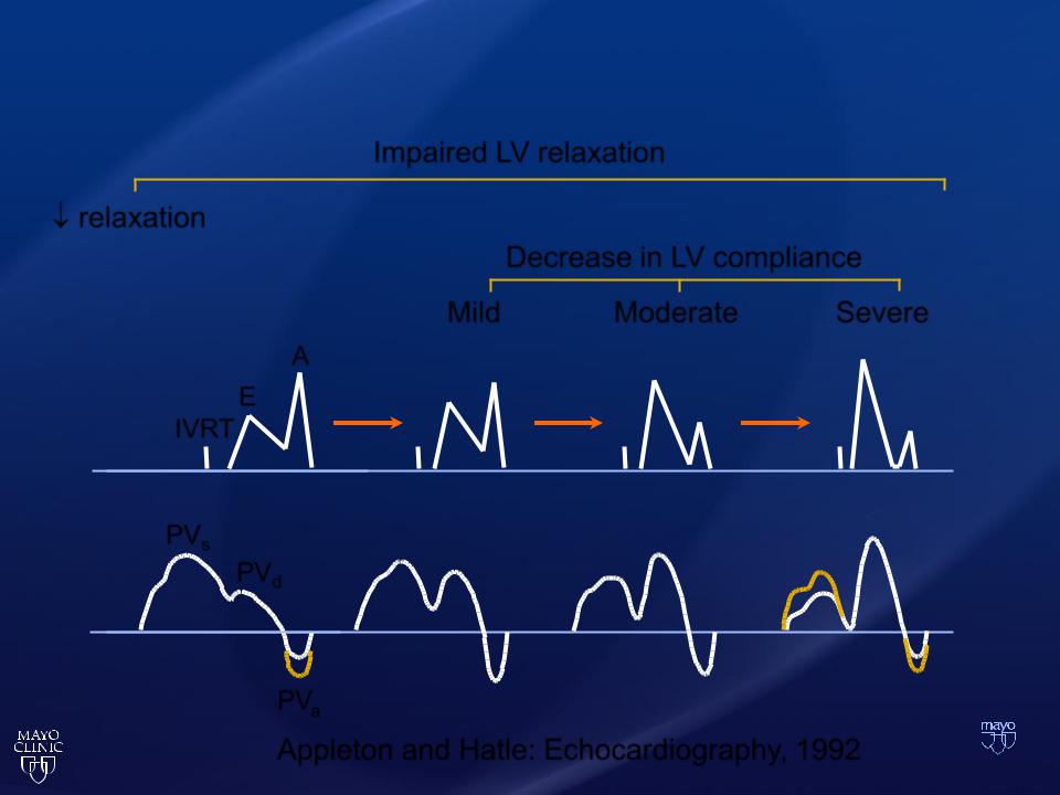
Diastolic Dysfunction
Impaired LV relaxation
relaxation
Decrease in LV compliance
Mild Moderate Severe
A
E
IVRT
PVs
PVd
PVa
Appleton and Hatle: Echocardiography, 1992
CP1018119-2

Pseudonormalization
•
•
•
2D CluesLV function
LV walls
LA volume
CP993639-26

Proof of Pseudonormalization
Uncovering Evidence of
• Delayed LV relaxation and/or
• Increased filling pressure
CP1018119-5

Pseudonormalization
Doppler Clues
(Delayed relaxation)
•
•
•
•
L-wave
DTI (E/e´)
Valsalva
Color M-mode
CP1018119-3

Mitral PW Doppler
83 Year Old woman with PAF
L-wave

Tissue Doppler Imaging
Pulsed Wave Spectral Doppler
Recording of Mitral Annulus Velocity
©2011 MFMER | slide-26

Mitral Annulus DTI
LV relaxation is impaired for most patients with
•
•
E' lateral < 8.5 cm/sec E' septal < 8 cm/sec
Journal of the American Society of Echocardiography
Volume 22(2); February 2009

Tissue Doppler Imaging for Diastolic Function
•Mitral Inflow PW Doppler pattern varies with preload
•Mitral annular DTI faithfully provides the velocity of relaxation through a wide range of preload

Nagueh SF; JASE 22(2); February 2009

Normal Mitral Annulus Velocities
•
•
•
Medial e’ ≥ 8 Lateral e’ ≥ 10
LA volume index < 34 cc/m2
Normal Diastolic function
©2011 MFMER | slide-30
