

ECHO 2013 / Pediatric Echocardiography Overview of Quantification and Lessons Learned
.pdfCurrently Available Normal
Pediatric Datasets
• Approximately 70 publications
– Ventricular dimensions/function |
21 |
– Atrial dimensions/function |
2 |
– Cardiac valves |
8 |
– Aortic dimensions/flow |
16 |
– Aortic arch |
6 |
– Coronary arteries |
5 |
– Pulmonary arteries |
12 |
•Online Z-score calculator: Parameter(z)
–http://parameterz.blogspot.com/

Currently Available Normal
Pediatric Datasets
•Nearly all single center studies
•Lacking agreement in methodology
–Nomenclature
–Adjustment for body size
–Many with inadequate statistics
•Lacking in geographical and/or ethnic diversity
•Discrepancy with adult methodology
JASE 2013

Normal Pediatric Echocardiogram
Lai et al. JASE 2006
• Skills and Knowledge
• Standardized views
– Subxiphoid
– Apical
– Parasternal
– Suprasternal
– Right Parasternal
• Measurements
• Reporting
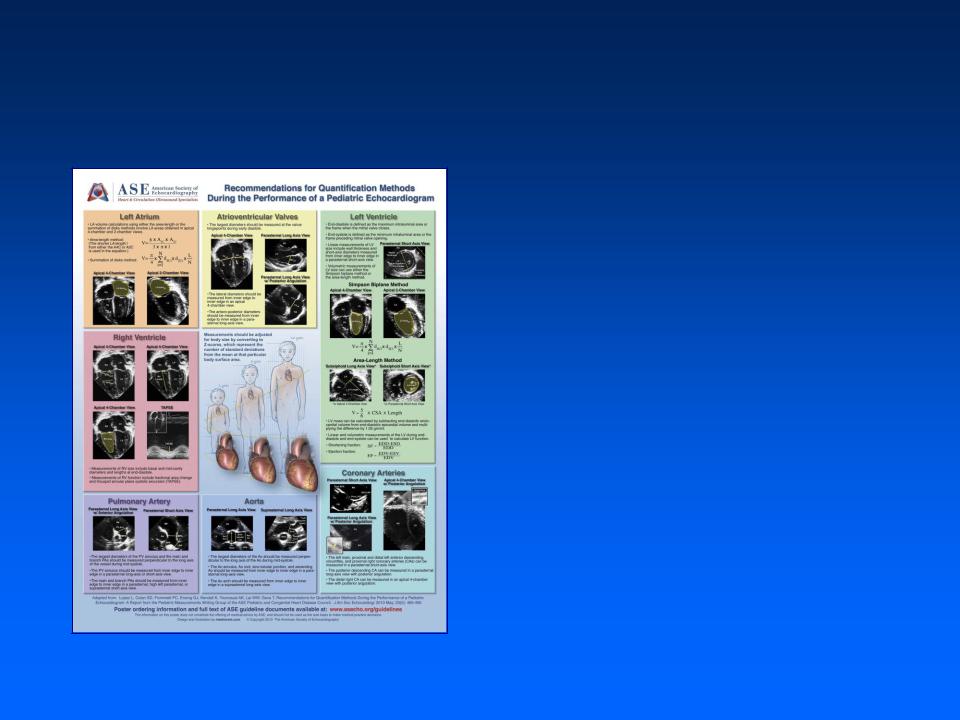
Pediatric Quantification Methods
Lopez et al. JASE 2010
• Veins and Atria
• Atrioventricular valves
• Left ventricle
• Right ventricle
• Ventricular outflow tracts and Semilunar valves
• Aorta, Coronary arteries, and Pulmonary arteries

Pediatric Quantification Methods
Lopez et al. JASE 2010
•LV size and function
–Timing of measurements
•End-diastole: maximum area or MV closure
•End-systole: minimum area or immediately preceding MV opening

Pediatric Quantification Methods
• LV size and function
– 
–Linear vs. volumetric measurements
•Assumes normal LV shape

Pediatric Quantification Methods
• LV size and function
– 
–Linear vs. volumetric measurements
•Assumes normal LV shape
•Normal data available

Pediatric Quantification Methods
• LV size and function
– 
– 
– Long-axis vs. short-axis: limitations to both

Pediatric Quantification Methods
Images courtesy of Dr. Leo Lopez
• LV size and function
– 
– 
–Long-axis vs. short-axis
•angle of measurement

Pediatric Quantification Methods
Images courtesy of Dr. Leo Lopez
•LV size
–Long-axis vs. short-axis
•
•recognition of septal flattening