

ECHO 2013 / Stress Echocardiography
.pdfStress Echocardiography
Michael H Picard, M.D.
Massachusetts General Hospital
Harvard Medical School
No disclosures

Stress Echo
•CAD
–Diagnosis
–Prognosis
–Special issues
•Valve disease
–Exercise induced pulmonary hypertension
•Diastolic function
–Dyspnea evaluation

Why add an echo to a stress test ?
• Addition of any imaging modality to ETT increases accuracy
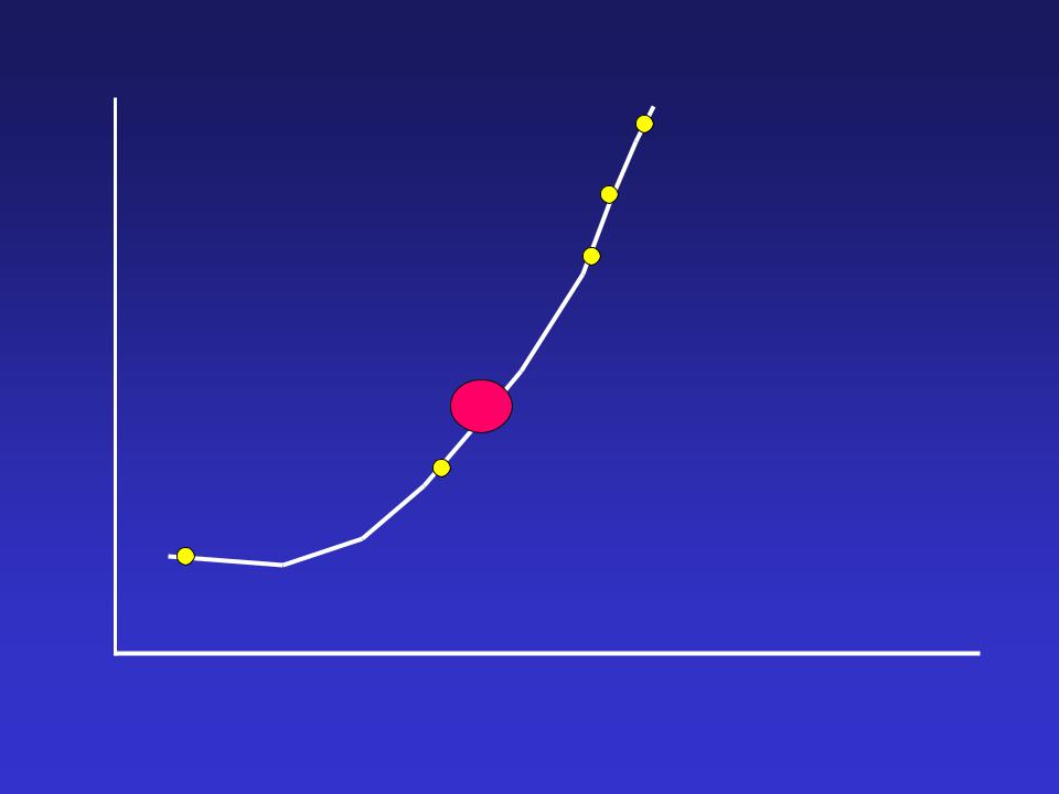
Rate-pressure product
Angina
Ischemic ST segment
PCW elevation
 Significant perfusion defect
Regional myocardial dysfunction
Flow heterogeneity
Rest
Stress Time
Modified from Beller, Am J Card 1988;61:2

Why add an echo to a stress test ?
•advantages of echo
–noninvasive, no radiation, portable
–20-30 min test; no delayed imaging, rapid results
–closely spaced, repeated testing is highly feasible
–beat by beat, stage by stage analysis
–global and segmental function (resting and stress)
–relative cost
–other info obtained - pericardium, valves, chambers, etc.
•for other sources of chest pain

Adding echo to a stress test
•Disadvantages
–< 100% imaging success rate
•improving with harmonics, contrast
–operator dependent
•sonographer and interpreter
–learning curve to interpretation (subjective)
•wall motion during ischemia is subtle and transient
–subjective interpretation
•quantitation under development
–assessing end result of ischemia
–? ischemia within region of prior infarct

Sept 2007
JACC +
JASE
March 2011
JASE Jan 2011

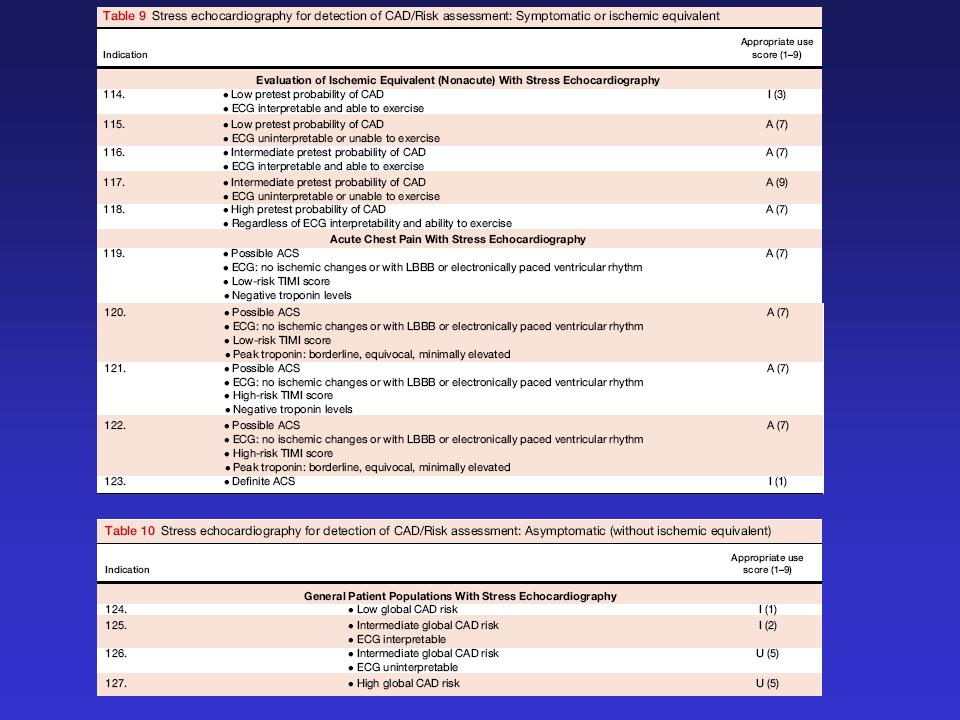
Appropriate use of stress echo
Douglas et al, 2011 March JACC and JASE
•Detection of CAD and Risk Assessment
–Symptomatic or ischemic equivalent
–Asymptomatic (without ischemic equivalent)
–Asymptomatic (without ischemic equivalent) in populations with defined co-morbidities
•After prior test results
•Risk Assessment
–Peri-op evaluation for non-cardiac surgery without active cardiac conditions
–Within 3 months of an ACS
–Postrevascularization (PCI or ACS)
•Assessment of viability/ischemia
•For hemodynamics (includes Doppler during stress)
•Contrast use

Wall motion abnormality due to CAD:
what is the eye and brain integrating?
•Decreased inward systolic motion
•Decreased systolic thickening
–Systolic thinning in extreme cases
•Regional diastolic thinning when chronic CAD
•Systolic motion is simplest to assess but Systolic Thickening is critical and most specific