

Tools and Applications of Biochemical Engineering Science
.pdf66 |
|
S. Nath |
1 |
Introduction and Brief History . . . . . . . . . . . . . . . . . . . |
. 68 |
2 |
The ATP Synthase . . . . . . . . . . . . . . . . . . . . . . . . . . |
. 69 |
2.1 |
Subunit Composition . . . . . . . . . . . . . . . . . . . . . . . . |
. 69 |
2.2 |
Molecular Machine Characteristics . . . . . . . . . . . . . . . . . |
. 70 |
2.3 |
Structure . . . . . . . . . . . . . . . . . . . . . . . . . . . . . . . |
. 70 |
2.3.1 |
The Walker Structure . . . . . . . . . . . . . . . . . . . . . . . . |
. 70 |
2.3.2 |
The Pedersen-Amzel Structure . . . . . . . . . . . . . . . . . . . |
. 71 |
3Molecular Mechanism of ATP Synthesis in the F1 and F0 Portions
|
of ATP Synthase . . . . . . . . . . . . . . . . . . . . . . . . . . . . |
71 |
3.1 |
The Binding Change Mechanism . . . . . . . . . . . . . . . . . . . |
71 |
3.1.1 |
Modification of the Binding Change Mechanism . . . . . . . . . . |
72 |
3.2Latest Experimental Evidence not in Agreement with any Version
of the Binding Change Mechanism . . . . . . . . . . . . . . . . . . 73
3.3Further Specific Difficulties Associated with the Binding Change
|
Mechanism . . . . . . . . . . . . . . . . . . . . . . . . . . . . . . . |
73 |
3.4 |
The Torsional Mechanism of ATP Synthesis . . . . . . . . . . . . . |
75 |
3.4.1Some Novel Predictions of the Torsional Mechanism
of ATP Synthesis . . . . . . . . . . . . . . . . . . . . . . . . . . . . |
79 |
3.4.2 Quantification of the Torsional Mechanism . . . . . . . . . . . . . |
80 |
3.4.3Resolution of Difficulties Achieved and Experimental Evidence
|
Supporting the Torsional Mechanism . . . . . . . . . . . . . . . . |
82 |
3.4.3.1 |
Optical Probes . . . . . . . . . . . . . . . . . . . . . . . . . . . . . |
83 |
3.4.3.2 Electron Microscopy and Image Analysis . . . . . . . . . . . . . . |
83 |
|
3.4.3.3 |
Single Molecular Spectroscopy . . . . . . . . . . . . . . . . . . . . |
83 |
3.4.3.4 Biochemical Nucleotide Site Occupancy Experiments . . . . . . . |
84 |
|
3.4.3.5 Biochemical Acid Quench/Cold Chase Experiments . . . . . . . . |
84 |
|
3.4.3.6 |
Structural Considerations . . . . . . . . . . . . . . . . . . . . . . . |
85 |
3.4.3.7 Site-Site Cooperativity in ATP Synthase . . . . . . . . . . . . . . . |
86 |
|
3.4.3.8 |
Irreversible Mode of Catalysis . . . . . . . . . . . . . . . . . . . . |
88 |
4Further Insights into the Molecular Mechanism in the F0 Portion
of ATP Synthase . . . . . . . . . . . . . . . . . . . . . . . . . . . . |
88 |
5Thermodynamic Analysis of Molecular Mechanisms
|
for ATP Synthesis . . . . . . . . . . . . . . . . . . . . . . . . . |
. . 93 |
5.1 |
Mechanistic P/O Stoichiometry in Oxidative Phosphorylation |
. . 94 |
6 |
Molecular Physiological Engineering: a New Frontier . . . . . |
. . 96 |
References . . . . . . . . . . . . . . . . . . . . . . . . . . . . . . . . . . |
. . 96 |
|
The Molecular Mechanism of ATP Synthesis by F1F0-ATP Synthase: A Scrutiny of the Major Possibilities |
67 |
|
Symbols and Abbreviations |
|
|
AO |
affinity of oxidation |
|
AP |
affinity of phosphorylation |
|
a,b,c |
subunits of the F0 portion of ATP synthase enzyme |
|
a,b,g,d,e |
subunits of the F1 portion of ATP synthase enzyme |
|
bO , bC , bTP , bDP |
open, closed, loose, tight conformations, respectively, of the |
|
|
catalytic site |
|
d |
channel width |
|
e |
dielectric constant |
|
x |
frictional coefficient |
|
C |
closed |
|
E |
enzyme |
|
F |
Faraday |
|
F0 |
hydrophobic, membrane-bound portion of ATP synthase |
|
F1 |
hydrophilic, extra-membrane portion of ATP synthase |
|
f |
fraction |
|
I |
moment of inertia |
|
I |
ion concentration |
|
K |
equilibrium constant |
|
Kqss |
quasi-steady state constant |
|
k |
torsional constant (modulus of rigidity) |
|
kr , kr¢ |
rate constants |
|
ks |
constant of proportionality |
|
kt |
transport rate constant |
|
L |
loose |
|
l |
horizontal distance |
|
L |
electrostatic coupling strength |
|
l |
screening distance |
|
DµH |
electrochemical potential difference |
|
n |
exponent |
|
O |
open |
|
q |
angle, angular displacement |
|
DpH |
pH difference |
|
Dy |
electrical potential difference |
|
D(Dy) |
change in electrical potential |
|
q |
Coulombic charge, degree of coupling |
|
R |
radial distance |
|
r |
radial distance, vertical distance |
|
T |
tight |
|
t |
time |
|
tM |
machine electrostatic torque |
|
V |
electrostatic potential |
|
Vmax |
maximum velocity |
|
vhyd |
rate of ATP hydrolysis |
|
vsyn |
rate of ATP synthesis |
|
w |
angular velocity |
|
68 |
S. Nath |
x |
distance, phosphate potential |
y |
distance |
Z |
mechanistic stoichiometry |
ADP |
adenosine diphosphate |
AMP-PNP |
non-hydrolyzable ATP analog |
Asp |
aspartic acid |
ATP |
adenosine triphosphate |
DELSEED |
seven-residue amino acid sequence in the b subunit |
Glu |
glutamic acid |
Met |
methionine |
P/O |
ATP/oxygen ratio |
Pi |
inorganic phosphate |
Phe |
phenylaniline |
Val |
valine |
1
Introduction and Brief History
From knowledge of metabolic pathways and the extent of the world’s biomass, it is estimated that the energy currency of the cell, the molecule adenosine triphosphate (ATP) and the adenosine diphosphate (ADP) and inorganic phosphate (Pi) from which it is formed, participate in more chemical reactions than any other compound on the surface of the earth with the exception of water. ATP was co-discovered by Fiske and SubbaRow and independently by Lohmann in 1929 [1, 2]. Over 60 years ago, the vital cellular process of oxidative phosphorylation was demonstrated by Belitzer and Kalckar. It was recognized that this was the major pathway by which our bodies captured energy from foodstuffs and used it to carry out a variety of essential cellular functions, but how it took place was largely unknown. In 1941, Lipmann postulated the key role of ATP in cellular metabolism and suggested that it possessed a “high group transfer potential” [3]. Lehninger identified the site of this reaction as the mitochondrion in 1948.
The synthesis of ATP is catalyzed by the enzyme ATP synthase (or F1F0-ATP synthase); the F1 portion of this enzyme was first isolated by Racker and coworkers in 1960 [4]. ATP synthase is present in abundance in the membranes of animal mitochondria, plant chloroplasts, bacteria and other organisms. ATP synthesized by our ATP synthase is transported out of mitochondria and used for the function of muscle, brain, nerve, liver and other tissues, for active transport, and for synthesizing myriad compounds needed by the cell. Since the pool of adenosine phosphates in the body is limited, the use of ATP must be continually compensated by its synthesis, and an active person synthesizes his own body weight of ATP every day. The synthesis of ATP is the most prevalent chemical reaction in the body [5]. This is indeed a very important reaction. How exactly does it occur?
Historically, a first step towards resolution of this problem was taken by Slater in 1953 through his formulation of the chemical hypothesis [6]. Un-
The Molecular Mechanism of ATP Synthesis by F1F0-ATP Synthase: A Scrutiny of the Major Possibilities |
69 |
fortunately, no chemical intermediate between oxidation and phosphorylation could be isolated despite frenzied efforts by several groups. Between 1961 and 1966, Mitchell formulated his then radical chemiosmotic hypothesis of oxidative phosphorylation [7, 8], while Williams proposed localized models coupling oxidation and ATP synthesis [9, 10]. Boyer presented the salvage conformational hypothesis in 1965 in which oxidation was directly coupled to phosphorylation via protein-protein conformational interactions [11]. In 1973, Boyer and colleagues proposed a precursor [12] to what later became the binding change mechanism of ATP synthesis, which he reviewed in great detail in 1993 [13]. Cross modified the mechanism and presented it in a review in 1996 [14], but still referred to it as the binding change mechanism.
Exactly a decade ago, after a long, rigorous and thorough education in both the engineering and biological sciences at IIT Kanpur, Princeton University, MIT, GBF/TU Braunschweig (under the inspiring guidance of Prof. Dr. W.-D. Deckwer), I decided, as a young Assistant Professor at IIT Delhi, to “work on something really challenging.” Basic studies in bioenergetics had intrigued scientists for a long time; yet the molecular mechanism of biological energy transduction remained an enigma. It appeared that a completely fresh and original hypothesis was needed that could explain the wealth of existing data and better withstand further experimental challenge. In a series of papers during 1998– 2001, Nath and co-workers proposed the torsional mechanism of energy transduction and ATP synthesis [16–20, 56].
In this review, I shall attempt to critically examine, as best as I can in the limited space available, some of the major candidate molecular mechanisms of ATP synthesis.
2
The ATP Synthase
2.1
Subunit Composition
ATP is synthesized by the enzyme ATP synthase (or F1F0 ATPase), which transforms energy from a transmembrane gradient of protons, or, in some cases, Na+ ions into the chemical energy of ATP. This enzyme, the smallest known molecular machine, consists of two major parts: a membrane-extrinsic, hydrophilic F1 containing three a, three b, and one copy each of g, d, and e subunits, and a membrane-embedded, hydrophobic F0 composed of one a, two b, and twelve c subunits (Fig. 1). The molecular masses of the a, b, g, d, and e subunits in E. coli measure about 55, 50, 31, 19, and 15 kDa, while the a, b, and c subunits have molecular masses of 30, 17, and 8 kDa, respectively [21–30]. The F0 and F1 domains are linked by two slender stalks [21–30]. The central stalk is formed by the e subunit and part of the g subunit, while the peripheral stalk is constituted by the hydrophilic portions of the two b subunits of F0 and the d subunit of F1. The ion channel is formed by the interacting regions of the a and c subunits in F0, while the catalytic binding sites are predominantly located in the b subunits of F1 at the a-b interface [21, 22, 28].
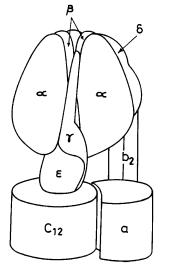
70 |
S. Nath |
Fig. 1. Schematic diagram of the Escherichia coli ATP synthase enzyme
2.2
Molecular Machine Characteristics
ATP synthesis takes place by conformational changes at the catalytic binding sites. Recent structural [21, 27], biochemical [13, 28, 31], spectroscopic [32, 33] and microscopic [34, 35] studies indicate that these conformational changes arise from rotation of the g-e subunit in a static barrel of a3b3 subunits in ATP synthase, making it the world’s smallest molecular machine with a rotor radius of ≈1 nm (Fig. 1).
2.3 Structure
2.3.1
The Walker Structure
The X-ray crystal structure of bovine heart mitochondrial F1 was solved by the group of Walker to 2.8 Å resolution in 1994, and represents the largest asymmetric structure ever solved [21]. The elongated a and b subunits are arranged alternately in the form of a hexagon.A 9-nm long a-helix of the g subunit (comprising of residues 209–272 at the carboxy terminal) extends from top to bottom of the central cavity of the hexagon. The lower part of the carboxy terminal helix forms a coiled coil with a a-helix consisting of residues 1–45 at the amino terminus of the g subunit. A short a-helix consisting of residues 73–90 of the g subunit projects from the bottom. The d and e subunits, and 145 residues of the g subunit (~50%), were not sufficiently ordered to diffract to high resolution and were not located in the electron density map.

The Molecular Mechanism of ATP Synthesis by F1F0-ATP Synthase: A Scrutiny of the Major Possibilities |
71 |
In the Walker crystal structure of F1-ATPase, the three non-catalytic a sites are liganded with the non-hydrolyzable ATP analog MgAMP-PNP. In contrast, the three catalytic b sites possess different conformations. One of the catalytic sites in the structure binds the analog MgAMP-PNP and is designated as bTP; another site binds MgADP and is denoted by bDP , while the third site is empty and distorted and is called bE [21]. In further contrast, the nucleotide-free a3b3 subcomplex of ATP synthase is a symmetric trimer [36].
2.3.2
The Pedersen-Amzel Structure
In 1998, Pedersen, Amzel, and colleagues solved the X-ray structure of rat liver mitochondrial F1-ATPase to 2.8 Å resolution and obtained a more symmetrical structure of the a and b subunits [27]. In this structure, catalytic as well as noncatalytic sites are occupied with bound nucleotide and the three a subunits and the three b subunits are in very similar but distinct closed conformations, with no indication of an open conformation as found in the Walker structure. The rat liver crystals were grown in the presence of substantially higher concentrations of nucleotides, and the crystallization medium contained only ATP (and not AMP-PNP), but no Mg2+.
3
Molecular Mechanism of ATP Synthesis in the F1 and F0 Portions of ATP Synthase
3.1
The Binding Change Mechanism
According to the binding change mechanism proposed by Boyer (Fig. 2, where O, L, T refer to open, loose and tight conformations, respectively), the synthesis of ATP occurs reversibly with an equilibrium constant close to unity in the high-affinity enzyme catalytic binding site; energy input from the so-called protonmotive force is needed not for synthesis of ATP at the catalytic site (which occurs spontaneously), but for the release of synthesized ATP from the high-affinity catalytic site [5, 13, 22, 37]. Thus, the step of chemistry is not an energy-requiring step according to this mechanism, while the high-affinity catalytic site opens due to provision of energy by the protonmotive force. Another
Fig. 2. Depiction of the binding change mechanism of ATP synthesis (simplified diagram redrawn from [13])

72 |
S. Nath |
central feature of the binding change mechanism is that the catalytic sites interact with each other, i.e., cooperativity exists among catalytic sites [5, 13]. Thus, substrate (ADP+Pi) binding at a low-affinity catalytic site promotes product ATP release from the high-affinity catalytic site; hence, substrate binding must precede product release or be simultaneous with it during steady operation. Moreover, a simultaneous negative cooperativity of binding and positive cooperativity of catalysis is hypothesized to occur in the F1 portion of ATP synthase in the physiological mode of operation [13]. Another tenet is the free rotation of the g subunit in the cavity of the a3b3 barrel in F1. According to the binding change mechanism, physiological rates of ATP synthesis occur when two of the three catalytic sites contain bound nucleotide (Fig. 2).
3.1.1
Modification of the Binding Change Mechanism
In the version of the binding change mechanism (1993) described in Sect. 3.1 (bi-site catalysis, Fig. 2) [13], (ADP+Pi) enter in the loose catalytic site (L), ATP forms spontaneously in the tight site (T) and is released from the open site (O). In the 1997 version, the mechanism remains bi-site, but now (ADP+Pi) enters in O,ATP forms in T and is released in L [22]. In the modification of the binding change mechanism by Cross and co-workers, all three catalytic sites were used (though bound nucleotide still occurs in only two sites, thereby making this modification effectively still a bi-site mechanism), and an intermediate enzyme state containing loosely bound (ADP+Pi) in L as well as tightly bound (ADP+Pi) in T was envisaged [14] (Fig. 3). Cross envisaged that ATP forms from tightly bound (ADP+Pi) in T only after ATP release has taken place from O (Fig. 3, steps 1, 2). However, recently [5, 13], the concept of the tightly bound (ADP+Pi) intermediate was abandoned by Boyer, though all the tenets given in Sect. 3.1 were still retained. A quasi-equilibrium of a catalytic site containing (ADP+Pi) (form 2-H) and ATP (form 2-S) was envisaged. In the latest modification in the year 2000, the requirement of (ADP+Pi) in the L site was deemed unnecessary as it “introduces another control”, and the conformation that a single catalytic site passes through during ATP synthesis was proposed to be O to T to L [37]. All other tenets were as in Sect. 3.1.
Fig. 3. Modified model of the binding change mechanism highlighting cooperativity and including tightly bound ADP and Pi as intermediates (simplified and redrawn from [14])
The Molecular Mechanism of ATP Synthesis by F1F0-ATP Synthase: A Scrutiny of the Major Possibilities |
73 |
3.2
Latest Experimental Evidence not in Agreement with any Version of the Binding Change Mechanism
Recently, in a significant development, Senior and colleagues designed an optical probe by inserting a tryptophan residue to directly monitor, for the first time, the occupancy of the catalytic sites by nucleotides [28, 38]. Their tryptophan fluorescence experiments in the hydrolysis mode established definitively that Vmax activity is attained by F1-ATPase only when all three catalytic sites are occupied by bound nucleotide, i.e., an enzyme species with all three b subunits occupied is the only catalytically competent species [38]. Due to the inviolability of microscopic reversibility, this result holds for ATP synthesis also, and agrees with an earlier assessment based on enzyme kinetic and radioactive nucleotide binding data [39]. This experimental evidence is in complete disagreement with the fundamental tenets of the binding change mechanism. Further, the tryptophan fluorescence experiments showed that Pi binding can not be spontaneous, as suggested in diagrams illustrating the binding change mechanism (e.g., Figs. 2 and 3). The recent X-ray structure of mitochondrial F1-ATPase in the transition state [39a] shows nucleotide bound to all three catalytic sites, which can never be explained by the binding charge mechanism.
Direct visualization of F1-ATPase by sophisticated single molecule spectroscopy techniques such as epifluorescence/confocal microscopy clearly and unequivocally demonstrates unidirectional and discrete motion of the g subunit in steps of 120° [34, 35, 40]. In various disciplines, from quantum chemistry to chemical/biochemical and mechanical engineering, such motion is considered irreversible. It is very difficult to conceive how a unidirectional, discrete motion can take place by a reversible mode of catalysis (E.ATP E.ADP.Pi), i.e., how can a subunit of a single enzyme molecule oscillate back and forth in the presence of a driving force in one direction? This irreversibility of operation in a single molecule mode contradicts the fundamental tenet of the binding change mechanism that ATP synthesis occurs reversibly (and spontaneously) in a catalytic site of the enzyme. In my view, it is imperative to relate the chemical kinetics (the arrows representing an elementary step in the kinetic scheme) to the mechanical aspects (structure and dynamics of the molecular machine). It is interesting to note that our approach to the problem, in contrast, has incorporated irreversibility from the very outset [16–20, 41, 42, 56].
3.3
Further Specific Difficulties Associated with the Binding Change Mechanism
In addition to the very serious recent evidence in total disharmony with the binding change mechanism discussed above (Sect. 3.2), we pointed out several other specific difficulties in a paper published in January 2000, and stated that it was most important “to recognize and understand the imperative need to consider novel ideas with an open mind, and to overcome the limitations imposed on our own thinking by currently accepted mechanisms, by paradigms that are no longer applicable” [18]. For instance, since the loose conformation, L (bTP), con-
74 |
S. Nath |
tains bound (MgADP+Pi), and the open site, O (bE), contains no bound nucleotide or Pi (Fig. 2), we found it difficult to conceive how both the binding steps could take place in the same catalytic site conformation (bTP), especially keeping in mind that the binding of Pi is not spontaneous and requires energy [18]. The spontaneity of inorganic phosphate binding was emphasized as a serious defect. In the latest version [37], in addition to competent binding of ADP and Pi to O, even ATP is synthesized in a single O Æ T binding change, which is even more problematic. We considered it highly unlikely that nucleotide could bind to the catalytic site in the O conformation as proposed by Boyer [13, 22, 37], because of the absence of a proper nucleotide binding site [18]. This is even more difficult to reconcile in the hydrolysis mode, as pointed out by other workers [27]. Moreover, in this mode, in Boyer’s bi-site mechanism, MgATP binds to the low affinity O site, but the medium affinity L site is left unfilled, which is a problem, as discussed [43]. Further, negative binding cooperativity and positive catalytic cooperativity need not occur simultaneously, as discussed by us [19], and as shown by recent experimental evidence [44].We have also shown from first principles that there is absence of site-site cooperativity in ATP synthase under physiological steady state operating conditions [19]; in fact, there is no need for cooperativity [18]. Moreover, the molecular basis of cooperativity in ATP synthase remains unelucidated, despite almost three decades of experimental effort! Finally, our kinetic analysis necessitates that in the steady state physiological mode of ATP synthesis, product ATP release must precede substrate ADP binding [19], in agreement with the latest experimental evidence by direct optical probes [38], while, according to all versions of the binding change mechanism from 1973 to 2000, ADP binding must precede ATP release or be simultaneous with it. In the latest modification [37], the L site has no real role to play. It appears to be there just to accommodate the existence of three catalytic sites. Further, in the synthesis mode, T contains bound ATP while in the hydrolysis mode, L has bound ATP. If, in the synthsis mode, release of ATP takes place from O, then, at any instant, there exist two bound ATP molecular in F1 . This contradicts experimental data [bottom of p. 255 in 37]. If, in the synthesis mode, ATP is released from L, why is it that in the hydrolysis mode [39a] the ATP, entering in O, is not released from L itself? A logical explanation of these numerous discrepancies is that the basic tenets of the binding charge mechanism are incorrect.
Cross’s modification of the binding change mechanism (Fig. 3) is also inadequate; in addition to the difficulties detailed above, it is beset with other serious problems. For instance, the open conformation (O) causes release of ATP, therefore, how can it cause binding of ADP, as envisaged? Moreover, it is hypothesized that ATP forms from tightly bound (ADP+Pi) in T only after ATP release has taken place from O (Fig. 3; steps 1, 2); since these conformational changes are caused by rotation of g-e, they should logically occur simultaneously at the different catalytic sites. How substrate (MgADP) binding enhances product (MgATP) release at a different catalytic site, which in turn promotes ATP synthesis at a third site, has never been proposed, i.e., how is this signal communicated, and, above all, how is the energy transmitted from one b subunit to another through the intervening a subunits? Further, in the nonequilibrium mode of functioning, tightly bound (ADP+Pi) should not be present in T; instead,
The Molecular Mechanism of ATP Synthesis by F1F0-ATP Synthase: A Scrutiny of the Major Possibilities |
75 |
bound MgATP should be present. Finally, in this mechanism, the loose (L) site is serving no real function; it contains loosely bound (ADP+Pi) throughout the cycle (Fig. 3). Moreover, the O site also contains (ADP+Pi), and the T site also has bound (ADP+Pi) at a particular instant!
In view of the numerous serious difficulties discussed above, it seemed clear that only by questioning our fundamental beliefs would it be possible to truly understand how ATP is made.
3.4
The Torsional Mechanism of ATP Synthesis
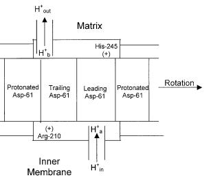
In a series of papers, we have proposed the torsional mechanism of energy transduction and ATP synthesis, the only unified and detailed molecular mechanism of ATP synthesis to date [16–20, 56] which addresses the issues of ion translocation in F0 [16, 20, 56], ionmotive torque generation in F0 [16, 20, 56], torque transmission from F0 to F1 [17, 18], energy storage in the enzyme [17], conformational changes in F1 [18], and the catalytic cycle of ATP synthesis [18, 19]. We have also studied the thermodynamic and kinetic aspects of ATP synthesis [19, 20, 41, 42, 56]. A kinetic scheme has been developed and mathematically analyzed to obtain a kinetic model relating the rate of ATP synthesis to pHin and pHout in the F0 portion and the adenine nucleotide concentrations in the F1 portion of ATP synthase.Analysis of these kinetic models reveals a wealth of mechanistic details such as the absence of cooperativity in the F1 portion of ATP synthase, order of substrate binding and product release events, and kinetic inequivalence of DpH and Dy.
Fig. 4. Schematic diagram of the DpH – Dy two mutually non-colinear half-channel model for torque generation by the F0 portion of ATP synthase forming a part of the torsional mechanism of energy transduction and ATP synthesis [16–20, 56]