

Tools and Applications of Biochemical Engineering Science
.pdf86 |
S. Nath |
represents the active conformation of the enzyme. In their view, the Walker structure is a non-physiological state induced by crystallization without sufficient total nucleotide present to occupy all three catalytic sites [27]. In a recent X-ray structure of the E. coli F1-ATPase at 4.4 Å resolution [50], the departure from exact threefold symmetry was confirmed, in agreement with the Walker structure. The crystallization conditions used in this study employed a higher (5 mM) concentration of nucleotide, which is above the physiological level (~3 mM). Hence, the asymmetry of F1 is not caused by an artificially low concentration of nucleotide as stated [27] but is an intrinsic property of the enzyme, as proposed [21], and as considered in the formulation of the torsional mechanism. Moreover, the crystallization medium employed for solving the Pedersen-Amzel structure contained 5 mM ATP but no Mg2+, and these workers proposed that “Mg2+ binding to ATP in the b subunits causes no major conformational changes” [27], and that the main difference between the two structures, the presence of the closed conformation in the rat liver enzyme [27], could not be attributed to the absence of Mg2+, as suggested [22]. According to the torsional mechanism [18], “Mg2+ is critical for the structure and mechanism, and, as shown and emphasized, Mg2+ has a crucial role in catalysis.” We therefore could not conceive “how a closed catalytic site conformation can occur in the absence of Mg2+” [18]. In fact, our argument implies that, since a closed conformation of the catalytic sites is indeed observed in the PedersenAmzel structure [27], Mg2+ must be present. Since no Mg2+ was contained externally in the crystallization medium, a possibility is that the Mg2+ may have been present internally in the rat liver enzyme source itself. A physiologically functional rat liver mitochondrial enzyme would be expected to contain enough Mg2+ to fill all sites. It is very interesting to note that the a subunits have been clearly shown to contain bound Mg2+ (Figs. 1, 5 of ref. 27) from careful analysis of the electron density in the Pedersen-Amzel X-ray structure. Thus, it can be inferred logically that the Mg2+ in the b subunits has originated from the same source (whatever that might be) that caused the a subunits of the rat liver enzyme to contain Mg2+, as observed in the structure [27]. This point may be responsible for the confusion and controversy that has been expressed in the bioenergetics literature, and our interpretation would readily explain and reconcile various conflicting viewpoints, without disregarding any of the experimental facts, all of which, in our view, have the capacity to significantly contribute to our understanding of the molecular mechanism of ATP synthesis.
3.4.3.7
Site-Site Cooperativity in ATP Synthase
The molecular basis of site-site cooperativity in ATP synthase still remains unelucidated [46], and the absence of any direct evidence for cooperativity (despite almost three decades of effort) is explained, within the framework of the torsional mechanism, by the fact that site-site cooperativity does not exist in the physiological, steady state mode of functioning. Since, according to the torsional mechanism, no rotation takes place in uni-site or bi-site catalysis
The Molecular Mechanism of ATP Synthesis by F1F0-ATP Synthase: A Scrutiny of the Major Possibilities |
87 |
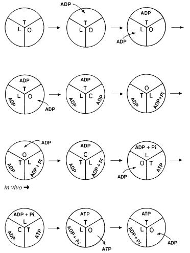
Fig. 9. Unsteady state start-up leading to physiological steady state operation of ATP synthase based on the torsional mechanism
modes of ATP synthesis, each substrate molecule enters and binds to the enzyme system that is in the same (unchanging) state in each catalytic cycle. Hence, there is no question of change in rate constant with substrate concentration in the physiological steady state mode of operation, and hence there can be no cooperativity in this mode of operation. The unsteady state (in vitro) filling up of the catalytic sites (start-up) and the progression to steady state operation are depicted in Fig. 9. Finally, the Michaellian nature of the results of our recent kinetic model of ATP synthesis based on the torsional mechanism (Eq. 11 of ref. 19 and Eq. 12 of ref. 20) clearly indicates the absence of site-site cooperativity during steady state ATP synthesis and suggests the presence of competitive inhibition of ATP synthase by product ATP as the inhibitor in the
88 |
S. Nath |
synthesis mode. An important consequence of the competitive product inhibition is the order imposed on binding and release events, i.e., product release must precede substrate binding [19].
3.4.3.8
Irreversible Mode of Catalysis
In the kinetic scheme for ATP synthesis (Fig. 2 of ref. 19), if the catalytic steps resulting in the formation of E.ADP.Pi and E.ATP are made reversible with equilibrium constants Kr and Kr¢, mathematical analysis of this modified kinetic scheme leads to the result:
vsyn = kt [ADPcy (K2KrKr¢/K1) – ATPcy]/(1 + K2KrKr¢/K1) |
(15) |
which predicts a linear (and not hyperbolic) relationship between the rate of ATP synthesis and the substrate ADP concentration, which is not in agreement with the experimental observations [51, 52]. A similar result is obtained for the F0 portion (Eq. 16), after similar modification of the kinetic scheme (Fig. 2 of ref. 20) to incorporate reversible catalysis.
v |
syn |
= k k [H+ |
(K K /K ) – H+ |
]/(1 + K K /K ) |
(16) |
||
|
s t |
in |
r 2 1 |
out |
r 2 1 |
|
|
Hence, the analysis reveals an irreversible mode of catalysis in both F0 and F1 portions of ATP synthase.
4
Further Insights into the Molecular Mechanism in the F0 Portion of ATP Synthase
To reach a delocalized Dy of ~200 mV for the macroscopic process of chemiosmotic coupling, Mitchell [8] estimated the charge transfer to be 0.8 µeq H+/g protein in rat liver mitochondria. From classical data [53], 1 mg mitochondrial protein contains 7.2 ¥109 mitochondria. To generate a delocalized Dy of 120 mV, the number of protons transferred per mitochondrion is {0.8¥(120/200)¥10–9 ¥6 ¥1023}/7.2 ¥109 = 40,000 H+/mitochondrion. Thus, the creation of a macroscopic, delocalized electrical potential difference of 120 mV across the inner mitochondrial membrane (to maintain the thermodynamic potential needed for ATP synthesis) requires the transfer of 40,000 protons in each mitochondrion, i.e., of 3333 cycles of electron transfer! Transfer of only 2 e– will create a negligible delocalized Dy in Mitchell’s chemiosmotic model. Hence, it may not be realistic to retain the analogy of the energy transduction process with a fuel cell [54]. In our conception, the ATP synthase should be viewed, not as a fuel cell, but as an enthalpic nonequilibrium molecular machine and mechano(electro)chemical transducer [17, 18]. Note that on taking the experimental value of 20,000 molecules per mitochondrion [53], we obtain a potential of 60 mV by translocation of one H+ discretely, in perfect agreement with our value obtained by energy balance. Our proposed mechanism postulates that the energy supplied by translocation of each ion is due to transport of a charge
The Molecular Mechanism of ATP Synthesis by F1F0-ATP Synthase: A Scrutiny of the Major Possibilities |
89 |
discretely irrespective of the rate of ion transport. The rate of ion transport itself is determined by the corresponding ion concentration gradient. Moreover,
the commonly employed expression RTln(Iin/Iout) for any ion I is a measure of the total energy available to the system through that ion I; it does not represent
the energy per ion of I translocated. If the ion concentration gradient is increased, the fraction of the enzyme complexes involved in ATP synthesis in its population and/or the rate of ion transport increases, and this is responsible for the experimentally observed increase in the rate of ATP synthesis with an increase in the ion concentration gradient. However, it should be emphasized that the energy obtained per ion translocated still remains the same.
The molecular mechanism and the kinetic model for the F0 portion of ATP synthase show that DpH and Dy are kinetically inequivalent driving forces for ATP synthesis [16, 20, 56]. This kinetic inequivalence has also been demonstrated by recent experiments [55]. DµH cannot be taken as the true driving force for ATP synthesis. Inequivalence can also be seen from the fact that DpH and Dy act at different elementary steps in our kinetic scheme [20, 56] as well as in our molecular mechanism [16, 20, 56]. Further, the diffusion potential generated as a result of diffusion of organic anions that are permeable to the membrane
(e.g., succinate) (included in Dyintrinsic , see Sect. 3.4) or that generated due to K+-valinomycin transport (Dyextrinsic) may be predicted to precede the translocation of protons along their concentration (pH) gradient. In such a situation,
each component of the driving force, DpH and Dy, can alter the rate of ATP synthesis independently of each other. A possible cause of this inequivalence is the existence of a delocalized DpH and a localized Dy, as conceived originally by us, though our molecular mechanism applies irrespective of the source of this Dy, whether localized or delocalized. This inequivalence has further implications that have been analyzed in detail [56]. Since protons that have been transported across the membrane to the matrix are transported back to the inner membrane by the redox enzyme complexes, DpH is delocalized. Similarly, Dp anion is delocalized. The energy of the anion gradient is converted to a Dy; both the
components of Dy discussed here – Dyintrinsic and Dyextrinsic – are created in the vicinity of the ATP synthase enzyme complex, in which case the Dy is a local-
ized phenomenon. If this is the case, then it is incorrect to combine DpH and Dy as is done in the definition of DµH . Also, a delocalized and a localized driving force cannot compensate for each other and have to be inequivalent. In such a situation, DµH is a mathematically defined quantity that does not have any physical meaning.
A more detailed molecular mechanism incorporating the above concepts is represented diagramatically in Fig. 10. Possibilities include the electrogenic movement of protons, on one hand, and the movement of a proton (along its concentration gradient) following a negative interior potential created by diffusion along its concentration gradient of a permeant anion, or by K+-valino- mycin transport, on the other. The former will lead to a D(Dy) of ~60 mV per unbinding and binding step, while the latter will yield ~30 mV (due to the DpH across the inlet half-channel) + ~30 mV due to the D(Dy) caused by proton binding and a similar contribution from the exit half-channel, providing in all ~120 mV in both cases per proton translocated across the membrane

90 |
S. Nath |
Fig. 10. Interaction of the ions with protein-in-the-membrane in the F0 portion of ATP synthase. Black dots are protons; the negative charge represents the Asp-61 residue in the c subunit. y is 120 mV for the electrogenic case or 60 mV for the dynamically electrogenic, overall electroneutral case discussed in the text. In the left most panel, the –y potential has been created either by the redox enzymes or by organic anions respectively. Following the rightmost arrow, a state is reached (not shown) when the entire potential has been discharged. This potential is re-created, and the cycle repeats
(Fig. 10). If respiration does not generate the delocalized Dy, then we strongly suggest consideration of the alternative, that the transport is dynamically electrogenic but electroneutral overall (i.e., transiently electrogenic, but electroneutral in the steady state). In such a situation, one cannot use the term “electrogenic” indiscriminately. Key sets of experiments to unambiguously distinguish between these possibilities need to be systematically designed and conducted.
Previous models of F0 place the essential Asp-61 carboxyl group at the interface of the a and c subunits [30, 57, 58]. However, the protonated carboxyl in the membrane, in its resting state, is positioned at the center of four a-helices of two interacting c subunits [59, 60]. In our view, the e subunit and the bottom of the g subunit do not move simultaneously with c; rather, movement of the latter precedes that of the former. As c rotates by 15° due to ion binding, the helices rotate and tilt, and the ion-protein interaction energy is stored as twist in the a-helices of the 10 membrane-embedded c subunits. The polar residues of c that are interacting with the g subunit remain more or less stationary, and the bottom of the c subunits move by 15°, thereby bringing the Asp-61 residues of all the c subunits to the periphery. The H+ in the membrane cannot exit, and the helices of the ten c subunits, present in their high energy state, will almost instantaneously untwist and return to their resting state. This impulse moves the polar residues by 15° thereby causing the c subunits to straighten.
The electrostatic interactions between the polar residues of these c subunits and the g subunit causes the bottom of the g subunit to rotate by 15°. Exposure of residues of the new c subunit to the aqueous medium at the a-c interface (helped by the bend in the C-terminal helix beyond Pro-64) and non-exposure of the helical face due to the excessive energy penalty in hydrating the hydrophobic residues such as Val-74, Met-75, or Phe-76 ensures that this tilted C-termi- nal helix of the c subunit is not allowed to untwist. The protonated Asp-61 lying in the middle of the helix is still not at the a-c interface; movement of the bottom of the g subunit forces this new trailing c subunit to straighten and expose the proton to the interface. The H+ exposed at the a-c interface now unbinds, rotates c, and the cycle of energy storage and release continues (Figs. 11, 12). In
The Molecular Mechanism of ATP Synthesis by F1F0-ATP Synthase: A Scrutiny of the Major Possibilities |
91 |
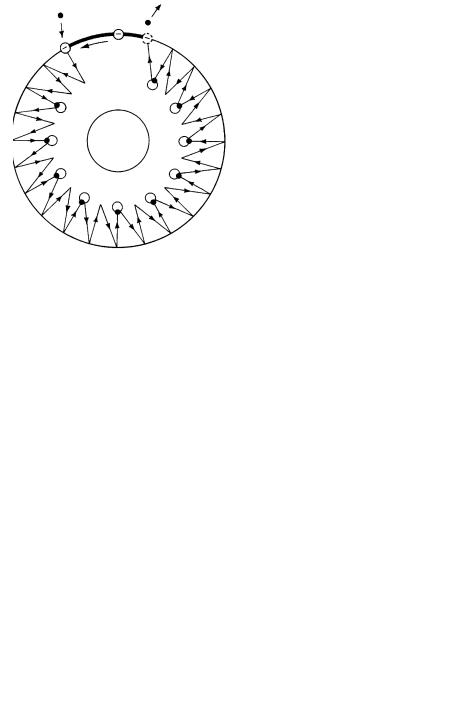
Fig. 11. Proposed schematic representation of the swiveling movement of helices in the F0 portion of ATP synthase. Since deprotonation of the Asp-61 residue can only occur at the periphery of the c-rotor at the a-c interface, twisting and swiveling of the helices composing the c subunits is caused by the rotation of the c-rotor. Since all protonated c subunits are identical, untwisted (protonated) c subunit helices should twist when the c-rotor rotates as a result of proton binding and unbinding during ATP synthesis
Fig. 12. Legend see page 92
92 |
S. Nath |
Fig. 12. Details of the torsional mechanism within F0 . The c subunits facing the a subunit are twisted and bent, while the remaining ten c subunits inside the membranous region being protonated, are untwisted, i.e. in their normal/resting state for the protonated Asp residues which lie in the interior of the c-rotor. In this state, the exposed Asp residues interact with the charges on the a-stator, and the system is in a local equilibrium (Fig. 4). When the incoming proton, which is transported along its concentration gradient, through the proton half channel facing the inner membrane (or outside) binds to the leading Asp-61 residue, the leading c subunit immediately attains an untwisted conformation characteristic of a protonated c subunit, and the now-protonated Asp-61 residue moves towards the center of the c-rotor. This alters the electrostatic interactions in the system and in order to attain a new local equilibrium state, the c-rotor rotates by 15°. As a result of this 15° rotation of the c-rotor, all the protonated c subunits get twisted and bent, keeping the polar residues of the c subunits, which are in contact with the bottom of the g subunit, stationary while the bottom of the c subunits move by a full 15° displacement thereby bringing all the Asp-61 residues to the periphery of the c-ro- tor. However, since this state is a metastable high-energy state for all the eleven protonated c subunits, all these c subunits except the new, incoming c subunit untwist simultaneously carrying all the Asp-61 residues of the ten protonated c subunits inside. The impulse thus created moves the polar residues of these ten protonated and untwisted c subunits by 15°, thereby causing these c subunits to straighten (untilt). The electrostatic interactions between the polar residues of these ten c subunits and the g subunit cause the bottom of the g subunit to rotate by 15°. Hence, the energy released during the proton binding step is transiently stored as strain energy in the c subunits as twist and is later stored as strain energy in the g subunit as torsion. The final state of the F0 portion attained after the 15° rotation has the c subunit immediately before the trailing c subunit at the interface of the membranous and the non-membranous region, and the leading c subunit at the other interface. The Escherichia coli c subunit contains a Pro-64 and a series of hydrophobic residues toward the end of the C-ter- minal helix. The portion of the C-terminal helix beyond Pro-64 bends such that the new, incoming trailing c subunit has part of the c subunit protruding out of the membranous region, with the rest of the helix still inside the membranous region. The twist causes the hydrophobic residues to face the N-terminal helix and keeps them away from the aqueous medium. When the remaining ten protonated c subunits untwist to release the strain, this c subunit is unable to do so, as this would require the hydrophobic residues to be in direct contact with the aqueous medium, which is energetically highly unfavorable. The untwisting of the ten c subunits drives the rotation of the bottom of the g subunit by 15°; rotation of the bottom of the g subunit forces this new, trailing c subunit to straighten. The straightening of this new, trailing c subunit brings its Asp-61 residue to the a-c interface. This Asp-61 residue then loses its proton to the matrix (or inside) through the exit half-channel to become unprotonated. The deprotonation of this Asp-61 residue keeps the already twisted c subunit in the same conformation characteristic of the unprotonated c subunit. The change in the electrostatics of the system again results in a rotation of the c-rotor by 15° in order to attain a new local equilibrium position. As a result of this 15° rotation, all the protonated and therefore untwisted c- subunits get twisted and tilted (keeping the polar residues and the g subunit stationary), thereby bringing their Asp-61 residues to the periphery of the c-rotor. As this state is a highenergy state for all the ten protonated c subunits, they untwist, and the impulse created causes these protonated c subunits to untilt by moving their polar residues by 15°. The movement of the polar residues causes the bottom of the g subunit to rotate by an equal 15° angular displacement. The e subunit interacts with the bottom of the g subunit and the polar residues of the c subunits, and therefore the e subunit rotates together with the bottom of the g subunit. In this way, each proton translocation drives the rotation of the e subunit and the bottom of the g subunit by 30° (in two steps of 15° each) and the consequent storage of energy as torsional strain in the g subunit. The final state of the system after these elementary steps is the same as the initial state, i.e. with both the Asp-61 residues facing the a-stator unprotonated, except that the e subunit and the bottom of the g shaft have rotated by 30° relative to the initial state.
The Molecular Mechanism of ATP Synthesis by F1F0-ATP Synthase: A Scrutiny of the Major Possibilities |
93 |
this schematic representation, the need for dynamic cyclical changes in protein structure in membrane-bound F0 is emphasized. Thus, the energy transduction processes can be represented as the energy of ion-protein interactions getting converted [via D(Dy)] to twist in the c-subunit helices, which is converted to torsional energy in g, and thereafter to the chemical energy of ATP by ATP synthase.
5
Thermodynamic Analysis of Molecular Mechanisms for ATP Synthesis
An extensive study is needed to understand the energetics of coupling in the F0 portion of ATP synthase. ATP synthesis has been demonstrated with the enzyme molecule isolated, purified, and reconstituted into liposomes, which do not contain any redox complexes [49, 51, 55]. Therefore, either Dy is not created, or it is created in the vicinity of the ATP synthase enzyme complex, i.e., Dy is localized. Since DpH supplies only half the energy requirement for mitochondrial ATP synthase, the rest has to be supplied by a locally present but independent source of Dy. One possible explanation suggested by several research groups is electroneutral transport of ions [61–64]; however, we postulate a dynamically electrogenic but overall electroneutral ion transport involving membrane-permeable anions (such as succinate), and protons, as discussed. In this way, through cation binding within the electrostatic potential field created by the transport of anions, the enzyme is able to utilize the energy of both the delocalized DpH and the localized Dy.
When the K+-valinomycin system is used, the H+ moves following K+ transport (antisequenceport), and, therefore, as the concentration of K+-valinomycin is increased, H+ transport increases and the rate of ATP synthesis correspondingly increases. However, when the K+ transport attains a maximum limit upon saturation, the rate of ATP synthesis also becomes constant. This is in complete agreement with the experimental observations that the K+/ATP ratio increases with an increase in K+ concentration and becomes constant at 4 for very high concentrations of K+ [61, 62]. The dynamically electrogenic but overall electroneutral ion transport implies that, if the transmembrane potential (Dy) is measured with nano/microelectrodes, the potential should be close to zero, as found by direct measurements on giant mitochondria [63, 64]. The anion and the proton are transported back to the outside against the concentration gradients by the redox complexes, thereby storing the energy of oxidative phosphorylation as proton and anion gradients. In this context, we predict that complex II in the respiratory chain (succinate ubiquinone oxidoreductase) transports anions against the concentration gradient and thus plays a key role in energy coupling.
94 |
S. Nath |
5.1
Mechanistic P/O Stoichiometry in Oxidative Phosphorylation
The question of mechanistic P/O stoichiometries has been the subject of extensive studies for over 50 years [65, 66]; yet it remains an unresolved issue [67]. To address this aspect, we consider the dynamically electrogenic but overall electroneutral ion transport for the complete process of oxidative phosphorylation in mitochondria. ATP synthesis is regulated by its demand for various cellular processes; when ATP4– is required, it is transported out from the matrix to the cytoplasm along its concentration gradient. The electrical potential thus created drives ADP3– along its concentration gradient to the mitochondrial matrix in exchange for ATP4– by the adenine nucleotide transporter. The resulting unbalanced potential causes HPO42– to move into the matrix, and the OH– produced during ATP synthesis in the F1 portion of ATP synthase [18, 68] is driven out of the mitochondria in exchange for the HPO42– by the HPO42–/OH– antiporter. The OH– released per ATP produced is neutralized by a proton from the external medium, forming water. Thus, we need four protons to synthesize one molecule of ATP and one proton to neutralize the released OH–, i.e., 5 H+/ATP overall. In each cycle of oxidative phosphorylation, twelve protons are pumped out by the redox complexes with NADH-related substrates such as 3-hydroxybuty- rate, and eight protons are transported from the matrix to the inner membrane against the concentration gradient with succinate as substrate. This implies that the mechanistic P/O ratio equals 12/5 (= 2.4) and 8/5 (= 1.6) for 3-hydroxybuty- rate and succinate, respectively, for the overall oxidative phosphorylation process. This P/O ratio corresponds to the physiological steady state mode of operation. However, if only the coupling between the redox complexes and ATP synthase is considered, then the mechanistic P/O ratio equals 12/4 (= 3) and 8/4 (= 2) for 3-hydroxybutyrate and succinate, respectively [41, 42]. The experimentally observed P/O ratio will be lower than the mechanistic P/O due to the presence of proton leak through the membrane, as found [42, 69, 70]. It is worthwhile to note that if unsteady state experiments (pulse mode experiments) are carried out, the observed P/O ratios will depend on the time of incubation with 2.4 (complete neutralization of OH–) and 3.0 (no neutralization of OH–) as the theoretical lower and upper limits (without considering proton leaks). Thus, if the system is incubated for short durations (<1 min), all the OH– released by the population of the ATP synthase molecules will not be neutralized by H+, and the observed P/O ratio will be higher than that observed for steady state operation or for systems incubated for long durations (>2 min). According to the above analysis, the observed P/O (or JP/JO) should show a continuously decreasing trend with time during the state 3 to state 4 transition due to the progressive neutralization by protons of the hydroxyl ions released into the external medium. This is in complete agreement with classical experimental data recorded by Slater and colleagues for respiring mitochondria [69]. The consistently observed increase in the magnitude of the phosphorylation affinity (AP) with time [69] during the state 3 to state 4 transition is explained by the progress of the system from the initial unsteady state non-optimal operation to its steady state optimal operation. The decrease of the P/O ratio with increase in (the magni-
The Molecular Mechanism of ATP Synthesis by F1F0-ATP Synthase: A Scrutiny of the Major Possibilities |
95 |
tude) of AP agrees with the nonequilibrium thermodynamic analysis of oxidative phosphorylation also [42]. In a systematic experimental investigation of mechanistic P/O ratios for mitochondrial oxidative phosphorylation [70], long incubation times of 4.5 min led to the lower values of the P/O stoichiometries of 2.27 for 3-hydroxybutyrate and 1.48 for succinate as substrate, as predicted by our analysis above. The same analysis can also be employed to explain the variation in observed P/O ratios with successive additions of substrate ADP to mitochondria [71]. In this study, for pyruvate + malate as substrates, P/O ratios as high as 3 and 2.6 were observed in the presence of EDTA and MgCl2 , respectively, for short incubation times of less than 60 s. The observed decrease in the P/O ratios (from 3.01 to 2.87 and from 2.58 to 2.51) occurs due to un-neutral- ized OH– remaining in the medium from the previous incubation. The longer incubation time during the second incubation (e.g. ~100 s instead of ~50 s for the EDTA case and ~75 s instead of ~50 s for MgCl2) step led to partial neutralization of the OH– remaining in the medium from the first and the second incubation steps, which was reflected in a slight increase (2.87 to 2.91 for EDTA) or a constant value (2.51 for MgCl2) of the P/O ratio during the third incubation step. Our mechanistic stoichiometry is also in agreement with experimental studies carried out by the engineering community [72].
One view to explain different P/O ratios for different classes of organisms is to consider variability in both the molecular mechanism as well as the stoichiometry of proton transport and ATP synthesis with the source of the enzyme [67]. However, considering our molecular mechanism and the energetics of the oxidative phosphorylation process, we believe that a universality in the mechanistic, kinetic and thermodynamic characteristics of the system is operative.
Since the phosphate potential (x = AP/AO) can be looked at as the fraction of the oxidative phosphorylation energy for synthesizing each ATP molecule, x can be taken as 1/3 and 1/2 for 3-hydroxybutyrate and succinate, respectively, which is in accordance with the experimental measurements for the state 3 phosphate potential in rat liver mitochondria [73, 74]. Hence, for short incubation times, the mechanistic stoichiometry in the absence of proton leak, Z, equals 3 for 3-hydroxybutyrate, which corresponds to a degree of coupling, q, of 0.982 [42, 73] or n = 5 [73], which implies that the system optimizes output power, efficiency, and developed phosphate potential. On the other hand, for steady state operation or for long incubation times, Z equals 2.4 for 3-hydroxybutyrate which corresponds to a q of 0.986 or n = 6, which implies that the system optimizes output power, efficiency, and developed phosphate potentials due to protons as well as anions. The occurrence of n = 6 for steady state operation, which is the physiological mode of operation, supports the dynamically electrogenic but overall electroneutral ion transport discussed in the previous section. Hence, knowledge of the underlying molecular mechanism of ion translocation permits the assessment of the final mechanistic stoichiometries for the oxidative phosphorylation process.