

Tools and Applications of Biochemical Engineering Science
.pdfCultivation of Hematopoietic Stem and Progenitor Cells: Biochemical Engineering Aspects |
119 |
mature erythroid and megacaryocytic cells, while mature granulocytes show higher proliferation at low oxygen levels [1]. Together with the choice of cytokine combination, this can be used to control and direct the proliferation and differentiation of the culture towards the cells of interest. This is a challenge for bioprocess engineering, as the oxygen concentration in the medium has to be measured accurately and controlled exactly to avoid oxygen limitations. This is especially important in the cultivation of stem and progenitor cells at low oxygen levels. According to the growth of the cells the oxygen transfer has to be adjusted by increasing the oxygen concentration in the gas phase, increasing the perfusion or increasing the agitation rates.
3.4 pH
The regulation of hematopoiesis in the bone marrow is not only controlled by the cytokine composition, the cells microenvironment and the oxygen tension, but, as shown recently [55], also by the local pH. For cells of different lineages deviating pH optima have been described. While CFU-GM proliferate best in a pH range 7.2–7.4 (the normal pH of blood), for erythroid cells an optimum of pH 7.6 was found. Below an acidic pH of 6.7 no differentiation or proliferation of any hematopoietic cell was observed. Cells of the erythroid lineage are even strongly inhibited at a pH below 7.1 [56].
To avoid inhibition of proliferation and differentiation the pH in hematopoietic cell cultures has to be controlled carefully. Especially in non-perfused culture the pH shifts during the cultivation due to secreted acidic metabolites like lactate. However is has been reported recently that pH control alone is not sufficient to eliminate inhibition of cell growth and metabolism as other inhibitory factors also accumulate in un-fed cultures [51].
3.5 Osmolality
Hematopoietic cells like all mammalian cells are quite sensitive to changes in osmolality. We have recently examined the effect of osmolality (0.26– 0.38 mOsmol/kg) on human hematopoietic proliferation and differentiation and found an optimum for the expansion of MNC and CD34+ cells in a range between 0.31 and 0.32 mOsmol per kg, as described earlier by McAdams [57]. While MNC show a symmetric decrease in proliferation for osmolalities beyond this range, for CD34+ cells a much greater sensitivity for hypertonic conditions was found [58]. At the level of colony-forming cells distinct differences have been observed. Progenitors of the granulocytic and macrophage lineage show maximum proliferation at slightly hypotonic osmolalities (0.29 mOsmol/kg), while BFU-E proliferation is enhanced at hypertonic levels (0.34 mOsmol/kg). This finding might offer an additional opportunity to direct the proliferation and differentiation by adjusting the appropriate culture conditions.
120 |
T. Noll et al. |
3.6
Biocompatibility of Materials
As outlined earlier, hematopoiesis is a complex process that is influenced and affected by a multitude of factors. Thus, the biocompatibility of materials is an important issue in hematopoietic culture. It has to be ensured that all materials that are in contact with hematopoietic cells or their culture medium do not leach any toxins or adhere medium components. In case of cell immobilization, additional effects of the materials surface (e.g., charge) have to be considered. LaIuppa investigated the influence of a wide range of materials on the proliferation and differentiation of hematopoietic cells in comparison to polystyrene (the standard material in cell culture) [59, 60]. She found that colony-forming- cell expansion is more affected than total cell expansion and CD34+ cells are more sensitive than MNC. In our own studies we compared different materials that are commonly used in the construction of bioreactors for their influences in hematopoietic culture, with and without serum [58]. Direct contact was investigated, as well as their potential to release toxins into the culture medium and to adhere essential medium components. By preincubating medium with a sample of the materials, it was clearly demonstrated that the inhibitory effects are much stronger in direct contact, and in the absence of serum. The protective effect of serum is probably caused by the material’s surface being coated with protein. A direct correlation was found between the inhibitory effect and the ratio of the material’s surface to medium volume (cm2/ml). For example, glass with a ratio of 1 showed no inhibitory effects either on the expansion of MNZ or CD34+ cells or on the CFC. If one increases the ratio to 3.8 the expansion of colony-forming cells compared to polystyrene (PS) dropped about 50%, while MNZ and CD34 expansion only slightly decreased. A further increase in the surface-to-volume ratio (11.3) resulted in a 95% decrease of CFC expansion when compared to PS. Expansion of mononuclear and CD34+ cells was also significantly reduced (75% and 85%, respectively). In most bioreactor concepts the material surface to medium volume is in a range that makes inhibitory effects unlikely, but this should be evaluated when developing a new hematopoietic culture system.
4
Concepts of Cultivation
As outlined earlier, hematopoietic cells have a widespread potential in medicine, but all of the envisaged applications necessitate the generation of large amounts of cells. This is true for stem and progenitor cells as well as for mature hematopoietic cells. In every case the bioprocess has to meet the following requirements:
Large Cell Expansion in Short Time. Most of the cells needed for therapy have to be patient-specific and therefore must be provided ‘on demand’ and in a number sufficient for one patient. A billion-fold expansion in a timeframe of months fails to meet the clinical requirements. For example, in the case of a
Cultivation of Hematopoietic Stem and Progenitor Cells: Biochemical Engineering Aspects |
121 |
stem cell transplantation after high-dose chemotherapy, the expansion of the cells has to be performed during the chemotherapy protocol, giving the cultivation no more than 7–10 d. An exception might be megacaryocytes, used for the treament of thrombocytopenia, which can be cryopreserved and thawed in the necessary amount prior to transfusion.
Considering the normally small amount of cells in the early stage of culture and the significant increase during cultivation, the bioreactors used should enable not only an increase in cell density but also in culture volume without changing the culture system.
Easy Cell Harvest. In this type of bioprocess the cells are the products so a method for an easy cell harvest must be provided. Neither a substantial loss in cell number nor damage to the cells is acceptable, because this would directly affect the therapeutic value of the transplant. A large number of labor-intensive isolation and purification steps is also unacceptable. This is not only because of the increase in time and costs, but also due to the increasing risk of contamination.
Controlled Culture Conditions. As described above, the culture conditions have an enormous influence on the proliferation and differentiation of hematopoietic (stem and progenitor) cells. Slight changes in the oxygen tension, the pH or the medium and growth factor composition can result in significant alterations in the differentiation pattern and the proliferative potential. This, in combination with the small starting volume in the early stage of culture, is a great challenge for bioprocess engineering. The materials used for the bioreactor setup, as well as the mechanical forces occurring during cultivation, have to be adapted to the intended subtype of cells.
Clinical Applicability. The regulatory conditions for cell therapy directly refer to the cells and the materials utilized during the cultivation. The use of cell lines, especially those of animal origin (e.g. stromal cell lines in cocultivation), and the use of animal serum should carefully be reconsidered, as this will make the seeking of clinical approval a difficult task.
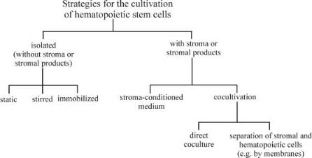
In case of stem and progenitor cells, which are the focus of this review, many strategies for cultivation and expansion have been developed, as shown in Fig. 3.
4.1
Cultivation of Isolated Stem and Progenitor Cells
Since the development of recombinant human cytokines, cultivation and significant expansion of isolated hematopoietic stem and progenitor cells is possible without a supporting feeder-layer of stromal cells. It is the simplicity of these culture systems that is their main attraction [29]. The cells are cultivated in chemically defined culture medium containing defined combinations of cytokines and without any stromal products. On the one hand this enables a better control of the culture and the investigation of the influence of single cytokines or culture conditions. On the other hand, as our understanding of stem cell regu-
122 |
T. Noll et al. |
Fig. 3. Strategies for the cultivation of hematopoietic stem and progenitor cells
lation is still incomplete and it is hypothesized that stromal influences regulate in vivo hematopoiesis, it is likely that the cultivation of isolated hematopoietic cells results in suboptimal expansion. This is supported by our own results, which show significantly better expansion, especially of early progenitors, in the presence of stromal cells [61].
Three different approaches for the cultivation of isolated hematopoietic cells have been described, the static, the stirred and the immobilized culture. Static cultivation takes place in very simple culture systems like well plates, tissue-cul- ture flasks or gas-permeable culture bags [62, 63]. As the first two systems do not allow cell cultivation on a clinical scale, the latter is actually the most often used technique for stem cell expansion. All these systems have the advantage of being easy to handle, single-use devices, which enable an uncomplicated cell harvest. But all of them do not offer possibilities for process control or continuous feeding. This causes variations in culture conditions during fermentation (e.g., oxygen tension, pH, substrate, metabolite and cytokine concentrations).
Stirred bioreactors are common in animal cell culture, as they offer a homogenous environment, representative sampling, better access to process control and an increased oxygen transfer. Several of these techniques (spinner flasks and stirred vessel bioreactors) have been tested successfully for the cultivation of hematopoietic cells [58, 64–67].
The immobilization of stem and progenitor cells is an attempt to reach local high cell densities and to imitate the three-dimensional structure of the bone marrow without the use of stroma. A number of porous microcarriers with and without additional coating of components of an extracellular matrix (e.g., collagen, fibronectin, laminin) have been investigated for this purpose. Bagley et al. compared different porous materials and described a greater than sixfold expansion of CFC in a long-term cultivation of CD34+ cells in tantalum-coated porous carriers, even without adding exogenous cytokines [68]. However, stem cell immobilization, especially in porous materials, requires a technique for
Cultivation of Hematopoietic Stem and Progenitor Cells: Biochemical Engineering Aspects |
123 |
detaching the cells from the matrix prior to transplantation, a significant disadvantage compared to suspension culture.
Hollow fiber modules and the microencapsulation of progenitor cells have been used in hematopoietic culture with less success [69, 70]. Furthermore, these approaches do not fit the clinical requirements, as the harvest of the cells is almost impossible.
The most sophisticated technique for stem cell expansion is the AastromReplicell system (Aastrom Biosciences Inc., Ann Arbor, MI, USA), which is an automated clinical system for the onsite expansion of stem cells in cancer therapy. It consists of a grooved perfusion chamber for the retention of the hematopoietic cells, with the medium flow perpendicular to the channel grooves resulting in a continuous supply of fresh nutrients while metabolites are simultaneously removed [47, 71, 72]. This technique has already been used in a number of clinical studies [73, 74]. No incompatibility of the expanded cells was found, but the expansion of the early progenitor cells was rather low [75].
Local high cell densities, as they are realized in the pores of microcarriers or in the grooves of the AastromReplicell, have been described to make bone marrow MNC essentially stroma-independent in terms of LTC-IC maintenance and expansion [44]. This might also be the reason for the good expansion of progenitors in the culture bags, where the cells accumulate in the wrinkles of the bag and reach local high cell densities.
4.2
Cultivation with Stromal Cells or Stroma-Derived Factors
Before the discovery and development of recombinant human cytokines, a feeder layer of stromal cells was essential for the cultivation of hematopoietic cells. The first system for the successful expansion of hematopoietic cells from murine origin was described by Dexter more than 20 years ago [76, 77]. Some years later this principle was transferred to human cells [78]. Stromal culture of hematopoietic cells is a generic term that covers a variety of cultivation concepts, as indicated in Fig. 3.
The most simple strategy is the application of exogenous stromal-condition- ed medium [79, 80]. Stromal cells secrete numerous different growth factors necessary for the maintenance and expansion of stem and progenitor cells and many of these substances are still unknown. Some attempts have already been made to characterize them [81] and, in the future, significant progress can be expected using techniques developed for proteomics analysis. The use of conditioned medium enables the application of all bioreactor systems developed for the cultivation of isolated stem and progenitor cells and allows the culture to be supplied with (unidentified) growth factors secreted from the stroma. The conditioned medium can be produced in large amounts exogenously and stored until application.
A variation of this approach is the in situ generation of stromal-conditioned medium in a stroma non-contact co-culture (e.g., seperated by microporous membranes). This permits an interaction between stromal and hematopoietic cells mediated by secreted molecules, which has been described to support the

124 |
T. Noll et al. |
expansion of the early progenitor cells more efficiently than the use of exogenous conditioned medium [82]. Disadvantageous in this technique is the increase in the technical requirements for the set-up and process control.
One important element in the in vivo regulation of hematopoiesis is missing in the approaches using conditioned medium: the cell-cell contact between hematopoietic and stromal cells. This mimicry of the bone marrow has been reproduced in several types of bioreactors providing direct coculture of progenitors on a twoor three-dimensional stromal feeder layer. Tissue-culture flasks were used as well as microcarriers (in spinner flasks, airlift or fixed-bed bioreactors), nonwoven fabrics disks, large pore-size cubes or nylon screens [50, 61, 70, 76, 83–85]. In many of these cultivations expansion of the early progenitors is superior to that obtained in stroma-free culture. In our own study we reached a 100-fold expansion of MNC and CFC and a more than 6-fold expansion of CAFC in a fixed-bed bioreactor within 14d [61]. Köhler et al. described a trend toward decreased apoptosis in culture with bone marrow stroma indicating the supportive character of the feeder layer [86].
For stromal culture, besides primary stroma from bone marrow, a variety of stromal cell lines are available. However, most are from murine origin, and human cell lines that support hematopoiesis are not efficiently available (Table 2). The human cell lines L87/4 and L88/5 are difficult to irradiate and favor differentiation and maturation over the expansion of early progenitors [87, 88].
The main attraction of stromal culture of hematopoietic cells is their superior ability for stem cell expansion especially in direct co-culture. However, despite this, a clinical use has not been realized until now. The major drawback is the origin of the stromal support. While autologous stroma would be feasible, in many cases this is not realizable. Furthermore, the use of cell lines (although irradiated to prevent further proliferation) is problematic as all stromal cells have to be removed completely prior to transplantation, a demand which is difficult to fulfill. In the case of murine cell lines a transfer of residue cells into a patient would be a xenotransplantation, which is faced with extensive regulatory hurdles.
Recently, a new approach has been reported that may offer co-culture a chance to be clinical applicable. Kawada et al. described a co-cultivation of stromal and hematopoietic cells separated by a porous membrane that enables a di-
Table 2. Stromal cell lines used in the cultivation of hematopoietic stem and progenitor cells
Cell line |
Origin |
Application |
References |
|
|
|
|
AC6.21 |
Murine |
Co-culture and SCM |
40, 89 |
FBMD-1 |
Murine |
Co-culture and SCM |
79, 90, 91 |
HESS-5 |
Murine |
Co-culture |
92–94 |
L87/4, L88/5 |
Human |
Co-culture and SCM |
79, 87, 95 |
MS-5 |
Murine |
Co-culture and SCM |
96–99 |
M2-10B4 |
Murine |
Co-culture |
50, 87, 100, 101 |
M2-10B4mod |
Murine |
Co-culture |
61, 102 |
Sl/Sl |
Murine |
Co-culture |
61, 102 |
|
|
|
|
Cultivation of Hematopoietic Stem and Progenitor Cells: Biochemical Engineering Aspects |
125 |
rect contact between the cells by their extracellular matrix, but prevents the stromal cells from entering the hematopoietic compartment [94]. This work was carried out on small scale in a transwell, but a scale-up to a clinical level seems possible.
5
Conclusions and Outlook
The ex vivo expansion of hematopoietic cells is a rapidly growing area of tissue engineering with many potential applications in medicine. During the last few years a variety of bioreactor concepts and cultivation strategies have been developed, but no final decision has been made about the optimal system for hematopoietic culture.
The cultivation of isolated stem and progenitor cells has some distinct advantages like its simplicity, clinical applicability and the possibility to use standard cell culture techniques, but the expansion of the early progenitor cells shown in different clinical studies was rather low. The use of stromal-condi- tioned medium promises better expansion and lower costs due to the reduced need for exogenous cytokine supplementation, but the culture medium is chemically undefined, which will render reproducibility more difficult and complicate the clinical permission. Co-cultivation of stromal and hematopoietic cells results in the best expansion of early progenitor cells, but, as nearly all stromal cell lines are of murine origin, a clinical application of this approach is hardly possible. Membrane-separated co-cultivation may offer a possibility to avoid this disadvantage.
What can be expected from the future? The cultivation of isolated hematopoietic cells will benefit from the identification of new cytokines, making the use of stromal-conditioned medium unnecessary. The recent development of proteomics techniques will expediate this process. The prospects of the co-cul- tivation will depend on the development of human stromal cell lines supporting the expansion of early progenitor cells, and the membrane approach has to prove its potential on a clinical scale.
In summary, for the near future it can be expected that culture systems of sufficient simplicity, productivity and reliablity will be developed and tested in clinical studies, making transplantation therapies utilizing cultured hematopoietic cells a more common application in medicine.
Acknowledgement. We thank Dr. Joy Burchell for careful revision of the manuscript.
References
1.McAdams TA, Miller WM, Papoutsakis ET (1996) TIBTECH 14:341
2.Wilkins BS (1992) J Clin Pathol 45:645
3.Verfaillie C, Hurley R, Bhatia R, McCarthy JB (1994) Crit Rev Oncol/Hematol 16:201
4.Mayani H, Guilbert LJ, Janowska-Wieczorek A (1992) Eur J Hematol 49:225
5.Deryugina EI, Muller-Sieburg CE (1993) Crit Rev Immunol 13:115
6.Koller MR, Palsson BO (1993) Biotechnol Bioeng 42:909
126 |
T. Noll et al. |
7.Sutherland HJ, Lansdorp PM, Henkelman DH, Eaves AC, Eaves CJ (1990) Proc Natl Acad Sci 87:3584
8.Breems DA, Blokland EAW, Neben S, Ploemacher RE (1994) Leukemia 8:1095
9.Ploemacher RE (1997) Baillière’s Clinical Hematology 10:429
10.Ramsfjell V, Bryder D, Björgvinsdottir H, Kornfält S, Nilsson L, Borge OJ, Jacobsen SEW (1999) Blood 94:4093
11.Lapidot T, Pflumio F, Doedens M, Murdoch B, Williams DE, (1992) Science 255:1137
12.Shimizu Y, Ogawa M, Kobyashi M, Almeida-Porada G, Zanjani ED (1998) Blood 91:3688
13.Brandt JE, Bartholomew AM, Fortman JD, Nelson MC, Brumo E, Chen LM, Turian FV, Davis TA, Chute JP, Hoffman R (1999) Blood 94:106
14.Krause DS, Fackler MJ, Civin CI, Stratford W (1996) Blood 87:1
15.Bhatia M, Bonnet D, Murdoch B, Gan OI, Dick JE (1998) Nature Med 4:1038
16.Zanjani ED, Almeida-Porada G, Livingston AG, Flake AW, Ogawa M (1998) Exp Hematol 26:353
17.Goodell MA (1999) Blood 94:2545
18.Sato T, Laver JH, Ogawa M (1999) Blood 94:2548
19.Yin AH, Miraglia S, Zanjani ED, Almeida-Porada G, Ogawa M, Leary AG, Olweus J, Kearney J, Buck DW (1997) Blood 90:5002
20.Ziegler BL,Valitieri M,Almeida-Porada G, De Maria R, Muller R, Masella B, Gabbianekki M, Casella I, Pelosi E, Bock T, Zanjani ED, Peschle C (1999) Science 285:1553
21.Kawabata K, Ujikawa M, Egawa T, Kawamoto H, Tachibana K, Iizasa H, Katsura Y, Kishimoto T, Nagasawa T (1999) Proc Natl Acad Sci 96:5663
22.Dorrell C, Gan OI, Pereira DS, Hawley RG, Dick JE (2000) Blood 95:102
23.Rowlings PA (1999) In: Schindhelm K, Norton R (eds) Ex vivo cell therapy. Academic Press, San Diego, p 85
24.McAdams TA, Winter JN, Miller WM, Papoutsakis ET (1996) TIBTECH 14:388
25.Kim DK, Fujiki Y, Fukushima T, Ema H, Shibuya A, Nakauchi H (1999) Stem Cells 17:286
26.Cairo MS, Wagner JE (1997) Blood 90:4665
27.Schindhelm K, Nordon R (1999) (eds) Ex vivo cell therapy. Academic Press, San Diego
28.Collins PC, Papoutsakis ET, Miller WM (1996) Curr Opin Biotechnol 7:223
29.Alcorn MJ, Holyoake TL (1996) Blood Rev 10:167
30.Bremers AJA, Parmiani G (2000) Crit Rev Oncol/Hematol 34:1
31.Linenberger ML, Jacobsen FW, Bebbett LG, Broudy VC, Martin FH, Abkowitz JL (1995) Exp Hematol 23:1104
32.Lisovsky M, Braun SE, Ge Y, Takahari H, Lu L, Savchenko VG, Lyman SD, Broxmeyer HE (1996) Leukemia 10:1012
33.Guerriero A, Worford L, Holland HK, Guo GR, Sheehan K, Walle EK (1997) Blood 90: 3444
34.Cottner TG, Fernandes RS, Verhaegen S, McCarthy JV (1994) Immunol Rev 142:93
35.Piacibello W, Sanavio F, Garetto L, Severino A, Bergandi D, Ferrario J, Fagioli F, Berger M, Aglietta M (1997) Blood 89:2644
36.Murray LJ, Young JC, Osborne LJ, Luens KM, Scollary R, Hill BL (1999) Exp Hematol 27:1019
37.Ramsfjell V, Bryder D, Björgvinsdottir H, Kornfält S, Nilsson L, Borge OJ, Jacobsen SEW (1999) Blood 94:4093
38.Möhle R, Bautz F, Rafii S, Moore MAS, Brugger W, Kanz L (1998) Blood 91:4523
39.Eichmann A, Corbel C, Nataf V, Vaigot P, Breánt C, LeDouarin N (1997) Proc Natl Acad Sci 94:5141
40.Shih CC, Hu MC, Wenig Y, Yazaki PJ, Medeiros J, Forman SJ (2000) Blood 95:1957
41.Thoma S, Schmidt S, Jelinek N, Herfurth C, Takors R, Wandrey C, Biselli M (1999) Onkologie 22 (suppl 1):180
42.Dybedal I, Jacobsen SE (1995) Blood 86:949
43.Sandstrom CE, Miller WM, Papoutsakis ET (1994) Biotechnol Bioeng 43:706
44.Koller MR, Manchel I, Palsson MA, Maher RJ, Palsson BO (1996) Biotechnol Bioeng 50:505
Cultivation of Hematopoietic Stem and Progenitor Cells: Biochemical Engineering Aspects |
127 |
45.Schwartz RM, Palsson BO, Emerson SG (1991) Proc Natl Acad Sci 88:6760
46.Sandstrom CE (1995) PhD thesis, Northwestern University, Evanston
47.Sandstrom CE, Bender JE, Miller WM, Papoutsakis ET (1996) Biotechnol Bioeng 50: 493
48.Wang TY, Brennan JK, Wu JHD (1995) Exp Hematol 23:26
49.Palsson BO, Paek SH, Schwartz RM, Palsson MA, Lee GM, Silver S, Emerson SG (1993) Bio/Technology 11:368
50.Meissner P, Schroeder B, Herfurth C, Biselli M (1999) Cytotechnology 30:227
51.Patel SD, Papoutsakis ET, Winter JN, Miller WM (2000) Biotechnol Prog 16:885
52.Ishikawa Y, Ito T (1988) Eur J Hematol 40:126
53.Rich IN, Kubanek B (1986) Br J Hematol 52:579
54.Broxmeyer HE, Cooper S, Lu L, Miller ME, Langefeld CD, Ralph P (1990) Blood 76:323
55.Hehevan DL, Papoutsakis ET, Miller WM (2000) Exp Hematol 28:267
56.McAdams TA, Papoutsakis ET, Miller WM (1995) Blood (suppl. 1) 86:674a
57.McAdams TA (1997) PhD thesis, Northwestern University, Evanston
58.Schmidt S (2000) PhD thesis, University RWTH, Aachen
59.LaIuppa JA, McAdams TA, Miller WM, Papoutsakis ET (1995) Blood (Suppl. 1) 86:231a
60.LaIuppa JA, McAdams TA, Papoutsakis ET, Miller WM (1997) Biomed Mat Res 36:347
61.Jelinek N (2000) PhD Thesis University RWTH Aachen
62.Brugger W, Heimfeld S, Berenson RJ, Mertelsmann R, Kanz L (1995) N Eng J Med 333:283
63.Alcorn MJ, Holyoake TL, Richmond L (1996) J Clin Oncol 14:1839
64.Collins PC, Miller WM, Papoutsakis ET (1998) Biotechnol Bioeng 59:534
65.Kim BS (1998) Biotechnol Lett 20:595
66.Zandstra PW, Petzer AL, Eaves CJ, Piret JM (1997) Biotechnol Bioeng 54:58
67.De León A, Mayani H, Ramirez OT (1998) Cytotechnology 28:127
68.Bagley J, Rosenzweig M, Marks DF, Pykett MJ (1999) Exp Hematol 27:496
69.Levee MG, Lee GM, Paek SH, Palsson BO (1994) Biotechnol Bioeng 43:734
70.Sardonini CA, Wu YJ (1993) Biotechnol Prog 9:131
71.Sandstrom CE, Bender JG, Papoutsakis ET, Miller WM (1995) Blood 86:958
72.Koller MR, Maher RJ, Manchel I, Oxender M, Smith AK (1998) J Hematotherapy 7:413
73.Bachier CR, Gokmen E, Teale J, Lanzkron S, Childs C, Franklin W, Shpall E, Douville J, Weber S, Muller T, Armstrong D, Lemaistre CF (1999) Exp Hematol 27:615
74.Chabannon C, Novakovitch G, Blache JL, Olivero S, Camerlo J, Genre D, Maraninchi D, Viens P (1999) Hematol Cell Ther 41:78
75.Chabannon C, Blache JL, Sielleur I, Douville J, Faucher C, Gravis G, Arnoulet C, OzielTaieb S, Blaise D, Novakovitch G, Camerlo J, Chabbert I, Genre D,Appel M,Armstrong D, Maraninchi D, Viens P (1999) Int J Oncol 15:511
76.Dexter TM, Wright EG, Krizsa F, Lajtha LG (1977) Biomedicine 27:344
77.Dexter TM, Allen TD, Laijtha LG (1977) J Cell Physiol 91:335
78.Gartner S, Kaplan HS (1980) Proc Natl Acad Sci 77:4756
79.Breems DA, Blokland EAW, Ploemacher RE (1997) Leukemia 11:142
80.Bhatia R, McGlave PB, Lin W, Wissink S, Miller JS, Verfaillie CM (1995) Blood (Suppl. 1) 86:294a
81.Gupta P, McCarthy JB, Verfaillie CM (1996) Blood 87:3229
82.Verfaillie CM, Catanzarro PM, Li W (1994) J Exp Med 179:643
83.Highfill JG, Haley SD, Kompala DS (1995) Biotechnol Bioeng 50:514
84.Naughton BA, Naughton GK (1991) J Biomech Eng 113:171
85.Tomimori Y, Tagaki M, Yoshida T (2000) Cytotechnology 34:121
86.Köhler T, Plettig R, Wetzstein W, Schaffer B, Ordemann R, Nagels HO, Ehninger G, Bornhauser M (1999) Stem Cells 17:19
87.Schroder B (1997) PhD thesis, University of Bonn
88.Bertolini F, Battaglia M, Soligo D, Corsini C, Curioni C, Lazzari L, Pedrazzoli P, Thalmeier K (1997) Exp Hematol 25:350
89.Shih CC, Hu MCT, Hu J, Medeiros J, Forman SJ (1999) Blood 94:1623
128 |
T. Noll et al. |
90.Breems DA, Van Driel EM, Hawley RG, Siebel KE, Ploemacher RE (1998) Leukemia 12:951
91.Breems DA, Blokland EAW, Siebel KE, Mayen AEM, Engels LJA, Ploemacher RE (1998) Blood 91:111
92.Tsuji T, Nishimura-Morita Y, Watanbe Y, Hirano D, Nakanishi S, Mori KJ, Yatsunami K (1999) Growth Factors 16:225
93.Nakamura Y, Ando K, Chargui J, Kawada H, Sato T, Tsuji T, Hotta T, Kato S (1999) Blood 94:4053
94.Kawada H, Ando K, Tsuji T, Shimakura Y, Nakamura Y, Chargui J, Hagihara M, Itagaki H, Shimizu T, Nokuchi S, Kato S, Hotta T (1999) Exp Hematol 27:904
95.Thalmeier K, Meissner P, Reisbach G, Falk M, Brechtel A, Dormer P (1994) Blood 83:1799
96.Bennaceur-Griscelli A, Tourino C, Izac B, Vainchenker W, Coulombel L (1999) Blood 94:529
97.Tordjman R, Ortega N, Coulombel L, Plouet J, Romeo PJ, Lemarchandel V (1999) Blood 94:2301
98.Nishi N, Ishikawa R, Inoue H, Bishikawa M, Yoneya T, Kakeda M, Tsumara H, Ohashi H, Mori LJ (1997) Leukemia (suppl. 3) 11:468
99.Kanai M, Ikeda H, Ikebuchi K (2000) Bone Marrow Trans 26:837
100.Lemoine FM, Humphries RK, Abraham SD, Krystal G, Eaves CJ (1988) Exp Hematol 16:708
101.Sutherland HJ, Eaves CJ, Landsdorp PM, Thaker JD, Hogge DE (1991) Blood 78:666
102.Sutherland HJ, Hogge DE, Cook D, Eaves CJ (1993) Blood 81:1465
Received: April 2001