

New, centrally acting dopaminergic agents with an improved oral bioavailability [2004]
.pdfIntroduction
expression of the dopamine D3 receptor in the recipient cells.17 Later studies have revealed functional coupling of dopamine D3 receptors to different transduction mechanisms in various cell lines. These mechanisms are: (1) inhibition of cAMP production,86 (2) aggregation of melanophore pigment,87 (3) acidification of the extracellular environment,88 (4) inhibition of Ca2+ currents,89 and (5) mitogen-activated protein kinase.90 It is interesting that the dopamine D3 receptors mimic the signalling output of dopamine D2 receptors in the same cell, but with a lower efficacy, implying a less efficient coupling of dopamine D3 receptors. The different efficacy of dopamine D2 and D3 receptor activation may be an important means for varying the information resulting from dopamine neurotransmission.86
1.4Structure-activity relationships of dopamine receptor agonists
Through the years a number of different structure-activity relationships (SARs) and receptor models have been developed (for reviews see refs. 91, 92). Therefore this introduction will only give a short overview of the general aspects of dopamine receptor agonist SARs.
The growing interest in unravelling the mechanism of action of dopamine and related compounds at the molecular level started with the considerations of the conformation(s) of the dopamine molecule when bound to the receptor site. Dopamine itself is a conformationally flexible molecule that can adopt specific conformation(s) needed to achieve appropriate three dimensional interaction with various groups located at or near the recognition (receptor) site(s).
When the conformation of the ethylamine side-chain with respect to the aromatic nucleus is considered as the starting-point, the dopamine molecule may exist either in a trans (extended) conformation, or in a cis (gauche) conformation. Single crystal x-ray analysis of dopamine hydrochloride indicates that the ethylamine chain is in a nearly completely extended conformation which resides on a plane almost perpendicular to the plane of the catechol ring.93
Cannon94,95 defined α- and β-conformers of dopamine, in which the catechol ring is coplanar with the ethylamine side-chain (chart 1.3). In the α-conformer the meta-OH group is on the edge of the ring closer to the ethylamine chain, whereas in the β-conformer the meta-OH is on the edge of the ring away from the side-chain. Concluding from several studies with dopamine analogues, which simulate the gauche conformation leading to inactive compounds, dopamine is assumed to interact with the receptor in the extended conformation.
11

Chapter 1
HO |
NH2 |
HO |
NH2 |
|
|||
NH2 |
HO |
HO |
|
HO |
OH |
|
|
|
|
|
|
1 (gauche) |
1 (α-conformer) |
|
1 (β-conformer) |
N |
HO |
N |
|
|
OH 9 |
10 |
Chart 1.3 |
Gauche conformation of |
dopamine (1) and the two rotamers of the extended |
|
|
conformation of dopamine (1 α-conformer and 1 β-conformer), and the corresponding |
||
|
2-aminotetralins S-(–)-5-OH-DPAT ( 9) and R-(+)-7-OH-DPAT (10). |
||
Studies with hydroxylated 2-aminotetralin analogues (among others using compounds 9 and 10) and phenylethylamine analogues revealed that the first possessed a higher potency.96,97
Apparently, the near co-planar arrangement is required for higher dopamine receptor agonist activity.
There are two main routes along which the development of dopamine agonists has initially proceeded, namely rigidification of the dopamine molecule (1), and dissection of one of the first known potent dopamine agonists, apomorphine (11).
|
|
|
|
|
|
|
R2 |
|
|
|
|
|
|
|
|
|
|
|
HO |
NH2 |
|
|
|
N |
||
|
HO |
|
R1 |
|
|
|
|
|
|
|
|
|
|
||||
|
|
|
|
|
OH |
|
|
|
|
1 |
|
11 R1 |
= OH, R2 = Me |
||||
|
|
|
|
12 R1 |
= H, R2 = n-Pr |
|||
|
|
|
|
|
||||
Chart 1.4 |
Chemical structures of |
dopamine |
(1), apomorphine (11) and 11-hydroxy-N-n- |
|||||
propylnoraporphine (12).
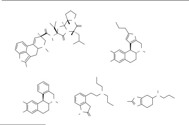
The rigidification of the dopamine molecule led to a number of dopamine receptor agonists in which the dopamine molecule, with its phenylethylamine moiety, is readily recognisable. In these dopamine agonists the amine moiety is either a part of a cyclic system, e.g. benzo[f]quinolines, naphthoxazines, and benzopyranoxazines, or an exocyclic amine, e.g. 2-aminotetralins and 2-aminoindans (Chart 1.5).
12

Introduction
|
|
|
|
7 |
|
8 |
N |
6 |
N |
|
7 |
R |
||
|
R |
|
5 |
|
|
6 |
|
|
4 |
|
5 |
|
|
|
|
2-aminotetralins |
|
2-aminoindans |
|
|
1 |
|
1 O |
1 O |
|
4 |
|
N 4 |
N 4 |
9 |
N |
9 |
||
|
|
9 |
||
R |
|
R |
|
R |
|
7 |
|
7 |
O |
|
|
7 |
||
|
benzo[f]quinolines |
|
naphthoxazines |
benzopyranoxazines |
Chart 1.5 Some approaches for the rigidification of the dopamine ethylamine side chain. R-groups
are usually -H or -OH.
SAR studies with these different classes of dopamine receptor agonists have led to the identification of a pharmacophore for activation of dopamine D2 (-like) receptors consisting of the meta-hydroxyphenylethylamine structure. 2-Aminotetralins, hydroxylated at the 5- or 7-posistion, are potent dopamine receptor agonists, but their 6- or 8-hydroxylated analogues are less potent.98 8-Hydroxy-(N,N-di-n-propylamino)tetralin (8-OH-DPAT) is a potent serotonergic agent. Also dihydroxylated compounds like 5,6-di-OH-DPAT, which possess the catechol moiety also present in dopamine, are potent dopaminergic agents.
McDermed et al.99,100 found that the interaction of dopamine receptor agonists with dopamine receptors is highly stereoselective. They observed that for compounds which are α-conformers the S-enantiomer is the most active (as is the case in S-(–)-5-OH-DPAT, 9), whereas for β-conformers the R-enantiomer is the most active (as is the case in R-(+)-7-OH- DPAT, 10). Their assumption then was that in order to achieve the proper orientation of the amino moiety and meta-OH groups, relative to the ethylamine side-chain, with respect to their presumed binding groups on the dopamine receptor surface, the β-conformer must be rotated with respect to the α-conformer (Chart 1.6). As a result of such a rotation the amine groups are pointing in the same direction.
13
Chapter 1
O
H O
N
9 and 10
Chart 1.6 Superimposition of S-(–)-5-OH-DPAT ( 9) with R-(+)-7-OH-DPAT (10).
McDermed and colleagues have also included a region in the receptor preventing the interaction with compounds having a steric bulk in this region.
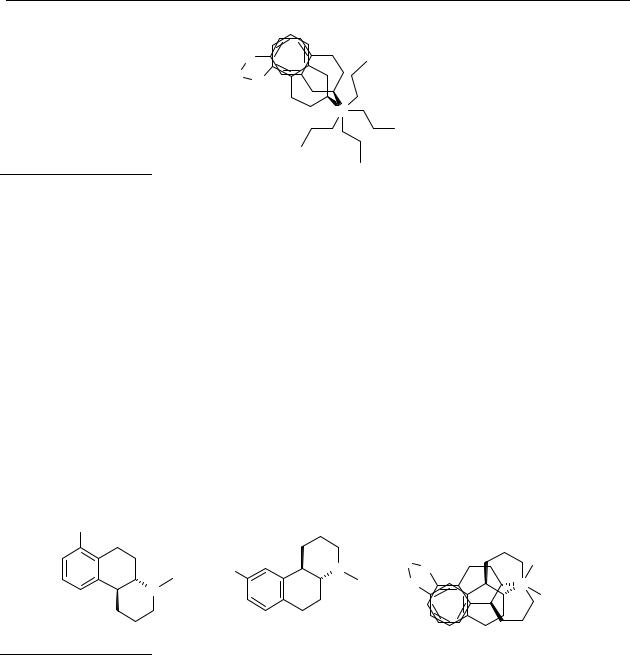
In an extension of the McDermed model Wikström and colleagues, 101,102 applied to the dopaminergic monohydroxylated 3-phenylpiperidines and the octahydrobenzo[f]quinolines, have found that there is a relationship between the absolute configuration, the ring position of the hydroxyl group and the size of the N-alkyl substituent. In their model there are now two different directions possible for the N-alkyl substituents, namely ‘upwards’ and ‘downwards’ (Chart 1.7). An assumption they made is that, due to the different directions in which the N-alkyl substituents point, the space available for the N-substituent of compound 14 might be more restricted than for the corresponding isomer 13. And indeed, they have found that the N- substituent ‘downwards’ is sterically restricted to maximally an n-propyl group, whereas the ‘upwards’ direction has less restricted demands.
HO |
|
|
|
|
|
HO |
N |
H O |
Rup |
|
Rup |
|
Rdown O |
N |
|
N |
|
||
|
|
|
Rdown |
|
|
|
|
|
|
|
13 |
14 |
|
|
Chart 1.7 |
Wikström’s modification of |
McDermed’s |
model. The structures |
of 7- ( 13) and 9- |
hydroxyoctahydrobenzo[f]quinolines (14) superimposed.
The distance from the amine-nitrogen to the hydroxyl-oxygen (meta or para) has been studied in several classes of dopamine agonists. The distances which were found vary considerably between these studies, which made clear that an optimal distance is hard to define. The nitrogen atom to meta-hydroxyl distance varied from 5.5 to 7.3 Å, the nitrogen to parahydroxyl varied from 7.0-7.8 Å. 91 Therefore, it can be concluded that any distance between the nitrogen and the meta-hydroxyl that is less than that of dopamine in its fully extended form may be acceptable to a minimum of 5.5 Å.
14

Introduction
Although some studies suggest the opposite,103 it is generally believed that the nitrogen atom of a dopamine agonist interacts with the receptor in the protonated (ammonium) form. This idea is supported by studies with permanently charged dopamine analogues.104
Furthermore, at physiological pH, dopamine exists mainly in the protonated form.105
In general, increasing the lipophilicity brings about increased potency for dopamine agonists. However, this is only true for compounds within the same series. When comparing compounds from different structural classes, other factors like conformation and stereochemistry are of primary importance for dopamine agonist activity. The oft-quoted rule106 that an octanol-water partition coefficient, logPoct, of approximately 2 log units is optimum for ready entry into the brain is derived from studies on biological activity and not from rates of permeation nor from equilibria. Gratton et al.,107 however, found that even if there is a parabolic relationship between the logarithm of a permeability surface area (logPS) and logPoct, this relationship is not good enough to use predictively.
The development of SARs for dopamine agonists was accompanied by the development of dopamine receptor models with computer programs. Basically, two kinds of dopamine receptor models emerged: indirect models and direct models. Indirect models take a series of (analogous) agents (agonists or antagonists) with certain receptor-binding characteristics as a starting point, and describe the receptor binding site as a collection of ‘areas’ with certain properties. 102,108-112
Later, the amino acid sequence and structure of the various dopamine receptor proteins became known with molecular biological techniques, which enabled the development of direct models.113-115 These models take the receptor protein as a starting point, and describe the receptor binding site in terms of amino acids with different physical and sterical properties. Despite the development of these models for the different dopamine D2-like receptors, it is still difficult to design selective ligands, because there is a high similarity in the transmembrane segments of the dopamine D2 like receptors and the crystal structure of the receptor is not known yet. Since the models developed are all based on known compounds, their predictability declines when new, not included, compounds are tested in this model.
1.5Pathogenesis of the dopaminergic system
Both movement and psychotic disorders in humans have been associated with disturbances in the functioning of the dopaminergic system. Examples of the motor disturbances are Parkinson’s disease, Huntington’s disease and Gilles de la Tourette’s syndrome. These movement diseases can be categorised into either hypoor hyperkinetic disorders. In this paragraph Parkinson’s disease and schizophrenia with their current treatment will be discussed. Also drug abuse and its possible treatment will be discussed, since selective dopamine D3 receptor agonists may be active as anti-addictive drugs.116
15

Chapter 1
1.5.1Parkinson’s disease
1.5.1.1 Pathology of Parkinson’s disease
Parkinson’s disease is a progressive neurodegenerative disorder of the basal ganglia, which most often becomes apparent after the age of 55. It is a prototypic hypokinetic disorder, with tremor, rigidity, bradykinesia, and akinesia as the most prominent features.117 Depression and a general slowing down of intellectual processes also occur, but are less well-defined. The neurological and psychiatric symptoms usually worsen with time (for review: ref. 118). The neuropathology of Parkinson’s disease reveals a striking loss of the dopaminergic neurons of the nigrostriatal pathway terminating preferentially in the caudate nucleus and putamen.119,120
Although the nigrostriatal system has a large reserve (only after a decline of approx. 70-80% of these cells, Parkinson’s disease symptoms arise), progressive loss of these systems ultimately leads to Parkinson’s disease, with its well-known movement disorders (‘shaking palsy’). In contrast with some other neurodegenerative diseases (e.g. Huntington’s disease), for Parkinson’s disease no genetic component has been discovered yet. Usually the disease is not diagnosed before the first symptoms appear.
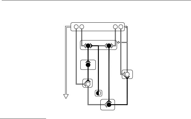
The control of normal motor behaviour is under influence of the basal ganglia through a so-called reinforcing basal ganglia-thalamocortical ‘motor circuit’. Based on current data from a variety of experimental fields, a functional model of this ‘motor circuit’ has been proposed, as depicted, greatly simplified, in Figure 1.2.
First, there is a pathway called the ‘direct’ pathway consisting of neurones containing GABA and substance P. Activation of this pathway tends to disinhibit the thalamic stage of the circuit. The second pathway is the ‘indirect’ pathway, which passes first to the external segment of the globus pallidus (GPe) via striatal projection neurones that contain both GABA and enkephalin, then from GPe to the subthalamic nucleus (STN) via a purely GABAergic pathway, and finally to the output nuclei via an excitatory, probably glutaminergic, projection from the STN.121 It should be apparent that dopamine D1 receptors are primarily present on the medium spiny neurones, forming the ‘direct’ pathway, whereas dopamine D 2 receptors are primarily present on the medium spiny neurones at the start of the ‘indirect’ pathway. 122
16
Introduction
Cerebral Cortex
(glu) |
|
(glu) |
|
Striatum |
|
D2 |
A2A |
D1 |
(GABA enk) |
(GABA subst P) |
|
|
|
(DA) |
GPe |
|
|
(GABA) |
|
|
|
|
Thal |
STN |
|
SNc |
(glu) |
|
|
Brainstem |
(GABA) |
Spinal cord
GPi/SNr
Figure 1.2 Schematic diagram of the circuitry and neurotransmitters of the basal gangliathalamocortical circuitry, indicating the parallel ‘direct’ and ‘indirect’ pathways from the striatum to the basal ganglia output nuclei in a normal situation. Inhibitory neurons are shown as filled symbols, excitatory neurons as open symbols. Abbreviations: DA, dopamine; D1, dopamine D1 receptor; D2, dopamine D2 receptor; A2A, adenosine A2A receptor; enk, enkephalin; GABA, γ-aminobutyric acid; GPe, external segment of globus pallidus; GPi, internal segment of globus pallidus; glu, glutamate; PPN, pedunculopontine nucleus; SNc, substantia nigra pars compacta; SNr, substantia nigra pars reticulata; subst P, substance P; STN, subthalamic nucleus; Thal, thalamus. Adapted from references 121-123.
Considerable evidence indicates that shifts in the balance between the activity in the ‘direct’ and ‘indirect’ striatal output pathways and the resulting alterations in the output of the SNr and the GPi may account for the hypoand hyperkinetic features of basal ganglia movement disorders. In summary, excessive inhibition of GPe within the indirect pathway leads to disinhibition of the STN, which in turn provides excessive excitatory drive to basal ganglia output nuclei (GPi/SNr), thus leading to excessive thalamic inhibition. This is reinforced by reduced inhibitory input to GPi/SNr through the direct pathway. Overall these effects are postulated to results in a reduction in the usual reinforcing influence of the motor circuit upon cortically initiated movements.123 (For more reviews and references see ref. 117, 124-126).
Based on the altered neuronal activity in the ‘motor’ circuit in Parkinson’s disease, it seems that parkinsonism symptoms can be treated by restoring the right balance between the activity in the ‘direct’ and ‘indirect’ pathways.
17

Chapter 1
Although the exact cause of the progressive degeneration of nigrostriatal dopamine neurons in Parkinson’s disease is still unknown, several mechanisms were proposed to explain the cell damaging processes at the molecular level that ultimately cause Parkinson’s disease.127
These mechanisms, which are probably related by interacting mechanisms, include:
(1) excitotoxic mechanism,128 (2) mitochondrial toxins,129 and (3) oxidative stress.130
There is considerable body of (indirect) evidence which makes oxidative stress one of the best accepted hypothesis for explaining the cause of Parkinson’s disease. For example, the Fe(II)/Fe(III) ratio in the substantia nigra is shifted from 2:1 in the normal brain to 1:2 in Parkinsonian brain.131,132 In the Parkinsonian brain several enzymes which constitute the antioxidative defence mechanisms (glutathione peroxidase, catalase) have a decreased activity, while the activity of superoxide dismutase is increased, relative to the normal brain.133
Furthermore, specific products of radical damage, such as lipid hydroperoxides, were detected at a 10-fold increased level in the Parkinsonian brain.134
1.5.1.2 Current treatment of Parkinson’s disease
As Parkinson’s disease is associated with a loss of dopamine, it is commonly treated with drugs, which replace or supply dopamine. Since dopamine itself cannot pass the blood-brain barrier, the most commonly used therapy is levodopa (L-DOPA), a precursor of dopamine. A complication of long-term treatment with L-DOPA, however, is the development of rapid fluctuations in clinical state where the patient switches suddenly between mobility and immobility; this phenomenon is known as the ‘on-off effect’. 135,136 This effect might be caused by the loss of feed-back mechanisms.
An alternative approach to the treatment with L-DOPA is the use of drugs that mimic the action of dopamine. Treatment with dopamine-agonists, such as bromocriptine (15),137 pergolide,138,139 and lisuride,138,140 has some advantages over treatment with L-DOPA. Dopamine agonists are effective in patients in the advanced stages of Parkinson’s disease, unlike L-DOPA, because their action at postsynaptic dopamine receptors is unaffected by the lack of dopamine producing nerve cells.
Furthermore, there is an increasing interest in the direct effect of dopamine agonists to be potential neuro-protective agents. A number of studies in biologic and non-biologic systems have shown that dopamine agonists have antioxidant effects and can trap a variety of radical species.141-143 Theoretically, such a protective effect might result from (a) a decrease in L-DOPA application, as L-DOPA itself may cause oxidative stress,144 (b) stimulation of dopamine autoreceptors resulting in decreased dopamine synthesis, release, and turnover, as dopamine metabolism leads to reactive oxygen species,145 and (c) direct anti-oxidant effects.141-143
18
Introduction
|
|
OH |
N |
|
O |
O |
H |
O |
|
N |
|
|
||
H |
|
|
S |
|
N |
|
H |
|
|
|
|
|
||
H |
|
O |
|
|
N |
|
|
AcO |
N |
|
|
|
||
|
|
|
|
H |
HN |
|
|
AcO |
|
15 |
|
|
|
|
|
|
|
|
|
Br |
|
|
|
16 |
|
|
|
N |
|
H |
|
|
|
S |
N |
|
|
|
|
|
||
|
|
|
|
|
|
HO |
N |
H |
|
H2N |
|
|
|
|
N |
|
|
|
|
|
|
|
|
HO |
|
|
N |
|
|
|
H |
|
|
|
|
|
17 |
O 18 |
|
19 |
|
|
|
|
Chart 1.8 Chemical structures of bromocriptine (15), ABT-431 (16), dihydrexidine (17), ropinirole
(18), and pramipexole (19).
Drug therapies or therapies which are in clinical development,146-149 nowadays, include treatment with (prodrugs of) dopamine receptor agonists that are either selective for the dopamine D1 receptor, such as ABT-431150 (16) and dihydrexidine151,152 (17), or are selective for the dopamine D2/D3 receptor subtypes, such as ropinirole153 (18) and pramipexole154,155 (19). An ideal antiparkinsonian treatment may require stimulation of both D1 and D2 dopamine receptors.156 Literature shows that antiparkinsonian effects can be exerted either by a dopamine D1 receptor agonist alone or by a dopamine D2 receptor agonist alone, whereas hyperactivity and aggressiveness manifested by dopamine receptor agonists require co-activation of the D1 and D2 receptors. The antiparkinsonian effect can be dissociated from the adverse effect by therapeutic strategy. It is implied that imbalances in activation of the D1 and D2 receptors may provide a favourable approach for long-term treatment of parkinsonian patients with dopamine drugs.157,158 Potent dopamine D1 receptor agonists with an intermediate half-life may prove to be better adjuncts in the treatment of Parkinson’s disease, because stimulation of the dopamine D 1 receptor may provide a better integration of neural inputs to the internal segment of the globus pallidus (referred to as the basal ganglia output) compared with L-DOPA and selective dopamine D2 receptor agonist.159,160 A drug with affinity for both the dopamine D1 and D2 receptors is apomorphine, which can be a good tool in restoring the imbalances in activation of the D1 and D2 receptors in Parkinson’s disease.
19

Chapter 1
Dopamine receptor agonists used in the therapy against Parkinson’s disease often possess phenolic or catecholic moieties, which lead to low oral bioavailabilities for these compounds since they undergo considerable metabolic degradation in the liver.161 Because of the low oral bioavailability of hydroxylated compounds, there has been a lot of interest in the development of prodrugs of such compounds, thereby circumventing the metabolic degradation.
Other approaches, some of which are still in clinical development, include restoration of the acetylcholine-dopamine balance in the basal ganglia, neuronal nicotinic receptor agonists, neurotrophic immunophilins, dopamine transport inhibitors, COMT-inhibitors, and adenosine A2A receptor antagonists.162,163 Also surgical therapies are used or under development, including stereotactic thalamotomy, continuous electric thalamus stimulation,164 posteroventral pallidotomy and transplantation of embryonal substantia nigra cells.
1.5.2Schizophrenia
1.5.2.1 Pathology of schizophrenia
Schizophrenia is a psychotic disorder of unknown aetiology in which patients suffer from a cluster of symptoms which may include both positive (delusions, hallucinations, disordered thoughts, and disorganised speech) and negative (flat effect, anhedonia, social withdrawal, emotional detachment, cognitive deficits, and poverty of speech) symptoms. This disease, which is relatively common (lifetime prevalence rate ~1 %), usually strikes its victims during adolescence or early adulthood. Since occupational and social function is severely affected, often leading to institutionalisation, the cost to society is very high.165 Unfortunately, it is very difficult to diagnose schizophrenia and many different diagnostic systems have so far been developed. Moreover, there are strong indications that schizophrenia is not a homogeneous disorder, but rather consists of subgroups.166
Although there has been extensive research during several decades, still no definite explanation for the development of schizophrenia in man can be given. There is still a controversy between the very diverse theories to explain the development of the disease. It is generally accepted that genetic predisposition plays a significant etiological role in schizophrenia.167 Although there has been a lot of research for finding genes for schizophrenia this search has not been successful yet. Crocq et al.168 found an association between schizophrenia and homozygosity at the dopamine D3 receptor gene. However, Yang et al.169 found that this association was not present in schizophrenics. These two studies show that more research is necessary to find genes. Other etiological factors in schizophrenia are infection and autoimmunity,170 and obstetric complications.171 Also theories involving neurochemical alterations are related to the pathogenesis of schizophrenia. From this point of view the dopamine hypothesis has provided a framework for understanding the disease and proposing approaches to the treatment of schizophrenia. This hypothesis suggests that schizophrenia
20