

New, centrally acting dopaminergic agents with an improved oral bioavailability [2004]
.pdfThiophene analogues of naphthoxazines and 2-aminotetralins
4.4Discussion
In the present study we investigated the effects of the bioisosteric replacement of a phenol moiety by a thiophene moiety. The effects of the compounds on dopamine release were determined using the microdialysis technique in freely moving rats. Systemic administration of compounds 34, 35 and 39 but not 38 induced a decrease in the release of dopamine in the striatum, which results from the dopamine receptor agonistic properties of the compounds, as the release of dopamine is under the control of dopamine autoreceptors.189 Compound 39 was less effective compared to compounds 34 and 35 in decreasing dopamine release in the striatum and compound 38 was without effect, which is in line with the differences of the four compounds in binding affinities found for the dopamine D2 and D3 receptor. All compounds were less potent than 5-OH-DPAT, again in agreement with the higher affinity at the dopamine D2 and D3 receptors of the latter compound. The role of the dopamine D3 receptor as an autoreceptor is still under debate,61,64 while it is generally accepted that the dopamine D2 receptor functions as an autoreceptor.63
The affinity of compounds 34 and 35 for the dopamine D2 and D3 receptors is lower than that of 5-OH-DPAT, probably due to the fact that the sulfur atom in the thiophene ring is only a weak hydrogen bond acceptor unlike the hydroxyl moiety of a phenol, which is a strong hydrogen bond acceptor and donor. The fact that compound 39 was more effective compared to compound 38 is most likely due to a better fit into the receptor of the n-propyl substituent of compound 39 than the hydrogen of compound 38.
For a compound to display dopamine receptor activity the distance between the nitrogen atom and the H-bond forming group is of importance. Previous studies indicated that the distance between the nitrogen and the hydroxyl moiety in dopamine receptor agents should be between 5.5 and 7.4 Å. 91,108 For 5-OH-DPAT and 8-OH-DPAT the distance between the nitrogen and the hydroxyl moiety in a minimised conformation using the computer program MacroModel is 6.6 and 5.2 Å, respectively (Chapter 2). For 5-OH-DPAT this has been formerly published by Malmberg et al.115 This difference in distances might explain the difference in dopamine receptor activity of the two compounds, i.e. 5-OH-DPAT fits into the dopamine receptor, while the distance in 8-OH-DPAT seems to be too small. The distances between the sulfur and the nitrogen atom of compounds 34 and 35 in a minimised conformation are 5.4 and 6.0 Å, respectively (Chapter 2). This might explain why compound 34 displays dopamine receptor activity beside its serotonin receptor activity different than 8-OH-DPAT.
The relative oral bioavailability of compounds 34, 35 and 5-OH-DPAT was determined by comparing the effects on the dopamine output after s.c. and p.o. administration, i.e. applying a pharmacodynamic method. Compounds 34 and 35 showed relative oral bioavailabilities of about 10 % and >10 %, respectively. The reference compound 5-OH-DPAT had a relative oral bioavailability of about 1 % (Table 4.2). Thus, the structural changes did influence the oral bioavailability in a positive manner. For hydroxylated 2-aminotetralins glucuronidation is the
81

Chapter 4
main route of metabolism.161 The thiophene ring is not a target for glucuronidation, which most likely explains the higher relative oral bioavailability of compounds 34 and 35, as compared to 5-OH-DPAT.
The effects of compounds 34 and 35 on postsynaptic dopamine receptors were determined using a locomotor activity measure and looking at the behavioural characteristics after administration of the drugs. Compounds 34 and 35 induced a significant increase in locomotor activity in reserpinised rats, which again confirms that these compounds are dopamine receptor agonists. The behavioural scoring (Table 4.3) showed that compound 34 induced dopamine receptor stereotyped behaviour (sniffing, licking and rearing), as well as the 5-HT behavioural syndrome (flat body posture and lower lip retraction). Compound 35, on the other hand, only induced dopamine receptor activity. Thus, the behavioural models confirm that both compounds are active at postsynaptic dopamine receptors. In the microdialysis experiments and in the locomotor activity experiments compound 34 was in low doses more potent than compound 35. The behavioural scoring, however, does not show this difference in potency. It is speculated that this might have been caused by the fact that the serotonergic activity of compound 34 attenuated the dopamine receptor activity of this compound which was not the case for compound 35.
Bioisosteres are groups or molecules, which have chemical and physical similarities producing broadly similar biological effects.225 The substitution of -CH=CH- by -S- in aromatic rings has been one of the most successful applications of classical isosterism.182 Since the dopamine D2 and D3 receptor binding affinity of compounds 34 and 35 are comparable to DPAT (73), it could be suggested that a thiophene moiety is just a bioisostere for a benzene moiety rather than for a phenol moiety. If this hypothesis were correct, the in vivo activity of compounds 34 and 35 should have been the same. However, compound 35 did not induce the 5- HT behavioural syndrome in reserpinised rats, whereas compound 34 and DPAT (73) both possess serotonin and dopamine receptor properties.241 When the thiophene moiety is considered as a bioisostere for a phenol it is clear that there are similarities between compounds
34 and 35 and their alleged corresponding hydroxylated 2-aminotetralins, i.e. 8- and 5-OH- DPAT, but not all pharmacological aspects are identical. Due to differences in distances between the nitrogen atom and H-bond accepting or donating moieties of the different compounds, there is not a hydroxyl position in a hydroxylated 2-aminotetralin that exactly corresponds with the sulfur position in the thiophene analogues. This is, however, a general phenomenon of isosteric replacement; even though it represents a subtle structural change it might result in a modified profile, i.e. some properties of the parent molecule remain unaltered, others will be changed.
Compounds 38 and 39 were synthesised as possible bioisosteric analogues for PHNO (27a) or one of its analogues. After changing the structure of the hexahydronaphthoxazines (27a, 75,
76) to the hexahydrothianaphthoxazines (38, 39) the position of the sulfur atom would suggest that the hexahydrothianaphthoxazines are bioisosteric analogues for trans-7-hydroxy-4-n-propyl- 2,3,4a,5,6,10b-hexahydro-4H-naphth[1,2b][1,4]oxazine (75) which is not a potent dopamine
82

Thiophene analogues of naphthoxazines and 2-aminotetralins
receptor ligand. However, small structural changes in the basic structure may have large influences on the dopamine receptor activity of compounds. For instance, in the series of the hydroxylated 2-aminotetralins the position of the hydroxyl moiety on the benzene ring determines the dopamine receptor activity of the compounds. For these compounds there is an order of dopamine receptor potency: 5-OH-DPAT > 7-OH-DPAT > 6-OH-DPAT > 8-OH- DPAT, the latter displaying negligible dopamine receptor affinity.251 On the other hand, in the series of the hexahydronaphthoxazines (27a, 75, 76) only the 9-hydroxy analogue possesses potent dopamine receptor activity,206 while in the series of the benzo[f]quinolines (77 and 78) both the 7- and 9-hydroxy isomers are potent dopamine receptor ligands.101,102 Also this study shows that the structural changes of compounds 34 and 35 result in a higher dopamine receptor activity of compound 34 compared to compound 35, which was unexpected based on the ranking of the monohydroxy 2-aminotetralins.
Still, the binding data indicate that indeed compounds 38 and 39 are ligands with low dopamine receptor affinity. Despite this low affinity for the dopamine D2 and D3 receptors the compounds were tested since it was not clear whether or not possible active metabolites could be formed in vivo. Sulfur atoms in molecules may be oxidised in vivo to sulfoxides, which may be active compounds. For instance, in the case of pergolide the sulfoxide metabolite retains its dopamine receptor activity.252 The pharmacological data, however, show that the dopamine receptor activity of compounds 38 and 39 resembles the low dopamine receptor efficacy of compound 75 or its non-hydroxylated analogue 76.206 Given the small difference in binding affinities between compound 75 and its non-hydroxylated analogue 76206 and their comparable, low efficacy, it is not possible to determine whether a thiophene moiety is a bioisostere for a phenol or a phenyl moiety using the hexahydrothianaphthoxazines 38 and 39.
Because of the diminished activity of compounds 34 and 35, compared to 5-OH-DPAT, it is now an interesting challenge to develop new compounds based on the structure of tetrahydrobenzo[b]thiophenes, which possess the same, improved oral bioavailability as do our compounds 34 and 35, but with a higher affinity and activity at the dopamine D2 and D3 receptor. These compounds will be of great interest for the development of new drugs in Parkinson’s disease therapy.
In conclusion, we have shown that a thiophene moiety may qualitatively function as a bioisostere for a phenol moiety in hydroxylated 2-aminotetralins. For the hexahydrothianaphthoxazines it was not possible to discriminate between bioisosterism for a phenyl or a phenol moiety. The tetrahydrobenzo[b]thiophenes (34 and 35) possess higher relative oral bioavailabilities than 5-OH-DPAT.
83

Chapter 4
84

Chapter 5
Dopamine D2 activity of R-(–)-apomorphine and selected analogues: a microdialysis study*
Abstract
In the present study, R-(–)-apomorphine and three of its analogues were studied for their potency in decreasing the release of dopamine in the striatum after subcutaneous administration and for their oral bioavailability using the microdialysis technique in freely moving rats.
The analogues R-(–)-N- n-propylnorapomorphine and R-(–)-11-hydroxy-N- n- propylnoraporphine displayed a higher potency than R-(–)-apomorphine in decreasing the release of dopamine in the striatum. A high dose of R-(–)-11-hydroxyaporphine, a dopamine D 2 receptor partial agonist, had a small effect on the release of dopamine in the striatum. The catechols R-(–)-N- n-propylnorapomorphine and R-(–)-apomorphine displayed a comparable oral bioavailability (1%), while the monohydroxy analogue R-(–)-11-hydroxy-N- n- propylnoraporphine displayed a slightly higher oral bioavailability (3%).
In conclusion, R-(–)-N- n-propylnorapomorphine and R-(–)-11-hydroxy-N- n- propylnoraporphine did not show a substantial improvement in bioavailability. However, due to the clear difference in their efficacy in decreasing dopamine release, in spite of the similar agonist binding affinity for the dopamine D2 receptor of the two analogues compared to R-(–)-apomorphine, they could be useful alternatives for apomorphine in the treatment of Parkinson’s disease.
* This chapter is based on: Rodenhuis, N.; Dijkstra, D; Vermeulen, E.S.; Timmerman, W.; Wikström, H.V. (2000) Dopamine D2 activity of R-(–)-apomorphine and selected analogs: a microdialysis study.
Eur. J. Pharmacol. 387, 39-45.
85

Chapter 5
5.1Introduction
Parkinson’s disease is a progressive neurodegenerative disorder of the basal ganglia, which most often becomes apparent after the age of 55. It is a prototypic hypokinetic disorder, with akinesia, bradykinesia, rigidity and tremor as the most prominent features.117 Depression and a general slowing of intellectual processes also occur, but are less well-defined. The neurological and psychiatric symptoms usually worsen with time (for review see ref. 118). The neuropathology of Parkinson’s disease reveals a striking loss of the dopaminergic neurons of the nigrostriatal pathway.119,120
As Parkinson’s disease is associated with a loss of dopamine, it is commonly treated with drugs which replace dopamine. Since dopamine itself cannot pass the blood-brain barrier, the most commonly used therapy is levodopa (L-DOPA), a precursor of dopamine. A complication of long-term treatment with L-DOPA, however, is the development of rapid fluctuations in clinical state where the patient switches suddenly between mobility and immobility; this phenomenon is known as the ’on-off’ effect. 135,136
An alternative approach to the treatment with L-DOPA is the use of drugs that mimic the action of dopamine. Treatment with dopamine receptor agonists has some advantages over treatment with L-DOPA. Dopamine receptor agonists are effective in patients in the advanced stages of Parkinson’s disease unlike L-DOPA, because their action at postsynaptic receptors is unaffected by the lack of dopamine producing nerve cells. Furthermore, there is an increasing interest in the potential of dopamine receptor agonists to provide a neuroprotective effect. Theoretically, such a protective effect might result from (a) a decrease in L-DOPA application, as L-DOPA may cause oxidative stress,144 (b) stimulation of dopamine autoreceptors resulting in decreased dopamine synthesis, release, and turnover, as dopamine metabolism leads to reactive oxygen species,145 and (c) direct anti-oxidant effects.141-143
The dopamine D1/D2 receptor agonist R-(–)-apomorphine has proven to be very effective in Parkinson’s disease. Subcutaneously administered R-(–)-apomorphine in combination with L-DOPA rapidly and consistently reverses the ‘off’ period motor deficits. 253-256 Beside its action as a dopamine D1/D2 receptor agonist, R-(–)-apomorphine can also act as a radical scavenger 257 and, therefore, may have neuroprotective properties. One of the major limitations of the clinical use of R-(–)-apomorphine, a catechol-aporphine, however, is its low oral activity. 258-260
86

Microdialysis study of R-(–)-apomorphine and analogues
|
|
N |
|
|
R2 |
|
HO |
H |
|
|
|
|
R1 |
|
11. R1=OH, R2=CH3 |
R-(–)-apomorphine |
|
79. R1=H, R2=CH3 |
R-(–)-11-hydroxy-aporphine |
|
80. R1=OH, R2=n-C3H7 |
R-(–)-N-n-propylnorapomorphine |
|
12. R1=H, R2=n-C3H7 |
R-(–)-11-hydroxy-N-n-propylnoraporphine |
|
|
|
|
Chart 5.1 Chemical structures of R-(–)-apomorphine and selected analogues.
With respect to the low bioavailability of R-(–)-apomorphine we initiated a study of three analogues (79, 80, 12) of R-(–)-apomorphine ( 11). The selected analogues all possess affinity for the dopamine D1 and D2 receptors comparable to R-(–)-apomorphine. It was postulated that the monohydroxy compounds would have a higher oral bioavailability, as compared to the catechols, because they are likely to be less sensitive to metabolic degradation. Although a great deal has already been reported on the in vitro and in vivo pharmacology of apomorphine (11) and selected analogues (79, 80, 12), no study has been undertaken to examine this series of compounds with respect to their oral bioavailabilities in vivo. We have now examined these compounds with respect to their potencies and relative bioavailabilities, using the microdialysis technique in freely moving rats. By measuring dopamine release in the striatum, information on the degree of dopamine D2 autoreceptor stimulation can be obtained. Dopamine D1 receptor stimulation was not investigated in this study. Comparisons were made after subcutaneous (s.c.) and per oral (p.o.) administration in an attempt to estimate the importance of the first-pass effect for this series of apomorphine analogues.
5.2Materials and Methods
5.2.1Animals
Male Wistar rats (from CDL, Groningen, The Netherlands) weighing 280-320 g were used for microdialysis experiments. The rats were housed in plexiglas cages, eight animals in each cage, with free access to water and food. The cages were placed in a room with controlled environmental conditions (21 °C; humidity 60-65%; lights on at 8 a.m. and off at 8 p.m.). The animals were housed at least one week after arrival prior to surgery. Animal procedures were conducted in accordance with guidelines published in the NIH Guide for the Care and Use of Laboratory Animals and all protocols were approved by the Groningen University Institutional Animal Care and Use Committee.
87

Chapter 5
5.2.2Drug treatment
The drugs were dissolved in degassed ultra pure water with approximately 0.5 mg/ml ascorbic acid to prevent oxidation of the compounds and stocked in a concentration of 300 nmol/ml for subcutaneous administration and 10 µmol/2 ml for oral administration and diluted, if necessary, with degassed ultra pure water before administration. To dissolve R-(–)-11- hydroxy-aporphine a drop of glacial acetic acid was added. Drugs used were R-(–)-apomorphine.HCl ( 11), R-(–)-11-hydroxyaporphine ( 79), R-(–)-N- n- propylnorapomorphine.HCl (80) and R-(–)-11-hydroxy-N- n-propylnoraporphine.HBr (12). R-(–)-apomorphine.HCl was purchased from RBI, compounds 80 and 12 were provided by Prof. J.L. Neumeyer (Harvard Medical School, MA), R-(–)-11-hydroxy-aporphine ( 79) was synthesised in Groningen.
5.2.3Surgery and brain microdialysis
On-line brain microdialysis in freely moving animals has previously been described.188 In brief, the rats were anaesthetised with midazolam (5 mg/kg s.c.), atropine nitrate (0.1 mg/kg s.c.), ketamine (50 mg/kg i.p.) and xylazine (8 mg/kg i.p.); 10% lidocaine was locally applied. The rats were then mounted into a stereotaxic frame (Kopf). The incisor bar was placed in position so that the skull was held horizontal. The skull was exposed and burr holes were drilled. A Y-shaped cannula was used for the experiments, with an exposed tip length of 3 mm. The dialysis tube (ID: 0.22 mm; OD: 0.31 mm) was prepared from polyacrylonitrile/sodium methallyl sulfonate copolymer (AN 69, Hospal, Bologna, Italy). The microdialysis membrane was implanted in the striatum. The dura was removed with a sharp needle. Two anchor screws were positioned in different bone plates nearby. The following co-ordinates were used according to the atlas of Paxinos and Watson:250 AP + 1.0, LM ± 3.0 relative to bregma, and VD − 6.0 below dura. Before insertion into the brain the dialysis probe was perfused successively with ultra pure water, methanol, ultra pure water and Ringer solution (1.2 mM Ca2+). The dialysis probe was positioned in the burr hole under stereotaxic guidance. The probe was cemented in this position with phosphatine dental cement. After the surgery the rats received buprenorphine (0.1 mg/kg i.m.), an analgesic agent. The rats were housed solitary.
The experiments were performed in conscious rats 17-48 h after implantation of the
cannula. The striatum was perfused with a Ringer solution (147 mmol/l NaCl, 4 mmol/l KCl, 1.2 mmol/l CaCl2, 1.1 mmol/l MgCl2) at 2 μl/min (CMA/102 microdialysis pump, Sweden).
After the experiments, the rats were sacrificed and the brains were removed. After removal the brains were kept in 4% paraformaldehyde solution until they were sectioned to control the location of the dialysis probe.
Dopamine was quantitated by high-performance liquid chromatography (HPLC) with electrochemical detection with a detection limit of approximately 5 fmol/sample. An HPLC
88

Microdialysis study of R-(–)-apomorphine and analogues
pump (LKB, Pharmacia) was used in conjugation with an electrochemical detector (Antec, Leiden) working at 625 mV versus an Ag/AgCl reference electrode. The analytical column was a Supelco Supelcosil LC-18 Column (3 µm particle size). The mobile phase consisted of a mixture of 4.1 g/l Na-acetate (Merck), 85 mg/l octane sulphonic acid (Aldrich), 50 mg/l EDTA (Merck), 8.5 % methanol (Labscan) and ultra pure water (pH=4.1 with glacial acetic acid).
5.2.4Data analysis
Data were converted into percentage of basal levels. The basal levels were determined from four consecutive samples (less than 20% variation), and set at 100%. During 180 min after administration of the compound the dopamine release was measured. This time course was chosen to be able to compare the effects and Areas Under the Curve (AUC) of the different compounds and routes of administration. The AUC was determined using GraphPad Prism for Windows (GraphPad Inc.). To determine the AUC the mean of the first 4 samples were taken as baseline and then the AUC was calculated from t=0 min to t=180 min. At t=180 min the program draws an imaginary vertical line and left from this line the AUC is calculated. The experiments were terminated after 180 minutes to be able to compare the AUCs. The relative oral bioavailabilities were determined by comparing the curves after p.o. and s.c. administration. When there was no significant difference between the effects on dopamine release the s.c. dose was divided by the p.o. dose and multiplied by 100 to give a percentage representing the relative oral bioavailability. Microdialysis data were compared using one-way analysis of variance (ANOVA) for repeated measurements, followed by Dunnett’s Method post-hoc test. A significance level of 0.05 was applied. Statistical analysis of the AUCs was performed by a t- test. For comparison with R-(–)-apomorphine ( 11) 30 nmol/kg equal variance test failed and than Rank Sum Test followed by Mann-Whitney test was performed.
5.3Results
The basal dialysate concentrations in the striatum for the experiments were 11.9 ± 0.7 (n = 79) fmol/min.
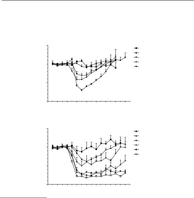
Figures 5.1A-D show that s.c. administration of R-(–)-apomorphine ( 11), R-(–)-N- n- propylnorapomorphine (80), and R-(–)-11-hydroxy-N- n-propylnoraporphine (12), but not R-(–)-11-hydroxyaporphine ( 79), induced a dose-dependent decrease in the release of dopamine in the striatum. R-(–)-apomorphine ( 11) induced a significant decrease in the release of dopamine in the striatum in a dose-range from 0.1 to 1 µmol/kg s.c. In a dose-range from 0.003 to 0.3 µmol/kg s.c., R-(–)-N- n-propylnorapomorphine (80) induced a significant decrease in the release of dopamine in the striatum. R-(–)-11-hydroxy-N- n-propylnoraporphine (12) induced a significant decrease in the release of dopamine in the striatum in a dose-range from 0.03 to 0.3 µmol/kg s.c. Although R-(–)-11-hydroxyaporphine ( 79) displays affinity for the dopamine D2
89
Chapter 5
receptor, it only induced a small significant decrease in the release of dopamine in the striatum in a dose of 1 µmol/kg s.c.
A
|
|
|
|
R-(–)-apomorphine s.c. |
|
|
|||
|
150 |
|
|
|
|
|
|
|
0.003 µmol/kg |
values) |
|
|
|
|
|
|
|
|
|
|
|
|
|
|
|
|
|
0.03 µmol/kg |
|
|
|
|
|
|
|
|
|
0.1 µmol/kg |
|
100 |
|
|
|
|
|
|
|
0.3 µmol/kg |
|
(% of basal |
|
|
|
|
|
|
|
||
|
|
|
|
|
|
|
1 µmol/kg |
||
|
|
|
|
|
|
|
|
||
|
|
|
|
|
|
|
|
|
|
DA release |
50 |
|
|
|
|
|
|
|
|
0 |
|
|
|
|
|
|
|
|
|
|
|
|
|
|
|
|
|
|
|
|
-60 |
-30 |
0 |
30 |
60 |
90 |
120 |
150 |
180 |
Time (min)
B
|
|
R-(–)-N- n-propylnorapomorphine s.c. |
|
||||||
|
150 |
|
|
|
|
|
|
|
0.001 µmol/kg |
values) |
|
|
|
|
|
|
|
|
|
|
|
|
|
|
|
|
|
0.003 µmol/kg |
|
|
|
|
|
|
|
|
|
0.01 µmol/kg |
|
100 |
|
|
|
|
|
|
|
0.03 µmol/kg |
|
basal |
|
|
|
|
|
|
|
||
|
|
|
|
|
|
|
0.1 µmol/kg |
||
|
|
|
|
|
|
|
|
||
|
|
|
|
|
|
|
|
0.3 µmol/kg |
|
(% of |
|
|
|
|
|
|
|
|
|
|
|
|
|
|
|
|
|
|
|
DA release |
50 |
|
|
|
|
|
|
|
|
0 |
|
|
|
|
|
|
|
|
|
|
|
|
|
|
|
|
|
|
|
|
-60 |
-30 |
0 |
30 |
60 |
90 |
120 |
150 |
180 |
Time (min)
Figure 5.1 Effects on striatal dopamine release in freely moving rats after s.c. administration. The results are the mean ± S.E.M. of data obtained from 4 rats. (A) R-(–)-apomorphine ( 11); changes are significant (p < 0.05) from t = 30 min to t = 90 min for 0.1, and 0.3 µmol/kg s.c., and from t = 30 min to t = 120 min for 1 µmol/kg s.c. (B) R-(–)-N- n- propylnorapomorphine (80); changes are significant (p < 0.05) from t = 30 min to t = 105 min for 0.003 and 0.01 µmol/kg s.c., from t = 15 min to t = 180 min for 0.03, 0.1, and 0.3 µmol/kg s.c.
90