

New, centrally acting dopaminergic agents with an improved oral bioavailability [2004]
.pdfMicrodialysis study of R-(–)-apomorphine and analogues
C
|
|
R-(–)-11-hydroxy-N- n-propylnoraporphine s.c. |
|||||||
|
150 |
|
|
|
|
|
|
|
0.003 µmol/kg |
values) |
|
|
|
|
|
|
|
|
|
|
|
|
|
|
|
|
|
0.01 µmol/kg |
|
|
|
|
|
|
|
|
|
0.03 µmol/kg |
|
100 |
|
|
|
|
|
|
|
0.1 µmol/kg |
|
(% of basal |
|
|
|
|
|
|
|
||
|
|
|
|
|
|
|
0.3 µmol/kg |
||
|
|
|
|
|
|
|
|
||
50 |
|
|
|
|
|
|
|
|
|
DA release |
|
|
|
|
|
|
|
|
|
0 |
|
|
|
|
|
|
|
|
|
|
|
|
|
|
|
|
|
|
|
|
-60 |
-30 |
0 |
30 |
60 |
90 |
120 |
150 |
180 |
Time (min)
D
|
|
R-(–)-11-hydroxy-aporphine s.c. |
0.003 µmol/kg |
|||||
|
200 |
|
|
|
|
|
|
0.01 µmol/kg |
|
|
|
|
|
|
|
0.03 µmol/kg |
|
values) |
|
|
|
|
|
|
|
|
|
|
|
|
|
|
|
0.1 µmol/kg |
|
150 |
|
|
|
|
|
|
0.3 µmol/kg |
|
|
|
|
|
|
|
1 µmol/kg |
||
of basal |
|
|
|
|
|
|
|
|
100 |
|
|
|
|
|
|
|
|
(% |
|
|
|
|
|
|
|
|
DA release |
50 |
|
|
|
|
|
|
|
0 |
|
|
|
|
|
|
|
|
|
|
|
|
|
|
|
|
|
|
-30 |
0 |
30 |
60 |
90 |
120 |
150 |
180 |
Time (min)
Figure 5.1 Effects on striatal dopamine release in freely moving rats after s.c. administration. The (continued) results are the mean ± S.E.M. of data obtained from 4 rats. (C) R-(–)-11-hydroxy-N- n- propylnoraporphine (12); changes are significant (p < 0.05) from t = 30 min to t = 180
min for 0.03 and 0.1 µmol/kg s.c., from t = 15 min to t = 180 min for 0.3 µmol/kg s.c.
(D) R-(–)-11-hydroxyaporphine ( 79); changes are significant (p < 0.05) from t = 45 min to t = 75 min for 1 µmol/kg s.c.
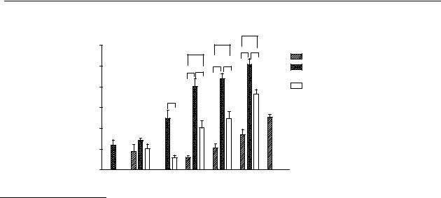
The dose-response relationships of the test compounds are given in Figure 5.2. The response of the compounds is given as the AUC. To compare the AUCs, the experiments were stopped after 180 min. The rank order in the potency upon s.c. administration of the compounds is: R-(–)-N- n-propylnorapomorphine (80) > R-(–)-11-hydroxy-N- n-propylnoraporphine (12) > R-
(–)-apomorphine ( 11).
91
Chapter 5
Dose-AUC curves
AUC (% x Time)
|
*** |
|
|
15000 |
** |
|
|
* |
*** |
** |
R-(–)-apomorphine |
|
|
|
|
12500 |
*** ** |
|
R-(–)-N-n-propylnor- |
* ** |
|
|
apomorphine |
|
|
|
|
10000 |
|
|
R-(–)-11-hydroxy-N- |
** |
|
|
n-propylnoraporhine |
|
|
|
|
7500 |
|
|
|
5000 |
|
|
|
|
|
|
2500 |
|
|
|
|
|
|
0 |
3 |
10 |
30 |
100 |
300 |
1000 |
1 |
Dose (nmol/kg sc)
Figure 5.2 Comparison of the dose-AUC relationship of R-(-)-apomorphine (11), R-(-)-N-n- propylnorapomorphine (80) and R-(-)-11-OH-N-n-propylnoraporphine (12). Data represent mean values ± S.E.M. of 4 animals. Statistical analysis by t-test: * p<0.05, ** p<0.01, *** p<0.001. For comparison with R-(-)-apomorphine (11) 30 nmol/kg equal variance test failed and than Rank Sum Test followed by Mann-Whitney test was performed.
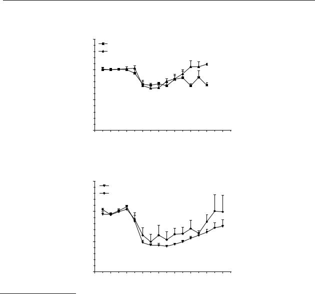
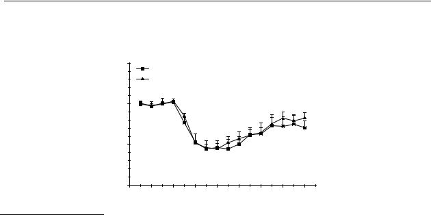
The relative oral bioavailabilities of R-(–)-apomorphine ( 11) and two of its analogues 80 and 12 can be found from Figures 5.3A-C. The relative oral bioavailability was determined by comparing the curves and the AUC after s.c. and p.o. administration. When the AUCs were not significantly different, the relative oral bioavailability was determined by dividing the s.c. dose by the p.o. dose and multiplying by 100. It was known that R-(–)-apomorphine ( 11) possessed a low oral bioavailability. It was expected that the oral bioavailability of the three compounds would be between 1 % and 10 %. Based on this assumption the oral doses were chosen. With this method, both the catechols R-(–)-apomorphine ( 11) and R-(–)-N- n-propylnorapomorphine (80) possess a relative oral bioavailability of about 1 %. The mono-hydroxy compound R-(–)-11-hydroxy-N- n-propylnoraporphine (12) possesses a relative oral bioavailability of about
3 %.
When the courses of the curves after s.c. administration of compounds 11, 80, and 12, respectively, are compared it can be seen that the duration of action of the N-n-propyl analogues is longer than that of R-(–)-apomorphine.
Compound 79 does not induce an effect on the release of dopamine from the striatum (Figure 5.1D). The binding data together with literature data suggest that this compound is a partial dopamine D2 receptor agonist and a dopamine D1 receptor antagonist.261 Therefore, the present pharmacodynamic method was not suitable to determine the relative oral bioavailability.
92
Microdialysis study of R-(–)-apomorphine and analogues
A
R-(–)-apomorphine comparison between p.o. and s.c.
|
150 |
10 µmol/kg p.o. |
|
|
|
|
|
|
||
values) |
|
|
|
|
|
|
|
|||
|
0.1 µmol/kg s.c. |
|
|
|
|
|
|
|||
|
|
|
|
|
|
|
|
|
|
|
(% of basal |
100 |
|
|
|
|
|
|
|
|
|
|
|
|
|
|
|
|
|
|
|
|
release |
50 |
|
|
|
|
|
|
|
|
|
|
|
|
|
|
|
dose |
|
|
AUC |
|
|
|
|
|
|
10 µmol/kg p.o. |
2748 ± 586 |
||||
|
|
|
|
|
0.1 µmol/kg s.c. |
2636 ± 482 |
||||
DA |
|
|
|
|
|
|||||
0 |
|
|
|
|
|
|
|
|
|
|
|
|
|
|
|
|
|
|
|
|
|
|
-60 |
-30 |
0 |
30 |
60 |
90 |
120 |
150 |
180 |
|
Time (min)
B
|
|
R-(–)-N- n-propylnorapomorphine comparison |
|||||||
|
|
|
|
between p.o. and s.c. |
|
|
|||
|
150 |
|
1 µmol/kg p.o. |
|
|
|
|
|
|
values) |
|
|
|
|
|
|
|
||
|
0.01 µmol/kg s.c. |
|
|
|
|
|
|||
|
|
|
|
|
|
|
|
|
|
(% of basal |
100 |
|
|
|
|
|
|
|
|
|
|
|
|
|
|
|
|
|
|
release |
50 |
|
|
|
|
|
|
|
|
|
|
|
|
|
|
dose |
|
AUC |
|
|
|
|
|
|
|
1 µmol/kg p.o. 7697 ± 534 |
|||
DA |
|
|
|
|
|
|
|||
0 |
|
|
|
|
0.01 µmol/kg s.c. 6208 ± 946 |
||||
|
|
|
|
|
|
|
|
|
|
|
-60 |
-30 |
0 |
30 |
60 |
90 |
120 |
150 |
180 |
Time (min)
Figure 5.3 Effects on striatal dopamine release in freely moving rats after s.c. and p.o. administration. The results are the mean ± S.E.M. of data obtained from 4 rats.
(A) R-(–)-apomorphine ( 11); changes are significant (p < 0.05) from t = 30 min to t = 90 min for 0.1 µmol/kg s.c. and 10 µmol/kg p.o. (B) R-(–)-N- n-propylnorapomorphine (80); changes are significant (p < 0.05) from t = 30 min to t = 105 min for 0.01 µmol/kg s.c. and for t = 15 min to t = 180 min for and 1 µmol/kg p.o.
93
Chapter 5
C
|
|
R-(–)-11-hydroxy-N- n-propylnoraporphine |
|||||||
|
|
|
comparison between p.o. and s.c. |
|
|||||
|
150 |
3 µmol/kg p.o. |
|
|
|
|
|
||
values) |
|
|
|
|
|
|
|||
|
0.1 µmol/kg s.c. |
|
|
|
|
|
|||
|
|
|
|
|
|
|
|
|
|
(% of basal |
100 |
|
|
|
|
|
|
|
|
|
|
|
|
|
|
|
|
|
|
release |
50 |
|
|
|
|
|
|
|
|
|
|
|
|
|
|
dose |
|
AUC |
|
|
|
|
|
|
3 µmol/kg p.o. 6992 ± 1523 |
||||
DA |
|
|
|
|
|
0.1 µmol/kg s.c. 6180 ± 813 |
|||
0 |
|
|
|
|
|
|
|
|
|
|
|
|
|
|
|
|
|
|
|
|
-60 |
-30 |
0 |
30 |
60 |
90 |
120 |
150 |
180 |
Time (min)
Figure 5.3 |
Effects on striatal dopamine release in freely moving rats after s.c. and p.o. |
(continued) |
administration. The results are the mean ± S.E.M. of data obtained from 4 rats. |
|
(C) R-(–)-11-hydroxy-N- n-noraporphine (12); changes are significant (p < 0.05) from |
|
t = 30 min to t = 180 min for 0.1 µmol/kg s.c., from t = 15 min to t = 180 min for 3 |
|
µmol/kg p.o. |
5.4Discussion
R-(–)-apomorphine ( 11) is a catechol and is known to have a low oral bioavailability.262
However, the drug is very useful in the treatment of Parkinson’s disease when L-DOPA treatment gives ‘on-off’ fluctuations. 253 An analogue which also displays dopamine D1 and D2 receptor agonistic properties, but possessing a higher oral bioavailability, would be beneficial as an alternative treatment in Parkinson’s disease. The analogues tested (R-(–)-11- hydroxyaporphine (79), R-(–)-N- n-propylnorapomorphine (80) and R-(–)-11-hydroxy-N- n- propylnoraporphine (12)) possess affinity for the dopamine D1 and D2 receptors comparable to R-(–)-apomorphine (Table 5.1). In our experiments we monitored the dopamine D 2 receptor agonistic properties of the compounds, as the release of dopamine is under the control of dopamine D2 autoreceptors.189
Figures 5.1A-C show that compounds 11, 80, and 12 act as dopamine D2 receptor agonists, because they all induce a decrease in the release of dopamine in the striatum. R-(–)-11- hydroxyaporphine (79) (Figure 5.1D) induces, in a dose of 1 µmol/kg, a small significant decrease in the release of dopamine in the striatum. This lack of biochemical activity of compound 79 was not expected from a structure-activity point of view. However, Schaus et al.261 already published that R-(–)-11-hydroxyaporphine ( 79) acts as a partial agonist at the dopamine D2 receptor. This would explain our findings that this compound has a very weak effect on the dopamine D2 autoreceptor.
94

Microdialysis study of R-(–)-apomorphine and analogues
Table 5.1 Affinities of R-(–)-apomorphine ( 11) and its selected analogues (79, 80, 12).
compound |
|
Ki (nM) |
|
|
[3H]-SCH23390 |
[3H]-spiperone [3H]-ADTN |
|
|
(D1) |
(D2-antagonist) (D2-agonist) |
|
R-(–)-apomorphine ( 11)a |
240 |
11.1 |
3.7 |
R-(–)-11-hydroxyaporphine ( 79)b |
107c |
58c |
- |
R-(–)-N- n-propylnorapomorphine (80)a |
340 |
0.8 |
1.5 |
R(–)-11-hydroxy-N- n- |
434 |
0.9 |
5.3 |
propylnoraporphine (12)d |
|
|
|
Footnotes: Affinity of R-(–)-apomorphine ( 11) and its selected analogues (79, 80, 12) as measured by their ability to displace in vitro [3H]-SCH23390 (D1-antagonist), [3H]-spiperone (D2-antagonist), and [3H]-ADTN (D2-agonist) from membrane preparations of rat brain corpus striatum tissue in order to measure the affinity for dopamine D1 and D2 receptors, respectively. a Values are taken from ref. 263; b values are taken from ref. 261; c IC50 in nM; d values are taken from ref. 264.
The dose-AUC relationships upon s.c. administration (Figure 5.2) clearly show that the N-n-propyl analogues 80 and 12 are more efficacious in the microdialysis experiments than the N-methyl analogue (R-(–)-apomorphine ( 11)). An explanation could be the presence of a propyl moiety on the nitrogen for R-(–)-N- n-propylnorapomorphine (80) because a propyl moiety has a better fit into the receptor than a methyl. This, however, should have been shown in differences in binding affinities, which are not observed. Although there is a clear distinction in efficiency in the microdialysis experiments between the three compounds, this difference cannot be explained by the affinity for the dopamine D2 receptor (Table 5.1). The relevant dopamine D2 receptor agonist binding is comparable for the three compounds.
Figures 5.3A and B show that both of the catechol-containing aporphines (R-(–)-apomorphine ( 11) and R-(–)-N- n-propylnorapomorphine (80)) possess a relative oral bioavailability of about 1%. The greater s.c. potency of R-(–)-N- n-propylnorapomorphine (80) over R-(–)-apomorphine ( 11) is not likely to be the result from different rates of metabolism in the periphery because the main route of metabolism for both compounds is glucuronidation of the catechol moiety.265 The greater potency of the mono-hydroxy analogue R-(–)-11-hydroxy-N- n-propylnoraporphine (12) (Figure 5.3) could possibly be explained from differences in metabolism, R-(–)-11-hydroxy-N- n-propylnoraporphine (12) possesses a relative oral bioavailability of 3%. Although this represents an increase, it is still not an optimal oral bioavailability for a therapeutic agent. The improvement of the relative oral bioavailability of the mono-hydroxy analogue 12 may be explained from the fact that a mono-hydroxy analogue is less sensitive to metabolic degradation than a catechol moiety.266 Beside glucuronidation, the catechol moiety is also sensitive to oxidative degradation, as well as to degradation by catechol- O-methyl transferase (COMT), which will predominantly result in 10-methoxy-11-
95

Chapter 5
hydroxyaporphine, an inactive compound.267,268 The catechol moiety is clearly not necessary for high affinity at the dopamine D2 receptors. The presence of a free 11-hydroxy moiety is enough to confer dopamine D2 receptor agonist-like activity, similar to that of 10,11-dihydroxyaporphines.266
An explanation could be differences in intrinsic efficacy and differences in the ability to pass the blood-brain barrier. For R-(–)-N- n-propylnorapomorphine (80) this difference in ability to pass the blood-brain barrier compared to R-(–)-apomorphine has previously been
published.265,269,270
Beside comparable effects on the release of dopamine in the striatum, R-(–)-N- n- propylnorapomorphine (80) and R-(–)-11-hydroxy-N- n-propylnoraporphine (12) could also possess the same neuroprotective effects as R-(–)-apomorphine. This neuroprotective effect resides in the phenolic moiety, which can act as a radical scavenger.141-143 Both compounds possess this moiety.
Based on our results, R-(–)-N- n-propylnorapomorphine (80) and R-(–)-11-hydroxy-N- n- propylnoraporphine (12) could be good targets for treatment of Parkinson’s disease. Although their oral bioavailabilities are low, their greater efficacy results in the possibility of administering lower doses. R-(–)-N- n-propylnorapomorphine (80) has proven to be a useful adjunct in the long-term management of patients with unsatisfactory response to L-DOPA and produced a significant therapeutic benefit at doses much lower than the dose at which sideeffects occur.271
In conclusion, this microdialysis study shows that R-(–)-N- n-propylnorapomorphine (80) and R-(–)-11-hydroxy-N- n-propylnoraporphine (12) are more potent than R-(–)-apomorphine (11) in inhibiting dopamine release in the striatum. This difference in potency on the presynaptic receptors resembles the difference in potency of these analogues on supersensitive postsynaptic receptors as described by Kelly et al.269
96

Chapter 6
Neuropharmacological evaluation of a new dopaminergic prodrug with anti-parkinsonian potential*
Abstract
In this study, a prodrug of hydroxylated 2-aminotetralins was tested against the prototypic dopamine receptor agonist S-(–)-5-OH-DPAT. The active S-enantiomer of the prodrug (DD9812) was able to decrease the release of dopamine in the striatum, but with a lower potency than S-(–)-5-OH-DPAT after s.c. administration. The R-enantiomer of the prodrug (DD9813) only had a limited effect on the release of dopamine in the striatum. The prodrug, however, showed an improved relative oral bioavailability, as compared to S-(–)-5-OH-DPAT.
* This chapter is based on: Rodenhuis, N.; Venhuis, B.J.; Timmerman, W.; Wikström, H.V.; Dijkstra, D; Meltzer, L.; Johnson, S.; Wise, L.D. (2000) Neuropharmacological evaluation of a new dopaminergic prodrug with anti-parkinsonian potential. In preparation.
97

Chapter 6
6.1Introduction
Parkinson’s disease is a neurodegenerative disease that is characterised by progressive damage of predominantly dopaminergic neurons in the substantia nigra. Damage to neurons in the substantia nigra causes a dopamine deficiency in the striatum, resulting in disturbed motor functioning. Only when approximately 70-80 % of these cells have degenerated, symptoms of Parkinson’s disease arise.
Current drug-based therapies for Parkinson’s disease are palliative therapies, i.e. they relieve the symptoms, but do not cure the disease. One of the current and expanding therapies is treatment with dopamine D2/D3 receptor agonists. There are a number of such therapeutics, e.g. bromocriptine, pergolide, apomorphine, ropinirole, and pramipexole.
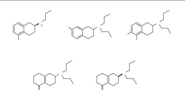
A class of compounds with a high affinity for e.g. dopamine D2 receptors are the hydroxylated 2-aminotetralins, e.g. 5-OH-DPAT (9), N-0437 (31), 7-OH-DPAT (10) and 5,6-di- OH-DPAT (81).272,273 Preclinical data show that these compounds display limited activity upon oral administration. A major disadvantage of the hydroxylated 2-aminotetralins and other phenolic compounds is that they undergo considerable inactivation by glucuronidation in the gut and the liver.161 One of the strategies to circumvent the problem of the low oral bioavailability of the hydroxylated 2-aminotetralins is to search for suitable prodrugs. Frequently investigated prodrugs of phenols are esters and carbamates.96,185-187
We have now synthesised 6-(N,N-di-n-propylamino-3,4,5,6,7,8-hexahydro-2H-naphthalen-1- one (PD148903), which is a dihydro analogue of 5-OH-DPAT and which may possibly act as a prodrug or a pharmacophore equivalent of 5-OH-DPAT. The individual enantiomers of racemic PD148903 were prepared in our laboratory (to be published). Johnson et al.274 published that the in vitro biochemistry of PD148903 showed that the compound itself did not have any affinity for the dopamine D2 receptor. However, in vivo experiments showed that it has potent dopamine receptor agonist-like activity. These two observations suggest that in vivo the prodrug is, at least partly, converted to hydroxylated derivatives.274 Therefore, we have pharmacologically evaluated the S-(–)-enantiomer (DD9812, 83) and the R-(+)-enantiomer (DD9813, 82) of the potential prodrug PD148903 against the active enantiomer of the prototypic dopamine receptor agonist S-(–)-5-OH-DPAT. The exact mechanism of conversion and metabolism is still under investigation.
98
Neuropharmacological evaluation of a new dopaminergic prodrug
N |
HO |
|
N |
R |
|
N |
|
|
|
|
|
|
|
|
HO |
OH |
|
|
OH |
9 R=C3H7 |
|
10 |
81 |
31 R=2-thienylethyl |
|
|
|
N N
O 82 |
O 83 |
|
|
|
|
Chart 6.1 Chemical structures of S-(–)-5-hydroxy-2-(N,N-di- n-propylamino)tetralin (S-(–)-5-OH- DPAT, S-(–)- 9), S-(–)-5-hydroxy-2-(N- n-propyl-N-2-thienylethylamino)tetralin (S-(–)-N-0437, S-(–)- 31), R-(+)-7-hydroxy-2-(N,N-di-n-propylamino)tetralin (R-(+)-7-OH-DPAT, R-(+)-10), 5,6-dihydroxy-2-(N,N-di-n-propylamino)tetralin (5,6- di-OH-DPAT, 81) and R- and S-6-(N,N-di-n-propylamino-3,4,5,6,7,8-hexahydro-2H- naphthalen-1-one (R-enantiomer, DD9813, 82; S-enantiomer, DD9812, 83).
6.2Material and methods
6.2.1Animals
Male Wistar rats (from CDL, Groningen, The Netherlands) weighing 280-320 g were used for microdialysis experiments. The rats were housed in Plexiglas cages, eight animals in each cage, with free access to water and food. The cages were placed in a room with controlled environmental conditions (21 °C; humidity 60-65%; lights on at 8 a.m. and off at 8 p.m.). The animals were housed at least one week after arrival prior to surgery and use in the experiments. Animal procedures were conducted in accordance with guidelines published in the NIH Guide for the Care and Use of Laboratory Animals and all protocols were approved by the Groningen University Institutional Animal Care and Use Committee.
6.2.2Drug treatment
The drugs were dissolved in saline and stocked in a concentration of 1 mg/ml for subcutaneous (s.c.) and 1 mg/2 ml for oral (p.o.) administration and diluted, if necessary, with saline before administration. A volume of 1 ml/kg was administered per s.c. injection and 2
99

Chapter 6
ml/kg per p.o. injection. Drugs used were DD9812, DD9813 and S-(–)-5-OH-DPAT and were synthesised at the Department of Medicinal Chemistry in Groningen. The amount of compound was recalculated to an amount of S-(–)-5-OH-DPAT, i.e. 1 mg/ml indicates and amount of compound equal to 1 mg/ml S-(–)-5-OH-DPAT.
6.2.3Surgery and brain microdialysis
On-line brain microdialysis in freely moving animals has previously been described.188 In brief, the rats were anaesthetised with midazolam (5 mg/kg s.c.), atropine nitrate (0.1 mg/kg s.c.), ketamine (50 mg/kg i.p.) and xylazine (8 mg/kg i.p.); 10% lidocaine was locally applied. The rats were then mounted into a stereotaxic frame (Kopf). The incisor bar was placed in position so that the skull was held horizontal. The skull was exposed and burr holes were drilled. A Y-shaped dialysis probe was used for the experiments, with an exposed tip length of 3 mm. The dialysis tube (ID: 0.22 mm; OD: 0.31 mm) was prepared from polyacrylonitrile/sodium methallyl sulfonate copolymer (AN 69, Hospal, Bologna, Italy). The microdialysis membrane was implanted in the striatum. The dura was removed with a sharp needle. Two anchor screws were positioned in different bone plates nearby. The following coordinates were used according to the atlas of Paxinos and Watson:250 AP + 1.0, LM ± 3.0 relative to bregma, and VD − 6.0 below dura. Before insertion into the brain the dialysis probe was perfused successively with ultra pure water, methanol, ultra pure water and Ringer solution (1.2 mM Ca2+). The dialysis probe was positioned in the burr hole under stereotaxic guidance. The probe was cemented in this position with dental cement. After the surgery, the rats received buprenorphine (0.1 mg/kg i.m.), an analgesic agent. The rats were housed solitary.
The experiments were performed in conscious rats 17-48 h after implantation of the cannula.
The striatum was perfused with a Ringer solution (147 mmol/l NaCl, 4 mmol/l KCl, 1.2 mmol/l CaCl2, 1.1 mmol/l MgCl2) at 2 μl/min (CMA/102 microdialysis pump, Sweden).
Dopamine was quantitated by high-performance liquid chromatography (HPLC) with electrochemical detection with a detection limit of approximately 5 fmol/sample. An HPLC pump (LKB, Pharmacia) was used in conjunction with an electrochemical detector (Antec, Leiden) working at 625 mV versus an Ag/AgCl reference electrode. The analytical column was a Supelco Supelcosil LC-18 Column (3 µm particle size). The mobile phase consisted of a mixture of 4.1 g/l sodium acetate (Merck), 85 mg/l octane sulphonic acid (Aldrich), 50 mg/l EDTA (Merck), 1 mM tetramethylammonium chloride (ACROS), 8.5 % methanol (Labscan) and ultra pure water (pH=4.1 with glacial acetic acid).
After the experiments the rats were sacrificed and the brains were removed. After removal the brains were kept in 4% paraformaldehyde solution until they were sectioned to control the location of the dialysis probes.
100