New, centrally acting dopaminergic agents with an improved oral bioavailability [2004]
.pdf
Chapter 1
Introduction
1.1Dopamine, a neurotransmitter in the central nervous system
Neurotransmitters serve to transmit signals between neurons, which are separated by a synaptic cleft. One of the neurotransmitters is dopamine (DA), or β-(3,4- dihydroxyphenyl)ethylamine (1). Until the mid-1950s dopamine was exclusively considered to be an intermediate in the biosynthesis of the catecholamines noradrenaline and adrenaline. Significant tissue levels of dopamine were first demonstrated in peripheral organs of ruminant species.1 A short time later it was found that dopamine was also present in the brain in about equal concentrations to those of noradrenaline.2
When a stimulus depolarises the transmembrane potential in a
HO |
NH2 |
spiking axon above the threshold level, an all-or-none action |
||
|
|
|||
HO |
|
potential |
in a spiking |
axon is activated. The action potential |
|
propagates unattenuated to the nerve terminal where ion fluxes |
|||
1 |
|
|||
|
activate a |
mobilisation |
process leading to transmitter secretion.3 |
|
The neurotransmitter binds reversibly to receptor proteins embedded in the membrane of a neuron, which triggers a certain effect. There are two types of receptors known, presynaptic receptors or autoreceptors which are present on the neurotransmitter ‘releasing neurons’, and postsynaptic or heteroreceptors, which are present on the neurotransmitter ‘receiving neuron’. The former are supposed to perform a feed-back function, and slow down the release of neurotransmitter from these neurons when they are stimulated.4
Dopamine receptors are G-protein coupled receptors. After a receptor has been activated, this G-protein may activate or inhibit the second messenger system causing certain biochemical reactions to occur. In contrast to the ionotropic receptors that are linked to an ion-channel and respond very fast to activation by a neurotransmitter (millisecond processes), G-protein coupled receptors mediate slower responses (seconds to minutes) and in general have a modulatory function on other signal transduction processes.4
Although dopamine also has important peripheral functions, e.g. within the regulation of cardiovascular homeostasis, this thesis will deal with the effects of ligands on dopamine receptors in the central nervous system (CNS). Dopamine is the predominant catecholamine neurotransmitter in the mammalian brain and is involved in the regulation of a number of physiological activities, i.e. movement, emotion and hormone secretion. These activities are associated with three principal dopaminergic pathways: (1) The nigrostriatal pathway controls movements; partial degeneration of this system contributes to the pathogenesis of Parkinson’s disease. (2) The mesocorticolimbic pathway is involved in emotion; imbalance in this pathway
1

Chapter 1
is thought to contribute to the aetiology of schizophrenia. (3) The tuberoinfundibular pathway regulates e.g. prolactin secretion from the pituitary and influences lactation and fertility.5
1.2General aspects of dopamine neurotransmission in the central nervous system
1.2.1Biosynthesis of dopamine
The synthesis of dopamine originates from the precursor the amino acid L-tyrosine, which must be transported across the blood-brain barrier into the dopaminergic neuron. The rate limiting step in the synthesis is the conversion of L-tyrosine to L-dihydroxyphenylalanine (L-DOPA) by the enzyme tyrosine hydroxylase (TH). L-DOPA is subsequently converted to dopamine by aromatic L-amino acid decarboxylase. The latter enzyme turns over so rapidly that L-DOPA levels in the brain are negligible under normal conditions.1
NH2 |
HO |
NH2 |
HO |
NH2 |
|
TH |
|
AAAD |
|
COOH |
HO |
COOH |
HO |
|
HO |
|
|
||
2 |
3 |
|
|
1 |
Chart 1.1 Biosynthesis of dopamine. Dopamine (1); L-Tyrosine (2); L-3,4-dihydroxyphenylalanine
(3, L-DOPA); TH, tyrosine hydroxylase; AAAD, aromatic L-amino acid decarboxylase.
Under normal conditions it is not feasible to augment dopamine synthesis significantly by increasing brain levels of L-tyrosine, since the levels in the brain are already above the Km for tyrosine hydroxylase. The activity of tyrosine hydroxylase is subject to four major regulatory influences: (1) Dopamine functions as end-product inhibitor of TH by competing with a tetrahydrobiopterin (BH-4) cofactor for a binding site on the enzyme. (2) The availability of BH- 4 may also play a role in regulating TH activity. (3) Presynaptic dopamine receptors also modulate the rate of tyrosine hydroxylation. These receptors are activated by dopamine released from the nerve terminal, resulting in feedback inhibition of dopamine synthesis. (4) Dopamine synthesis also depends on the rate of impulse flow in the nigrostriatal pathway. On the other hand it is possible to enhance dramatically the formation of dopamine by increasing the levels of L-DOPA because of the high activity of aromatic L-amino acid decarboxylase and the low endogenous levels of L-DOPA normally present in the brain.1
1.2.2Dopamine re-uptake and metabolism
Once the dopamine that is released into the synaptic cleft has exerted its action on the various dopamine receptors, these actions have to be terminated to prevent continuous
2

Introduction
stimulation of these receptors. This inactivation is brought about by re-uptake mechanisms and by metabolism of dopamine.
Dopamine nerve terminals possess high-affinity dopamine uptake sites that are important in termination transmitter action and in maintaining transmitter homeostasis. Uptake is accomplished by a membrane carrier that is capable of transporting dopamine in either direction, depending on the existing concentration gradient. The dopamine transporter recycles extracellular dopamine by actively pumping it back into the nerve terminal. About 70-80 % of the dopamine, which is present in the synaptic cleft, is inactivated by this process. Certain drugs, such as cocaine, are able to block the action of the dopamine transporter, thereby sustaining the presence of dopamine in the synaptic cleft and its action on dopamine receptors.
After re-uptake by the nerve terminal a part of the released dopamine is converted to dihydroxyphenylacetic acid (DOPAC, 5) by intraneuronal monoamine oxidase (MAO) and aldehyde dehydrogenase (AD). Released dopamine is also converted to homovanillic acid (HVA, 6), probably at an extraneuronal site through the sequential action of catechol-O- methyltransferase (COMT) and MAO. In rat brain, DOPAC is the major metabolite and considerable amounts of DOPAC and HVA are present in sulfate-conjugated as well as free forms.1
|
HO |
|
HO |
COOH |
|
CHO |
AD |
|
|
|
|
|
|
|
MAO |
HO |
|
HO |
COMT |
4 |
|
5 |
||
|
|
|||
|
|
|
||
HO |
NH2 |
|
|
MeO |
|
|
|
|
COOH |
HO |
|
|
|
HO |
1 |
|
|
|
6 |
COMT |
|
|
|
AD |
|
|
|
|
|
MeO |
NH2 |
|
MeO |
CHO |
|
MAO |
|
|
|
HO |
|
|
HO |
|
|
7 |
|
8 |
|
Chart 1.2 Neuronal metabolism of dopamine. Dopamine (1); 3,4-dihydroxyphenyl acetaldehyde
(4); 3,4-dihydroxyphenylacetic acid (DOPAC, 5); homovanillic acid (HVA, 6); 3-methoxytyramine (3-MT, 7); 3-methoxy-4-hydroxyphenyl acetaldehyde (8); MAO, monoamine oxidase; AD, aldehyde dehydrogenase; COMT, catechol-O- methyltransferase.
3

Chapter 1
1.3Dopamine receptors
1.3.1General structural features of G-protein-coupled receptors
Upon release from dopaminergic neurons, dopamine exerts its action by interacting with specific dopamine receptors. With molecular biological techniques five subtypes of dopamine receptors have been identified so far. These receptors have been characterised anatomically, and to a certain extent biochemically and pharmacologically. All currently identified dopamine receptor subtypes belong to the superfamily of G-protein-coupled receptors (GPCRs).
A wide variety of membrane receptors for hormones and neurotransmitters are coupled to guanine-nucleotide-binding regulatory ‘G’ proteins, which upon activation by receptors, stimulate or inhibit various effectors such as enzymes or ion channels. Among the family of receptors coupled to G-proteins are those for catecholamines, acetylcholine and related muscarinic ligands, tachykinins, and for the pituitary glycoprotein hormones and many more.6
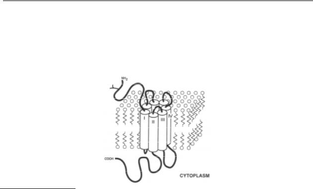
The exact molecular structures of GPCRs are unknown, since attempts to crystallise these proteins have failed thus far. Nevertheless, biophysical, biochemical and molecular biological studies on various GPCRs suggest that these receptors have many structural features in common. The common features are: (1) a cell surface receptor; (2) an effector, such as an ion channel or the enzyme adenylyl cyclase; and (3) a G-protein, that is coupled to both the receptor and its effector.7 All these receptors are made up of a single chain containing amino acid residues of which the number of residues differ for each receptor subtype. The amino-terminus, which has no signal sequence, may contain sites for N-linked glycosylation, which is the case for the dopamine receptors; the carboxy-terminus has typical sites for phosphorylation by protein kinase A and other kinases. Seven stretches of 22-28 hydrophobic conserved residues, separated by hydrophilic segments, are found in each of the members of the family of proteins, as has been seen earlier in the well-characterised rhodopsins, which are also coupled to a GTP binding protein. This similarity has led to the suggestion that these receptors share with bacteriorhodopsin its peculiar membrane topology by which the seven conserved hydrophobic segments form transmembrane domains, possibly constituting α-helices, although other configurations are also imaginable. The N-terminal region, by virtue of its glycosylation, is extracellular and the carboxy-terminal domain is intracellular. The various portions between the seven hydrophobic stretches are either extraor intracellular. The third intracellular and the carboxy-terminal segments display an extensive variability in length and sequence, which has led to the hypothesis that these parts of the 7 transmembrane G-protein coupled receptors are responsible for the selective interaction with the various regulatory G-proteins.6 All GPCRs cloned so far have been shown to possess a substantial degree of homology in their amino acid sequences, especially in the transmembrane regions.
The binding site for the endogenous ligand (the ‘active site’) is believed to be situated within the core formed by the seven transmembrane domains.7 Binding of the endogenous
4

Introduction
ligand to the active site presumably induces conformational changes in the receptor molecule, which trigger via the G-protein an intracellular response, e.g. the activation of a second messenger system. In this way, the ‘information’ carried by the ligand is transduced over the plasma membrane into the cell (for reviews and references on GPCRs, see refs. 6-8).
Figure 1.1 Schematic model for the insertion of G-protein-coupled receptors in the plasma membrane. The seven transmembrane domains are shown as cylinders spanning the lipid bilayer. The intraand extracellular loops are represented by black ribbons. The ligand binding site is formed by the interaction of several transmembrane domains. Coupling to transducing and desensitisation systems involves the cytoplasmic loops. Glycosylation (represented with a Y) of the N-terminus is required for proper insertion of the receptors in the membrane but not for ligand binding. Figure adapted from ref. 6.
1.3.2Dopamine receptor classification
The application of biochemical, pharmacological and physiological techniques to the study of dopamine receptors showed clearly that there were multiple receptors for dopamine, and in 1978 it was proposed that there were two subtypes of the dopamine receptor (D1 and D2).9 The application of molecular biological techniques in the late 1980s showed that there were at least five dopamine receptor subtypes (D1, D2, D3, D4 and D5).10-18
On the basis of structural, pharmacological, functional and distributional similarities, all dopamine receptor subtypes fall into one of the two initially recognised receptor categories, here designated dopamine D1- or dopamine D2-like receptors. Dopamine D5 receptors share extensive similarities with dopamine D1 receptors, while dopamine D3 and D4 receptors more closely conform to the features of dopamine D2 receptors. The properties of the two subfamilies closely resemble those of the dopamine D1 and D2 receptor subtypes as originally defined by Kebabian and Calne.9 The most important characteristics of the cloned human dopamine receptor subtypes are summarised in Table 1.1.
5

Chapter 1
Table 1.1 Summary of the characteristics of cloned human dopamine receptor subtypes.
|
D1-like |
|
D2-like |
|
|
|
D1 |
D5 |
D2 |
D3 |
D4 |
Gene |
|
|
|
|
|
Chromosome localisation |
5q35.119 |
4p16.319 |
11q22-2319 |
3q13.319 |
11p15.519 |
Introns |
no12-14 |
no10,18 |
yes15 |
yes17 |
yes16 |
Expression |
– |
– |
D 2A/D2B |
– |
D 4.2-D4.10 |
Protein |
|
|
|
|
|
Amino acids |
44612-14 |
47710,18,20 |
443/41415 |
40017 |
38716 |
3rd Cytoplasmatic loop |
short |
short |
long |
long |
long |
C-terminus |
long |
long |
short |
short |
short |
Sequence homologya D1 |
100 |
82 |
47 |
45 |
42 |
D5 |
|
100 |
44 |
40 |
45 |
D2 |
|
|
100 |
77 |
51 |
D3 |
|
|
|
100 |
40 |
D4 |
|
|
|
|
100 |
Localisationb, 21 |
Cput, NAc, |
Hipp, Hyp |
Cput, NAc, |
NAc, ICj, |
FC, OT, |
|
ICj, OT |
|
ICj, OT, |
Sept, Thal, |
Amyg, |
|
|
|
Pit, SN, |
Hyp, Cer |
Mes, MO |
|
|
|
VTA |
|
|
Pharmacology |
|
|
|
|
|
Dopamine affinity (nM)c |
2300 |
230 |
2000 |
30 |
450 |
Agonist |
SKF 38393 |
SKF 38393 |
N-0923 |
PD 128907 |
PD 168077 |
Antagonist |
SCH 23390 |
SCH 23390 |
Raclopride |
S 14297 |
L-745,870 |
Biochemistryd |
|
|
|
|
|
G-protein coupled |
yes |
yes |
yes |
yes/? |
yes |
cAMP |
+ |
+ |
– |
n.e./– |
n.e./– |
IP3 |
+ |
? |
+ |
n.e. |
n.e. |
Ca2+ |
+ |
? |
– |
– |
– |
Arachidonic acid |
? |
? |
+ |
n.e./– |
+ |
Dopamine release |
? |
? |
– |
n.e./– |
n.e. |
Mitogenesis |
? |
? |
+ |
n.e./+ |
? |
Acidification |
? |
? |
+ |
+ |
+ |
Footnotes: a Sequence homology in transmembrane domains, expressed as percentages. b Based on rat brain mRNA distribution data, only the areas with a high density are mentioned. Abbreviations: Cput,
6

Introduction
caudate putamen; NAc, nucleus accumbens; ICj, islands of Calleja; OT, olfactory tubercle; Hipp, hippocampus; Hyp, hypothalamus; Thal, thalamus; Pit, pituitary; SN, substantia nigra; VTA, ventral tegmental area; Sept, septum; Cer, cerebellum; FC, frontal cortex; Amyg, amygdala; Mes, mesencephalon; MO, medulla oblongata. c Values in the presence of Gpp(NH)p (taken from ref. 22). d Abbreviations: cAMP, cyclic adenosine monophosphate; IP3, inositol triphosphate; +, increase; –, decrease; n.e., no effect; ?, unknown; n.e./– and n.e./+, in the literature there is a controversy about the effect (taken from ref. 23).
In addition to elucidating the molecular features of GPCRs, molecular cloning techniques have also allowed for the expression of GPCRs in cells that normally do not express such receptors. Thus, mammalian cell lines can be transiently or permanently transfected with cDNAs encoding the different dopamine receptor subtypes. Because these cells usually express a single receptor subtype in high density, they are very suitable for determining receptor binding affinities of drug candidates. Since most newly synthesised target compounds presented in the subsequent chapters of this thesis have been evaluated for their ability to bind to cloned human dopamine D2 and D3 receptors, the characteristics of these two receptor subtypes will be described in more detail in the next sections. For reviews and references on the other dopamine receptor subtypes, see refs. 21,24-26.
1.3.3Dopamine D2 receptors
Based on the presumed structural homology between different GPCRs a rat genomic library was screened by Bunzow et al.15 They used the DNA sequence encoding the hamster β2- adrenergic receptor as a hybridisation probe. Ultimately a cDNA was isolated which coded for a 415 amino acid protein, which possessed a relative molecular mass similar to that for the deglycosylated form of the dopamine D2 receptor as determined by SDS-PAGE. Another conformation that this cloned receptor was the dopamine D2 receptor was the fact that the mRNA distribution parallels that of the dopamine D2 receptor. Several structural features of the protein deduced from this cDNA demonstrated that it belongs to the family of GPCRs. First, a hydrophobicity plot of the protein sequence shows the existence of seven stretches of hydrophobic amino acids, which could represent seven transmembrane domains. Second, the primary amino acid sequence shows a high degree of similarity with other GPCRs. Third, the protein has several structural characteristics common to the other members of the family of GPCRs. There are three consensus sequences for N-linked glycosylation in the N-terminus with no signal sequence. Aspartate 80 found in transmembrane domain II is conserved in all known GPCRs. In transmembrane domain III Asp114 corresponds to an Asp residue found in receptors that bind cationic amines. Phosphorylation has been proposed as a means of regulating receptor function. A potential site for phosphorylation by protein kinase A exists at Ser228 in the third cytoplasmic loop. The cloned protein contains a large cytoplasmic loop between transmembrane
7

Chapter 1
domains V and VI with a short C terminus. This structural organisation is similar to other receptors, which are coupled to Gi.15
In situ hybridisation revealed the distribution of dopamine D2 receptor mRNA in the rat brain. High abundance was found in regions which are classically associated with dopaminergic neurotransmission, including the caudate putamen, nucleus accumbens, olfactory tubercle, pituitary, substantia nigra pars compacta and ventral tegmental area.27,28
When the receptors were expressed in mouse fibroblast cells it was possible to determine the pharmacological features of the receptor. It turned out that the expressed receptors possessed features typical of the native striatal dopamine D2 receptor. When all of the drug Ki values were compared, their rank order of potency (spiperone > (+)-butaclamol > haloperidol > sulpiride >>
(–)-butaclamol) agrees closely with the published values for the dopamine D 2 receptor.29
Expression of the receptor in other cell lines has revealed that the receptor interacts productively with a G-protein, probably Gi to inhibit adenylyl cyclase activity,30 and also appeared to inhibit prolactin secretion.29 Taken together, these findings strongly suggest that the cloned protein indeed corresponded to the classical dopamine D2 receptor (for reviews and references see ref. 29).
After cloning of the rat dopamine D2 receptor by Bunzow et al.15 the next step was the cloning of the human dopamine D2 receptor.11,31 The human and rat dopamine D2 receptors are encoded by highly related genes, and are pharmacologically highly related.11 The two receptors differ in only 18 amino acid substitutions and by 1 amino acid in length, the human form lacking an isoleucine in the third cytoplasmic loop.11 In order to determine which amino acids were relevant in binding and effector-coupling and to test these models, dopamine D2 receptor mutants were generated. Initially, the highly conserved Asp 80 was recognised as serving a central role in normal receptor coupling to adenylyl cyclase.21,32 Ser 193, in particular, seems to play a critical role in binding dopamine21,33 and Asp 114 was a prerequisite for agonist as well as antagonist binding.21
A number of different studies have revealed that the dopamine D2 receptor exists in alternate splice forms in rats31,34-38 and humans.31,39 The two forms of the dopamine D2 receptor exist both in human and rat, and are generated by alternative splicing. Specifically, these forms differ by a 29 residue peptide sequence located in the predicted intracellular domain between transmembrane domain V and VI (cytoplasmic loop 3). The presence of this peptide characterises the more abundant, larger form (D2L receptor); its absence defines the rarer, shorter form (D2S receptor). The fact that the difference between the two isoforms lies in the third cytoplasmic loop may be important for G-protein coupling. This suggests that alternative splicing is used to fine-tune receptor interaction with Gi and Go proteins. In fact, in preliminary expression studies, the D2S receptor inhibited adenylyl cyclase activity to a greater extent than did the D2L receptor form, corresponding with the notion that the shorter form more effectively couples to a Gi protein.31 Several groups tried to identify properties that may differentiate the two dopamine D2 receptor variants by studying the mRNA expression of the two receptor
8

Introduction
isoforms in brain and pituitary, and as expressed in different cell lines.35-37,39-41 Both splice variants have been detected in human anterior pituitary31,42 and in a variety of brain regions.42 In general, the mRNA distribution of both splice variants agrees with the distribution pattern of total dopamine D2 mRNA. However, the D2L/D2S mRNA ratio varies in different brain regions and the short isoform is the least abundant of the two. Receptor binding experiments show that for D2S and D2L the Ki values for the high and low affinity agonist binding are rather similar in three cell lines tested, it is the proportions of the sites that differ. However, some compounds seem to have a higher affinity for the dopamine D2S receptor.43,44
Presumably via coupling to different G-proteins, dopamine D2 receptors in various cell lines revealed that they utilise different signal transduction systems (for reviews see refs. 21, 25). Inhibition of adenylyl cyclase has been detected in all cellular environments,30,45-51 but cellspecific signalling pathways may be present as well. Beside inhibition of intracellular cAMP production, stimulation of dopamine D2 receptors may result in: (1) enhancement of phosphatidylinositol (PI) hydrolysis by activation of the enzyme phospholipase C;49,51
(2) increase48,49,51 or decrease51 in the intracellular Ca2+ concentration; (3) opening of K+ channels;51 and (4) extracellular release of arachidonic acid.48,52
1.3.4Dopamine D3 receptors
In 1990 Sokoloff et al.17 cloned the cDNA encoding a novel dopamine D2-like receptor, which was designated as dopamine D3 receptor. The human dopamine D3 receptor consisted of 400 amino acid residues and displayed a homology of 46 % with the human dopamine D2 receptor and a homology of 78 % if only the presumed transmembrane domains are considered.53 In addition, the subtypes have more structural features in common, such as the presence of introns in the coding sequence, a long third intracellular loop, a short carboxylic acid terminal segment and several glycosylation sites. The existence of introns may give rise to the expression of splice variants encoded by the same gene. Various truncated forms of the dopamine D3 receptor mRNA, generated by alternative splicing, have been detected in rat and human brain which do not correspond to a functional receptor.19,54
The anatomical distribution of the dopamine D3 receptor mRNA partially overlaps but markedly differs from that of dopamine D2 receptor mRNA. The dopamine D3 receptor is mainly expressed in discrete brain areas belonging to or related to the limbic system, whereas dopamine D1 and D2 receptors are widely expressed in all major dopaminoceptive areas.17 Both dopamine D2 and D3 receptors are expressed by dopamine neurons belonging to the A9 and A10 cell groups and it has been suggested that both act as autoreceptors. Such a role for the dopamine D3 receptor is consistent with its high apparent affinity for dopamine. The pharmacological profile of the dopamine D3 receptor is comparable to that of the dopamine D2 receptor, and supports a possible role as an autoreceptor. Thus, all dopamine D2 receptor agonists and antagonists bind with good affinities to dopamine D3 receptors as well, but some
9

Chapter 1
compounds, previously designated as putative dopamine D2 autoreceptor agonists (e.g. 7-OH- DPAT) or antagonists (e.g. (+)-AJ 76 and (+)-UH 232), show preference for the dopamine D3 receptor. Given this pharmacological profile it would favour a role for the dopamine D3 receptor as an autoreceptor. If so, it could play a role in regulating impulse flow, as well as neurotransmitter synthesis and release. However, the function of the dopamine D3 receptor is still subject of debate. Several studies have given some insight into this debate, using the dopamine D3 receptor preferring ligands R-(+)-7-OH-DPAT and PD128907 (agonists) or U-99194A (antagonist). Some studies indicate that the dopamine D3 receptor is an autoreceptor involved in the presynaptic regulation of dopamine release.55-62 These data, however, could not be confirmed by dopamine D2 and D3 knock-out mice. Experiments with dopamine D2 receptordeficient mice strongly suggest that only D2 but not D3 receptors are involved in the autoreceptor-mediated inhibition of the evoked release of [3H]-dopamine.63 These data are confirmed by experiments with D3-receptor knock-out mice.64
Other studies indicate that the dopamine D3 receptor is located postsynaptically and is involved in locomotor activity and behaviour. Experiments with dopamine D3 receptor agonists65-72 and antagonists73-76 show that these receptors have inhibitory effects on locomotor activity and behaviour. In the literature it is also described that administration of dopamine D3 receptor antisense67 and dopamine D3 mutant mice77 give hyperactivity in the animals. Svensson et al.72,78 showed that the decrease in locomotor activity after agonist administration is not likely to be dependent upon effects on dopamine release or synthesis.
Using [3H]-7-OH-DPAT and [3H]-PD 128907 it was possible to determine the localisation of the dopamine D3 receptor in the brain with autoradiographic techniques.79,80 The abundance of the dopamine D3 receptor mRNA seems to be several orders of magnitude lower than that of dopamine D2 receptor mRNA.81 Furthermore, the dopamine D3 receptor mRNA is mainly expressed in discrete brain areas belonging or related to the limbic system,82 whereas dopamine D2 receptor mRNA is widely expressed in all major dopaminoceptive areas.17 Northern blot81 and in situ hybridisation17 analyses showed that dopamine D3 receptor mRNA is widely expressed in the olfactory tubercle-islands of Calleja complex, antero-medial part of the nucleus accumbens, the bed nucleus of the stria terminalis, amygdaloid, septal, medial mammillary or anterior thalamic nuclei and hippocampal formation. Since the dopamine D3 receptors are mainly expressed in these ’limbic’ areas of the brain it is suggested that these receptors mediate cognitive, emotional, neuro-endocrine and autonomic functions.81 Hence, these receptors are major targets for antipsychotic drug action and anti-addictive drug therapy.
Whereas the signal transduction pathways of the dopamine D2 receptor have been unravelled to a large extent, the biochemistry of the dopamine D3 receptor is much less clear. Initial studies failed to demonstrate any coupling to G-proteins. Binding of agonists to dopamine D3 receptors, expressed in various cell types, was not or only weakly affected by guanine nucleotides, and no second messenger generation was observed.17,22,50,83-85 Such a lack of response may, however, be due to the absence of a suitable G-protein or to an inappropriate
10
