

Enzymes (Second Edition)
.pdf340 TIME-DEPENDENT INHIBITION
Figure 10.13 Chemical structures of DuP697 and the generic form of a PGHS2 selective inhibitor. [Based on the data from Copeland et al. (1995).]
for more potent and more selective compounds. The results of these studies identified the structural component labeled ‘‘generic PGHS2 inhibitor’’ in Figure 10.13 as the critical pharmacophore for selective PGHS2 inhibition (Copeland et al., 1995). Within this general class of compounds we were able to prepare inhibitors that showed complete discrimination between the two isozymes: that is, inhibitors that demonstrated potent, time-dependent inhibition of PGHS2, while showing no inhibition of PGHS1 at any concentration
EXAMPLES OF SLOW BINDING ENZYME INHIBITORS |
341 |
up to their solubility limits (Copeland et al., 1995). The information obtained from these studies, and similar studies from other laboratories, provided a clear direction for the development of PGHS2 specific inhibitors. These compounds have proved useful in the design of new NSAIDs that are now in clinical use, with significant benefits to patients suffering from inflammatory diseases.
10.5.3 Chemical Modification as Probes of Enzyme Structure and
Mechanism
The use of chemical modifiers has provided a wealth of structural insights for a wide variety of enzymes and receptors. These reagents act as irreversible inactivators, conforming to Scheme D of Figure 10.1, that covalently modify a specific amino acid (or group of amino acids) that is critical to the catalytic function of the enzyme. Quantitative analysis of such inactivation can provide information on the number of residues modified and their structural type. Proteolytic mapping of the covalently modified enzyme can allow the researcher to identify the specific residue(s) modified, and thus obtain some insight into the structural determinants of catalysis.
10.5.3.1 Amino Acid Selective Chemical Modification. A number of chemicals are known to selectively modify specific amino acid side chains within proteins (Glazer et al., 1975; Lundblad, 1991); some of these that are commonly used to study enzyme inactivation are summarized in Table 10.2. These compounds covalently modify the accessible amino acids in a general way, so that treatment of an enzyme with such reagents will lead to modification of both catalytically critical residues and nonessential residues as well. The reagents listed in Table 10.2 either produce chromophoric labels on the modified enzyme or can be obtained in radiolabeled versions, so that by one of these means the total number of covalent labels incorporated into each molecule of enzyme (z) can be quantified. Knowing the concentrations of enzyme and modifying reagent used in such experiments, the researcher can titrate the enzyme with modifying reagent to determine the mole ratio of modifier required to inactivate the enzyme (i.e., the number of moles of modifier required to inactivate one mole of enzyme molecules).
Suppose, for example, that there are n accessible amino acid residues that react equally with a chemical modifying reagent, such as those listed in Table 10.2. Of these, x residues are essential for catalyic activity. If we incubate the enzyme with the modifying reagent for a period of time so an average of z residues on each enzyme molecule are modified, the probability that any particular residue has been modified is z/n, and likewise the probability that any particular residue remains unmodified is 1 z/n. For the enzyme to continue to display activity, all the x essential residues must remain unmodified. The probability of this occurrence is given by (1 z/n) . Thus, the fractional activity remaining after modification of z groups per molecule is

342 TIME-DEPENDENT INHIBITION
Table 10.2 Some examples of amino acid--selective chemical modifying agents
Preferred Amino |
|
|
Acid Modified |
Modifying Reagent(s) |
Comments |
|
|
|
Carboxylates |
Isoxazolium salts (e.g., |
|
|
trimethyloxonium |
|
|
fluoroborate), carbodiimides |
|
Cysteine |
Iodoacetamide, maleimides, |
Iodoacetamide can also modify |
|
Ellman’s reagent, |
histidine and lysine residues |
|
p-hydroxymercuribenzoate |
|
Histidine |
Diethyl pyrocarbonate |
Can also react with lysines, |
|
|
cysteines, and tyrosines |
Lysine |
Acid anhydrides, succinimidyl |
Reagents react with primary |
|
esters, isothiocyanates, |
amines. Thus modification of |
|
trinitrobenzenesulfonic acid |
the amino terminus of |
|
|
proteins can also occur |
Serine |
Halomethyl ketones, peptidic |
Attack serine nucleophiles, |
|
aldehydes |
useful for modification of |
|
|
active sites of serine |
|
|
proteinases. Peptide |
|
|
aldehydes also modify active |
|
|
site cysteines of cysteine |
|
|
proteinases |
Tryptophan |
N-Bromosuccinimide, |
|
|
nitrobenzyl halides |
|
Tyrosine |
Tetranitromethane, |
Chloramine T also modifies |
|
chloramine T, NaI, and |
histidine and methionine |
|
peroxidases |
residues |
|
|
|
given by: |
|
|
|
|
|
|
|
|
|
|
|
|
|
|
|
|
|
v |
|
|
|
|
z |
|
|
|
|||
|
|
v 1 n |
|
|
|
(10.18) |
||||||||
therefore |
|
|
|
|
|
|
|
|
|
|
|
|
|
|
|
|
|
|
v |
|
|
|
|
|
z |
|
|||
|
|
|
|
|
|
1 |
|
|
(10.19) |
|||||
|
|
v |
|
n |
||||||||||
|
|
|
|
|
|
|
|
|
|
|
|
|
||
The value of v |
/v |
can be measured at each concentration of modifying agent, |
||||||||||||
and the value of z for each experiment can be determined from measuring the |
||||||||||||||
amount of spectroscopic or radioactive label associated with the enzyme. A plot of (v /v ) as a function of z yields a straight line according to Equation
EXAMPLES OF SLOW BINDING ENZYME INHIBITORS |
343 |
10.19. Since, however, we do not know the value of x, we construct a series of plots for v /v , (v /v ) , (v /v ) , and so on against z and evaluate them to determine the value of x that gives the best linear fit. Plots of this type are known as Tsou plots (Tsou, 1962), and they provide a good measure of the number of catalytically critical residues that are modified by a specific inactivator.
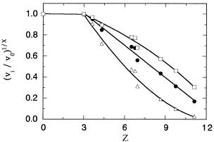
One complication with the foregoing approach is that not all amino acid residues will necessarily be modified at equal rates by a particular chemical modifier (see Tsou, 1962, for a detailed discussion of this complication). It commonly happens in experiments that some number of nonessential residues are modified at a faster rate than the catalytically essential residues. The effect of this situation is that an initial region of the Tsuo plot will occur where no decrease in enzymatic activity is realized, followed by a region of the expected linear decrease in activity with increasing value of z (Figure 10.14). The number of essential residues modified can still be ascertained from evaluation of the linear portion of such plots, as discussed by Tsou (1962). Norris and Brockelhurst (1976) extended this approach to the evaluation of multisubunit enzymes, where residues on each subunit are modified.
To clarify this approach, let us walk through an example of the experimental details of such a chemical modification study. Paterson and Knowles (1972) wished to determine the number of carboxylic acid groups required for catalytic activity in the proteolytic enzyme pepsin. To quantify this they treated the enzyme with [ C]trimethyloxonium fluoroborate, a reagent that esterifies carboxylate groups in proteins, hence imparting a C label to the protein after
Figure 10.14 Tsou plot of (vi /v0)1/x as a function of z for modification of the carboxylate groups of pepsin by trimethyloxonium fluoroborate. The data are plotted for x 1 (triangles), x 2 (solid circles), and x 3 (squares). Below z 3 modification of carboxylates has no effect on enzymatic activity. The data above z 3 are fit to a linear equation for x 2 and to third-order polynomials for x 1 and 3. The linear fit of the data for x 2 suggests that two carboxylates are critical for enzymatic activity. See text for further details. [Data adapted from Paterson and
Knowles (1972).]
344 TIME-DEPENDENT INHIBITION
each esterification reaction. Knowing the specific radioactivity of the modifying reagent (see Chapter 7), the researchers could quantify the number of C atoms incorporated into the enzyme after each reaction.
Varying concentrations of [ C]trimethyloxonium fluoroborate were added to samples of a solution of pepsin (20 mg/mL), the pH was maintained by addition of NaOH, and the sample was incubated until the modification reaction was complete. The radiolabeled protein was then separated from free reactants and by-products by size exclusion chromatography (see Chapter 7), after which the radioactivity associated with the protein was quantified by scintillation counting. Enzymatic activity of the samples after size exclusion chromatography was assessed by the ability of the enzyme to catalyze the hydrolysis of N-acetyl- -phenylalanyl- -phenylalanylglycine, a known substrate for pepsin.
The results of the experiments by Paterson and Knowles (1972) are summarized in Figure 10.14, where (v /v ) is plotted as a function of z (the number of C atoms incorporated per mole of enzyme). We see immediately from this figure that a fraction of nonessential carboxylates is rapidly modified without effect of enzymatic activity. From Figure 10.14 we can estimate that approximately three such groups occur in pepsin. After this, the activity of the enzyme decreases with increasing esterification of the carboxylates. To determine the number of carboxylates essential for catalysis, the fractional activity data are plotted in three different ways in Figure 10.14: as v /v (i.e., for x 1, triangles); as (v /v ) (i.e., x 2, solid circles); and as (v /v ) (i.e., x 3, squares). Paterson and Knowles fit the data in each form to both linear and polynomial functions, from which they concluded that the best fit to a straight line was obtained for x 2. From this analysis they were able to conclude that two carboxylate residues are essential for catalysis in the enzyme pepsin.
10.5.3.2 Substrate Protection Experiments. When catalytically essential groups are identified by chemical modification studies, a question that often arises is whether these groups are located within the substrate binding pocket (i.e., active site) of the enzyme. This issue can often be addressed by substrate protection experiments, in which one assesses the ability of the substrate, product, or a reversible competitive inhibitor to protect the enzyme against inactivation by the modifying reagent. If an essential amino acid side chain is located in the active site of an enzyme, formation of the reversible binary enzyme—substrate, enzyme—product, or enzyme—inhibitor (for competitive inhibitors) complex may occlude the amino acid so that it is no longer exposed to the chemical modifying reagent during inactivation studies. In this case, removal of free modifying reagent and protectant (i.e. substrate, etc.) by dialysis, size exclusion chromatography, and so on will reveal that enzymatic activity has been retained where the comparable experiment in the absence of protectant resulted in irreversible inactivation.
If the rate of inactivation is followed as described earlier (Scheme D of Figure 10.1, Figure 10.4, and Section 10.2.3) in the presence of varying

EXAMPLES OF SLOW BINDING ENZYME INHIBITORS |
345 |
concentrations of substrate, the observed rate constant for inactivation is found to depend on both substrate and inactivator concentrations as follows:
|
|
|
|
|
k [I] |
|
|
||
k |
|
|
|
|
|
K |
|
(10.20) |
|
|
|
K |
|
|
1 |
[S] |
|
[I] |
|
|
|
|
|
|
|
||||
Similar equations can be derived when a product or reversible competitive inhibitor is used as the protectant. Equation 10.20 provides a simple test for whether a catalytically essential group that is chemically modified by a particular reagent is localized to the active site. By measuring the diminution in rate of inactivation with increasing substrate concentration, the researcher can fit the experimental data to Equation 10.20 to determine whether the results are quantitatively consistent with this hypothesis (Figure 10.15). Equation 10.20 additionally provides a means of estimating the value of K for a
substrate when k and K are known from previous experiments. This is most useful in the case of multisubstrate enzymes that follow sequential-
mechanisms (see Chapter 11). If the first substrate to bind to the enzyme is varied in an inactivation experiment, its binding is in equilibrium with the free enzyme and the inactivator molecule. Hence, the K term in Equation 10.20 is replaced by K in this case, and the value of K for the substrate can be determined directly. An advantage of this approach over more conventional
Figure 10.15 Substrate protection against inactivation by chemical modification of an active site amino acid residue. As the substrate concentration is raised, the ability of the active site—directed inactivator to compete for binding and chemical modification of the enzyme is diminished. The symbols represent different [S]/K ratios at fixed concentrations of inactivator and enzyme. The lines drawn through the data were obtained by fitting to Equation 10.20 with K , [I], and k set to 1.0 M, 1.0 M, and 0.1 min , respectively.
346 TIME-DEPENDENT INHIBITION
equilibrium methods, such as equilibrium dialysis (Chapter 4) is that here only catalytic amounts of the enzyme are required for the determination of K . Thus, when enzyme supplies are limited, this type of experiment can be used to great advantage. See Malcolm and Radda (1970) and Anderton and Rabin (1970) for examples of this approach.
10.5.3.3 Affinity Labels. Covalent modifying groups can often be incorporated into substrate analogues and other ligands (cofactors, inhibitors, activators, etc.) to direct covalent modification to specific functional sites on the enzyme molecule. The work of Tang et al. (1995), discussed in Section 10.5.2, introduced the concept of affinity labeling an enzyme with a covalent modifier as a probe of enzyme structure and mechanism. This approach can help to identify key residues within a ligand binding pocket of an enzyme or receptor through a combination of covalent modification and subsequent peptide mapping studies.
As we just saw in Section 10.5.3.2, certain chemical functionalities will react selectively with specific amino acid side chains, and some of these functionalities can be synthetically incorporated into substrate molecules. Maleimides and succinyl esters, for example, have been incorporated into substrate and/or inhibitor analogues to specifically modify active site cysteine and lysine residues, respectively. Peptidic substrates of serine and cysteine proteinases can have halomethyl ketones and aldehydes incorporated into them to covalently modify the active site nucleophiles of these enzymes specifically. Likewise, metal chelating groups such as carboxylic and hydroxamic acids can be incorporated into the peptidic substrates of metalloproteinases to bind the active site metal in a slowly reversible manner. Alternatively, more permissive crosslinking agents can be incorporated into ligand analogues to determine the identify of amino acids in the ligand binding pocket. A particularly useful strategy is the use of nonselective photoaffinity labels for this purpose.
Photoaffinity labels are molecules that form highly reactive excited states when illuminated with light of an appropriate wavelength, leading to covalent modification of groups within the binding site of the protein. The value of these reagents is that they can be mixed with proteins under varying conditions, and the researcher can control the initiation of crosslinking by illuminating the sample. Two examples of such functionalities are aryl azides and the benzophenone group (Figure 10.16). For both groups, excitation into the * excited state results in formation of reactive centers that will combine with nearby methylene groups in the target enzyme or receptor. (In the case of the aryl azides, photolysis leads to formation of an aryl nitrene functionality, which then reacts with carbon—hydrogen or, preferably, oxygen—hydrogen bonds.) By incorporating such functionalities into a ligand molecule, photocrosslinking is targeted to the ligand binding pocket of the target protein. After photolysis, the ligand analogue is covalently attached to a group or groups within the binding pocket. In addition to the photocrosslinker, researches usually incorporate a chromophore, radiolabel, or other affinity label (e.g., biotin) into the

EXAMPLES OF SLOW BINDING ENZYME INHIBITORS |
347 |
Figure 10.16 Examples of photoaffinity labels that can be incorporated into substrate and inhibitor analogues to covalently modify residues within the ligand binding pocket of proteins:
(A) reaction of the benzophenone group and (B) reaction of the aryl azide group.
ligand structure to facilitate detection of the covalently linked species. The crosslinked protein—ligand complex can then be treated with an appropriate proteinase to cleave the target protein into a number of peptide fragments. These fragments are separated by HPLC or electrophoretic methods, and the fragment containing the crosslinked group is collected. The amino acid sequence of the labeled peptide can then be determined by mass spectroscopic methods or by traditional Edman sequencing chemistry. In this way the amino acids located within the ligand binding pocket can be identified.
An example of this approach comes from the work of DeGrado and coworkers (Kauer et al., 1986; O’Neil et al., 1989). This group wished to determine the location of the peptide binding pocket on the protein calmodulin. They had previously identified a peptide of the following sequence that bound tightly (K of 400 pM) and specifically to calmodulin:
Lys-Leu-Trp-Lys-Lys-Leu-Leu-Lys-Leu-Leu-Lys-Lys-Leu-Leu-Lys-Leu-Gly
They next synthesized a peptide of similar sequence in which the tryptophan at position 3 was replaced by a benzophenone. Mixing this peptide with
348 TIME-DEPENDENT INHIBITION
calmodulin and subsequent photolysis led to a covalent peptide—calmodulin complex that could be separated from free calmodulin by SDS-PAGE or reversed phase HPLC. The same peptide was also synthesized with a H- containing acetyl cap on the N-terminal lysine to impart a radiolabel to the peptide and photolysis product. Cleavage of the photoproduct with cyanogen bromide or S. aureus V8 proteinase led to selective cleavage of amide bonds within the calmodulin polypeptide without any cleavage of the peptide ligand. The tritium-containing cleavage product was separated by reversed phase HPLC and subjected to N-terminal amino acid sequence analysis. From these studies DeGrado and coworkers were able to identify Met 144 and Met 71 as the primary sites of photolabeling. These results allowed the researchers to build a model of the three-dimensional structure of the peptide binding pocket in calmodulin.
Affinity labeling of enzymes is a common and powerful tool for studying enzyme structure and mechanism. We have barely scratched the surface in our brief description of these methods. Fortunately there are several excellent in-depth reviews of these methods in the literature. General affinity labeling is covered in a dedicated volume of Methods in Enzymology (Jakoby and Wilchek, 1977). General chemical modification of proteins is covered well in the texts by Lundblad (1991) and Glazer et al. (1975). Photoaffinity labeling is covered in the Methods in Enzymology volume edited by Jakoby and Wilchek (1977) and also in review articles by Dorman and Prestwich (1994) and by Chowdhry (1979). These references should serve as good starting points for the reader who wishes to explore these tools in greater detail.
10.6 SUMMARY
In this chapter we have described the behavior of enzyme inhibitors that elicit their inhibitory effects slowly on the time scale of enzyme turnover. These slow binding, or time-dependent, inhibitors can operate by any of several distinct mechanisms of interaction with the enzyme. Some of these inhibitors bind reversibly to the enzyme, while others irreversibly inactivate the enzyme molecule. Irreversible enzyme inactivators that function as affinity labels or mechanism-based inactivators can provide useful structural and mechanistic information concerning the types of amino acid residue that are critical for ligand binding and catalysis.
We discussed kinetic methods for properly evaluating slow binding enzyme inhibitors, and data analysis methods for determining the relevant rate constants and dissociation constants for these inhibition processes. Finally, we presented examples of slow binding inhibitors and irreversible inactivators to illustrate the importance of this class of inhibitors in enzymology.
REFERENCES AND FURTHER READING |
349 |
REFERENCES AND FURTHER READING
Anderton, B. H., and Rabin, B. R. (1970) Eur. J. Biochem. 15, 568.
Chowdhry, V. (1979) Annu. Rev. Biochem. 48, 293.
Copeland, R. A. (1994) Methods for Protein Analysis: A Practical Guide to L aboratory Protocols, Chapman & Hall, New York, pp. 151—160.
Copeland, R. A., Williams, J. M., Giannaras, J., Nurnberg, S., Covington, M., Pinto, D., Pick, S., and Trzaskos, J. M. (1994) Proc. Natl. Acad. Sci. USA, 91, 11202.
Copeland, R. A., Williams, J. M., Rider, N. L., Van Dyk, D. E., Giannaras, J., Nurnberg, S., Covington, M., Pinto, D., Magolda, R. L., and Trzaskos, J. M. (1995) Med. Chem. Res. 5, 384.
Dorman, G., and Prestwich, G. D. (1994) Biochemistry, 33, 5661.
Glazer, A. N., Delange, R. J., and Sigman, D. S. (1975) Chemical Modification of Proteins, Elsevier, New York.
Jakoby, W. B., and Wilchek, M., Eds. (1977) Methods in Enzymology, Vol. 46, Academic Press, New York.
Kauer, J. C., Erickson-Viitanen, S., Wolfe, H. R., Jr., and DeGrado, W. F. (1986) J. Biol. Chem. 261, 10695.
Kettner, C., and Shervi, A. (1984) J. Biol. Chem. 259, 15106.
Kitz, R., Wilson, I. B. (1962) J. Biol. Chem. 237, 3245.
Lundblad, R. (1991) Chemical Reagents for Protein Modification, CRC Press, Boca Raton, FL. Malcolm, A. D. B., and Radda, G. K. (1970) Eur. J. Biochem. 15, 555.
Morrison, J. F. (1982) Trends Biochem. Sci. 7, 102.
Morrison, J. F., and Walsh, C. T. (1988) Adv. Enzymol. 61, 201.
Norris, R., and Brocklehurst, K. (1976) Biochem. J. 159, 245.
O’Neil, K. T., Erickson-Viitanen, S., and DeGrado, W. F. (1989) J. Biol. Chem. 264, 14571. Paterson, A. K., and Knowles, J. R. (1972) Eur. J. Biochem. 31, 510.
Picot, D., Loll, P. J., and Garavito, M. R. (1994) Nature, 367, 243.
Rome, L. H., and Lands, W. E. M. (1975) Proc. Natl. Acad. Sci. USA, 72, 4863.
Silverman, R. B. (1988a) Mechanism-Based Enzyme Inactivation: Chemistry and Enzymology, Vols. I and II, CRC Press, Boca Raton, FL.
Silverman, R. B. (1988b) J. Enzyme Inhib. 2, 73.
Tang, M. S., Askonas, L. J., and Penning, T. M. (1995) Biochemistry, 34, 808. Tian, W.-X., and Tsou, C.-L. (1982) Biochemistry, 21, 1028.
Tipton, K. F. (1973) Biochem. Pharmacol. 22, 2933.
Trzaskos, J. M., Fischer, R. T., Ko, S. S., Magolda, R. L., Stam, S., Johnson, P., and Gaylor, J. L.
(1995) Biochemistry, 34, 9677.
Tsou, C.-L. (1962) Sci. Sin. Ser. B (English ed.) 11, 1536.
Vane, J. R. (1971) Nature New Biol. 231, 232.
Weissman, G. (1991) Sci. Am. January, p. 84.