

Enzymes (Second Edition)
.pdf290 REVERSIBLE INHIBITORS
Figure 8.12 (A) Yonetani—Theorell plot for two inhibitors I and J that bind in a mutually exclusive fashion ( ) to a common enzyme. (B) Yonetani—Theorell plot for two nonexclusive inhibitors for which 1. Open circles are data points for [J] 0; solid circles are data points for [J] KJ .
enzyme. While this is often true, the caveat described for competitive inhibition with substrate (Section 8.2.1) holds here as well: mutually exclusive binding is observed when the two inhibitors bind to a common site on the enzyme, but it can potentially be observed if the two inhibitors bind at independent sites that strongly affect each other through conformational communication, so that ligand binding at one site precludes ligand binding at the second site. Hence, some caution is required in the interpretation of the results of studies of these types.
STRUCTURE—ACTIVITY RELATIONSHIPS AND INHIBITOR DESIGN |
291 |
8.6 STRUCTURE‒ACTIVITY RELATIONSHIPS AND INHIBITOR DESIGN
Modern attempts to identify inhibitors of specific enzymes have largely focused on elucidating the stereochemical and physicochemical features of inhibitory molecules that allow them to bind well to the enzyme (Suckling, 1991). Measures of inhibitor potency, such as K or IC , reflect the change in free energy that accompanies transfer of the inhibitor from the solvated aqueous state to the bound state in the enzyme binding pocket (i.e., the G of binding). We have already discussed the types of physicochemical force that are important in protein structure and ligand binding: hydrophobic interactions, hydrogen bonding, electrostatic interactions, and van der Waals forces. The same forces determine the strength of interaction between an inhibitor and an enzyme.
Likewise, we have seen that the shape or topology of a substrate will determine its ability to fit well into the binding pocket of an enzyme, based on the structural complementarity between the enzyme binding pocket and the substrate molecule. It stands to reason that the same structural complementarity should be important in inhibitor binding as well, and this is what is observed empirically. Thus if one can somehow identify a reasonably potent inhibitor of an enzyme, one can begin to make analogues of that molecule with varied structural and physicochemical properties to determine what effect these changes have of inhibitor potency. Attempts to correlate these structural changes with inhibitor potency are referred to as structure—activity relationship (SAR) studies.
Today SAR studies can be divided into two major strategic categories: SAR in the absence of structural information on the target enzyme and SAR that utilizes structural information about the enzyme that is obtained from x-ray crystallographic or multidimensional NMR studies. This latter category is also referred to as rational or structure-based inhibitor design. Section 8.6.1 and 8.6.2 introduce some of the techniques used for both these strategies.
8.6.1 SAR in the Absence of Enzyme Structural Information
Any SAR study begins with the identification of a lead compound that shows some potency for inhibiting the target enzyme. This lead compound might be identified by random screening of a compound library, such as a natural products library, or it might be based on the known structures of the substrate or product of a particular enzymatic reaction. With a lead compound in hand, analysts subject the substance to small structural perturbations and test these analogues for inhibitor potency. This most basic form of SAR study has been conducted in one way or another since the nineteenth century. The goals of these studies are to determine what structural changes will lead to improved inhibitor potency and to identify the pharmacophore, the minimal structure required for inhibition. Once identified, the pharmacophore serves as a template for further inhibitor design.
292 REVERSIBLE INHIBITORS
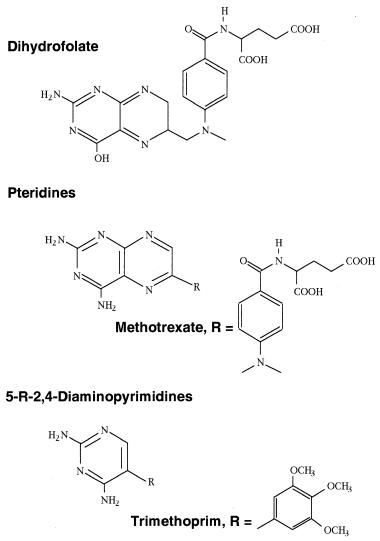
Consider the enzyme dihydrofolate reductase (DHFR), which catalyzes a key step in the biosynthesis of deoxythymidine. Inhibition of this enzyme blocks DNA replication and thus acts to inhibit cell growth and proliferation. DHFR inhibitors are therefore potentially useful therapeutic agents for the control of aberrant cell growth in cancer, and as antibiotics for the control of bacterial growth. Early attempts to identify inhibitors of this enzyme were based on synthesizing analogues of the substrate dihydrofolate. Figure 8.13 illustrates the chemical structures of dihydrofolate and two classes of DHFR inhibitors; the pteridines, exemplified by methotrexate, and the 5-substituted 2,4-diaminopyrimidines. Methotrexate was identified as a potent inhibitor of DHFR because of its striking structural similarity to the substrate dihydrofolate.
Next, the question of what portions of the methotrexate molecule were critical for DHFR inhibition was addressed by synthesizing various structural analogues of methotrexate. From these studies it was determined that the critical pharmacophore (i.e., the minimal structural component required for inhibition) was the 2,4-diaminopyrimidine ring system. This discovery led to the development of the second class of inhibitors illustrated in Figure 8.13, the 5-substituted 2,4-diaminopyrimidines, of which trimethoprim is a well-known example. Methotrexate is now a prescribed drug for the treatment of human cancers and certain immune-based diseases. Trimethoprim also is a prescribed drug, but its use is in the control of bacterial infections.
An unexpected outcome of the studies on these inhibitors was the finding that the pteridines, such as methotrexate, are potent inhibitors of both mammalian and bacterial DHFR, while trimethoprim and its analogues are much better inhibitors of the bacterial enzymes. The K values for trimethoprim for E. coli, and human lymphoblast DHFR are 1.35 and 170,000 nM, respectively (Li and Poe, 1988), a selectivity for the bacterial enzyme of about 126,000-fold! The reason for this spectacular species selectivity was not clear until the crystal structures of mammalian and bacterial DHFR were solved. (See Matthews et al., 1985, for a very clear and interesting account of these crystallographic studies and their interpretation.)
Today the enzymologist attempts to develop higher potency inhibitors not simply by random replacement of structural components on a molecule, but rather by systematic and rational changes in stereochemical and physicochemical properties of the substituents. Some properties to be changed are obvious from the structure of the lead compound. If, for example, the lead inhibitor contains a carboxylic acid group, one immediately wonders whether acid— base-type interactions with a group on the enzyme are involved in binding. One might substitute the carboxylate moiety with an ester, for example, to determine the importance of the carboxylate in binding. More general properties of chemical substituents can be examined as well. These studies call for quantitative measures of the different physicochemical properties to be considered. Among the relevant general properties of chemical substituents, steric bulk, hydrophobicity, and electrophilicity/nucleophilicity are generally agreed
STRUCTURE—ACTIVITY RELATIONSHIPS AND INHIBITOR DESIGN |
293 |
Figure 8.13 Chemical structures of the substrate (dihydrofolate) and two types of inhibitor of the enzyme dihydrofolate reductase (DHFR).
to be important factors, and chemists have therefore developed quantitative measures of these parameters.
Several measures have been suggested to quantify steric bulk or molecular volume. One of the earliest attempts at this was the Taft steric parameter E , which was defined as the logarithm of the rate of acid-catalyzed hydrolysis of a carboxymethyl-substituted molecule relative to the rate for the methyl acetate
294 |
REVERSIBLE INHIBITORS |
|
analogue (Taft, 1953; Nogrady, 1985): |
|
|
|
E log(k ) log(k ) |
(8.27) |
A more geometric measure of steric bulk is provided by the Verloop steric parameter, which basically measures the bond angles and bond lengths of the substituent group (Nogrady, 1985). Chemists also have used the molar refractivity as a measure of molecular volume of substituents (Pauling and Pressman, 1945; Hansch and Klein, 1986). The molar refractivity, MR, is defined as
follows: |
|
||||
MR |
n 1 MW |
(8.28) |
|||
|
|
|
|
||
n 1 |
d |
||||
where n is the index of refraction, MW is the molecular weight, and d is the density of the substituent under consideration. Since n does not vary widely among organic molecules, MR is mainly a measure of molecular volume.
The relative hydrophobicity of molecular substituents is most commonly measured by their partition coefficient between a polar and nonpolar solvent. For this purpose, chemists have made water and octanol the solvents of choice. The molecule is dissolved in a 1:1 mixture of the two solvents, and its concentration in each solvent is measured at equilibrium. The partition coefficient is then calculated as the equilibrium constant:
P |
[I] |
|
(8.29) |
[I] |
|||
|
|
|
|
In measuring relative hydrophobicity, the effect of different substituents, on the partition coefficient of benzene in octanol/water is used as a standard. The hydrophobic parameter is used for this purpose, and is defined as follows (Hansch and Klein, 1986):
log(P ) log(P ) |
(8.30) |
where P is the partition coefficient for a monosubstituted benzene with substituent x, and P is the partition coefficient of benzene itself.
The most widely used index of electronic effects in inhibitor design is the Hammett constant. Originally developed to correlate quantitatively the relationship between the electron-donating or -accepting nature of a parasubstituent on the ionization constant for benzoic acid in water (Hammett, 1970; Nogrady, 1985); this index is defined as follows:
log(K ) log(K ) |
(8.31) |
where K is the ionization constant for the parasubstituted benzoic acid with |
|
substituent x and K is the ionization constant for benzoic acid. Groups that |
|
are electron acceptors (e.g., COOH, NO , NR ) withdraw electron density |
|
|
|
STRUCTURE—ACTIVITY RELATIONSHIPS AND INHIBITOR DESIGN |
295 |
from the ring system, hence stabilize the ionized form of the acid; such groups have positive values of . Electron-donating groups (e.g., OH, OCH , NH ) have the opposite effect on the ionization constant and thus have negative values of . Values of for a very large number of organic substituents have been tabulated by several authors. One of the most comprehensive list of values can be found in the text by Martin (1978).
The ability to quantify these various physicochemical properties has led to attempts to express the inhibitor potency of molecules as a mathematical function of these parameters. This strategy of quantitative structure—activity relationships (QSAR) was first championed by Hansch and his coworkers (Hansch, 1969; Hansch and Leo, 1979). In a typical QSAR study, a series of analogues of a lead inhibitor is prepared with substituents that systematically vary the parameters described earlier. The experimentally determined potencies of these compounds are then fit to varying linear and nonlinear weighted sums of the parameter indices to obtain the best correlation by regression analysis. Equations 8.32—8.34 illustrate forms typically used in QSAR work.
1 |
|
|
|
||
log |
|
|
|
a b cMR d |
(8.32) |
K |
|||||
|
|
|
|
|
|
1 |
|
|
|
||
log |
|
a b cMR d |
(8.33) |
||
K |
|||||
|
|
|
|
|
|
1 |
|
|
|
||
log |
|
a b c log( MR) d |
(8.34) |
||
K |
|||||
|
|
|
|
|
|
Equation 8.32 is a simple linear relationship, while Equations 8.33 and 8.34 have nonlinear components. In these equations the values a, b, c, d, and are proportionality constants, determined from the regression analysis. In developing a mathematical expression for the correlation relationship here, one hopes to predict the inhibitory potency of further compounds prior to their synthesis, based on the equation established from the QSAR. In practice, the predictive power of these QSAR equations varies dramatically. When such predictions fail, it is usually because additional factors that influence inhibitor potency were not quantitatively included in the functional expression. In some cases these additional factors are neither well understood nor easily quantified.
As a simple example of QSAR, let us again consider the inhibition of bacterial DHFR by pteridines and 5-substituted 2,4-diaminopyrimidines. Coats et al. (1984) studied the QSARs of both classes of compounds for their ability to inhibit the DHFR from the bacterium L actobacillus casei. They measured the IC values for 25 pteridine analogues with different R substituents and also for 33 5-substituted 2,4-diaminopyrimidine analogues with different R groups (Figure 8.13). From these data they determined the QSAR equations given by Equations 8.35 and 8.36 for the pteridines and 5-R-2,4-
296 |
REVERSIBLE INHIBITORS |
|
||||
diaminopyrimidines, respectively: |
|
|||||
|
1 |
|
|
|
||
|
log |
|
|
|
0.23 0.004 0.77I 3.39 |
(8.35) |
|
IC |
|||||
|
|
|
|
|
|
|
|
1 |
|
|
|
||
|
log |
|
0.38 0.007 0.66I 2.15 |
(8.36) |
||
|
IC |
|||||
|
|
|
|
|
|
|
In these equations, the parameter refers to the hydrophobicity of the R group, and the index I is an empirical parameter related to the presence of a —N—C— or —C—N— bridge between the parent ring system and an aromatic ring on the substituent (Coats et al., 1984). The relationships between the IC values calculated from these equations and the experimentally determined IC values are illustrated in Figure 8.14. Again, one must keep in mind that the correlations illustrated are for the molecules used to establish the QSAR equations. The value of these equations in predicting the inhibitor potency of other molecules will depend on how significantly other unaccount- ed-for factors influence potency. Nevertheless, QSAR provides a means of rationalizing the observed potencies of structurally related compounds in terms of familiar physicochemical properties. An up-to-date volume by Kubinyi (1993) provides a detailed and practical introduction to the field of QSAR. This text should be consulted as a starting point for acquiring a more in-depth treatment of the subject.
Another approach to designing potent inhibitors of enzymes is to consider the probable structure of the transition state of the chemical reaction catalyzed by the enzyme. As described in Chapter 6, the catalytic efficiency of enzymes is due largely to their ability to achieve transition state stabilization. If this stabilization is equated with binding energy, a stable analogue that mimics the structure of the transition state should bind to an enzyme some 10 —10 times greater than the corresponding ground state substrate molecule (Wolfenden, 1972). Since typical substrate K values are in the millimolar-to- nanomolar range, a true transition state mimic may bind to its target enzyme with a K value between 10 and 10 M! With such incredibly tight binding affinities, such inhibitors would behave practically as irreversible enzyme inactivators.
The foregoing approach to inhibitor design has been hindered by the great difficulty of obtaining information on transition state structure by traditional physical methods. Because they are so short-lived, the transition state species of most enzymatic reactions are present under steady state conditions at very low concentrations (i.e., femtomolar or less). Hence, attempts to obtain structural information on these species from spectroscopic or crystallographic methods have been largely unsuccessful. Information on transition state structure can, however, be gleaned from analysis of kinetic isotope effects on enzyme catalysis, as recently reviewed by Schramm et al. (1994). As discussed in Chapter 7, kinetic isotope effects are observed because of the changes in

STRUCTURE—ACTIVITY RELATIONSHIPS AND INHIBITOR DESIGN |
297 |
Figure 8.14 QSAR correlation plots for the potencies of pteridines (A) and 5-substituted 2,4-diaminopyrimidines (B) as inhibitors of the dihydrofolate reductase from L. casei. [Data from
Coats et al. (1984).]
vibrational frequencies for the reactant and transition state species that accompany heavy isotope incorporation. By synthesizing substrate analogues with heavy isotopes at specific locations, one can determine the kinetic isotope effects imparted by each replacement. From this type of information, one can use vibrational normal mode calculations to identify the vibrational modes that are most strongly perturbed in the transformation from reactant to transition state of the substrate, hence to map out the structural changes that have occurred in the molecule. The information thus obtained can then be used to design molecules that mimic the structure of the reaction transition state.

298 REVERSIBLE INHIBITORS
This approach has been applied to the design of transition state analogues of renin, an aspartyl protease (Blundell et al., 1987).
In vivo, renin is responsible for the proteolytic processing of angiotensinogen to angiotensin I, the progenitor of the vasoconstrictor peptide angiotensin II. The substrate is hydrolyzed at a Leu-Val peptide bond, and the hydrolysis reaction is proposed to utilize an active site water molecule as the attacking nucleophile to produce the tetrahedral transition state illustrated in Figure 8.15A. The peptide sequence of the renin substrate angiotensinogen is shown in Figure 8.15C. In their first attempt at an inhibitor of renin, Blundell and coworkers replaced the P1 carbonyl group by a methylene linkage, yielding the reduced isostere (—CH —NH—) containing peptide inhibitor 1 (Figure 8.15C). The investigators next noted that the pepstatins are naturally occurring protease inhibitors that contain the unusual amino acid statine (Figure 8.15B), which in turn contains a —CH(OH)— moiety that resembles the proposed transition state of renin. Pepstatin is a poor renin inhibitor; but, reasoning that the statine group was a better transition state mimic than the reduced peptide isostere (—CH —NH—), Blundell et al. incorporated this structure into their peptide inhibitor to produce 2 (Figure 8.15C). The closest analogue to the true transition state of the reaction would be one incorporating
Figure 8.15 (A) Proposed structure of the tetrahedral transition state of the renin proteolysis reaction. (B) Chemical structure of the statine moiety of natural protease inhibitors, such as pepstatin. (C) Structures and IC values for the peptide substrate and inhibitors of renin, incorporating various forms of transition state analogues. [Data taken from Blundell et al.
(1987).]
STRUCTURE—ACTIVITY RELATIONSHIPS AND INHIBITOR DESIGN |
299 |
a —CH(OH)—NH— group at P1—P1 of the peptide. Since, however, synthesis of this transition state analogue was hampered by the instability of the resulting compound, Blundell’s group instead synthesized a closely related analogue containing a hydroxyethylene moiety [(—CH(OH)—CH —), 3, which proved to be an extremely potent inhibitor of the enzyme (Figure 8.15C).
To date, the de novo design of transition state analogues as enzyme inhibitors has been applied only to a limited number of enzymes by a handful of laboratories. With improvements in the computational methods associated with this strategy, however, more widespread use of this approach is likely to be seen in the future.
8.6.2 Inhibitor Design Based on Enzyme Structure.
In the search for potent enzyme inhibitors, knowledge of the three dimensional structure of the inhibitor binding site on the enzyme provides the ultimate guide to designing new compounds. The structures of enzyme active sites can be obtained in atomic detail from x-ray crystallography and multidimensional NMR spectroscopy. A detailed discussion of these methods is beyond the scope of the present text. Our discussion will focus instead on the use of the structural details obtained from these techniques. The reader interested in learning about protein crystallography and NMR spectroscopy can find many excellent review articles and texts (see McRee, 1993, and Fesik, 1991, for good introductions to protein crystallography and NMR spectroscopy, respectively).
The crystal or NMR structure of an enzyme with an inhibitor bound provides structural details at the atomic level on the interactions between the inhibitor and the enzyme that promote binding. Hydrogen bonding, salt bridge formation, other electrostatic interactions, and hydrophobic interactions can be readily inferred from inspection of a high resolution structure. Figure 8.16 provides simplified schematic representations of the binding interactions between DHFR and its substrate dihydrofolate and inhibitor methotrexate, illustrating the involvement of common amino acid residues in the binding of both ligands. These structural diagrams also indicate that the orientation and hydrogen bonding patterns are not identical for the substrate and the inhibitor. Nevertheless, the major forces involved in binding of both ligands to the enzyme are hydrogen bonds between amino acid residues of the active site and the 2,4-diaminopyrimidine ring of the ligands. Visual inspection by means of molecular graphics methods suggested that this ring constituted the critical pharmacophore and led to the design of trimethoprim, the prototypical 5-substituted 2,4-diaminopyrimidine (Marshall and Cramer, 1988). As we have seen, these structural inferences are consistent with the SAR and QSAR studies of DHFR inhibitors.
The example of trimethoprim suggests a straightforward, if tedious, means of utilizing structural information in the design of new enzyme inhibitors: namely, the iterative design, synthesis, and crystallization of inhibitor—enzyme complexes. In this approach, one starts with the crystal structure of the free