

Color Atlas of Neurology
.pdfParkinson Disease: Treatment
|
|
|
|
|
|
|
|
|
|
|
|
|
Glial cell |
|
|
3-0-methyldopamine |
|
DOPAC (dihydroxyphenylacetic acid) |
|
|
|
|
|
|
|||||||||||
|
|
|
|
|
|
||||||||||||
|
|
|
|
(converted to homo- |
|||||||||||||
|
|
|
|
|
|
|
|
|
|
|
|
|
|
|
|
|
|
Dopamine reabsorption |
|
|
|
|
|
|
|
|
|
|
vanillic acid) |
||||||
|
|
|
|
|
|
|
|
|
|
||||||||
Dopamine release |
|
|
|
|
|
|
|
|
|
MAO-B |
|||||||
|
|
|
|
|
|
|
|
|
|||||||||
Presynaptic terminal |
|
|
|
|
|
|
|
|
|
|
|
COMT |
|||||
|
|
|
|
|
|
|
|
|
|
|
|||||||
|
|
|
|
|
|
|
|
|
|
|
|
|
|
|
|
|
|
|
|
|
|
|
|
|
|
|
|
|
|
|
|
|
|
|
|
|
|
Tyrosine |
|
|
|
||||||||||||
Phenylalanine |
|
L-Dopa |
Dopamine |
|
|
|
|||||||||||
Phenylalanine |
Tyrosine |
Dopa |
|
|
D2 receptor |
||||||||||||
hydroxylase |
hydroxy- |
decar- |
|
|
|
||||||||||||
|
|
|
lase |
boxylase |
|
|
|
||||||||||
D1 receptor
Transport protein |
|
|
|
|
|
|
|
|
|||
Dopamine vesicle |
|
|
Postsynaptic ending |
||
|
|||||
|
|
|
|
|
|
Striatal dopaminergic synapse (schematic) |
|
||||
Occupational therapy
Speech therapy
Physical therapy
Central Nervous System
213
Rohkamm, Color Atlas of Neurology © 2004 Thieme
All rights reserved. Usage subject to terms and conditions of license.

Multiple Sclerosis
|
Multiple sclerosis (MS) is characterized by mul- |
|
|
tiple symptoms and signs of brain and spinal |
|
|
cord dysfunction that are disseminated in both |
|
|
time and space. Its pathological hallmark is in- |
|
|
flammatory demyelination and axonal lesions; |
|
|
its etiology remains unknown at present despite |
|
|
decades of intensive investigation. |
|
|
A relapse is the appearance of a new neurologi- |
|
|
cal disturbance, or the reappearance of one pre- |
|
|
viously present, lasting at least 24 hours. All |
|
|
such disturbances arising within a one-month |
|
System |
period are counted as a single relapse. The re- |
|
lapse rate is the number of relapses per year. |
||
Clear improvement of neurological function is |
||
|
||
|
termed remission. |
|
Nervous |
The course of MS varies greatly from one in- |
|
(66–85%; most common when onset is before |
||
|
dividual to another, but two basic types of |
|
|
course can be identified: relapsing-remitting |
|
Central |
age 25; well-defined relapses separated by peri- |
|
ods of nearly complete recovery with or without |
||
|
||
|
residual symptoms; does not progress during |
|
|
remission) and chronic progressive. The latter |
|
|
can be divided into three subtypes: primary |
|
|
chronic progressive (9–37%; most common |
|
|
when onset is after age 40; progresses from dis- |
|
|
ease onset onward); secondary progressive (seen |
|
|
in over 50% of cases 6–10 years after onset; ini- |
|
|
tially remitting-relapsing, later chronically pro- |
|
|
gressive; recurrences, mild remissions, and |
|
|
plateau phases may occur); and progressively re- |
|
|
mitting-relapsing (rare; complete remission may |
|
|
or may not occur after relapses; symptoms tend |
|
|
to worsen from one relapse to the next). |
|
|
Clinical Manifestations |
|
|
The symptoms and signs of MS reflect dysfunc- |
|
|
tion of the particular areas of the nervous sys- |
|
|
tem involved and are not specific for this dis- |
|
|
ease. Typical MS manifestations include paraly- |
|
|
sis, paresthesiae, optic neuritis (retrobulbar |
|
|
neuritis), diplopia, and bladder dysfunction. |
|
|
Paresis, spasticity, fatigability. Upper-motor- |
|
|
neuron type paralysis of the limbs either is pre- |
|
|
sent at onset or develops during the course of |
|
|
MS. Involvement is often asymmetrical and |
|
|
mainly in the legs, especially in the early stage of |
214the disease. Spasticity makes its first appearance in the form of extensor spasms; flexor
spasms develop later. The latter are often pain-
ful, cause frequent falls, and, if severe and persistent, can cause flexion contractures (paraplegia in flexion). Many patients complain of abnormal fatigability.
Sensory manifestations. Episodic or continuous paresthesiae (sensations of tingling or numbness, tightness of the skin, heat, cold, burning, prickling) are common, particularly in the early stage of the disease, with or without other manifestations of neurological dysfunction. As the disease progresses, such positive phenomena usually recede and are replaced by sensory deficits affecting all sensory modalities. A constant or only slowly rising sensory level (“sensory transverse cord syndrome”) is uncharacteristic of MS and should prompt the search for a spinal cord lesion of another kind. Many MS patients have Lhermitte’s sign (which is actually a symptom), an electric or coldlike paresthesia traveling from the nuchal region down the spine, sometimes as far as the legs, on flexion of the neck (p. 49). If no other symptoms or signs are present, other causes should be considered (e. g., a cervical spinal cord tumor).
Pain in MS most often appears in the form of trigeminal neuralgia (p. 186), severe pain in the limbs (p. 108), tonic spasms (p. 204), or backaches, sometimes with radiation in a radicular pattern. Other painful phenomena include flexor spasms due to spasticity, contractures, and dysuria due to urinary tract infection.
Visual impairment in MS is usually due to optic neuritis (mostly unilateral), which also produces pain in or around the eye. The impairment begins as blurred or clouded vision and progresses to cause reading impairment and visual field defects (central scotoma or diffuse defects). Marcus Gunn pupils (p. 92) may be observed. Physical exercise, high ambient temperature, menstruation, or cigarette smoking can aggravate existing visual problems (Uhthoff’s phenomenon). Optic neuritis, as an isolated finding, is not necessarily the first manifestation of MS; patients with bilateral optic neuritis have a much lower risk of developing MS than those with the unilateral form. Diplopia is usually due to internuclear ophthalmoplegia (p. 86). Nystagmus (p. 88).
Rohkamm, Color Atlas of Neurology © 2004 Thieme
All rights reserved. Usage subject to terms and conditions of license.

Multiple Sclerosis
|
|
|
|
|
|
|
|
|
|
|
|
|
|
|
|
|
|
|
|
|
|
Sensory disturbances |
|
|
|||||
Test for visual field defects (confrontation test) |
|
System |
|||||||
|
|
|
|
|
|
|
|
|
|
|
|
|
|
|
|
|
|
|
Nervous |
|
|
|
|
|
|
|
|
|
Central |
|
|
|
|
|
|
|
|
|
|
|
|
|
|
|
|
|
|
Motor disturbances |
|
|
|
|
|
|
|
|
|
(central paresis, spasticity, |
|
|
|
|
|
|
|
|
|
abnormal fatigability) |
|
Central scotoma (optic neuritis) |
Atrophy |
Nystagmus of abducting eye
|
Adductor |
|
|
paralysis |
|
Dissociated nystagmus |
Temporal papillary atrophy |
|
(internuclear ophthalmoplegia, |
215 |
|
patient looking to right) |
(after optic neuritis) |
|
Rohkamm, Color Atlas of Neurology © 2004 Thieme
All rights reserved. Usage subject to terms and conditions of license.

Multiple Sclerosis
Incoordination. Intention tremor, dysarthria, truncal ataxia, and oculomotor dysfunction are common. Gait unsteadiness due to motor incoordination is often experienced by the patient as dizziness or lightheadedness. Acute vertigo with nausea, vomiting, and nystagmus can also occur.
|
Autonomic |
dysfunction. |
Bladder |
dysfunction |
||
|
(p. 156) frequently develops in the course of |
|||||
|
MS, causing problems such as urinary urgency, |
|||||
|
incomplete voiding, or urinary incontinence. |
|||||
System |
Urinary tract infection is a not infrequent re- |
|||||
sult. Fecal incontinence (p. 154) is rare, but con- |
||||||
stipation is common. Sexual dysfunction (e. g., |
||||||
|
||||||
|
erectile dysfunction or loss of libido) is also |
|||||
Nervous |
common and may be aggravated by spasticity |
|||||
rity, and |
marital conflict |
often play a role as |
||||
|
or sensory deficits in the genital region. Psy- |
|||||
|
chological factors such as depression, insecu- |
|||||
Central |
well. If its cause is organic, sexual dysfunction |
|||||
in MS is usually accompanied by bladder dys- |
||||||
|
||||||
|
function. |
|
|
|
|
|
|
Behavioral changes. Mental changes (depres- |
|||||
|
sion, marital conflict, anxiety) and cognitive |
|||||
|
deficits of variable severity can occur both as a |
|||||
|
reaction to and as a result of the disease. |
|||||
|
Paroxysmal phenomena in MS include epileptic |
|||||
|
seizures, trigeminal neuralgia, attacks of dy- |
|||||
|
sarthria with ataxia, tonic spasms, episodic dy- |
|||||
|
sesthesiae, pain, and facial myokymia. |
|||||
|
Differential Diagnosis |
|
|
|||
|
There is no single clinical test, imaging study, or |
|||||
|
laboratory finding that alone establishes the di- |
|||||
|
agnosis of MS (p. 218; Table 27, p. 375). A metic- |
|||||
|
ulous differential diagnostic evaluation is |
|||||
|
needed in every case. |
|
|
|||
|
(Cerebral) Vasculitis (p. 180). Systemic lupus |
|||||
|
erythematosus, Sjögren syndrome, Behçet syn- |
|||||
|
drome, |
granulomatous |
angiitis, |
polyarteritis |
||
|
nodosa, antiphospholipid syndrome, chronic in- |
|||||
|
flammatory |
demyelinating polyradiculoneuro- |
||||
|
pathy (CIDP, p. 328). |
|
|
|||
Inflammatory diseases. Neurosarcoidosis, neuroborreliosis, neurosyphilis, Whipple disease, postinfectious acute disseminated encephalomyelitis (ADEM), progressive multifocal
216leukoencephalopathy (PML), subacute sclerosing panencephalitis (SSPE), HIV infection,
HTLV-1 infection.
Neurovascular disorders. Arteriovenous fistula of spinal dura mater, cavernoma, CADASIL (p. 172).
Hereditary/metabolic disorders. Spinocerebellar ataxias, adrenoleukodystrophy, endocrine diseases, mitochondrial encephalomyelopathy, vitamin B12 deficiency (funicular myelosis).
Tumors of the brain or spinal cord (e. g., lymphoma, glioma, meningioma).
Skull base anomalies. Arnold–Chiari malformation, platybasia.
Myelopathy. Cervical myelopathy (spinal stenosis).
Somatoform disturbances in the context of mental illness.
Prognosis
Favorable prognostic indicators in MS include onset before age 40, monosymptomatic onset, absence of cerebellar involvement at onset, rapid resolution of the initial symptom(s), a re- lapsing-remitting course, short duration of relapses, and long-term preservation of the ability to walk. A relatively favorable course is also predicted if, after the first 5 years of illness, the MRI reveals no more than a few, small lesions without rapid radiological progression and the clinical manifestations of cerebellar disease and central paresis are no more than mild. A benign course, defined as a low frequency of recurrences and only mild disability in the first 15 years of illness, is seen in 20–30% of patients. The disease takes a malignant course, with major disability within 5 years, in fewer than 5% of patients. Half of all MS patients have a second relapse within 2 years of disease onset.
Rohkamm, Color Atlas of Neurology © 2004 Thieme
All rights reserved. Usage subject to terms and conditions of license.

Multiple Sclerosis
Autonomic dysfunction
(urinary/fecal incontinence, sexual dysfunction)
Impaired coordination
Paroxysmal symptoms
(trigeminal neuralgia) Behavioral changes
Central Nervous System
217
Rohkamm, Color Atlas of Neurology © 2004 Thieme
All rights reserved. Usage subject to terms and conditions of license.

Multiple Sclerosis
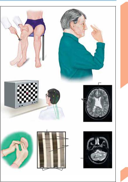
Diagnosis
|
Patients with MS are evaluated by clinical ex- |
|
|
amination, laboratory testing, neuroimaging, |
|
|
and neurophysiological studies. The clinical |
|
|
manifestations of MS and the lesions that cause |
|
|
them vary over the course of the disease (dis- |
|
|
semination in time and space). Diagnostic classi- |
|
|
fication is problematic (p. 216) if only one lesion |
|
|
is found (e. g., by MRI), if symptoms and signs |
|
|
are in only one area of the CNS (e. g., spinal |
|
|
cord), or if only one attack has occurred (Table |
|
System |
27, p 375). |
|
! Clinical Manifestations |
||
|
||
Nervous |
Sensory deficits, upper-motor-neuron paresis, |
|
incoordination, visual impairment (field de- |
||
|
||
|
fects), nystagmus, internuclear ophthalmople- |
|
|
gia, and/or bladder dysfunction are common |
|
Central |
signs of MS. Complaints of pain, paresthesiae, |
|
abnormal fatigability, or episodic disturbances |
||
are often, by their nature, difficult to objectify. |
||
Clinical examination may reveal no abnormality |
||
|
because of the episodic nature of the disease it- |
|
|
self. |
|
|
! Laboratory Tests and Special Studies |
|
|
Evoked potentials. Visual evoked potential (VEP) |
|
|
studies reliably detect optic nerve lesions, but |
|
|
neuroimaging is better for detecting lesions of |
|
|
the optic tract or optic radiation. VEP reveals |
|
|
prolongation of the P100 latency in one eye and/ |
|
|
or an abnormally large discrepancy between the |
|
|
latencies in the two eyes in roughly 40% of MS |
|
|
patients without known optic neuritis, and in al- |
|
|
most half of those with early optic neuritis. So- |
|
|
matosensory evoked potential (SEP) studies of |
|
|
the median or tibial nerve typically reveal pro- |
|
|
longed latencies in MS. Low amplitude of |
|
|
evoked potentials, on the other hand, often indi- |
|
|
cates a pathological process of another type, e. g. |
|
|
tumor. SEP abnormalities are found in up to 60% |
|
|
of MS patients with predominantly sensory |
|
|
manifestations. Auditory evoked potential (AEP) |
|
|
studies are less sensitive in MS than VEP or SEP. |
|
|
The most common AEP change is prolongation |
|
|
of latency. AEP studies are helpful for the further |
|
|
classification of vertigo, tinnitus, and hearing |
|
218 |
loss. Motor evoked potential (MEP) studies re- |
|
veal prolonged central conduction times when |
||
|
||
|
CNS lesions involve the pyramidal pathway. The |
|
|
|
sensitivity of MEP in MS is approximately the same as that of SEP. MEP studies can provide supporting evidence for MS in patients with latent paresis, gait disturbances, abnormal reflexes, or movement disorders that are difficult to classify.
Tests of bladder function. The residual urine volume can be measured by ultrasound. It should not exceed 100 ml in patients with a normal bladder capacity of 400–450 ml; in general, it should normally be 15–20% of the cystomanometrically determined bladder volume.
Urodynamic electromyography (EMG) provides more specific data concerning bladder dysfunction.
Neuroimaging. CT may reveal other diseases that enter into the differential diagnosis of MS (e. g., brain tumor) but is insufficiently sensitive (ca. 25–50%) to be useful in diagnosing MS itself. MRI scans reveal the characteristic foci of demyelination disseminated in the CNS (MS plaques); contrast enhancement is seen in acute but not in chronic lesions. The sensitivity of MRI for MS is greater than 90%, but its specificity is considerably lower; thus, the MRI findings alone cannot establish the diagnosis.
CSF examination. CSF abnormalities are found in more than 95% of MS patients. The cell count rarely exceeds 20 cells/mm3. The total protein concentration is elevated in ca. 40% of patients, and intrathecal IgG synthesis (IgG index) in ca. 90%. Oligoclonal IgG is found in 95% of MS patients, and antibodies to mumps, measles and herpes zoster in 80%.
Pathogenesis
The early course of MS varies among patients in accordance with the variable extent of the inflammatory lesions and disturbances of the blood–brain-barrier. The severity of late manifestations is correlated with the number of plaques. It is hypothesized that MS is caused by a combination of genetic (polygenic) predisposition and exogenous factors (viral or bacterial infection?) that induces an inappropriate immune response to one or more CNS autoantigens (see p. 220) that have not yet been identified.
Rohkamm, Color Atlas of Neurology © 2004 Thieme
All rights reserved. Usage subject to terms and conditions of license.
Multiple Sclerosis
Central paresis |
Clinical findings |
|
|
Impaired |
(right hyperreflexia) |
|
|
||
|
|
|
|
coordination |
|
|
|
|
Ventricle |
VEP measurement |
|
Lesions |
|
|
|
|
|||
|
|
|
|
|
|
|
|
|
MRI (T2-weighted image |
|
Oligoclonal bands |
|
|
of cerebral hemispheres) |
|
|
|
|
Serum
CSF
Lesions
Lumbar puncture |
IEF in MS |
IEF in normals |
MRI (T2-weighted |
|
|
|
image of cerebellum) |
Special tests in MS (IEF = isoelectric focusing)
Central Nervous System
219
Rohkamm, Color Atlas of Neurology © 2004 Thieme
All rights reserved. Usage subject to terms and conditions of license.

Central Nervous System
Multiple Sclerosis
Activation. Circulating autoreactive CD4+ T lymphocytes bear antigen-specific surface receptors and can cross the blood–brain barrier (BBB) when activated, e. g., by neurotropic viruses, bacterial superantigens, or cytokines. In MS, activated T lymphocytes react with MBP, PLP, MOG, and MAG. Circulating antibodies to various components of myelin can also be detected (for abbreviations, see below1).
Passage through the BBB. Activated lymphocytes and myelinotoxic antibodies penetrate the BBB at the venules (perivenous distribution of inflammation).
Antigen presentation and stimulation. In the CNS, antigen-presenting cells (microglia), recognition molecules (MHC class II antigens), and co-stimulatory signals (CD28, B-7.1) trigger the renewed activation and clonal proliferation of incoming CD4+ T lymphocytes into TH1 and TH2 cells. Proinflammatory cytokines elaborated by the TH1 cells (IL-2, IFN-γ, TNF-α, LT)2 induce phagocytosis by macrophages and microglia as well as the synthesis of mediators of inflammation (TNF-α, OH–, NO)2 and complement factors. The TH2 cells secrete cytokines (IL-4, IL-5, IL-6)2 that activate B cells ( myelinotoxic autoantibodies, complement activation), ultimately causing damage to myelin. The TH2 cells also produce IL-4 and IL-10, which suppress the TH1 cells.
Demyelination. Lesions develop in myelin sheaths (which are extensions of oligodendroglial cell membranes) and in axons when the inflammatory process outstrips the capacity of repair mechanisms.
Scar formation. The inflammatory response subsides and remyelination of damaged axons begins once the autoreactive T cells die (apoptosis), the BBB is repaired, and local anti-inflam- matory mediators and cells are synthesized. Astroglia form scar tissue that takes the place of the dead cells. Axonal damage seems to be the
main cause of permanent neurological deficits, as dystrophic axons apparently cannot be remyelinated.
Treatment
Relapse is treated with high-dose corticosteroids, e. g., methylprednisolone, 1 g/day for 3–5 days, which produce (unselective) immunosuppression, reduce BBB penetration by T cells, and lessen TH1 cytokine formation. Plasmapheresis may be indicated in refractory cases.
Drugs that reduce the frequency and intensity of relapses. Azathioprine p.o. (immunosuppression via reduction of T cell count), interferon beta-1b and beta-1a s.c. or i.m. (cytokine modulation, alteration of T-cell activity), glatiramer acetate s.c. (copolymer-1; blocks/competes at binding sites for encephalitogenic peptides on MHC-II molecules), IgG i. v. (multiple modes of action), and natalizumab (selective adhesion inhibitor).
Drugs that delay secondary progression. Interferon beta-1b and beta-1a. Mitoxantrone suppresses B cells and decreases the CD4/CD8 ratio. Methotrexate and cyclophosphamide
(different dosage schedules) delay MS progression mainly by unselective immunosuppression and reduction of the T-cell count.
Slowing of primary progression. No specific therapy is known at present.
Symptomatic therapy/rehabilitation. Medications, physical, occupational, and speech therapy, social, psychological, and dietary counseling, and mechanical aids (e. g., walking aids, wheelchair) are provided as needed. The possible benefits of oligodendrocyte precursor cell transplantation for remyelination, and of growth factors and immunoglobulins for the promotion of endogenous remyelination, are currently under investigation in both experimental and clinical studies.
1MBP, myelin basic protein; MOG, myelin-oligodendro- cyte glycoprotein; MAG, myelin-associated glycoprotein; PLP, proteolipid protein; S100 protein, CNPase, α#-crys- tallin, transaldolase
2IL, interleukin; IFN-γ, interferon-gamma; TNF-α, tumor necrosis factor-alpha; LT, lymphotoxin; OH–: hydroxyl
220radical; NO, nitric oxide
3p.o., orally; s.c., subcutaneously; i.m., intramuscularly; i. v., intravenously.
Rohkamm, Color Atlas of Neurology © 2004 Thieme
All rights reserved. Usage subject to terms and conditions of license.

Multiple Sclerosis
|
|
|
|
|
|
Trimolecular interaction (MHC protein, antigen protein, T-cell receptor) |
|||||||||||||||
Major histocompatibility complex |
|
Antigen peptides |
|
|
|
|
|
||||||||||||||
|
|
|
|
|
|
||||||||||||||||
(MHC) protein |
|
|
|
|
|
|
|
Antigen |
|
|
|
|
|
||||||||
MHC/antigen protein complex |
|
|
|
|
|
|
|
|
|
T-cell activation |
|||||||||||
|
|
|
|
|
|
|
|
|
|||||||||||||
|
|
|
|
|
|
|
|
|
|
|
|
|
|
|
|
|
|||||
Macrophage |
|
|
|
|
|
|
|
|
|
|
|
|
|
|
|
|
|
|
|
|
|
|
|
|
|
|
|
|
|
|
|
|
|
|
|
|
|
|
|
|
|
|
|
MHC protein-bound peptide |
|
|
|
|
|
|
|
|
|
|
|
|
|
|
|
|
|||||
|
|
|
|
|
|
|
|
|
|
|
|
|
|
|
|
||||||
(antigen presentation) |
|
|
|
|
|
|
|
|
|
|
|
|
|
|
|
|
|||||
|
|
|
|
|
|
|
|
|
|
|
|
|
|
|
|
|
|
T-cell receptor |
|||
Astrocyte |
|
|
|
|
|
|
|
|
|
|
|
|
|
|
|
|
|||||
|
|
|
|
|
|
|
|
|
|
|
|
|
Blood vessel |
|
|
|
|
|
|||
|
|
|
|
|
|
Antigen-presenting cell |
|
|
|
|
|
|
|
|
|
|
|
||||
|
|
|
|
|
|
(microglia, astrocyte) |
|
|
Neuron |
|
|
|
|
|
|||||||
|
|
|
|
|
|
|
|
|
|
|
|
|
|
|
|
|
|
||||
Autoreactive |
|
|
|
|
B cell |
|
|
|
|
Complement-activated |
|||||||||||
|
|
|
|
|
|
|
|
|
|||||||||||||
|
|
|
|
|
|
|
|
|
|||||||||||||
T cell |
|
|
|
|
|
|
|
|
|
|
|
|
|
|
complexes |
||||||
|
|
|
|
|
|
|
|
|
|
|
|
|
|
|
|
|
|
|
Complement |
||
|
|
|
|
|
|
|
|
|
|
|
|
|
|
|
|
|
|
|
|||
|
|
|
|
|
|
|
|
|
|
Antibody |
|
|
|
|
|
|
|
Demyelination |
|||
|
|
|
|
|
|
|
|
|
|
|
|
|
|
|
|
|
|||||
|
|
|
|
|
|
|
|
|
|
|
|
|
|
|
|
|
|
|
|
|
|
|
|
|
|
|
|
|
|
|
|
|
|
|
|
|
|
|
Oligodendrocyte |
||||
|
|
|
|
|
|
|
|
|
|
|
|
|
|
|
|
|
|||||
|
|
|
|
|
|
|
|
|
|
|
|
|
|
|
|
|
|
|
|
Myelinated |
|
|
|
|
|
|
|
|
|
|
|
|
|
|
|
|
|
|
|
|
|
||
|
|
|
|
|
|
|
|
|
|
|
|
|
|
|
|
|
|
|
|
axon |
|
Activated T cell (ad- |
|
|
|
|
|
|
|
|
|
|
|
|
|
|
|
|
|||||
hering to cell wall) |
|
|
|
|
|
|
|
Antigen-presenting |
|
|
|
|
|
||||||||
|
|
|
|
|
|
|
|
|
|
|
|
||||||||||
Crossing the blood- |
|
|
|
|
|
|
|
Cerebral cortex |
|||||||||||||
|
|
|
|
|
|
||||||||||||||||
brain barrier (BBB) |
|
|
|
|
|
cell in CNS (macrophage) |
|||||||||||||||
Endothelium (BBB) |
|
|
|
|
Lesion of myelin |
|
|
|
|
|
|
|
|
|
|
|
|||||
|
|
|
|
|
|
|
|
|
|
|
|
||||||||||
T-cell (TH1/TH2) |
|
|
|
|
|
sheath/axon |
|
|
|
|
|
|
|
|
|
|
|
||||
|
|
|
|
|
|
|
|
|
|
|
|
|
|
|
|
|
|
||||
|
|
|
|
|
|
|
|
|
|
|
|
|
|
|
|
|
|||||
proliferation and activation |
|
|
|
|
|
|
|
|
|
|
|
|
|
|
|
|
|||||
Cerebral lesion (plaque)
Pathogenesis of MS (schematic) |
White matter |
Central Nervous System
221
Rohkamm, Color Atlas of Neurology © 2004 Thieme
All rights reserved. Usage subject to terms and conditions of license.

CNS Infections
Syndromes
|
! Localization |
|
||
|
CNS infection may involve the leptomeninges |
|||
|
and CSF spaces (meningitis), the ventricular sys- |
|||
|
tem (ventriculitis), the gray and white matter of |
|||
|
the brain (encephalitis), or the spinal cord (my- |
|||
|
elitis). A focus of bacterial infection of the brain |
|||
|
is called a brain abscess, or cerebritis in the early |
|||
|
stage before a frank abscess is formed. Pus lo- |
|||
|
cated between the dura mater and the |
|||
System |
arachnoid membrane is called a subdural empy- |
|||
ema, while pus outside the dura is called an |
||||
epidural abscess. |
|
|||
|
|
|||
Nervous |
! Course |
|
||
The clinical manifestations may be acute |
||||
(purulent meningitis, CNS listeriosis, herpes |
||||
simplex |
encephalitis), subacute (cerebral ab- |
|||
Central |
scess, focal encephalitis, neuroborreliosis, neu- |
|||
rosyphilis, tuberculous meningitis, actinomyco- |
||||
|
||||
|
sis, nocardiosis, rickettsiosis, neurobrucellosis), |
|||
|
or chronic (tuberculous meningitis, neurosy- |
|||
|
philis, neuroborreliosis, |
Whipple encephalitis, |
||
|
Creutzfeldt–Jakob disease). The epidemiological |
|||
|
pattern of infection may be sporadic, endemic or |
|||
|
epidemic, depending on the pathogen. |
|||
|
! Clinical Manifestations |
|
||
|
Meningitis and encephalitis rarely occur as en- |
|||
|
tirely distinct syndromes; they usually present |
|||
|
in mixed form (meningoencephalitis, en- |
|||
|
cephalomyelitis). CSF examination establishes |
|||
|
the diagnosis. |
|
||
|
These disorders may present in specific ways in |
|||
|
certain patient groups. Neonates and children |
|||
|
commonly manifest failure to thrive, fever or |
|||
|
hypothermia, restlessness, breathing disorders, |
|||
|
epileptic seizures, and a bulging fontanelle. The |
|||
|
elderly may lack fever but frequently have be- |
|||
|
havioral |
abnormalities, |
confusion, epileptic |
|
seizures, generalized weakness, and impairment of consciousness ranging to coma. Immunodeficient patients commonly have fever, headache, stiff neck, and drowsiness in addition to the manifestations of their primary illness.
Meningitic syndrome is characterized by fever, severe, intractable headache and backache, pho-
222tophobia and phonophobia, nausea, vomiting, impairment of consciousness, stiff neck, and hy-
perextended posture, with opisthotonus or neck
pain on flexion. Kernig’s sign (resistance to passive raising of leg with extended knee) and Brudzinski’s sign (involuntary leg flexion on passive flexion of the neck) are signs of meningeal involvement. Painful neck stiffness is due to (lepto)meningeal irritation by infectious meningitis, septicemia, subarachnoid hemorrhage, neoplastic meningitis, or other causes. Isolated neck stiffness not caused by meningitis (meningism) may be due to cervical disorders such as arthrosis, fracture, intervertebral disk herniation, tumor, or extrapyramidal rigidity. Papilledema is usually absent; when present, it indicates intracranial hypertension (p. 158).
Encephalitic syndrome is characterized by headache and fever, sometimes accompanied by epileptic seizures (often focal), focal signs (cranial nerves deficits, especially of CN III, IV, VI, and VII; aphasia, hemiparesis, hemianopsia, ataxia, choreoathetosis), behavioral changes, and impairment of consciousness (restlessness, irritability, confusion, lethargy, drowsiness, coma). The neurological signs may be preceded by limb pain (myalgia, arthralgia), a slight increase in body temperature, and malaise. For acute cerebellitis ( ataxia), see p. 276. Brain stem encephalitis produces ophthalmoplegia, facial paresis, dysarthria, dysphagia, ataxia, and hearing loss.
Myelitic syndrome. Myelitis presents with severe local pain, paraparesis, paresthesiae, or some combination of these. Incomplete or complete paraplegia or quadriplegia (p. 48) develops within a few hours (acute) or days (subacute). The differential diagnosis may be difficult.
Rohkamm, Color Atlas of Neurology © 2004 Thieme
All rights reserved. Usage subject to terms and conditions of license.