

Color Atlas of Neurology
.pdfTrauma
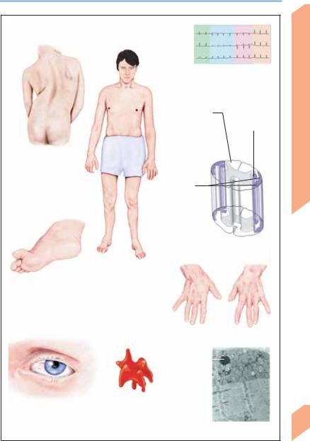
Whiplash injury of cervical spine |
|
|
|
|
|
|
|
|
|
|
||
(traumatic cervical distortion) |
|
|
|
|
|
|
|
|
Middle column |
|||
|
|
|
|
|
|
|
|
|
|
|
||
|
|
|
|
|
|
|
|
|
|
Posterior column |
||
|
|
|
|
|
Anterior |
|||||||
|
|
|
|
|
column |
|||||||
|
|
|
Anterior longitudinal |
|||||||||
|
|
|
ligament |
|||||||||
|
|
|
Posterior longitudinal |
|||||||||
|
|
|
ligament |
|||||||||
Normal cervical spine |
|
|
|
|
Three-column model of spinal stability |
|||||||
|
|
|
|
|
|
|
|
|
|
|||
|
|
|
|
|
Vertebral |
|
|
|
|
|
||
|
|
|
|
|
|
|
||||||
|
|
|
|
|
luxation |
|||||||
|
|
|
|
|
Ruptured ligament |
|||||||
|
|
|
|
|
||||||||
|
|
|
|
|
Fracture in poste- |
|||||||
|
|
|
|
|
rior column |
|||||||
|
|
|
|
|
Spinal cord |
|||||||
|
|
|
|
|
compression |
|||||||
|
|
Burst fracture |
|
|
|
|
|
|
|
|
|
Spinal cord |
|
|
|
|
|
|
|
|
|
|
|
||
|
|
|
|
|
|
|
|
|
|
|
|
contusion |
|
|
|
|
|
Syringomyelia |
|
|
|||||
|
|
|
|
|
|
|||||||
|
|
|
|
|
(posttraumatic) |
|||||||
|
|
|
|
|
Gunshot |
|||||||
|
|
|
|
|
wound |
|||||||
Spinal injuries
Central Nervous System
273
Rohkamm, Color Atlas of Neurology © 2004 Thieme
All rights reserved. Usage subject to terms and conditions of license.

Central Nervous System
Trauma
Spinal Cord Trauma
Open spinal cord trauma, by definition, involves penetration of the dura mater by a stab wound, gunshot wound, bone fragment, or severely dislocated vertebra. Closed spinal cord trauma (with dura intact) is the indirect effect of a nonpenetrating injury. The result may be a complete or incomplete spinal cord transection syndrome
(p. 48; Table 37, p. 380).
Acute stage (spinal shock). The acute manifestations of spinal cord transection syndrome are
seen below the level of the injury and include the total loss of voluntary and reflex motor function (flaccid paraplegia or quadriplegia, areflexia) and sensation, and autonomic dysfunction (urinary retention overflow incontinence, intestinal atony paralytic bowel obstruction, anhidrosis hyperthermia, cardiovascular dysfunction orthostatic hypotension, cardiac arrhythmia, paroxysmal hypertension). Patients are usually stable enough to begin rehabilitation in 3–6 weeks (rehabilitation stage, see below). For acute treatment, see p. 380 (Table 38).
Rehabilitation stage. The neurological deficits depend on the level of the lesion.
Level1 |
Motor Deficit |
Sensory Deficit2 |
Autonomic Deficit3 |
|
|
|
|
C1–C34 |
Quadriplegia, neck muscle |
Sensory level at back of |
Voluntary control of bladder, |
|
paresis, spasticity, respira- |
head/edge of lower jaw; pain |
bowel, and sexual function |
|
tory paralysis |
in back of head, neck, and |
replaced by reflex control; |
|
|
shoulders |
Horner syndrome |
C4–C5 |
Quadriplegia, diaphragmatic |
Sensory level at clavicle/ |
Same as above |
|
breathing |
shoulder |
|
C6–C85 |
Quadriplegia, spasticity, flac- |
Sensory level at upper chest |
Same as above |
|
cid arm paresis, diaphrag- |
wall/back; arms involved, |
|
|
matic breathing |
shoulders spared |
|
T1–T5 |
Paraplegia, diminished respi- |
Sensory loss from inner sur- |
Voluntary control of bladder, |
|
ratory volume |
face of lower arm, upper |
bowel, and sexual function |
|
|
chest wall, back region |
replaced by reflex control |
|
|
downward |
|
T5–T10 |
Paraplegia, spasticity |
Sensory level on chest wall |
Same as above |
|
|
and back corresponding to |
|
|
|
level of spinal cord injury |
|
T11–L3 |
Flaccid paraplegia |
Sensory loss from groin/ven- |
Same as above |
|
|
tral thigh downward, de- |
|
|
|
pending on level of injury |
|
L4–S26 |
Distal flaccid paraplegia |
Sensory loss at shin/dorsum |
Flaccid paralysis of bladder |
|
|
of foot/posterior thigh |
and bowel, loss of erectile |
|
|
downward, depending on |
function |
|
|
level of injury |
|
S3–S57 |
No motor deficit |
Sensory loss in perianal re- |
Flaccid paralysis of bladder |
|
|
gion and inner thigh |
and bowel, loss of erectile |
|
|
|
function |
|
|
|
|
1 Spinal cord level (not the same as vertebral level). 2 See p. 32ff. 3 Disturbance of bladder, bowel, rectal, and erectile function, sweating, and blood pressure regulation; p. 140ff. 4 High cervical cord lesion. 5 Low cervical cord lesion. 6 Epiconus. 7 Conus medullaris.
Chronic stage—late sequelae. Persistence of
274neurological deficits; assorted complications including venous thrombosis, pulmonary embolism, respiratory insufficiency, bowel obstruc-
tion, urinary tract infections, sexual dysfunction, cardiovascular disturbances, spasticity, chronic pain, bed sores, heterotopic ossification, and syringomyelia.
Rohkamm, Color Atlas of Neurology © 2004 Thieme
All rights reserved. Usage subject to terms and conditions of license.

Trauma
Trapezius m. (C2-C4)
Latissimus dorsi m. (C6-C8)
Triceps brachii m. (C7-C8)
Adductor magnus m. (L2-L4)
Pectoralis major m. (C7-T1)
|
|
|
|
Diaphragm (C3-C5) |
|
||||||||
|
|
|
|
|
|||||||||
|
|
|
|
|
|
|
Deltoid m. |
1 |
|||||
|
|
|
|
|
|
|
(C4-C6) |
||||||
|
|
|
|
|
|
|
|
|
|
|
|
|
2 |
|
|
|
|
|
|
|
Biceps brachii |
3 |
|||||
|
|
|
|
|
|
|
4 |
||||||
|
|
|
|
|
|
|
|||||||
|
|
|
|
|
|
|
m. (C5-C6) |
5 |
|||||
|
|
|
|
|
|
|
|
|
|
|
|
|
6 |
|
|
|
|
|
|
|
|
|
|
|
|
|
7 |
|
|
|
|
|
|
|
|
|
|
Flexor digi- |
8 |
||
|
|
|
|
|
|
|
|
|
|
torum pro- |
1 |
||
|
|
|
|
|
|
|
|
|
|
2 |
|||
|
|
|
|
|
|
|
|
|
|
fundus m. |
|||
|
|
|
|
|
|
|
|
|
|
3 |
|||
|
|
|
|
|
|
|
|
|
|
(C8-T1) |
|||
|
|
|
|
|
|
|
|
|
|
4 |
|||
|
|
|
|
|
|
|
|
|
|
|
|
|
|
|
|
|
|
|
|
|
|
|
|
|
|
Brachioradialis |
5 |
|
|
|
|
|
|
|
|
|
|
|
|
6 |
|
|
|
|
|
|
|
|
|
|
|
|
|
m. (C5-C6) |
|
|
|
|
|
|
|
|
|
|
|
|
|
7 |
|
|
|
|
|
|
|
|
|
|
|
|
|
|
|
|
|
|
|
|
|
|
|
|
|
|
|
Abductor |
8 |
|
|
|
|
|
|
|
|
|
|
|
|
9 |
|
|
|
|
|
|
|
|
|
|
|
|
|
pollicis |
|
|
|
|
|
|
|
|
|
|
|
|
|
brevis m. |
10 |
|
|
|
|
|
|
|
|
|
|
|
|
(C8-T1) |
11 |
|
|
|
|
|
|
|
|
|
|
|
|
|
|
|
|
|
|
|
|
|
|
|
|
|
|
|
12 |
|
|
|
|
|
|
|
|
|
|
|
|
|
1 |
|
|
|
|
|
|
|
|
|
|
|
|
|
2 |
|
|
|
|
|
|
|
|
|
|
|
|
|
3 |
|
|
|
|
|
|
|
|
|
|
|
|
|
4 |
|
|
|
|
|
|
|
|
|
|
|
|
|
5 |
|
|
Interossei |
|||||||||||
|
|
|
|||||||||||
|
|
(C8-T1) |
|
||||||||||
|
|
Quadriceps |
|
||||||||||
|
|
|
|||||||||||
|
|
m. (L2-L4) |
|
||||||||||
|
|
Gastrocnemius |
|
||||||||||
|
|
m. (L5-S1) |
|
||||||||||
|
|
Tibialis anterior |
|
||||||||||
|
|
|
|||||||||||
|
|
m. (L4-L5) |
|
||||||||||
|
|
Extensor |
|
||||||||||
|
|
|
|||||||||||
|
|
hallucis longus |
|
||||||||||
|
|
m. (L5-S1) |
|
||||||||||
Cervical cord lesion
Thoracic cord lesion
Lumbar cord lesion
Segment-indicating muscles
Lesion of conus/cauda equina
Topography of spinal cord lesions
Central Nervous System
275
Rohkamm, Color Atlas of Neurology © 2004 Thieme
All rights reserved. Usage subject to terms and conditions of license.

Central Nervous System
276
Cerebellar Diseases
! Signs of Cerebellar Dysfunction
Loss of coordination and balance. Ataxia is uncoordinated, irregular, and poorly articulated movement (dyssynergy). The typical patient sways while sitting (truncal ataxia) or standing (postural ataxia), undershoots or overshoots an intended target of movement (dysmetria = hypometria or hypermetria), and walks with quick, irregular steps in an unsteady, swaying, broadbased gait reminiscent of alcohol intoxication (gait ataxia, p. 54). Pointing tests are used to detect dysmetria, incoordination, and tremor that is worst as a movement approaches its target (intention tremor); the finger–nose, finger–fin- ger, and heel–knee–shin tests should be carried out with the eyes open and closed. Bárány’s pointing test: The patient is asked to close his or her eyes, touch the doctor’s finger with his or her own index finger, then lower and raise the still outstretched arm and touch the doctor’s finger again; the patient’s finger deviates laterally from the target, and the direction of deviation is toward the side of the lesion. Unsteadiness of stance of cerebellar origin, which may be so severe as to make standing impossible (astasia), is not influenced by opening or closing the eyes (Romberg sign) and differs in this respect from spinal (sensory) ataxia. Stepping in place for 30–60 seconds with the eyes closed causes the body to turn to the side of the lesion. Patients with mild ataxia find it difficult or impossible to walk a straight line (abasia; detected by heel-to- toe walking, tandem gait). The patient may be unable to perform rapid alternating movements (dysdiadochokinesia). The handwriting is enlarged (macrographia), coarse, and shaky, and the patient’s drawing of parallel lines or a spiral is unsatisfactory.
Dysarthria. The patient’s speech (p. 130) is slow, unclear (babbling, slurred), and monotonous (dysarthrophonia), and possibly also discontinuous (choppy, faltering, or scanning speech). There is poor coordination of breathing with the flow of speech, resulting in a sudden transition from soft to loud speech (explosive speech).
Oculomotor disturbances. Gaze-evoked nystagmus is a frequent finding in cerebellar disease. Voluntary saccades are too short or too long (ocular dysmetria) and are therefore followed by afterbeats. Slow pursuit movements are jerky
(saccadic). Patients are frequently unable to suppress the vestibulo-ocular reflex (p. 26), i.e., the normal visual suppression of nystagmus is impaired. The result is impaired visual fixation on turning of the head.
Muscle tone. Decreased muscle tone is mainly found in patients with acute unilateral lesions of the cerebellum. The examiner can detect it by passively swinging or shaking the patient’s limbs, or by testing for the rebound phenomenon. The patient is asked to extend the arms with the eyes closed (posture test) and the examiner lightly taps on one wrist, causing deflection of the arm. The rebound movement undershoots or overshoots the original arm position. Alternatively, the patient can be asked to flex the elbow against resistance. When the examiner suddenly releases the resistance, the affected arm rebounds unchecked.
! Topography of Cerebellar Lesions
Lesions of the cerebellum and its afferent and efferent connections (p. 54) produce characteristic signs of cerebellar disease. Expanding lesions may go on to produce further, extracerebellar deficits (e. g., cranial nerve palsies, hemiparesis, sensory loss).
! Special Diagnostic Studies
The diagnostic studies to be obtained depend on the clinical findings (to be described below) and may include imaging studies (MRI, CT), neurophysiological studies (nerve conduction studies, electromyography), ECG, pathological studies (of tissue, blood, CSF, bone marrow, muscle, or nerve biopsy specimens), and/or ophthalmological consultation (optic nerve atrophy, Kay- ser–Fleischer ring, tapetoretinal degeneration).
! Idiopathic Cerebellar Ataxia (IDCA)
This group of disorders includes various forms of nonfamilial cerebellar ataxia of unknown cause with onset in adulthood (generally age 25 years or older). IDCA occurs as an isolated disturbance or as a component of multiple system atrophy (MSA; p. 302).
Rohkamm, Color Atlas of Neurology © 2004 Thieme
All rights reserved. Usage subject to terms and conditions of license.

Cerebellar Diseases
Dysdiadochokinesis
Finger-finger test
(intention tremor)
Gait ataxia with “tandem” gait
Dysmetria (hypermetria)
Postural test for position sense
Test for gaze-evoked nystagmus
Rebound phenomenon
Saccades; gaze-evoked and rebound nystagmus
Central Nervous System
277
Rohkamm, Color Atlas of Neurology © 2004 Thieme
All rights reserved. Usage subject to terms and conditions of license.

Central Nervous System
278
Cerebellar Diseases
Acquired Cerebellar Syndromes
Onset |
Etiology |
Symptoms and Signs |
||
Acute (minutes to |
! |
Infection1 |
! |
Viral infection: varicella-zoster virus, Epstein–Barr virus, |
hours) |
|
|
|
rubella, mumps, influenza, parainfluenza, echovirus, |
|
|
|
|
coxsackievirus, cytomegalovirus, FSME, herpes simplex virus. |
|
|
|
|
Children are more commonly affected than adults. Special |
|
|
|
! |
type: opsoclonus-ataxia syndrome2. |
|
|
|
Abscess |
|
|
|
|
! Miller Fisher syndrome (ataxia, ophthalmoplegia, areflexia; |
|
|
|
|
|
p. 395) |
|
! |
Vascular |
! |
Brainstem signs (pp. 70ff., 170) predominate |
|
|
|
! Infarcts can be differentiated from hemorrhages by imaging |
|
|
|
|
|
studies |
|
|
|
! Early treatment, often neurosurgical, may be needed to pre- |
|
|
|
|
|
vent rapid development of life-threatening complications |
|
|
|
|
(p. 174 f) |
|
! |
Toxic |
! |
Alcohol, barbiturates, phenytoin, lithium |
Subacute |
! |
Tumor3 |
! |
Occipital pain (radiating to forehead, nuchal region, and |
(days to weeks) |
|
|
|
shoulders), recurrent vomiting, stiff neck, vertigo, truncal |
|
! |
Paraneoplastic4 |
! |
ataxia; obstructive hydrocephalus |
|
Cerebellar dysfunction may appear months or years before |
|||
|
|
|
|
the tumor is discovered. Anti-Purkinje-cell antibodies are pre- |
|
|
|
|
sent in the serum and CSF of patients with neuron loss |
|
! |
Toxic |
! |
Alcohol |
|
|
|
! Medications (anticonvulsants, e. g., phenytoin; lithium, 5- |
|
|
|
|
|
fluorouracil, cytosine arabinoside) |
|
|
|
! Heavy metals (mercury, thallium, lead) |
|
|
|
|
! Solvents (toluene, carbon tetrachloride) |
|
|
! |
Other |
! |
Hypoxia, heat stroke, hyperthermia |
Chronic (months to |
! |
Infection |
! |
Progressive rubella panencephalitis (very rare complication of |
years) |
|
|
|
congenital rubella infection in boys; onset at age 8 to 19 |
|
|
|
|
years; characterized by ataxia, dementia, spasticity, and dys- |
|
|
|
! |
arthria) |
|
|
|
Creutzfeldt–Jakob disease (p. 252) |
|
|
! |
Vascular |
! |
Meningeal siderosis causes ataxia and partial or complete |
|
|
|
|
hearing loss (leptomeningeal deposition of hemosiderin in |
|
|
|
|
chronic subarachnoid hemorrhage vascular malformations, |
|
|
|
|
oligodendroglioma, ependymoma of the cauda equina, post- |
|
! |
|
! |
operative occurrence) |
|
Metabolic |
Hypothyroidism, malabsorption syndrome (vitamin E defi- |
||
|
|
|
|
ciency), thiamin deficiency (acute Wernicke encephalo- |
|
|
|
|
pathy) |
|
|
|
! Refsum disease5 ( serum phytanic acid level, p. 332) |
|
|
|
|
! Wilson disease5 (ataxia, tremor, dysarthrophonia, dysphagia, |
|
|
|
|
|
dystonia, behavioral disturbances, p. 307) |
Intermittent |
! |
Metabolic5 |
! |
Hereditary metabolic disorders in neonates, children, and ju- |
|
|
|
|
veniles (see also pp. 306 f, 386 f) |
|
|
|
! Disorders of amino acid metabolism (hyperammonemia, |
|
Hartnup syndrome, maple syrup urine disease)
! Storage diseases (metachromatic leukodystrophy, neuronal ceroid lipofuscinosis, sialidosis, GM2 gangliosidosis)
1 Partial listing; numerous infections can cause ataxia as part of the syndrome of encephalomyelitis. 2 Highfrequency bursts of saccades in all directions of gaze without an intersaccadic interval. 3 See p. 254 ff; cerebellar astrocytoma, medulloblastoma, ependymoma, hemangioblastoma (von Hippel–Lindau disease), meningioma of the cerebellopontine angle, metastases (lung cancer, breast cancer, melanoma). 4 Antibodies (p. 388) against Hu, Yo, TR, CV2, Ma1, CRD1, CRD2, Ma2, and mGluR1. 5 Genetic; listed here for differential diagnostic purposes.
Rohkamm, Color Atlas of Neurology © 2004 Thieme
All rights reserved. Usage subject to terms and conditions of license.

Cerebellar Diseases
Drug-induced cerebellar syndromes
Cerebellar infections |
|
Normal |
|
|
|
|
Alcoholic cerebellar |
cortex |
|
|
|
|
degeneration |
|
|
Purkinje cell |
Cortical |
|
atrophy |
|
|
lesions |
|
|
|
Lesion of Hyperthermia-related cerebellar cortex cerebellar dysfunction
Vascular cerebellar lesion
Cerebellar atrophy
Malabsorptive and metabolic |
Paraneoplastic and hypoxic |
cerebellar syndromes |
cerebellar syndromes |
Central Nervous System
279
Rohkamm, Color Atlas of Neurology © 2004 Thieme
All rights reserved. Usage subject to terms and conditions of license.

Central Nervous System
280
Cerebellar Diseases
Hereditary Cerebellar Syndromes
" Autosomal Recessive Cerebellar Syndromes (partial listing)
Syndrome |
Symptoms and Signs |
CL1/Gene Product |
|
Friedreich ataxia2,7 |
Usual manifestations: |
9q13, 9p23-p11/ |
|
|
! |
Progressive limb/gait ataxia |
frataxin |
|
! Age of onset !30 years |
Mutation: Extended |
|
|
! |
Areflexia in legs |
GAA-trinucleotide |
|
! Neurophysiological evidence of sensory neuropathy |
repeat |
|
|
|
Variable manifestations: |
|
|
! Dysarthria, distal muscular atrophy/paresis (ca. 50%), pes cavus (ca. |
|
|
|
|
50%), scoliosis, optic nerve atrophy (ca. 25%), nystagmus (ca. 20%), |
|
|
|
oculomotor disturbances (p. 276), hearing loss (ca. 10%), cardiomy- |
|
|
|
opathy (ca. 65%), diabetes mellitus (ca. 10%) |
|
Ataxia with vitamin |
! |
Onset in childhood or adulthood |
8q13.1-q13.3/α-to- |
E deficiency7 |
! |
Gait ataxia |
copherol transfer |
(serum: vitamin |
! |
Dysarthria |
protein |
E, cholesterol/ |
! Other symptoms similar to those of Friedreich ataxia |
|
|
triglycerides) |
|
|
|
Abetalipoprotein- |
! |
Steatorrhea, other symptoms similar to those of Friedreich ataxia |
4q24/triglyceride |
emia3,7 (p. 300) |
|
|
transfer protein |
Ataxia-telangiec- |
! |
Ataxia first seen when child learns to walk |
11q22.3/phosphatidyl- |
tasia4,7 |
! |
Choreoathetosis |
inositol-3’- |
|
! |
Oculomotor disturbances5 |
kinase and rad36 |
|
! |
Oculocutaneous telangiectases |
|
|
! |
Immunodeficiency (frequent infections) |
|
! Increased risk of malignant tumors ! Elevated serum α-fetoprotein
1 Chromosome location (CL). 2 Classic form. 3 Bassen–Kornzweig syndrome; vitamin A and E deficiency, low cholesterol/ triglyceride levels, acanthocytosis. 4 Louis-Bar syndrome. 5 Oculomotor apraxia. 6 DNA repair kinase/cell cycle control; ataxia-telangiectasia-mutated (ATM) gene. 7 A direct gene test is available.
For mitochondrial syndromes with ataxia, see p. 403.
" Autosomal Dominant Cerebellar Syndromes (partial listing)
Syndrome |
Symptoms and Signs |
CL/Gene Product |
|
|
|
Autosomal domi- |
! ADCA1: Ataxia, ophthalmoplegia, pyramidal/extrapyramidal distur- |
SCA1: 6p23/ataxin1 |
nant cerebellar |
bances (p. 44); SCA15, SCA25, SCA32,5, SCA4, SCA85, SCA12, SCA13, |
SCA2: 12q24/ataxin2 |
ataxia (ADCA); |
SCA17 |
SCA3: 14q24.3-q31/ |
spinocerebellar |
! ADCA2: Ataxia, retinopathy, SCA75 |
MJD1 protein |
ataxia (SCA)1 |
! ADCA3: Predominant cerebellar ataxia; SCA5, SCA65, SCA10, SCA11, |
SCA4: 16q22.1 |
|
SCA125, SCA14, SCA15, SCA16 |
SCA5: 11p11-q11 |
|
|
SCA6: 19p13/α-1A |
|
|
calcium channel |
|
|
SCA7: 3p21.1-p12 |
Episodic ataxia |
! EA1: Episodes of ataxia lasting seconds to minutes, 1 to 10 times daily; |
! 12p135/potassium |
(EA)3 |
provoked by abrupt changes of position, emotional or physical stress, |
channel (point |
|
and caloric vestibular stimulation; myokymia in face and hands be- |
mutation) |
|
tween attacks; continuous spontaneous activity in resting EMG |
|
|
! EA2: Episodes of ataxia lasting minutes to hours (rarely days) of varia- |
! 19p135/voltage- |
|
ble frequency (daily to yearly); headache, tinnitus, vertigo, ataxia, |
gated calcium |
|
nausea, vomiting, nystagmus; induced by same stimuli as EA1; ataxia, |
channel4 (point |
|
nystagmus, and head tremor between attacks |
mutation) |
Gerstmann– |
Onset between the ages 40 and 50 years; presents with cerebellar ataxia; |
20pter-p12/P102L |
Sträussler– |
dysarthrophonia, dementia, nystagmus, rigor, visual disturbances, and |
|
Scheinker syn- |
hearing loss develop in the course of the disease |
|
drome (p. 252) |
|
|
Fatal familial in- |
Progressive insomnia, autonomic dysfunction (arterial hypertension, |
20pter-p12/D178N |
somnia (p. 252) |
tachycardia, hyperthermia, hyperhidrosis), myoclonus, tremor, ataxia |
|
|
|
|
1 Definitive identification of the SCA types listed is possible only with molecular genetics tests (examples in right column, see OMIM for details). 2 Machado–Joseph disease (MJD). 3 Other forms: EA3 and EA4. 4 Other mutations of this gene are associated with SCA6 and familial hemiplegic migraine. 5 A direct genetic test is available.
Rohkamm, Color Atlas of Neurology © 2004 Thieme
All rights reserved. Usage subject to terms and conditions of license.
Cerebellar Diseases
I |
aVR |
V1 |
V4 |
II |
aVL |
V2 |
V5 |
III |
aVF |
V3 |
V6 |
Cardiomyopathy in FA
(ECG shows repolarization disturbances and left axis deviation)
Ataxia and loss of |
Paresis due to |
position sense |
pyramidal |
due to posterior |
tract lesion |
column lesion |
|
Scoliosis in FA
Ataxia due to lesion of posterior and anterior
spinocerebellar tracts
Spinal degeneration in FA
Friedreich ataxia (FA)
Pes cavus/clawfoot
|
|
Distal muscular atrophy |
||
|
|
Lipid |
|
|
|
|
|
||
|
|
Mitochondria |
||
|
Acanthocyte |
|
|
|
Ocular telangiectasia |
(crenated erythrocyte) |
|
|
|
in abetalipoproteinemia |
Myofibrils |
|||
|
|
Mitochondrial encephalomyopathy |
||
Central Nervous System
281
Rohkamm, Color Atlas of Neurology © 2004 Thieme
All rights reserved. Usage subject to terms and conditions of license.

Myelopathies
The clinical differential diagnosis of myelopathies is based on the level of the spinal cord lesion, the particular structures affected, and the temporal course of the disorder (p. 48, Table 39, p. 381).
Acute Myelopathies
Symptoms and signs develop within minutes, hours, or days.
System |
! Spinal Cord Trauma |
|
|
||
(See p. 274) |
|
|
|||
! Myelitis |
|
|
|||
|
|
|
|||
Nervous |
Viral |
myelitis (p. 234 |
ff). |
Enteroviruses |
|
(poliovirus, coxsackievirus, |
echovirus), herpes |
||||
|
|||||
|
zoster virus, varicella zoster virus, FSME, rabies, |
||||
|
HTLV-1, HIV, Epstein–Barr virus, cytome- |
||||
Central |
galovirus, herpes simplex virus, postvaccinial |
||||
myelitis. |
|
|
|||
|
|
|
|||
|
Nonviral myelitis (p. 222 ff). Mycoplasma, neu- |
||||
|
roborreliosis, abscess (epidural, intramedul- |
||||
|
lary), |
tuberculosis, parasites |
(echinococcosis, |
||
cysticercosis, schistosomiasis), fungi, neurosyphilis, sarcoidosis, postinfectious myelitis, multiple sclerosis/neuromyelitis optica (Devic syndrome), acute necrotizing myelitis, connective tissue disease (vasculitis), paraneoplastic myelitis, subacute myelo-optic neuropathy (SMON), arachnoiditis (after surgical procedures, myelography, or intrathecal drug administration).
! Vascular Syndromes (p. 22)
Anterior spinal artery syndrome. Segmental paresthesia and pain radiating in a bandlike distribution may precede the development of motor signs by minutes to hours. A flaccid paraparesis or quadriparesis (corticospinal tract, anterior horn) then ensues, along with a dissociated sensory loss from the level of the lesion downward (spinothalamic tract impaired pain and temperature sensation, with intact perception of vibration and position) and urinary and fecal incontinence. Often only some of these signs are present.
Posterior spinal artery syndrome is rare and difficult to diagnose. It is characterized by pain in
282the spine, paresthesiae in the legs, a loss of position and vibration sense below the level of the
lesion, and global anesthesia with segmental
loss of deep tendon reflexes at the level of the lesion. Larger lesions cause paresis and sphincter dysfunction.
Sulcocommissural artery syndrome. Segmental pain at the level of the lesion, followed by flaccid paresis of ipsilateral arm/leg; loss of proprioception, position sense, and touch perception with contralateral dissociated sensory loss (Brown– Séquard syndrome). Sphincter dysfunction is rare.
Complete spinal infarction. Acute spinal cord transection syndrome with flaccid paraplegia or quadriplegia, sphincter dysfunction, and total sensory loss below the level of the lesion. Autonomic dysfunction may also occur (e. g., vasodilatation, pulmonary edema, intestinal atony, disordered thermoregulation). The cause is often an acute occlusion of the great radicular artery (of Adamkiewicz).
Central spinal infarction. Acute paraplegia, sensory loss, and sphincter paralysis.
Claudication of spinal cord. Physical exercise (running, long walks) induces paresthesiae or paraparesis that resolves with rest and does not occur when the patient is lying down.
Cause: Exercise-related ischemia of the spinal cord due to a dural arteriovenous fistula or highgrade aortic stenosis (see also p. 284).
Dural/perimedullary arteriovenous (AV) fistula is an abnormal communication (shunt) between an artery and vein between the two layers of the dural mater. An arterial branch of a spinal artery feeds directly into a superficial spinal vein, which therefore contains arterial rather than venous blood, flowing in the opposite direction to normal. Paroxysmal stabbing pain and/or episodes of slowly progressing paraparesis and sensory loss separated by periods of remission occur in the early stage of the disorder, which usually affects men between the ages of 40 and 60. If the suspected diagnosis cannot be confirmed by MRI scans (because of low shunt volume), myelography may be helpful ( dilated veins in the subarachnoid space).
Spinal hemorrhage can occur in epidural, subdural, subarachnoid, and intramedullary locations (intramedullary hemorrhage = hematomyelia). Possible causes: intradural/intramedullary AV malformation, cavernoma, tumor, aneurysm, trauma, lumbar puncture, and coagulopathy.
Rohkamm, Color Atlas of Neurology © 2004 Thieme
All rights reserved. Usage subject to terms and conditions of license.